70
3/27/2018 Obstetrics Board Review 2018 Obstetrics Board Review 2018 Diane M. Twickler, M.D. Diane M. Twickler, M.D.

3/27/2018
Obstetrics Board Review 2018Obstetrics Board Review 2018Diane M. Twickler, M.D.Diane M. Twickler, M.D.

The most likely diagnosis is
1. Normal2. Anencephaly3. Acrania4. Chiari II malformation5. Encephalocele

MSAFP is elevated with
1. Open neural tubedefects
2. Gastroschisis3. Down syndrome4. Incorrect dating5. All of the above6. 1,27. 1,2,38. 1,2,4

The most likely diagnosis is1. Omphaloclee2. Two-vessel cord3. Gastroschisis4. Short umbilical cord5. Normal

Gastroschisis is
1. Associated with aneuploidy2. Usually right-sided3. Associated with other organ system anomalies4. Associated with GI anomalies5. All of the above6. 2,47. 1,2,38. 2,3, 4

Color mapping 1. Increases the TIB, but decreases TIS2. Increases the MI3. Increases both TIB and TIS4. Increases frame rate

Findings are most suggestive of
1. Incomplete placenta previa2. Low lying placenta3. Vasa previa4. Complete placenta previa5. Succenturiate lobe

Transvaginal Ultrasound usually
1. Is a lower frequency than trans abdominal2. Provides better resolution3. Is contraindicated in in the setting of previa4. Provides better penetration5. All of the above6. 1,2, 47. 2,4

The focal zone 1. Increased depth increases frame rate2. Increased depth decreases frame rate3. Increased depth does not change frame rate

Findings suggest
1. Complete duplication2. Posterior urethral valves3. Ectopic ureterocele4. UPJ5. All of the above6. 1&47. 1&28. 1&3

The Color Mapping imageidentifies the
1. Umbilical veins2. Umbilical arteries3. Iliac arteries4. Femoral veins

Aliasing is seen in the vesselsbecause
1. The arterial red blood cell flowexceeds the color scale
2. The color scale is too high3. The insonating angle is
too high1. 1&3

Findings suggest
1. Twin peak2. Monochorionic pregnancy3. Diamniotic pregnancy4. Dichorionic pregnancy5. 1, 3 &46. 1, 2 & 37. All of the above

Which of the following are true?1. All dizygotic twins are dichorionic2. All monochorionic twins are monozygotic3. All monozygoitc twins are monochorionic4. All dichorionic twins are dizygotic5. 1&26. 3&4

Findings are most consistent with
1. Dandy walker2. Choroid plexus papilloma3. Encephalocele4. Agenesis of the corpus
callosum

Findings on the images1. Can bee seen in hydrops fetalis2. Are more common in males3. Are not associated with increased pleural
lymphocytes4. All of the above5. 1&3

The most likely diagnosis is
1. Thanatophoric dwarf2. Homozygous achondroplasia3. Osteogenesis Imperfecta4. Achondrogenesis

The most likely diagnosis is1. Autosomal dominant polycystic kidneys2. Autosomal recessive polycystic kidneys3. Posterior urethral valves4. Multicystic dysplastic kidneys

Bilateral multicystic dysplastickidneys1. Are inherited2. Cause pulmonary hyoplasia3. Are associated with polyhydramnios4. All of the above5. 1&2

Findings are most consistent with1. Placenta previa2. Focal myometrial contraction3. Lower uterine segment leiomyoma4. Full bladder mimicking placenta previa

Based on images what would you do next?1. Evaluate face2. Evaluate extremities3. Evaluate spine4. Recommend an MRI

The previous and present findings suggest
1. Encephalocele2. Chiari 2 with
myelomeningocele3. Aqueductal stenosis4. Dandy Walker

What change(s) resulted an increased in frame rate between the two images?Why is an increase in frame rate preferred for cardiac imaging?

The most likely finding is
1. Fetal teratoma2. Small bowel obstruction3. Normal abdominal fat4. Echogenic bowel5. Gall bladder tumor

1. Esophageal atresia2. Duodenal atresia3. Jejunal atresia4. Colonic atresia
Images are most consistent with

Duodenal atresia
1. Causes polyhydramniosmost of the time
2. Is associated with Downsyndrome
3. Usually does not requiresurgical intervention
4. All of the above5. 1&2

This finding is
1. A marker for Turner’ssyndrome
2. Often associated withhydrops fetalis
3. Not associated withother anomalies oraneuploidy
4. All of the above5. 1&2

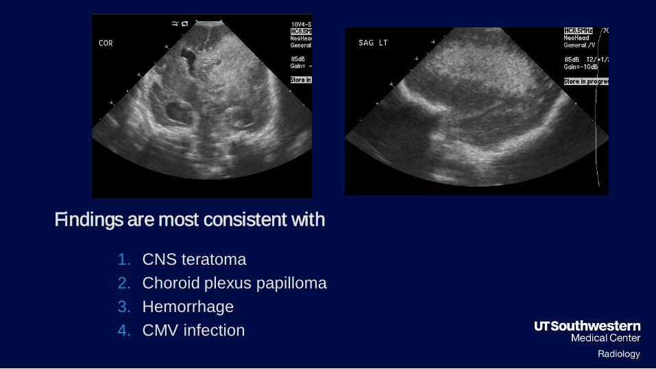
Findings are most consistent with1. CNS teratoma2. Choroid plexus papilloma3. Hemorrhage4. CMV infection
Findings are most consistent with
1. CNS teratoma2. Choroid plexus papilloma3. Hemorrhage4. CMV infection

Images are suggestive of1. Megacystis microcolon hypoperistalsis syndrome2. Prune Belly syndrome3. Posterior Urethral Valves4. MURCS syndorme

Image is mostconsistent with
1. Hypoplastic Left Heart2. Atrial-ventricular canal3. Ventriculoseptal defect4. Ebsteins anomaly

1. Esophageal atresia2. Duodenal atresia3. Jejunal atresia4. Colonic atresia
Images are most consistent with

Which is/ are true?
1. The endocervical length is 19mm2. The endocervical length is 48mm3. The measurement is normal4. The measurement is abnormal5. 1&46. 2&37. 1&3 x
+

The most like diagnosis is
1. Vasa Pevia2. Succenturiate Lobe3. Placenta Accreta4. Normal placenta

Images show1. Placenta Previa2. Placenta Accreta3. Vasa Pevia4. All of the above5. 1&2

Placenta Accreta is associated with
1. Previous cesarean delivery2. Placenta previa3. Major increased delivery risks4. Bleeding complications5. All of the above6. 1, 3, 4

MR Criteria for accreta include
1. Thickened darkened nodular contour of placentauterine interface with extensions of dark bandswithin the placenta
2. Mass effect on the placenta causing an outerbulge
3. Heterogeneous placental signal with large lakesor vessels
4. All of the above

The most like diagnosis is1. Vasa Pevia2. Succenturiate Lobe3. Placenta Accreta4. Normal placenta

Just Images………


What additional imaging?What additional imaging?


Which of the following is least likely?Vein of Galen Aneurysm
1. Is the venous finding of a cranial AVM2. Doppler findings suggest high arterial impedance3. Can result in cardiomegaly4. Will cause an increase in superior vena cava flow5. Is not associated with aneuploidy


The peak systolic velocity of the MCA§ Is angle independent?
§ Is used to diagnose ….
Doppler is the velocity of …..
Increased viscosity (increases or decreases)
the peak systolic velocity of the MCA.
Therefore, anemia (increases or decreases)
the peak systolic velocity of the MCA

MCA PSV in 111 fetuses at risk for anemia due to RBC alloimmunization.Solid curve represents median PSV. Dotted curve represents 1.50 MOM.Open circles indicate no anemia or mild anemia. Triangles indicate moderate orsevere anemia. Solid circles indicate hydrops. Mari. N Engl J Med 2000;342:9


Diagnosis
§Additional Images?


AFI=0

AFI =40mm

Oligohydramnios is§Defined as a largest vertical pocket less than ….. Or amniotic
fluid index less than…….
§Differential diagnosis
§D
§R
§ I
§P
§P
§C

Doppler waveform
§Progressive findings of………suggest…………..


Dandy Walker “Complex”§Differential diagnosis includes:§M§A§D§E§C


Good Luck !!!!!!!


