70
Obstructive Lung Diseases Restrictive Pulmonary Diseases Pulmonary infection Lung Tumors Diseases of the Pleura
| Date post: | 22-Dec-2015 |
| Category: |
Documents |
| Upload: | moris-fitzgerald |
| View: | 235 times |
| Download: | 2 times |
Obstructive Lung DiseasesRestrictive Pulmonary Diseases
Pulmonary infectionLung Tumors
Diseases of the Pleura
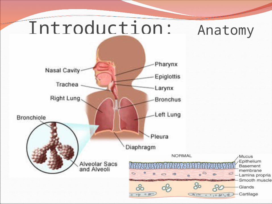
Introduction: Anatomy
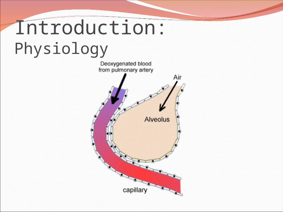
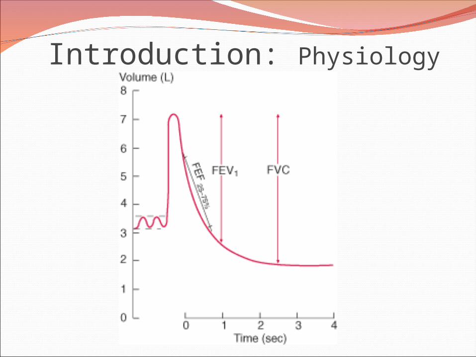
Introduction: Physiology

Pathology of lung diseasesVery important in clinical medicineComplication of air pollution Common symptoms:
Dyspnea: difficulty with breathing Decrease compliance, fibrosis Increased airway resistance , ch. bronchitis Chest wall disease, obesity Fluid accumulation, left sided heart failure
Cough Postnasal discharge, GERD, Br. Asthma, ch.
Bronchitis, pneumonia, bronchiectasis, drug inducedHemoptysis
Ch. Bronchitis, pneumonia, TB, bronchiectasis, aspergilloma

Atelectasis (collapse)Incomplete expansion of the lungs
or collapse of previously inflated lung substance.
Significant atelectasis reduce oxygenation and predispose to infection.

Types of Atelectasis1. Resorption atelectasis.2. Compression atelectasis.3. Contraction atelectasis.
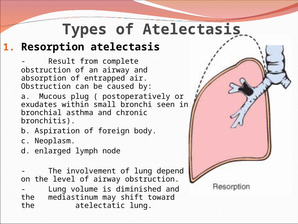
Types of Atelectasis1. Resorption atelectasis
- Result from complete obstruction of an airway and absorption of entrapped air. Obstruction can be caused by:
a. Mucous plug ( postoperatively or exudates within small bronchi seen in bronchial asthma and chronic bronchitis).
b. Aspiration of foreign body. c. Neoplasm.
d. enlarged lymph node
- The involvement of lung depend on the level of airway obstruction.- Lung volume is diminished and the mediastinum may shift toward the atelectatic lung.
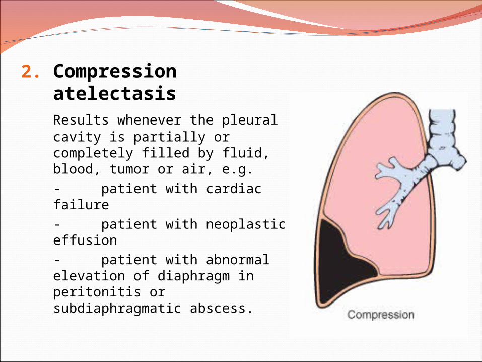
2. Compression atelectasisResults whenever the pleural cavity is partially or completely filled by fluid, blood, tumor or air, e.g. - patient with cardiac failure- patient with neoplastic effusion- patient with abnormal elevation of diaphragm in peritonitis or subdiaphragmatic abscess.

3. Contraction atelectasis.
Local or generalized fibrotic changes in pleura or lung preventing full expansion of the lung.
AtelectasisAtelectatic lung is prone to develop superimposed
infection.
It is reversible disorder except for contraction atelectasis.
It should be treated promptly to prevent hypoxemia.
Obstructive and Restrictive Pulmonary DiseasesDiffuse pulmonary diseases are divided into:
1. Obstructive disease: characterized by limitation of airflow owing to partial or complete obstruction at any level from trachea to respiratory bronchioles.Pulmonary function test: limitation of maximal airflow rate during forced expiration (FEVI).
2. Restrictive disease: characterized by reduced expansion of lung parenchyma with decreased total lung capacity while the expiratory flow rate is near normal. Occur in: 1. Chest wall disorder. 2. Acute or chronic, interstitial and infiltrative diseases, e.g. ARDS and pneumoconiosis.

Introduction: Physiology
Introduction: Physiology
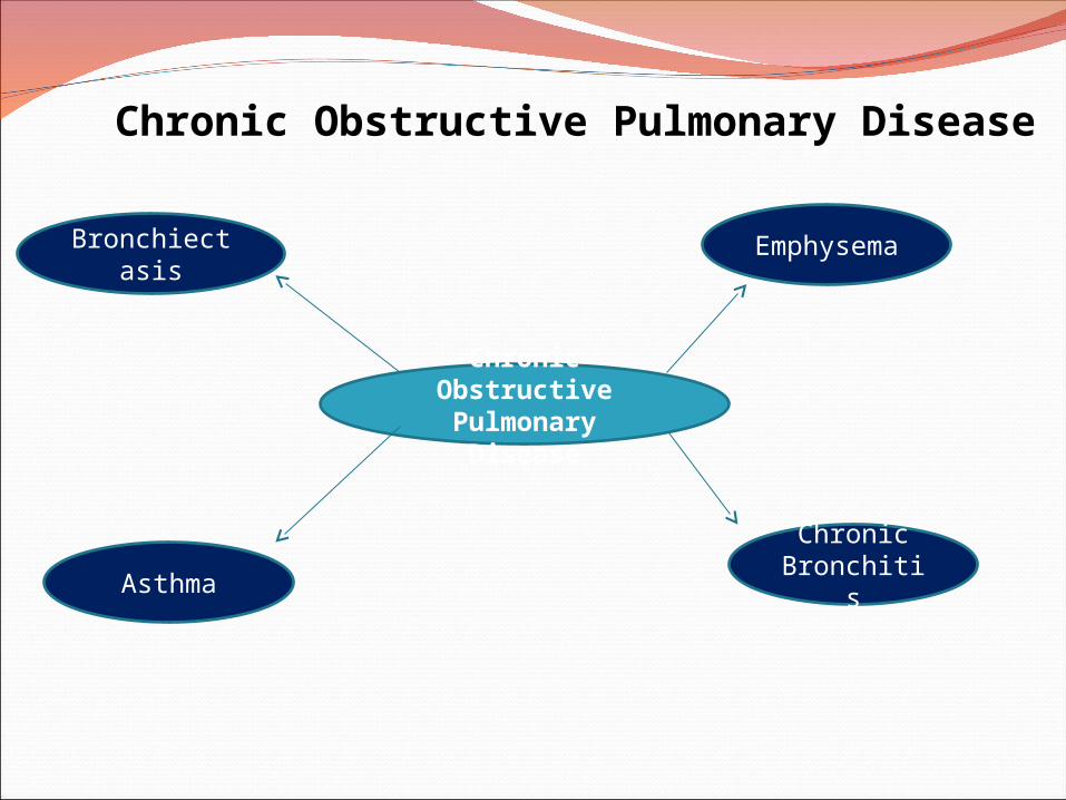
Chronic Obstructive Pulmonary Disease (COPD)
Share a major symptom: dyspnea with chronic or recurrent obstruction to airflow within the lung.
The incidence of COPD has increased dramatically in the past few decades.
Chronic Obstructive Pulmonary
Disease
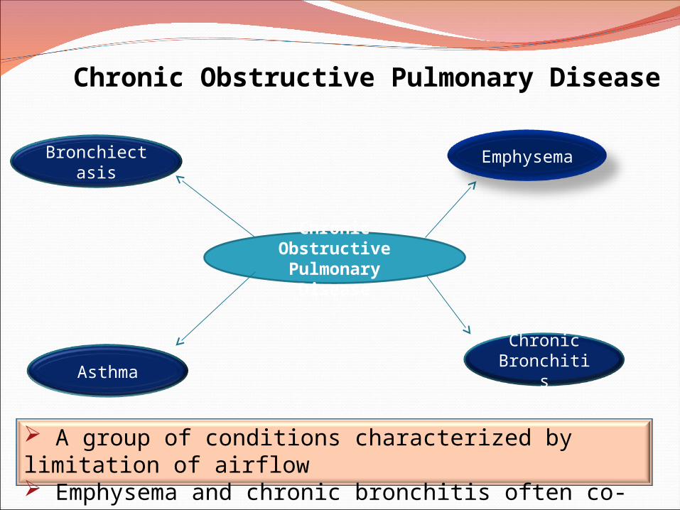
Emphysema
Bronchiectasis
Chronic BronchitisAsthma
A group of conditions characterized by limitation of airflow Emphysema and chronic bronchitis often co-exist.
Chronic Obstructive Pulmonary Disease

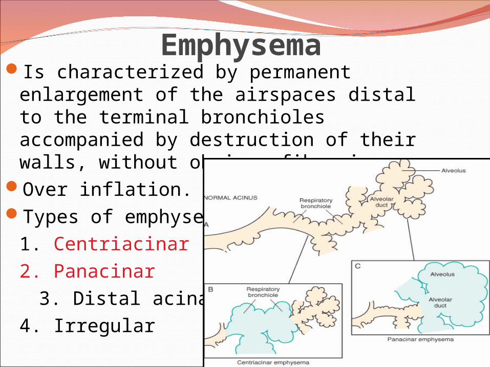
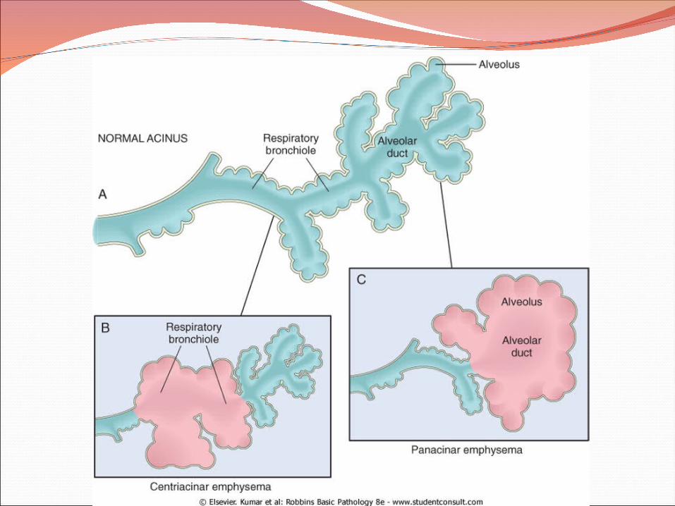
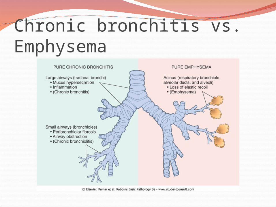
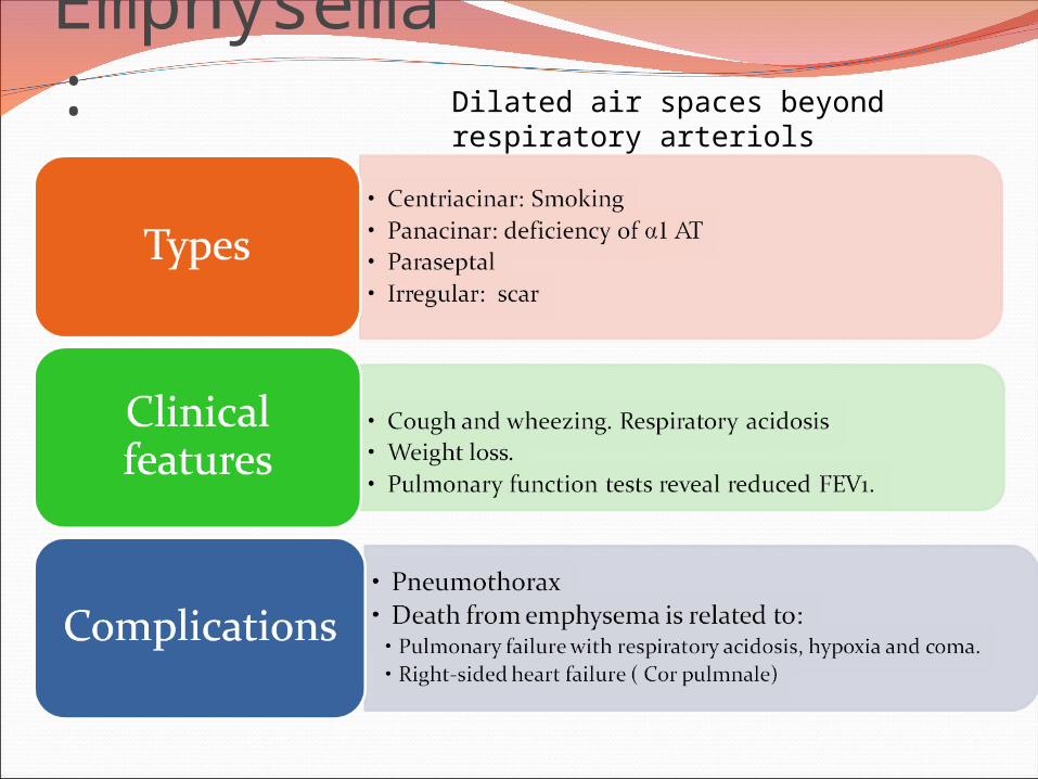
EmphysemaIs characterized by permanent
enlargement of the airspaces distal to the terminal bronchioles accompanied by destruction of their walls, without obvious fibrosis.
Over inflation.Types of emphysema:
1. Centriacinar (20x)2. Panacinar
3. Distal acinar4. Irregular
EmphysemaIncidenceEmphysema is present in approximately
50% of adults who come to autopsy.Pulmonary disease was considered to be
responsible for death in 6.5% of these patients.
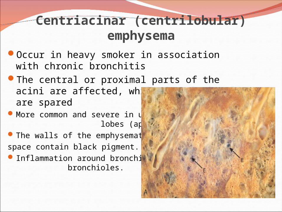
Centriacinar (centrilobular) emphysemaOccur in heavy smoker in association with
chronic bronchitisThe central or proximal parts of the acini are
affected, while distal alveoli are sparedMore common and severe in upper
lobes (apical segments)The walls of the emphysematous space contain black pigment. Inflammation around bronchi &
bronchioles.
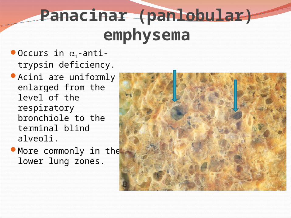
Panacinar (panlobular) emphysemaOccurs in 1-anti-
trypsin deficiency.Acini are uniformly
enlarged from the level of the respiratory bronchiole to the terminal blind alveoli.
More commonly in the lower lung zones.
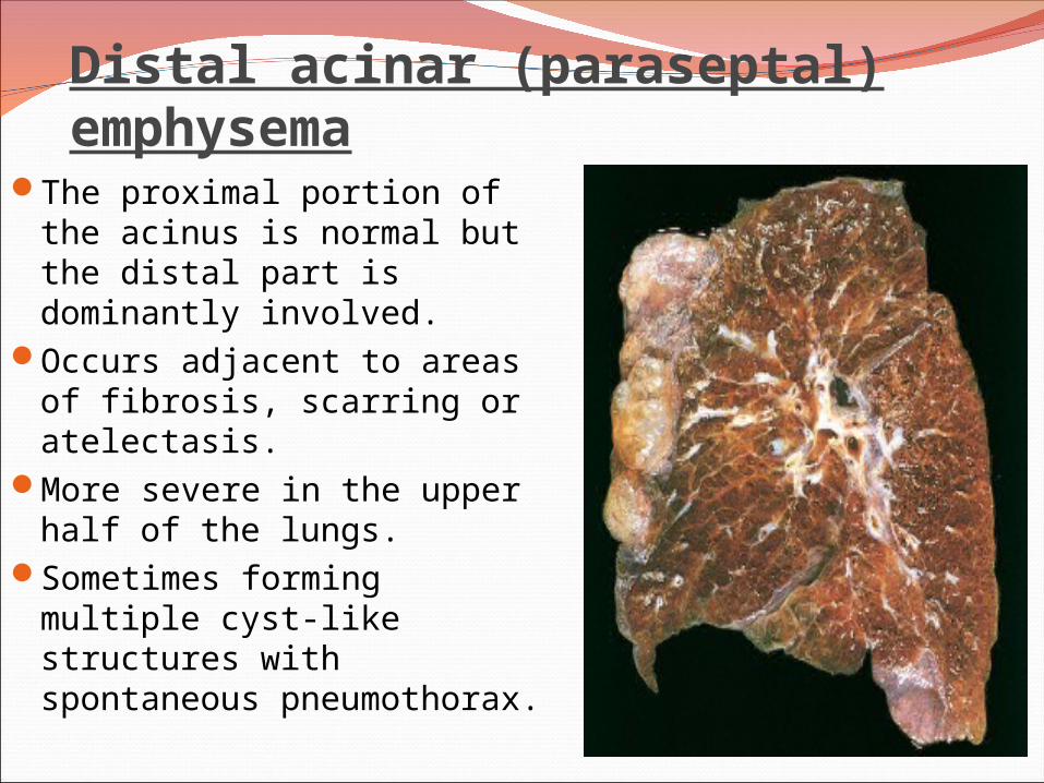
Distal acinar (paraseptal) emphysemaThe proximal portion of the
acinus is normal but the distal part is dominantly involved.
Occurs adjacent to areas of fibrosis, scarring or atelectasis.
More severe in the upper half of the lungs.
Sometimes forming multiple cyst-like structures with spontaneous pneumothorax.
Irregular Emphysema
The acinus is irregularly involved, associated with scarring.
Most common form found in autopsy.Asymptomatic.
Pathogenesis of Emphysema Is not completely understood.Alveolar wall destruction and airspace enlargement invokes
excess protease or elastase activity unopposed by appropriate antiprotease regulation (protease-antiprotease hypothesis)
2 key mechanisms: 1. excess cellular proteases with low antiprotease level 2. excess ROS from inflammation
Element of ch. Bronchitis coexists

Pathogenesis of EmphysemaProtease-antiprotease imbalance occur in 1% of emphysema1-antitrypsin, normally present in serum, tissue fluids and
macrophages, is a major inhibitor of proteases secreted by neutrophils during inflammation.
Encoded by codominantly expressed genes on the proteinase inhibitor (Pi) locus on chromosome 14.
Pi locus is extremely pleomorphic (M , Z)Any stimulus that increase neutrophil or macrophages in the
lung with release of protease lead to elastic tissue damage.
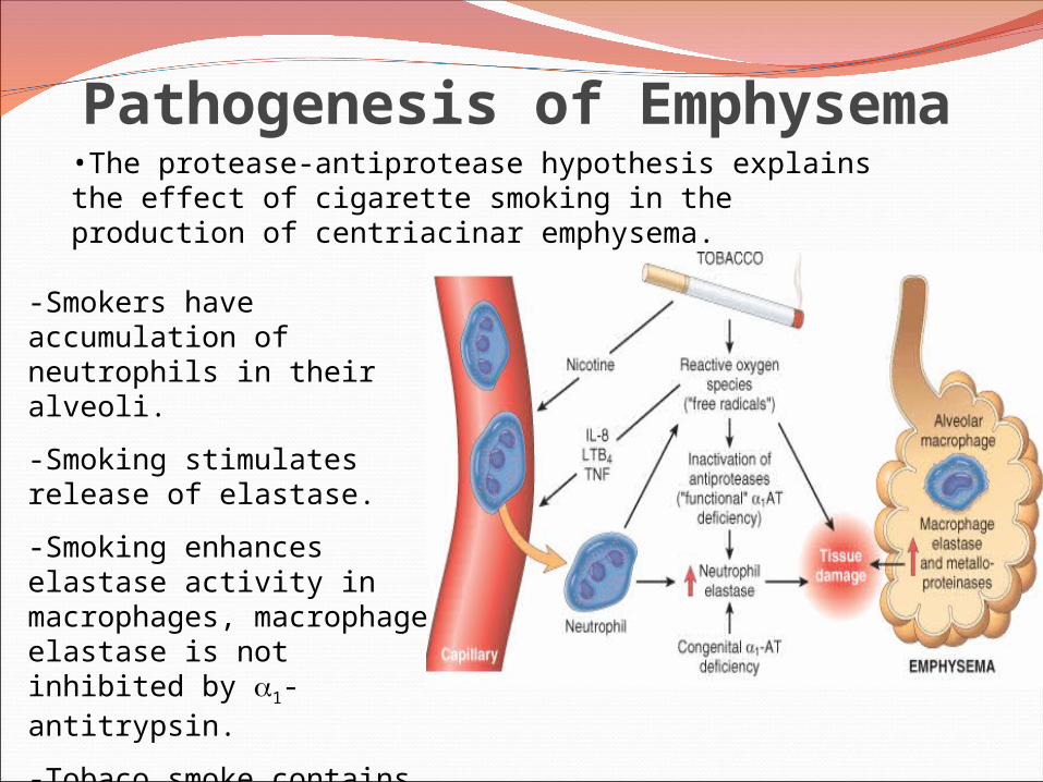
-Smokers have accumulation of neutrophils in their alveoli.
-Smoking stimulates release of elastase.
-Smoking enhances elastase activity in macrophages, macrophage elastase is not inhibited by 1-antitrypsin.
-Tobaco smoke contains reactive oxygen species with inactivation of proteases.
Pathogenesis of Emphysema•The protease-antiprotease hypothesis explains the effect of cigarette smoking in the production of centriacinar emphysema.

EmphysemaMorphologyThe diagnosis depend largely on the
macroscopic appearance of the lung.The lungs are pale, voluminous.Histologically, thinning and destruction of
alveolar walls creating large airspaces.
Loss of elastic tissue. Reduced radial traction on the small airways. Alveolar capillaries is diminished. Fibrosis of respiratory bronchioles. Accompanying bronchitis and bronchiolitis.

Emphysema: Clinical course
Cough and wheezing. Weight loss. Pulmonary function tests reveal reduced
FEV1.Death from emphysema is related to:1. Pulmonary failure with respiratory
acidosis, hypoxia and coma.2. Right-sided heart failure.

Chronic Obstructive Pulmonary
Disease
Emphysema
Bronchiectasis
Chronic BronchitisAsthma
Chronic Obstructive Pulmonary Disease
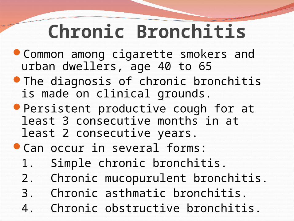
Chronic BronchitisCommon among cigarette smokers and
urban dwellers, age 40 to 65The diagnosis of chronic bronchitis is made
on clinical grounds.Persistent productive cough for at least 3
consecutive months in at least 2 consecutive years.
Can occur in several forms:1. Simple chronic bronchitis.2. Chronic mucopurulent bronchitis.3. Chronic asthmatic bronchitis.4. Chronic obstructive bronchitis.

Chronic bronchitisPathogenesisHypersecretion of mucus that starts in the large
airways.Causative factor are cigarette smoking and
pollutants.MorphologyEnlargement of the mucus-secreting glands,
increased number of goblet cells, loss of ciliated epithelial cells, squamous metaplasia, dysplastic changes and bronchogenic carcinoma.
Inflammation, fibrosis and resultant narrowing of bronchioles.
Coexistent emphysema.
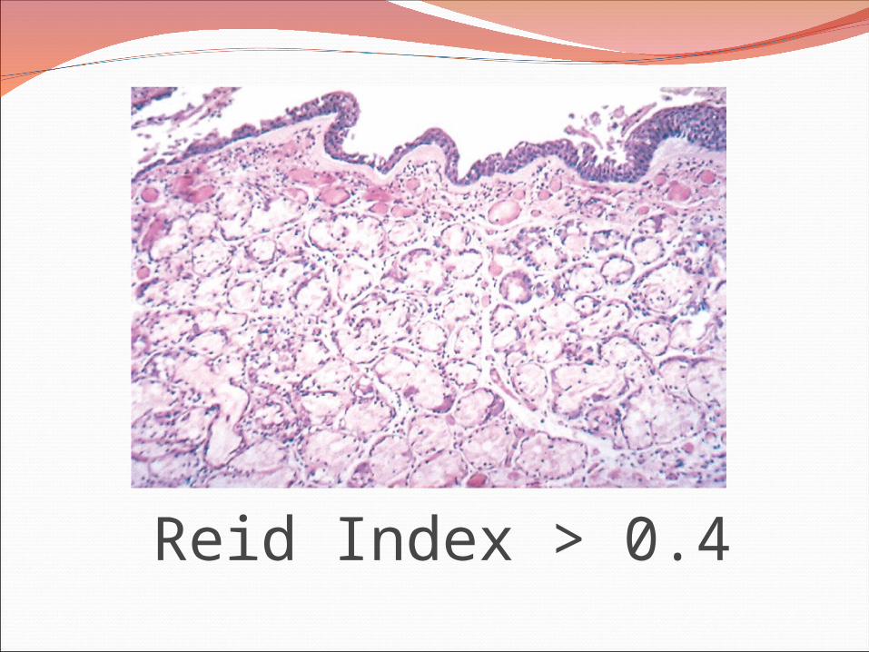
Reid Index > 0.4
Chronic bronchitis
Clinical CourseProminent cough and the production of sputum.COPD with hypercapnia, hypoxemia and cyanosis.Cardiac failure.
Chronic bronchitis vs. Emphysema

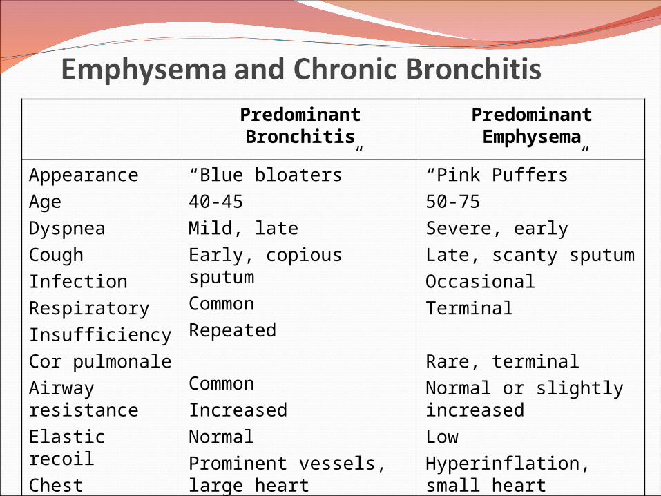
Predominant Bronchitis Predominant Emphysema
Appearance
Age
Dyspnea
Cough
Infection
Respiratory
Insufficiency
Cor pulmonale
Airway resistance
Elastic recoil
Chest radiography
“Blue bloaters”
40-45
Mild, late
Early, copious sputum
Common
Repeated
Common
Increased
Normal
Prominent vessels, large heart
“Pink Puffers”
50-75
Severe, early
Late, scanty sputum
Occasional
Terminal
Rare, terminal
Normal or slightly increased
Low
Hyperinflation, small heart

Chronic Obstructive Pulmonary
Disease
Emphysema
Bronchiectasis
Chronic BronchitisAsthma
Chronic Obstructive Pulmonary Disease
Chronic obstructive pulmonary diseasesBronchial asthma
Chronic relapsing inflammatory disorder characterized by hyperactive airways leading to episodic, reversible bronchoconstriction owing to increased responsiveness of the tracheobronchial tree to various stimuli.
It has been divided into two basic types:1. Extrinsic asthma.2. Intrinsic asthma.

Extrinsic AsthmaInitiated by type 1
hypersensivity reaction induced by exposure to extrinsic antigen.
Subtypes include:a. atopic (allergic) asthma.b. occupational asthma.c. allergic bronchopulmonary aspergillosis.
Develop early in life
Intrinsic Asthma
• Initiated by diverse, non-immune mechanisms, including ingestion of aspirin, pulmonary infections, cold, inhaled irritant, stress and exercise.• No personal or family history of allergic reaction.• Develop later in life
CLASSIFICATION OF ASTHMA

Extrinsic Asthma Atopic (allergic) asthma is the most
common form, begins in childhoodOther allergic manifestation: allergic
rhinitis, urticaria, eczema.Skin test with antigen result in an
immediate wheel and flare reaction Other family member is also affectedSerum IgE and eosinophil are increasedimmune related, TH2 subset of CD4+ T cells
Pathogenesis of Bronchial AsthmaEXAGGERATED BROCHOCONTRICTION Two components:
1. Chronic airway inflammation.2. Bronchial hyperresponsiveness.
The mechanisms have been best studied in atopic asthma.
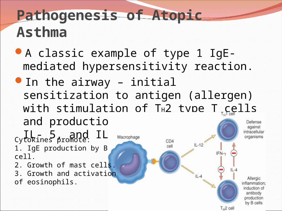
Pathogenesis of Atopic AsthmaA classic example of type 1 IgE-mediated
hypersensitivity reaction.In the airway – initial sensitization to
antigen (allergen) with stimulation of TH2 type T cells and production of cytokines (IL-4, IL- 5, and IL-13).
Cytokines promote:1. IgE production by B cell.2. Growth of mast cells.3. Growth and activation of eosinophils.
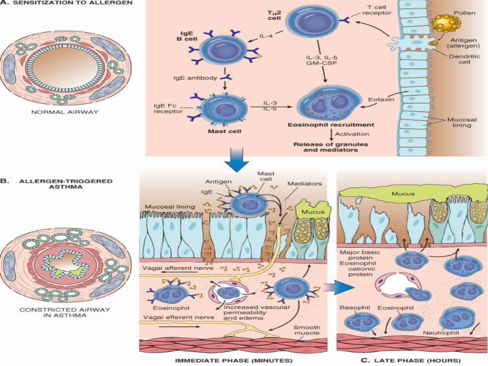

Pathogenesis of Atopic Asthma• IgE-mediated reaction to inhaled allergens
elicits: 1. acute response (within minutes) 2. a late phase reaction (after 4-8 hours)
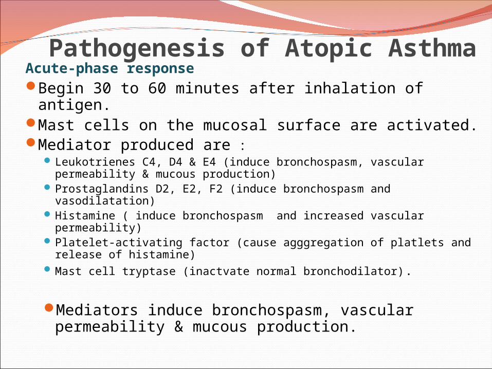
Pathogenesis of Atopic AsthmaAcute-phase response Begin 30 to 60 minutes after inhalation of antigen.Mast cells on the mucosal surface are activated.Mediator produced are :
Leukotrienes C4, D4 & E4 (induce bronchospasm, vascular permeability & mucous production)
Prostaglandins D2, E2, F2 (induce bronchospasm and vasodilatation) Histamine ( induce bronchospasm and increased vascular
permeability) Platelet-activating factor (cause agggregation of platlets and release
of histamine) Mast cell tryptase (inactvate normal bronchodilator).
Mediators induce bronchospasm, vascular permeability & mucous production.
Pathogenesis of Atopic Asthma Late phase reaction:
recruitment of leukocytes mediated by product of mast cells including:
1. Eosinophil and neutophil chemotactic factors
2 . IL-4 & IL-5 and induceTH2 subset ofCD4+ T cells3. Platelet-activating factor
4. Tumor necrosis factor.
Other cell types are involved: activated epithelial cells, macrophages and smooth muscle.
Pathogenesis of Atopic AsthmaLate phase reaction:The arrival of leukocytes at the site of mast cell
degranulation lead to: 1. Release of more mediators to activate more mast cells2. Cause epithelial cell damage .
Eosinophils produce major basic protein, eosinophilic cationic protein and eosinophil peroxidase ( toxic to epithelial cells).
These amplify and sustains injury without additional antigen.

Non-Atopic AsthmaTriggered by respiratory tract infection
including viruses and inhaled air pollutants e.g. sulfur dioxide, ozone.
Positive family history is uncommon.Serum IgE – normal.No other associated allergies.Skin test – negative.Hyperirritability of bronchial tree.Subtypes:
1. Drug-induced asthma. 2. Occupational asthma.
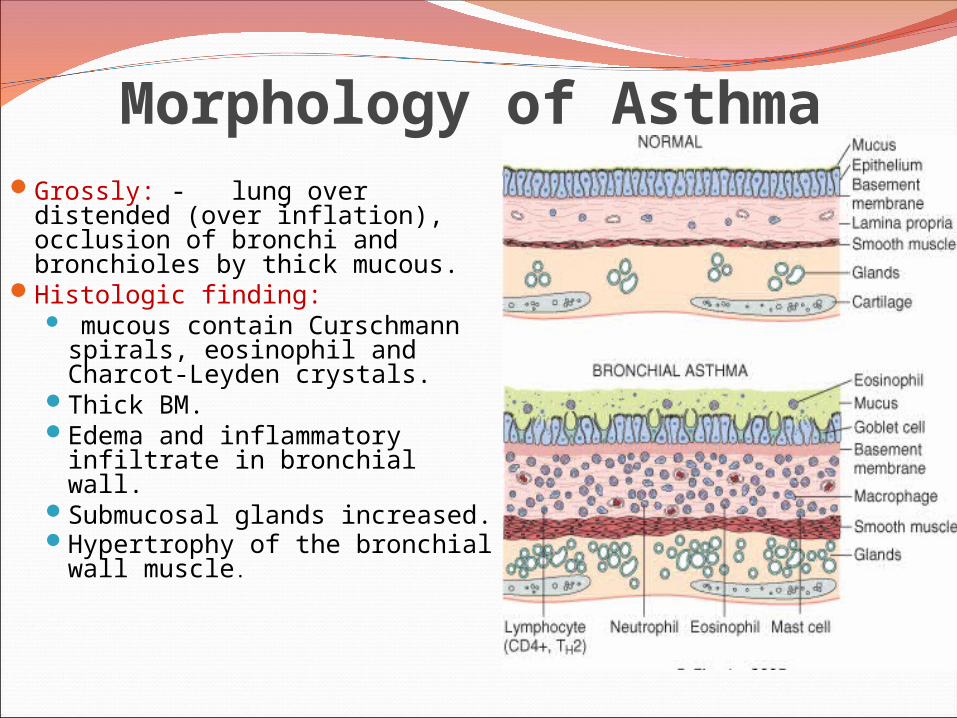
Morphology of AsthmaGrossly: - lung over distended
(over inflation), occlusion of bronchi and bronchioles by thick mucous.
Histologic finding: mucous contain Curschmann
spirals, eosinophil and Charcot-Leyden crystals.
Thick BM.Edema and inflammatory
infiltrate in bronchial wall.Submucosal glands increased.Hypertrophy of the bronchial
wall muscle.
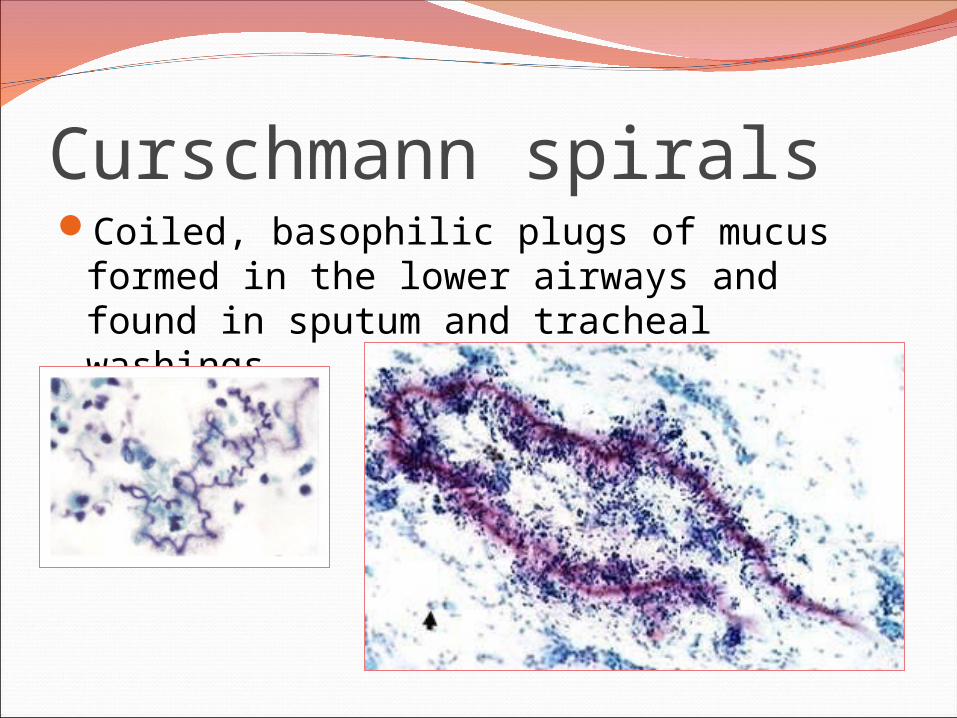
Curschmann spiralsCoiled, basophilic plugs of mucus formed
in the lower airways and found in sputum and tracheal washings

Charcot-Leyden crystals.Eosinophilic needle-shaped crystalline
structures.
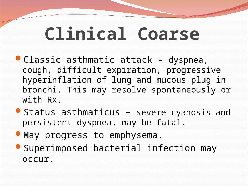
Clinical CoarseClassic asthmatic attack – dyspnea, cough,
difficult expiration, progressive hyperinflation of lung and mucous plug in bronchi. This may resolve spontaneously or with Rx.
Status asthmaticus – severe cyanosis and persistent dyspnea, may be fatal.
May progress to emphysema.Superimposed bacterial infection may occur.

Chronic Obstructive Pulmonary
Disease
Emphysema
Bronchiectasis
Chronic BronchitisAsthma
Chronic Obstructive Pulmonary Disease

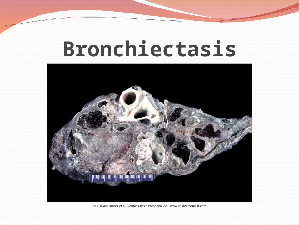
BronchiectasisChronic necrotizing infection of the bronchi
and bronchioles leading to or associated with abnormal dilatation of these airways.
Bronchial dilatation should be permanent.

Conditions associated with Bronchiectasis1. Bronchial obstruction
Localized:- tumor, foreign bodies or mucous impaction Generalized: - bronchial asthma- chronic bronchitis
2. Congenital or hereditary conditions:- Congenital bronchiactasis- Cystic fibrosis.- Intralobar sequestration of the lung.- Immunodeficiency status.- Immotile cilia and kartagner syndrome.
3. Necrotizing pneumonia.Caused by TB, staphylococci or mixed infection.
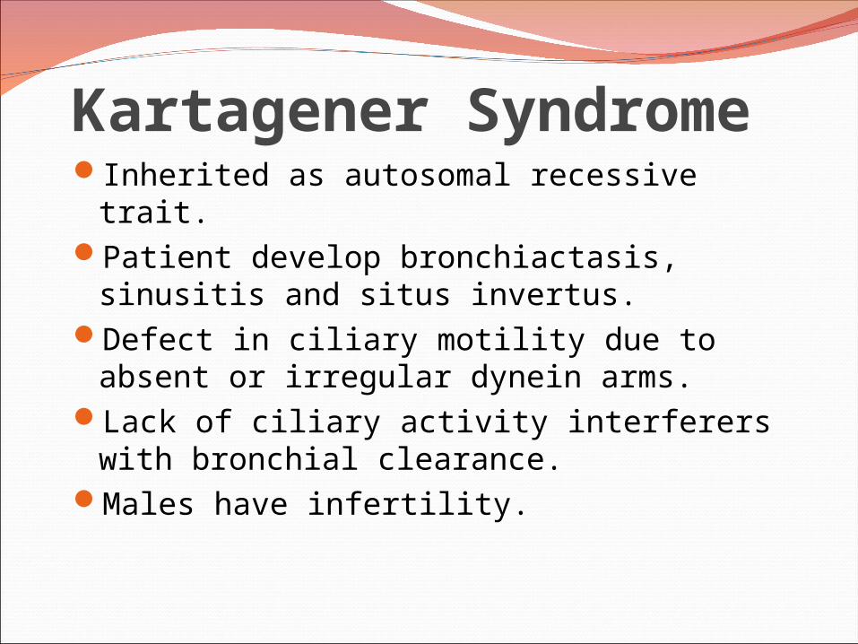
Kartagener SyndromeInherited as autosomal recessive trait.Patient develop bronchiactasis, sinusitis
and situs invertus.Defect in ciliary motility due to absent or
irregular dynein arms.Lack of ciliary activity interferers with
bronchial clearance.Males have infertility.

BronchiectasisEtiology and pathogenesisObstruction and infection.
Bronchial obstruction (athelectasis of airway distal to obstruction) – bronchial wall inflammation.
These changes become irreversible:1. If obstruction persist.2. If there is added infection.
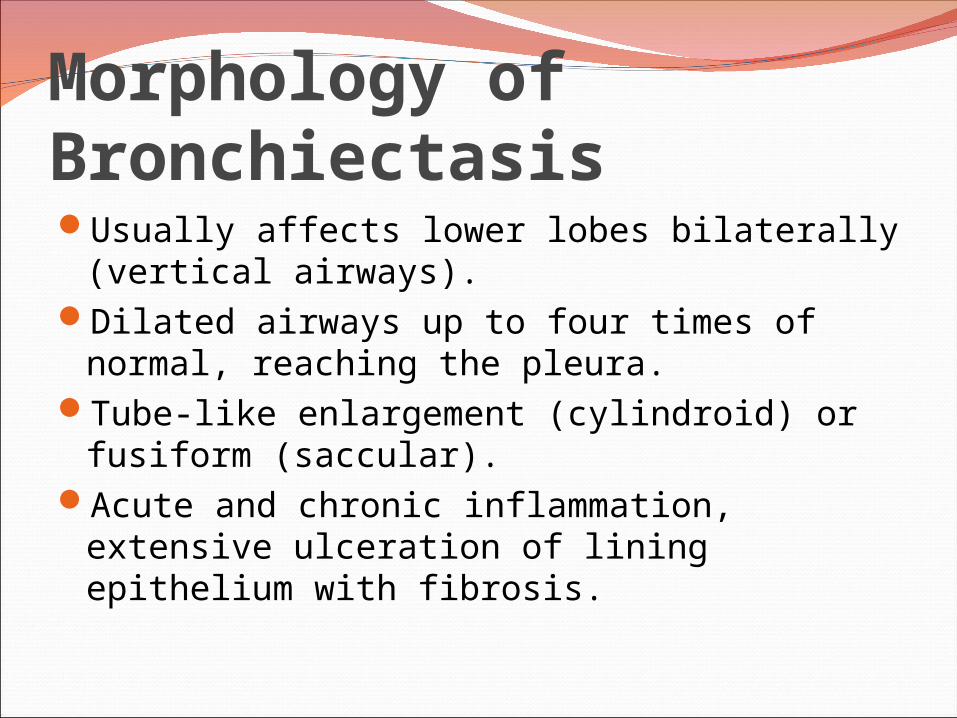
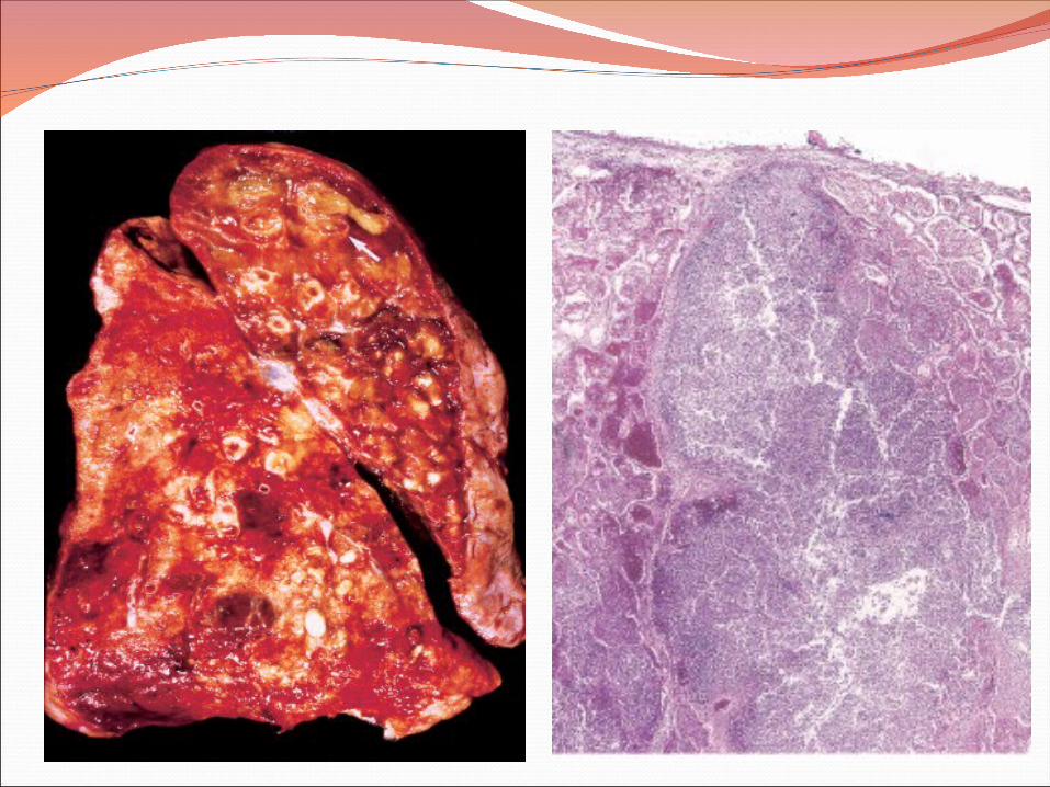
Morphology of BronchiectasisUsually affects lower lobes bilaterally
(vertical airways).Dilated airways up to four times of normal,
reaching the pleura.Tube-like enlargement (cylindroid) or
fusiform (saccular).Acute and chronic inflammation, extensive
ulceration of lining epithelium with fibrosis.
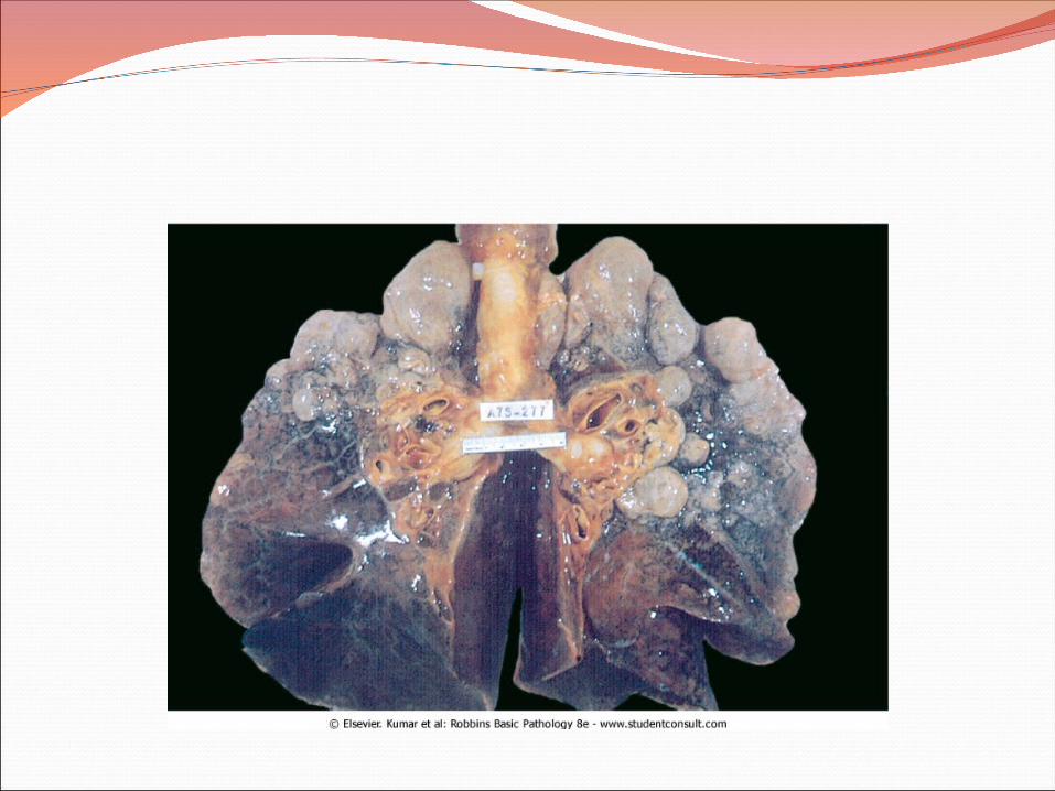
Bronchiectasis
BronchiectasisClinical course:
Sever persistent cough with sputum (mucopurulent, fetid sputum) sometime with with blood.
Clubbing of fingers.If sever, obstructive pulmonary function
develop.Rare complications: metastatic brain abscess
and amyloidosis.

Chronic Obstructive Pulmonary
Disease
Emphysema
Bronchiectasis Chronic
Bronchitis
Asthma
Summary: Athelectasis
Chronic Obstructive Pulmonary Disease
TypesPathogenesisPathologyClinical features
DefinitionCausesPathogenesisPathologyClinical Features
DefinitionCausesPathogenesisclassificationClinical Features
DefinitionCausesPathogenesisPathologyClinical Features
Emphysema: Dilated air spaces beyond respiratory arteriols

Chronic Bronchitis: Persistent productive cough for at least 3 consecutive months in at least 2 consecutive years, smoking related

Asthma: Dyspnea and wheezing
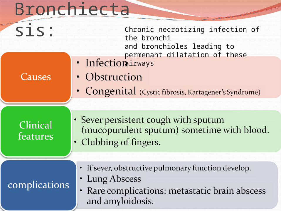
Bronchiectasis: Chronic necrotizing infection of the bronchi and bronchioles leading to permenant dilatation of these airways