Occupational exposure to carbon blacknanoparticles increases inflammatoryvascular disease risk: an implication of anex vivo biosensor assayJinglong Tang1, Wenting Cheng1, Jinling Gao1, Yanting Li1, Ruyong Yao2, Nathaniel Rothman3, Qing Lan3,Matthew J. Campen4, Yuxin Zheng1* and Shuguang Leng1,5,6*
Abstract
Background: Among manufactured or engineered nanoparticles, carbon black (CB) has largest productionworldwide and is also an occupational respiratory hazard commonly seen in rubber industry. Few studies haveassessed the risk for cardiovascular disease in carbon black exposed populations. An endothelial biosensor assaywas used to quantify the capacity of sera from 82 carbon black packers (CBP) and 106 non-CBPs to induceendothelial cell activation ex vivo. The mediation effect of circulatory proinflammatory factors on the associationbetween carbon black exposure and endothelial cell activation was assessed and further validated using in vitrointervention experiments.
Results: The average elemental carbon level inside carbon black bagging facilities was 657.0 μg/m3, which was 164-fold higher than that seen in reference areas (4.0 μg/m3). A global index was extracted from mRNA expression ofseven candidate biosensor genes using principal component analysis and used to quantify the magnitude ofendothelial cell activation. This global index was found to be significantly altered in CBPs compared to non-CBPs(P < 0.0001), however this difference did not vary by smoking status (P = 0.74). Individual gene analyses identifiedthat de novo expression of key adhesion molecules (e.g., ICAM and VCAM) and chemotactic factors (e.g., CCL2,CCL5, and CXCL8) responsible for the recruitment of leukocytes was dramatically induced in CBPs with CXCL8showing the highest fold of induction (relative quantification = 9.1, P < 0.0001). The combination of mediationanalyses and in vitro functional validation confirmed TNF-α, IL-1β, and IL-6 as important circulatory factorsmediating the effects of carbon black exposure on endothelial cell activation responses.
Conclusions: Inflammatory mediators in sera from CBPs may bridge carbon black exposure and endothelial cellactivation response assessed ex vivo. CBPs may have elevated risk for cardiovascular diseases when comorbidityexists. Our study may serve as a benchmark for understanding health effects of engineered carbon basednanoparticles with environmental and occupational health relevance.
Keywords: Carbon black nanoparticles, Biosensor, Endothelial cell activation, Mediation effect
* Correspondence: [email protected]; [email protected] of Occupational and Environmental Health, School of PublicHealth, Qingdao University, Qingdao 266021, ChinaFull list of author information is available at the end of the article
Tang et al. Particle and Fibre Toxicology (2020) 17:47 https://doi.org/10.1186/s12989-020-00378-8
IntroductionAmong manufactured or engineered carbon containingnanoparticles, carbon black (Chemical Abstracts ServiceRegistry No. 1333-86-4) has the highest productionworldwide and consists of pure elemental carbon [1–3].Carbon black has been recognized as an occupational re-spiratory hazard commonly seen in industries thatmanufacture it or apply it in the formulation of rubber,printing inks, and paints. Extensive epidemiological andanimal studies have established a causal link between ex-posure to air pollution of inhalable particulate mattersand morbidity and mortality of cardiovascular diseases[4–6]. Several studies have also found that carbon blackexposure may impair vasomotor function and accelerateplaque area development in the aorta of genetically ma-nipulated mice susceptible to the development of athero-sclerosis [7, 8]. However, although carbon black hasbeen used as a carbonaceous core analog of airborneparticulate matters in many in vitro and in vivo animalstudies [9, 10], evidence supporting its cardiovasculartoxicity in humans is still inconclusive [11].As one category of poorly soluble nanoparticles, the
main health concerns in humans associated with carbonblack exposure are lung effects resulting from inhalationexposure [12–15]. However, we found dramatically ele-vated multiple pro-inflammatory cytokines and chemo-kines in serum of carbon black packers (CBP) and in maleBALB/c mice exposed to carbon black aerosol [14]. More-over, our study identified elevated genomic instability inperipheral lymphocytes in CBPs that could be mediatedmostly by circulatory TNF-α [16], suggesting a systemicinflammation mediated mechanism for extra-pulmonaryeffects of carbon black inhalation exposure. Compellingevidence supports inflammation processes that pivotallyparticipate in all stages of atherosclerosis as a bridge link-ing classic risk factors to altered cellular behavior withinthe arterial wall. Many circulatory inflammatory markershave been identified in clinical studies to be associatedwith propensity for developing ischemic events and prog-nosis after acute coronary syndromes [17, 18]. However,although elevated inflammation was observed in the circu-lation in our cohort of CBPs, the young age of this popula-tion (average age 45.7 years) made it impossible to observeany incidence of cardiovascular diseases.Vascular disease is an inflammatory condition wherein
activated endothelial cells mediate the recruitment ofimmune cells into damaged areas of the vessel wall, pro-moting oxidative injury, pathological lesion growth, andhistological complexity [19]. Endothelial cell activation,often induced by cytokines such as CRP and TNF-α viathe nuclear factor-kappa beta pathway, leads to de novoexpression or presentation of key adhesion moleculesand chemotactic factors in cell surfaces for the recruit-ment of leukocytes [20]. Dr. Campen’s group has
developed a novel endothelial biosensor assay in whichthe capacity of a serum sample to induce endothelial cellactivation is quantified by assessing mRNA expression ofkey adhesion molecules and chemotactic factors in pri-mary human coronary artery endothelial cells (hCAEC)treated ex vivo with diluted serum samples from studysubjects. This method has been validated by showing agreater potential for inducing de novo expression ofendothelial cell adhesion molecules and chemokines inhCAECs treated with serum samples from patients withcoronary artery disease or obstructive sleep apnea [21,22]. Moreover, in a controlled diesel engine exhaust ex-posure study, serum from human subjects was found toexhibit greater inflammatory bioactivity on culturedendothelial cells compared to serum obtained followingsham exposures; while nitrogen dioxide exposure reca-pitulated the early inflammatory bioactivity, the inflam-matory effect of diesel engine exhaust was sustained forat least 24 h after exposure [23].In this study, we used the endothelial biosensor assay
to assess holistic inflammatory potential in serum sam-ples from 82 CBPs and 106 non-CBPs. We hypothesizedthat exposure to carbon black aerosol could increaseendothelial cell activation ex vivo through elevating cir-culatory levels of proinflammatory factors. The findingswould provide strong evidence supporting the effect ofcarbon black exposure on the risk of developing cardio-vascular diseases in carbon black exposed populationsand may serve as a benchmark for understanding healtheffects of engineered carbon based nanoparticles withenvironmental and occupational health relevance.
ResultsPhysicochemical characteristics of carbon black particlesPhysicochemical characteristics of carbon black particleshave been assessed extensively in reference [14]. A ther-mal decomposition of acetylene technique was used tomanufacture carbon black in the studied facility. Accord-ingly, carbon black product from this facility has a veryhigh carbon purity (> 99.8%). Under a transmission elec-tron microscope, the primary carbon black unit consistsof globular shaped particles with diameters ranging from30 to 50 nm. A scanning electron microscope identifiedformation of aggregates and agglomerates with hundredsto thousands of nanometers in at least one dimensionand forming an aciniform morphology [14]. Carbonblack has a surface area of 74.85 m2/g measured usingthe Brunauer-Emmett-Teller method. Carbon black sus-pension in water has a zeta potential of − 15.37 mV.
Characterization of study subjects and carbon blackaerosol in working environmentAll study subjects were male with Han Chinese ethnicity.The distribution of age, overweight or obesity, and
Tang et al. Particle and Fibre Toxicology (2020) 17:47 Page 2 of 11
smoking history was similar in CBP and non-CBP con-trol groups (all Ps > 0.065, Table 1). Because acetylenecarbon black is almost pure elemental carbon [14], thesummed level of urinary hydroxyl polycyclic aromatichydrocarbons was not different between CBPs and non-CBPs. The average level of particulate matter with diam-eters of 2.5 μm or less (PM2.5) in carbon black baggingareas was 800.0 μg/m3 with elemental carbon level at657.0 μg/m3 (Table 1) [14, 24], well within the recom-mended long term exposure limit of carbon black (3.5–4 mg/m3) in North America, European Union, andChina [25]. Size distribution analysis of carbon blackaerosol inside the bagging areas showed that up to 99.6%of the carbon black particles had aerodynamic diameterless than 2.5 μm with 96.7% of the particulates less than1.0 μm [14].
Effect of carbon black exposure on hCAEC activationAll biosensor genes except CCL5 and SELP had mRNAabundance substantially higher than the endogenouscontrol gene (TBP, Table 2). Principal component ana-lysis based on delta Ct of mRNA expression of 7 genesidentified a major principal component (i.e., biosensorPC1) that explained 41% of total variance and had posi-tive loading from delta Ct of all 7 genes with major load-ings (score > 0.3) from CCL2, CCL5, CXCL8, ICAM, andVCAM (Supplemental Table 1). Thus, we defined bio-sensor PC1 as a global score for hCAEC activation. Bio-sensor PC1 was significantly reduced in cultures treatedwith sera from CBPs versus non-CBPs (i.e., estimate = −0.92), indicating increased mRNA expression (i.e., rela-tive quantification = 1.89) of the 5 inflammatory genesmentioned above (Table 2). Findings from individual
gene association analyses substantiated results from theabove analyses with CXCL8 having the largest fold of in-crease in expression (relative quantification = 9.06, P <0.0001, Table 2). An interaction term between smokingstatus and carbon black exposure was included in thegeneralized linear model to assess whether the associ-ation between carbon black exposure and endothelialcell activation (i.e., biosensor PC1) varied by smokingstatus. The P value for the interaction term was 0.72(not shown), suggesting smoking status did not affectthe association between carbon black exposure andendothelial cell activation. Carbon black exposure his-tory was not associated with biosensor PC1 (P = 0.35,not shown).
Correlation between mRNA expression and proteinsecreted in culture medium for CXCL8Every 2-fold increase in mRNA expression of CXCL8was associated with 41.4 pg/ml (95%CI = 16.3–66.6 pg/ml, P = 0.0018, n = 52) increase in net CXCL8 proteinlevels in supernatants using a generalized linear modelwith adjustment for age, overweight or obesity, currentsmoking status, packyears, passage of cells, and batch ofassay. Net increase of CXCL8 protein level was also sig-nificantly (P < 0.0001) higher in cultures spiked withserum from CBPs (least square mean = 820.8 with95%CI as 723.5–918.1, n = 19) versus non-CBPs (leastsquare mean = 145.2 with 95%CI as 79.8–210.6, n = 33).
Circulatory inflammation mediating the effect of carbonblack exposure on hCAEC activationOur previous study identified that carbon black exposureincreased levels of multiple circulatory pro-inflammatory
Total RNA (μg, Mdn, Q1-Q3) 0.82 (0.47–1.62) 0.75 (0.44–1.69) 0.65c
CBP Carbon black packer, Q Quartile, M Mean, Mdn Median, PM Particulate matter, EC Elemental carbon, SD Standard deviationaStudent t testbChi square testc Wilcoxon Rank sum testdValues in current smokerseUrinary OH-PAHs were the sum of 1-hydroxynaphthalene, 2-hydroxynaphthalene, 2-hydroxyfluorene, 2-hydroxyphenanthrene, 9-hydroxyphenanthrene, and 1-hydroxypyrene in urine
Tang et al. Particle and Fibre Toxicology (2020) 17:47 Page 3 of 11
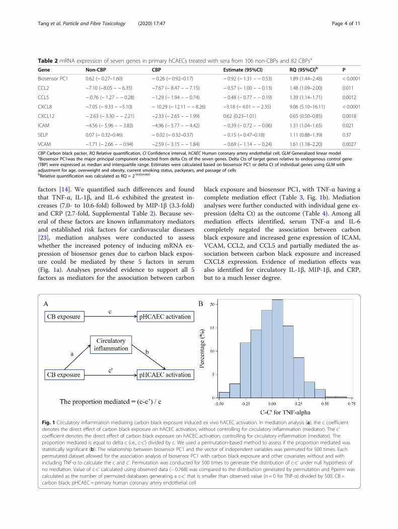
factors [14]. We quantified such differences and foundthat TNF-α, IL-1β, and IL-6 exhibited the greatest in-creases (7.0- to 10.6-fold) followed by MIP-1β (3.3-fold)and CRP (2.7-fold, Supplemental Table 2). Because sev-eral of these factors are known inflammatory mediatorsand established risk factors for cardiovascular diseases[23], mediation analyses were conducted to assesswhether the increased potency of inducing mRNA ex-pression of biosensor genes due to carbon black expos-ure could be mediated by these 5 factors in serum(Fig. 1a). Analyses provided evidence to support all 5factors as mediators for the association between carbon
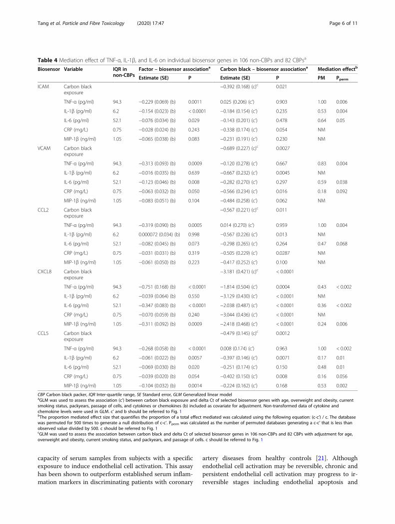
black exposure and biosensor PC1, with TNF-α having acomplete mediation effect (Table 3, Fig. 1b). Mediationanalyses were further conducted with individual gene ex-pression (delta Ct) as the outcome (Table 4). Among allmediation effects identified, serum TNF-α and IL-6completely negated the association between carbonblack exposure and increased gene expression of ICAM,VCAM, CCL2, and CCL5 and partially mediated the as-sociation between carbon black exposure and increasedCXCL8 expression. Evidence of mediation effects wasalso identified for circulatory IL-1β, MIP-1β, and CRP,but to a much lesser degree.
Table 2 mRNA expression of seven genes in primary hCAECs treated with sera from 106 non-CBPs and 82 CBPsa
CBP Carbon black packer, RQ Relative quantification, CI Confidence interval, hCAEC Human coronary artery endothelial cell, GLM Generalized linear modelaBiosensor PC1was the major principal component extracted from delta Cts of the seven genes. Delta Cts of target genes relative to endogenous control gene(TBP) were expressed as median and interquartile range. Estimates were calculated based on biosensor PC1 or delta Ct of individual genes using GLM withadjustment for age, overweight and obesity, current smoking status, packyears, and passage of cellsbRelative quantification was calculated as RQ = 2-(estimate)
Fig. 1 Circulatory inflammation mediating carbon black exposure induced ex vivo hACEC activation. In mediation analysis (a), the c coefficientdenotes the direct effect of carbon black exposure on hACEC activation, without controlling for circulatory inflammation (mediator). The c’coefficient denotes the direct effect of carbon black exposure on hACEC activation, controlling for circulatory inflammation (mediator). Theproportion mediated is equal to delta c (i.e., c-c’) divided by c. We used a permutation-based method to assess if the proportion mediated wasstatistically significant (b). The relationship between biosensor PC1 and the vector of independent variables was permuted for 500 times. Eachpermutated dataset allowed for the association analysis of biosensor PC1 with carbon black exposure and other covariates without and withincluding TNF-α to calculate the c and c’. Permutation was conducted for 500 times to generate the distribution of c-c’ under null hypothesis ofno mediation. Value of c-c’ calculated using observed data (− 0.768) was compared to the distribution generated by permutation and Pperm wascalculated as the number of permuted databases generating a c-c’ that is smaller than observed value (n = 0 for TNF-α) divided by 500. CB =carbon black; pHCAEC = primary human coronary artery endothelial cell
Tang et al. Particle and Fibre Toxicology (2020) 17:47 Page 4 of 11
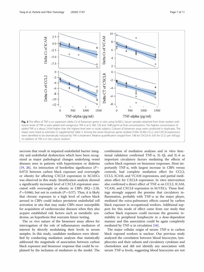
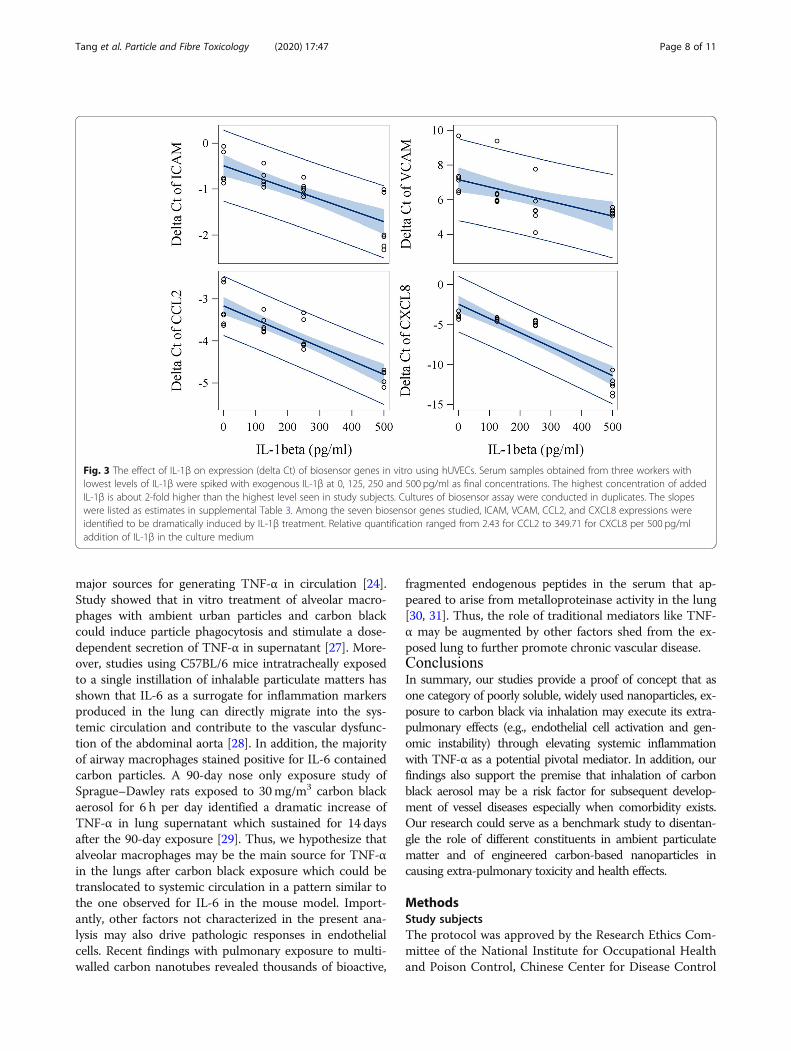
In vitro intervention studyWe further conducted an in vitro intervention experi-ment in which various concentrations of TNF-α, IL-1β,IL-6, MIP-1β and CRP were applied to endothelial cellsto assess the relationship between concentrations ofthese mediators and the magnitude of increased expres-sion of biosensor genes. Physiologically relevant concen-trations for these 5 mediators were selected based oncirculating levels seen in studied subjects. Addition ofIL-1β and TNF-α to subjects’ media with the lowestbackground level for tested factors significantly in-creased mRNA expression of ICAM, VCAM, CCL2, andCXCL8 in primary hUVECs (Figs. 2 and 3, SupplementalTable 3). We also found that the addition of IL-6 to themedium significantly increased mRNA expression ofCCL5 in hUVEC (Supplemental Figure 1, SupplementalTable 3). The potency of inducing CXCL8 gene expres-sion was significantly higher for IL-1β than TNF-αin vitro (P < 0.0001). In addition, although responses var-ied among the three subjects probably due to back-ground levels of other cofactors in serum, the slopes oflinear curves between levels of added cytokines or che-mokines and gene expression (delta Ct) were similaramong three serum samples from different individuals.Importantly, the induction of biosensor gene expressionby TNF-α (for ICAM, VCAM, CCL2, and CXCL8), IL-1β (for ICAM), and IL-6 (for CCL5) observed in in vitrostudies was highly consistent with the results of the me-diation analyses at the population level (Table 4). Specif-ically, the magnitude of associations of biosensor ICAMand CCL2 with TNF-α, and of biosensor CCL5 with IL-6 in the in vitro studies were almost identical to thatseen in the population study (Supplemental Table 3).
Inspection of dose-response curves (Figs. 2 and 3) identi-fied that the induction of CCL2 expression by theaddition of IL-1β or TNF-α showed the least variationacross sera from different subjects and higher proportionof variance explained by the model (R2 = 0.95 for IL-1βand R2 = 0.76 for TNF-α). Thus, based on the dose-response curves, the difference of CCL2 expression be-tween CBPs and non-CBPs (estimate = − 0.57, Table 2)was equivalent to the effect caused by an increase of177.5 pg/ml TNF-α or 222.9 pg/ml IL-1β in circulation.
DiscussionIn this study, the methodology of an established biosen-sor assay was further advanced by the creation of a glo-bal score quantifying the magnitude of endothelial cellactivation that was extracted from mRNA expression ofseven biosensor genes using principal component ana-lysis and was highly responsive to carbon black expos-ure. Our study provides strong evidence of ex vivoactivation of coronary artery endothelial cells caused byinflammation mediators in serum from CBPs as a resultof chronic inhalation exposure to high levels of carbonblack nanoparticles. Moreover, an in vitro interventionstudy estimated that the expression difference of the bio-sensor gene CCL2 between CBPs and non-CBPs wasequivalent to the effect caused by an increase of 177.5pg/ml for TNF-α or 222.9 pg/ml for IL-1β in circulation.Inflammatory activation of endothelial cells following
stress or injury is essential to both early vascular diseaseinitiation as well as progression of advanced plaques[19]. By assessing the expression of adhesion moleculesand chemotactic factors in hCAECs in vitro, this biosen-sor assay provided a holistic approach to quantify the
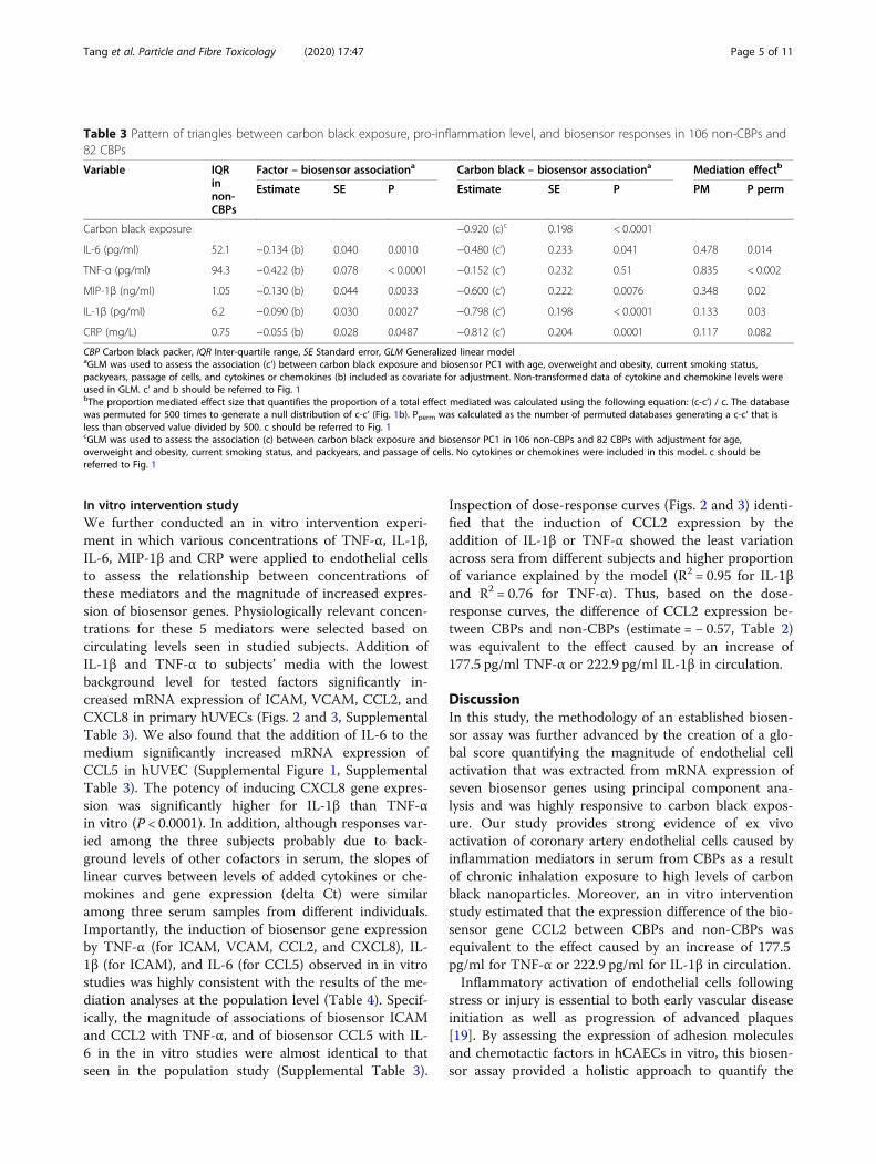
Table 3 Pattern of triangles between carbon black exposure, pro-inflammation level, and biosensor responses in 106 non-CBPs and82 CBPs
CBP Carbon black packer, IQR Inter-quartile range, SE Standard error, GLM Generalized linear modelaGLM was used to assess the association (c’) between carbon black exposure and biosensor PC1 with age, overweight and obesity, current smoking status,packyears, passage of cells, and cytokines or chemokines (b) included as covariate for adjustment. Non-transformed data of cytokine and chemokine levels wereused in GLM. c’ and b should be referred to Fig. 1bThe proportion mediated effect size that quantifies the proportion of a total effect mediated was calculated using the following equation: (c-c’) / c. The databasewas permuted for 500 times to generate a null distribution of c-c’ (Fig. 1b). Pperm was calculated as the number of permuted databases generating a c-c’ that isless than observed value divided by 500. c should be referred to Fig. 1cGLM was used to assess the association (c) between carbon black exposure and biosensor PC1 in 106 non-CBPs and 82 CBPs with adjustment for age,overweight and obesity, current smoking status, and packyears, and passage of cells. No cytokines or chemokines were included in this model. c should bereferred to Fig. 1
Tang et al. Particle and Fibre Toxicology (2020) 17:47 Page 5 of 11
capacity of serum samples from subjects with a specificexposure to induce endothelial cell activation. This assayhas been shown to outperform established serum inflam-mation markers in discriminating patients with coronary
artery diseases from healthy controls [21]. Althoughendothelial cell activation may be reversible, chronic andpersistent endothelial cell activation may progress to ir-reversible stages including endothelial apoptosis and
Table 4 Mediation effect of TNF-α, IL-1β, and IL-6 on individual biosensor genes in 106 non-CBPs and 82 CBPsa
CBP Carbon black packer, IQR Inter-quartile range, SE Standard error, GLM Generalized linear modelaGLM was used to assess the association (c’) between carbon black exposure and delta Ct of selected biosensor genes with age, overweight and obesity, currentsmoking status, packyears, passage of cells, and cytokines or chemokines (b) included as covariate for adjustment. Non-transformed data of cytokine andchemokine levels were used in GLM. c’ and b should be referred to Fig. 1bThe proportion mediated effect size that quantifies the proportion of a total effect mediated was calculated using the following equation: (c-c’) / c. The databasewas permuted for 500 times to generate a null distribution of c-c’. Pperm was calculated as the number of permuted databases generating a c-c’ that is less thanobserved value divided by 500. c should be referred to Fig. 1cGLM was used to assess the association between carbon black and delta Ct of selected biosensor genes in 106 non-CBPs and 82 CBPs with adjustment for age,overweight and obesity, current smoking status, and packyears, and passage of cells. c should be referred to Fig. 1
Tang et al. Particle and Fibre Toxicology (2020) 17:47 Page 6 of 11
necrosis that result in impaired endothelial barrier integ-rity and endothelial dysfunction which have been recog-nized as major pathological changes underlying vesseldiseases seen in patients with hypertension or diabetes[19, 26]. An interaction of borderline significance (P =0.072) between carbon black exposure and overweightor obesity for affecting CXCL8 expression in hCAECswas observed in this study. Stratification analysis showeda significantly increased level of CXCL8 expression asso-ciated with overweight or obesity in CBPs (RQ = 2.58,P = 0.046), but not in controls (P = 0.57). Thus, it is likelythat chronic exposure to a high level of carbon blackaerosol in CBPs could induce persistent endothelial cellactivation in situ that may make CBPs more susceptiblefor acquisition of cardiovascular diseases when they alsoacquire established risk factors such as metabolic syn-drome, an hypothesis that warrants future testing.The ex vivo nature of the biosensor assay allows for
interrogation of the role of inflammatory mediators ofinterest by directly modulating their levels in serumsamples. In this study, candidate mediators were identi-fied by conducting mediation analyses that statisticallyaddressed the magnitude of association between carbonblack exposure and biosensor response that could be ex-plained by the inclusion of mediators in the model. The
combination of mediation analyses and in vitro func-tional validation confirmed TNF-α, IL-1β, and IL-6 asimportant circulatory factors mediating the effects ofcarbon black exposure on biosensor responses. Most im-portantly TNF-α, with largest increase in CBPs versuscontrols, had complete mediation effect for CCL2,CCL5, ICAM, and VCAM expressions, and partial medi-ation effect for CXCL8 expression. In vitro interventionalso confirmed a direct effect of TNF-α on CCL2, ICAM,VCAM, and CXCL8 expression in hUVECs. These find-ings strongly support the premise that circulatory in-flammation, probably with TNF-α as the major player,mediated the extra-pulmonary effects caused by carbonblack exposure in occupational workers. Additional sup-port for this mode of effect came from our study thatcarbon black exposure could increase the genomic in-stability in peripheral lymphocyte in a dose-dependentmanner and this association could be again completelymediated by TNF-α in circulation [16].The major cellular origin of serum TNF-α in carbon
black exposed workers is unclear. Our previous studyanalyzed the correlation between blood leucocytes, lym-phocytes and their subsets and circulatory cytokines andchemokines and did not identify any association withserum TNF-α levels, suggesting blood leucocytes are not
Fig. 2 The effect of TNF-α on expression (delta Ct) of biosensor genes in vitro using hUVECs. Serum samples obtained from three workers withlowest levels of TNF-α were spiked with exogenous TNF-α at 0, 360, 720 and 1440 pg/ml as final concentrations. The highest concentration ofadded TNF-α is about 2-fold higher than the highest level seen in study subjects. Cultures of biosensor assay were conducted in duplicates. Theslopes were listed as estimates in supplemental Table 3. Among the seven biosensor genes studied, ICAM, VCAM, CCL2, and CXCL8 expressionswere identified to be dramatically induced by TNF-α treatment. Relative quantification ranged from 1.88 for CXCL8 to 3.05 for CCL2 per 500 pg/ml addition of TNF-α in the culture medium
Tang et al. Particle and Fibre Toxicology (2020) 17:47 Page 7 of 11
major sources for generating TNF-α in circulation [24].Study showed that in vitro treatment of alveolar macro-phages with ambient urban particles and carbon blackcould induce particle phagocytosis and stimulate a dose-dependent secretion of TNF-α in supernatant [27]. More-over, studies using C57BL/6 mice intratracheally exposedto a single instillation of inhalable particulate matters hasshown that IL-6 as a surrogate for inflammation markersproduced in the lung can directly migrate into the sys-temic circulation and contribute to the vascular dysfunc-tion of the abdominal aorta [28]. In addition, the majorityof airway macrophages stained positive for IL-6 containedcarbon particles. A 90-day nose only exposure study ofSprague–Dawley rats exposed to 30mg/m3 carbon blackaerosol for 6 h per day identified a dramatic increase ofTNF-α in lung supernatant which sustained for 14 daysafter the 90-day exposure [29]. Thus, we hypothesize thatalveolar macrophages may be the main source for TNF-αin the lungs after carbon black exposure which could betranslocated to systemic circulation in a pattern similar tothe one observed for IL-6 in the mouse model. Import-antly, other factors not characterized in the present ana-lysis may also drive pathologic responses in endothelialcells. Recent findings with pulmonary exposure to multi-walled carbon nanotubes revealed thousands of bioactive,
fragmented endogenous peptides in the serum that ap-peared to arise from metalloproteinase activity in the lung[30, 31]. Thus, the role of traditional mediators like TNF-α may be augmented by other factors shed from the ex-posed lung to further promote chronic vascular disease.ConclusionsIn summary, our studies provide a proof of concept that asone category of poorly soluble, widely used nanoparticles, ex-posure to carbon black via inhalation may execute its extra-pulmonary effects (e.g., endothelial cell activation and gen-omic instability) through elevating systemic inflammationwith TNF-α as a potential pivotal mediator. In addition, ourfindings also support the premise that inhalation of carbonblack aerosol may be a risk factor for subsequent develop-ment of vessel diseases especially when comorbidity exists.Our research could serve as a benchmark study to disentan-gle the role of different constituents in ambient particulatematter and of engineered carbon-based nanoparticles incausing extra-pulmonary toxicity and health effects.
MethodsStudy subjectsThe protocol was approved by the Research Ethics Com-mittee of the National Institute for Occupational Healthand Poison Control, Chinese Center for Disease Control
Fig. 3 The effect of IL-1β on expression (delta Ct) of biosensor genes in vitro using hUVECs. Serum samples obtained from three workers withlowest levels of IL-1β were spiked with exogenous IL-1β at 0, 125, 250 and 500 pg/ml as final concentrations. The highest concentration of addedIL-1β is about 2-fold higher than the highest level seen in study subjects. Cultures of biosensor assay were conducted in duplicates. The slopeswere listed as estimates in supplemental Table 3. Among the seven biosensor genes studied, ICAM, VCAM, CCL2, and CXCL8 expressions wereidentified to be dramatically induced by IL-1β treatment. Relative quantification ranged from 2.43 for CCL2 to 349.71 for CXCL8 per 500 pg/mladdition of IL-1β in the culture medium
Tang et al. Particle and Fibre Toxicology (2020) 17:47 Page 8 of 11
and Prevention. Design details including inclusion andexclusion criteria were published before [14, 15]. Briefly,the CBP study was established in 2012 by recruitingmale CBPs who have bagged newly manufactured car-bon black for more than 6 months and male non-CBPcontrols from a local water authority with no specific ex-posure to carbon black (Table 1). Written informed con-sent was acquired from all participants prior to theinterview and any procedures.
Exposure assessmentPhysicochemical characteristics of carbon black particles,ambient levels of carbon black aerosol (e.g., PM2.5,PM2.5 related elemental carbon, organic carbon, andtotal carbon) inside the bagging facilities and referenceareas, and particle size distributions of carbon blackaerosol inside the bagging facilities have been reportedin our previous studies [14–16, 24]. Urinary hydroxylmetabolites of six polycyclic aromatic hydrocarbons weredetermined using HPLC-MS/MS [32].
Cell culture and biosensor assayPrimary hCAECs (ATCC PCS-100-020) between pas-sages 4 and 6 were grown to confluence in 24-well platesin vascular cell basal media (ATCC PCS-100-030) sup-plemented with endothelial cell growth kit-BBE (ATCCPCS-100-040). Cells were serum-starved with basalmedia for 24 h prior to the incubation in basal mediumspiked with 10% serum obtained from study subjects for4 h at 37 °C. Each plate of cells was treated with a pro-portional number of non-CBPs and CBPs in a random-ized and blind fashion. mRNA expression levels of seventarget genes including CCL2 (Hs00234140_m1), CCL5(Hs00982282_m1), CXCL8 (Hs00174103_m1), CXCL12(Hs00171022_m1), ICAM (Hs00164932_m1), SELP(Hs00174583_m1), and VCAM (Hs01003372_m1), andone endogenous control gene (TBP, Hs00427620_m1)were measured using TaqMan assay (ThermoScientificApplied Biosystems). These seven genes were selectedbased on the key role of expressed proteins on the sur-face of endothelial cells upon activation for chemotaxisand adhesion of leukocytes [19]. Two serum pools werecreated by combining serum aliquots from 15 non-CBPsand 10 CBPs, respectively and were assayed with eachbatch of experiments as quality assessment samples for20 and 12 times, respectively. Coefficients of variationfor Cts of eight genes ranged from 2.8 to 11.1% with anaverage of 6.7%. In addition, Cts of TBP were not associ-ated with smoking status, packyears, and carbon blackexposure status, further supporting its appropriatenessas an endogenous control gene. The average RNA yieldfrom hCAEC cultures treated with sera from non-CBPsand CBPs was very similar (Table 1).
CXCL8 protein level in mediumSupernatants were collected for the first 52 samples (19CBPs and 33 non-CBPs) which provided sufficientpower for assessing the correlation between mRNA ex-pression and secreted protein of CXCL8. CXCL8 levelsin the supernatants were analyzed by CXCL8 human un-coated enzyme-linked immunosorbent assay Kits (Ther-moScientific Invitrogen). Net increase of CXCL8 level insupernatants was calculated by subtracting 10% serumlevel from supernatant level.
Circulatory inflammatory markersThree pro-inflammatory cytokines (i.e., IL-1β, IL-6, andTNF-α) and two chemokines (i.e., CXCL8 and MIP-1β)were measured in serum using cytometric bead array(BD Biosciences, USA) [14]. Serum C-reactive protein(CRP) was measured with an immunoturbidimetric assay(DiaSys, Germany) [14].
In vitro intervention studyBiologically verified primary human umbilical vein endo-thelial cells (hUVEC) were used to assess the effect of se-lected cytokines and chemokines on gene expression. Asimilar procedure for the biosensor assay as describedabove was followed except that hUVECs were serum-starved for 2 h due to elevated sensitivity. Serum samplesobtained from three workers with lowest levels of tar-geted cytokines were spiked with different concentra-tions of cytokines and chemokines for interference. Theinterference cytokines and chemokines were determinedby mediation analysis, and included CRP (0, 12.5, 25 and50 μg/mL; Sino Biological, 11,250-HNAH), TNF-α (0,360, 720 and 1440 pg/ml; Pepro Tech., 300-01A), IL-1β(0, 125, 250 and 500 pg/ml; Pepro Tech., 200-01B), IL-6(0, 430, 860 and 1720 pg/ml; Pepro Tech., 200–06), andMIP-1β (0, 1.25, 2.5 and 5 ng/ml, Sino Biological, 10,899-H08Y). The highest concentration of added cyto-kines is about 2-fold higher than the highest level seenin the serum of workers.
Statistical analysisFirst, generalized linear model (GLM) was used to assessthe association between carbon black exposure andhCAECs activation with adjustment for age, overweightand obesity, current smoking status, packyears, and pas-sage of cells. Principal component analysis was con-ducted based on delta Cts of seven biosensor genes toextract a major PC defining a global score quantifyingthe magnitude of hCAECs activation which was assessedin association with carbon black exposure to minimizethe effect of multiple comparisons. Individual gene asso-ciation with carbon black exposure was further assessedwith delta Ct as the outcome. Principal component ana-lysis identified two PCs with each explaining a
Tang et al. Particle and Fibre Toxicology (2020) 17:47 Page 9 of 11
proportion of total variance larger than average (1/7 =0.142, Supplemental Table 1). Thus, we applied two in-dependent analyses in calculating Bonferroni corrected Pvalues (0.05/2 = 0.025) for claiming a significant associ-ation in individual gene analyses. Second, the differenceof magnitude of the association between models with(c’) and without (c) adjustment for circulatory inflamma-tion (Fig. 1a) was calculated to quantify the mediationeffect of circulatory inflammatory factors on the associ-ation between carbon black exposure and hCAECs acti-vation. A permutation-based method was used to assesswhether the proportion mediated was statistically signifi-cant or not and was entailed in figure legend of Fig. 1b.Third, GLM was used to quantify the change of expres-sion (delta Ct) of biosensor genes by TNF-α, IL-1β, orIL-6 treatments in media in an in vitro interventionstudy with adjustment for serum ID. An interaction termbetween treatments and serum ID was included in theGLM to assess whether the slopes of dose-response weredifferent among the three individuals. Plots were createdusing SAS ODS Graphics Designer and R × 64 3.6.1. Allstatistical analyses were conducted using SAS (version9.4, NC, USA, site 70,239,492 and site 70,080,753).
Supplementary informationSupplementary information accompanies this paper at https://doi.org/10.1186/s12989-020-00378-8.
Additional file 1.
AcknowledgementsThe authors thank Ms. Shaobo Gao (Registered Nurse) from Department ofGynecology, The Affiliated Hospital of Qingdao University for providingnewborn cord for isolating primary human umbilical vein endothelial cells.The authors would like to thank Ms. Maria Picchi from Lovelace Biomedicalfor proof reading the manuscript.
Authors’ contributionsJT and SL conceived of and designed the study; JT, WC, JG, YL, and RYperformed the experiments and data collection; JT and SL conducted thedata analyses and tabulated the results; JT and SL interpreted the results anddrafted the manuscript; and JT, NR, QL, MJC, YZ, and SL critically edited themanuscript. All authors have read the manuscript and approved itssubmission.
Authors’ informationThe biosensor experiment was completed during Dr. Leng’s affiliation withQingdao University prior to 2020 through a collaboration with Dr. Campenfrom University of New Mexico. Finalized data analyses and critical revisionand submission of the manuscript were conducted during Dr. Leng’semployment at University of New Mexico since 2020.
FundingThis study was primarily supported by the National Natural ScienceFoundation of China (91643203, 81872600, and 91943301) and GuangdongProvincial Natural Science Foundation Team Project (2018B030312005). Dr.Tang received support from the National Natural Science Foundation ofChina (81872651). Finalizing data analysis and critical revision and submissionof the manuscript received Cancer Center Support Grant National CancerInstitute P30CA118100.
Availability of data and materialsDetailed assay protocols, SAS codes, and datasets used and/or analyzedduring the current study are available from the corresponding author onreasonable request.
Ethics approval and consent to participateWritten informed consent was acquired from all participants prior to theinterview and any procedures. The protocol was approved by the MedicalEthical Review Committee of the National Institute for Occupational Healthand Poison Control, Chinese Center for Disease Control and Prevention(protocol number: NIOHP201604).
Consent for publicationNot applicable.
Competing interestsThe authors declare that they have no competing interests.
Author details1Department of Occupational and Environmental Health, School of PublicHealth, Qingdao University, Qingdao 266021, China. 2Department of CentralLaboratory, Affiliated Hospital of Medical College of Qingdao University,Qingdao University, Qingdao 266021, China. 3Division of CancerEpidemiology and Genetics, National Cancer Institute, National Institutes ofHealth, Rockville, MD, USA. 4Department of Pharmaceutical Sciences, Collegeof Pharmacy, University of New Mexico, Albuquerque 87131, USA.5Department of Internal Medicine, School of Medicine, University of NewMexico, Albuquerque, NM 87131, USA. 6Cancer Control and PopulationSciences, University of New Mexico Comprehensive Cancer Center,Albuquerque, NM 87131, USA.
Received: 20 May 2020 Accepted: 4 September 2020
References1. Gray CA, Muranko H. Studies of robustness of industrial aciniform
aggregates and agglomerates--carbon black and amorphous silicas: areview amplified by new data. J Occup Environ Med. 2006;48(12):1279–90.https://doi.org/10.1097/01.jom.0000251477.40643.2a http://www.ncbi.nlm.nih.gov/pubmed/17159644.
2. Association ICB. Carbon black user’s guide, vol. 36; 2016.3. WHO guidelines on protecting workers from potential risks of manufactured
nanomaterials. https://apps.who.int/iris/bitstream/handle/10665/259671/9789241550048-eng.pdf;sequence=1 (2017). Accessed 23 Dec 2019.
4. Peng RD, Chang HH, Bell ML, McDermott A, Zeger SL, Samet JM, et al.Coarse particulate matter air pollution and hospital admissions forcardiovascular and respiratory diseases among Medicare patients. Jama.2008;299(18):2172–9. https://doi.org/10.1001/jama.299.18.2172 http://www.ncbi.nlm.nih.gov/pubmed/18477784.
5. Pelucchi C, Negri E, Gallus S, Boffetta P, Tramacere I, La Vecchia C. Long-term particulate matter exposure and mortality: a review of Europeanepidemiological studies. BMC Public Health. 2009;9:453. https://doi.org/10.1186/1471-2458-9-453 http://www.ncbi.nlm.nih.gov/pubmed/19995424.
6. Zanobetti A, Schwartz J. The effect of fine and coarse particulate airpollution on mortality: a national analysis. Environ Health Perspect. 2009;117(6):898–903. https://doi.org/10.1289/ehp.0800108 http://www.ncbi.nlm.nih.gov/pubmed/19590680.
7. Niwa Y, Hiura Y, Murayama T, Yokode M, Iwai N. Nano-sized carbon blackexposure exacerbates atherosclerosis in LDL-receptor knockout mice. Circ J.2007;71(7):1157–61. https://doi.org/10.1253/circj.71.1157 https://www.ncbi.nlm.nih.gov/pubmed/17587728.
8. Vesterdal LK, Folkmann JK, Jacobsen NR, Sheykhzade M, Wallin H, Loft S,et al. Pulmonary exposure to carbon black nanoparticles and vasculareffects. Part Fibre Toxicol. 2010;7:33. https://doi.org/10.1186/1743-8977-7-33https://www.ncbi.nlm.nih.gov/pubmed/21054825.
9. Swafford DS, Nikula KJ, Mitchell CE, Belinsky SA. Low frequency ofalterations in p53, K-ras, and mdm2 in rat lung neoplasms induced by dieselexhaust or carbon black. Carcinogenesis. 1995;16(5):1215–21. https://doi.org/10.1093/carcin/16.5.1215 http://www.ncbi.nlm.nih.gov/pubmed/7539340.
10. Long CM, Nascarella MA, Valberg PA. Carbon black vs. black carbon andother airborne materials containing elemental carbon: physical and
Tang et al. Particle and Fibre Toxicology (2020) 17:47 Page 10 of 11
chemical distinctions. Environ Pollut. 2013;181:271–86. https://doi.org/10.1016/j.envpol.2013.06.009 http://www.ncbi.nlm.nih.gov/pubmed/23850403.
11. Morfeld P, Mundt KA, Dell LD, Sorahan T, McCunney RJ. Meta-analysis ofcardiac mortality in three cohorts of carbon black production workers. Int JEnviron Res Public Health. 2016;13:3. https://doi.org/10.3390/ijerph13030302https://www.ncbi.nlm.nih.gov/pubmed/27005647.
12. Harber P, Muranko H, Solis S, Torossian A, Merz B. Effect of carbon blackexposure on respiratory function and symptoms. J Occup Environ Med.2003;45(2):144–55 http://www.ncbi.nlm.nih.gov/pubmed/12625230.
13. Gardiner K, van Tongeren M, Harrington M. Respiratory health effects fromexposure to carbon black: results of the phase 2 and 3 cross sectionalstudies in the European carbon black manufacturing industry. OccupEnviron Med. 2001;58(8):496–503. https://doi.org/10.1136/oem.58.8.496http://www.ncbi.nlm.nih.gov/pubmed/11452043.
14. Zhang R, Dai Y, Zhang X, Niu Y, Meng T, Li Y, et al. Reduced pulmonaryfunction and increased pro-inflammatory cytokines in nanoscale carbonblack-exposed workers. Part Fibre Toxicol. 2014;11:73. https://doi.org/10.1186/s12989-014-0073-1 https://www.ncbi.nlm.nih.gov/pubmed/25497989.
15. Yang M, Li Y, Meng T, Zhang L, Niu Y, Dai Y, et al. Ultrafine CB-inducedsmall airway obstruction in CB-exposed workers and mice. Sci Total Environ.2019;671:866–73. https://doi.org/10.1016/j.scitotenv.2019.03.033 https://www.ncbi.nlm.nih.gov/pubmed/30947057.
16. Cheng W, Liu Y, Tang J, Duan H, Wei X, Zhang X, et al. Carbon content inairway macrophages and genomic instability in Chinese carbon blackpackers. Arch Toxicol. 2020;94(3):761–71. https://doi.org/10.1007/s00204-020-02678-6 https://www.ncbi.nlm.nih.gov/pubmed/32076763.
17. Packard RR, Libby P. Inflammation in atherosclerosis: from vascular biologyto biomarker discovery and risk prediction. Clin Chem. 2008;54(1):24–38.https://doi.org/10.1373/clinchem.2007.097360 http://www.ncbi.nlm.nih.gov/pubmed/18160725.
18. Libby P. Inflammation in atherosclerosis. Nature. 2002;420(6917):868–74.https://doi.org/10.1038/nature01323 http://www.ncbi.nlm.nih.gov/pubmed/12490960.
19. Zhang J, Defelice AF, Hanig JP, Colatsky T. Biomarkers of endothelial cellactivation serve as potential surrogate markers for drug-induced vascularinjury. Toxicol Pathol. 2010;38(6):856–71. https://doi.org/10.1177/0192623310378866 http://www.ncbi.nlm.nih.gov/pubmed/20716788.
20. Collins T, Read MA, Neish AS, Whitley MZ, Thanos D, Maniatis T.Transcriptional regulation of endothelial cell adhesion molecules: NF-kappaB and cytokine-inducible enhancers. FASEB J. 1995;9(10):899–909 http://www.ncbi.nlm.nih.gov/pubmed/7542214.
21. Cung H, Aragon MJ, Zychowski K, Anderson JR, Nawarskas J, Roldan C, et al.Characterization of a novel endothelial biosensor assay reveals increasedcumulative serum inflammatory potential in stabilized coronary arterydisease patients. J Transl Med. 2015;13:99. https://doi.org/10.1186/s12967-015-0457-5 http://www.ncbi.nlm.nih.gov/pubmed/25890092.
22. Zychowski KE, Sanchez B, Pedrosa RP, Lorenzi-Filho G, Drager LF, PolotskyVY, et al. Serum from obstructive sleep apnea patients inducesinflammatory responses in coronary artery endothelial cells. Atherosclerosis.2016;254:59–66. https://doi.org/10.1016/j.atherosclerosis.2016.09.017 http://www.ncbi.nlm.nih.gov/pubmed/27693879.
23. Channell MM, Paffett ML, Devlin RB, Madden MC, Campen MJ. Circulatingfactors induce coronary endothelial cell activation following exposure toinhaled diesel exhaust and nitrogen dioxide in humans: evidence from a noveltranslational in vitro model. Toxicol Sci. 2012;127(1):179–86. https://doi.org/10.1093/toxsci/kfs084 https://www.ncbi.nlm.nih.gov/pubmed/22331494.
24. Dai Y, Niu Y, Duan H, Bassig BA, Ye M, Zhang X, et al. Effects of occupationalexposure to carbon black on peripheral white blood cell counts andlymphocyte subsets. Environ Mol Mutagen. 2016;57(8):615–22. https://doi.org/10.1002/em.22036 https://www.ncbi.nlm.nih.gov/pubmed/27671983.
25. Liang Y-x, Su Z, Wu WA, Lu BQ, Fu WZ, Yang L, et al. New trends in thedevelopment of occupational exposure limits for airborne chemicals inChina. Regul Toxicol Pharmacol. 2003;38(2):112–23. https://doi.org/10.1016/s0273-2300(03)00077-1.
26. Flammer AJ, Anderson T, Celermajer DS, Creager MA, Deanfield J, Ganz P,et al. The assessment of endothelial function: from research into clinicalpractice. Circulation. 2012;126(6):753–67. https://doi.org/10.1161/CIRCULATIONAHA.112.093245 http://www.ncbi.nlm.nih.gov/pubmed/22869857.
27. van Eeden SF, Tan WC, Suwa T, Mukae H, Terashima T, Fujii T, et al.Cytokines involved in the systemic inflammatory response induced by
exposure to particulate matter air pollutants (PM(10)). Am J Respir Crit CareMed. 2001;164(5):826–30. https://doi.org/10.1164/ajrccm.164.5.2010160http://www.ncbi.nlm.nih.gov/pubmed/11549540.
28. Kido T, Tamagawa E, Bai N, Suda K, Yang HH, Li Y, et al. Particulate matterinduces translocation of IL-6 from the lung to the systemic circulation. Am JRespir Cell Mol Biol. 2011;44(2):197–204. https://doi.org/10.1165/rcmb.2009-0427OC http://www.ncbi.nlm.nih.gov/pubmed/20378751.
29. Chu C, Zhou L, Xie H, Pei Z, Zhang M, Wu M, et al. Pulmonary toxicitiesfrom a 90-day chronic inhalation study with carbon black nanoparticles inrats related to the systemical immune effects. Int J Nanomedicine. 2019;14:2995–3013. https://doi.org/10.2147/IJN.S198376 https://www.ncbi.nlm.nih.gov/pubmed/31118618.
30. Aragon MJ, Topper L, Tyler CR, Sanchez B, Zychowski K, Young T, et al.Serum-borne bioactivity caused by pulmonary multiwalled carbonnanotubes induces neuroinflammation via blood-brain barrier impairment.Proc Natl Acad Sci U S A. 2017;114(10):E1968–E76. https://doi.org/10.1073/pnas.1616070114 https://www.ncbi.nlm.nih.gov/pubmed/28223486.
31. Mostovenko E, Young T, Muldoon PP, Bishop L, Canal CG, Vucetic A, et al.Nanoparticle exposure driven circulating bioactive peptidome causessystemic inflammation and vascular dysfunction. Part Fibre Toxicol. 2019;16(1):20. https://doi.org/10.1186/s12989-019-0304-6 https://www.ncbi.nlm.nih.gov/pubmed/31142334.
32. Zhang X, Duan H, Gao F, Li Y, Huang C, Niu Y, et al. Increased micronucleus,nucleoplasmic bridge, and nuclear bud frequencies in the peripheral bloodlymphocytes of diesel engine exhaust-exposed workers. Toxicol Sci. 2015;143(2):408–17. https://doi.org/10.1093/toxsci/kfu239 https://www.ncbi.nlm.nih.gov/pubmed/25370840.
Publisher’s NoteSpringer Nature remains neutral with regard to jurisdictional claims inpublished maps and institutional affiliations.
Tang et al. Particle and Fibre Toxicology (2020) 17:47 Page 11 of 11