37
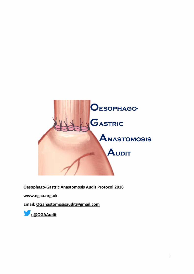
1 Oesophago-Gastric Anastomosis Audit Protocol 2018 www.ogaa.org.uk Email: [email protected] : @OGAAudit
1
Oesophago-Gastric Anastomosis Audit Protocol 2018
www.ogaa.org.uk
Email: [email protected]
: @OGAAudit
OGAA Protocol 2018
Steering Group
Name Organisation Email
Mr Richard Evans General Surgery Trainee – West Midlands Deanery
Mr James Bundred University of Birmingham [email protected]
Dr Dmitri Nepogodiev University of Birmingham [email protected]
Mr Pritam Singh General Surgery Trainee – West Midlands Deanery
Ms Siobhan McKay General Surgery Trainee – West Midlands Deanery
Mr Kasun Wanigsooriya General Surgery Trainee – West Midlands Deanery
Dr Tony Whitehouse University Hospitals Birmingham NHS Foundation Trust
Mr James Gossage Guy’s and St. Thomas’ NHS Foundation Trust
Prof Richard van Hillegersberg
UMC Utrecht [email protected]
Mr Ravinder Vohra Nottingham University Hospitals NHS Trust
Mr Ewen Griffiths University Hospitals Birmingham NHS Foundation Trust
OGAA Protocol 2018
Supporting organisations
West Midlands Surgical Research Collaborative
Providing REDCap Access
Academic Department of Surgery
Association of Laparoscopic Surgeons GB&I

OGAA Protocol 2018
Lay Summary
Oesophageal cancer is the sixth leading cause of cancer related death affecting up to 450,000 people
globally each year. The main surgical treatment for oesophageal cancer is an oesophagectomy - an
operation to remove part of the oesophagus and stomach followed by a join between the remaining
oesophagus and stomach. The techniques involved to create this join vary and can involve various
stitching methods and stapling devices. A proportion of these joins will breakdown and this results in
the patients becoming very unwell with a resulting increase in the risk of death. The strategies to
manage this complication again vary and include:
No surgical intervention
An endoscopic intervention or
A further surgical procedure.
This international audit will look at the rates of breakdown of these joins, commonly termed a ‘leak’,
how they are managed and the effect on the patient outcomes. The information collected from this
audit will help to develop recommendations on how to prevent and manage this serious complication.
OGAA Protocol 2018
Abstract
Background
Oesophageal cancer is the sixth leading cause of cancer related mortality affecting up to 450,000
people globally each year. The incidence continues to increase rapidly and despite advances in modern
treatment 5-year survival remains at around 15 to 20%. Oesophagectomy is a mainstay in curative
treatment for those with oesophageal cancer however the technique and outcome varies greatly.
Aim
The aim is to audit current oesophagectomy outcomes against the standards identified in current
literature.
Audit Standard
1- Anastomotic leak rate should be less than 10%
2- Major post-operative morbidity should be less than 20%
3- 30 day mortality rate should be less than 5% and 90 day mortality rate should be less than
10%
Primary Audit Objectives
1- Assess the variation in anastomotic leak rates
2- Assess which anastomotic technique is associated with optimal patient outcome
3- When anastomotic leak occurs what treatment options are associated with improved clinical
outcomes
OGAA Protocol 2018
Endpoints
A staged data collection protocol will identify patient demographics, operative and peri-operative
details and outcome markers. Key outcome measures will include post-operative mortality, morbidity
including grade of leak and length of stay. Management techniques used for anastomotic leaks will
also be assessed (e.g. conservative management, oesophageal stent, endo-luminal VAC therapy and
re-operation).
Methods
A nine month multicentre prospective audit will be performed globally starting in April 2018 and co-
ordinated by University Hospitals Birmingham. This will include patients undergoing oesophagectomy
over 6 months and encompassing a 90-day follow up period. A pilot data collection period will occur
at University Hospitals Birmingham and 3 other UK hospitals in 2017. Sites will be required to pre-
register for the audit and obtain local study approval prior to commencement of the study.
During the study sites will be required to record data contemporaneously via a dedicated encrypted
server through the Research Electronic Data Capture (REDCap) web application secure online
database. The REDCap database will provide a standardised data collection proforma assessing key
information to answer the primary audit question. The report of the audit will be prepared in
accordance with the guidelines as set by the STROBE (strengthening the reporting of observational
studies in epidemiology) statement for observational studies. All unit results will be anonymised to all
but the auditors and the specific unit. Unit results will not be shared to other units or the collaborators
as a whole. The study will be defined as audit not research in concordance with NHS Health research
authority (Appendix 2).
OGAA Protocol 2018
Discussion
This multicentre international audit will be collected by both surgeons and trainees alike to provide
greater insight into the complexities of oesophagectomy and outcome. This observational study may
highlight trends in improved survival associated with specific operative techniques which can be
further assessed and analysed through research to improve outcomes in oesophageal cancer.
OGAA Protocol 2018
Introduction
The techniques used for oesophagectomy can vary greatly amongst countries, units and surgeons. This
is also true for outcomes and historically oesophagectomy has been associated with significant
morbidity and mortality. Achieving a “textbook outcome” for patients undergoing oesophagectomy is
exceptionally challenging. The Dutch Upper Gastrointestinal Cancer Audit (DUCA) group found that
despite tailored oesophageal cancer care only 29.7% of patients undergoing oesophagectomy would
achieve a “textbook outcome” (25). In the UK 90-day mortality for oesophagectomy has improved
markedly from 5.7% in 2007-09 to 3.2% in 2013-15 and through further prospective analysis we seek
to identify current mortality rates and trends in oesophagectomy technique that could be further
analysed to potentially improve outcome (2). Leak rates post oesophagectomy are currently in the
region of 10% and are a significant contributor to morbidity and mortality (3, 10, 12-19). 30 day
mortality in patients with a demonstrable leak is around 17-35% whereas the 30 day mortality of
patients with an intact anastomosis is 2-3% (4, 5). Operative and anastamotic technique has long been
evaluated as a potential mechanism by which to minimise leak and improve patient outcomes.
Numerous studies have advocated varying techniques comparing handsewn and mechanical options
for anastomoses (6, 7). There is some evidence to show that a mechanical anastomosis using a linear
stapler has a reduced leak rate and reduced stricture rate as compared to a handsewn anastomosis
however results vary markedly between surgeons and units (8). Site of anastomosis much like
anastomotic technique is also a key factor in leak rate. There is evidence to suggest that cervical
anastomoses are associated with an increased leak rate as compared to thoracic anastomoses (9, 10).
Management of leaks much like anastamotic techniques is a continued area of controversy with a very
varied spectrum of practice. Early identification of an anastamotic leak can potentially speed clinical
intervention and improve patient outcome. With the advent of newer conservative techniques such
OGAA Protocol 2018
as endo-luminal vacuum and some evidence not advocating stent usage, it will be important to identify
potential trends in leak management that may improve patient outcome (11, 12).
An international multicentre audit will enable a larger number of patients’ data to be obtained over a
given time period. It will potentially obtain a greater overview of the variances in practice across units
and countries. While such an audit will not provide true evidence of efficacy or the impact of a specific
variable, it will provide data to narrow the spectrum of variables we can investigate to improve
outcomes post oesophagectomy.
Access and anastomosis have been continued areas of disagreement amongst oesophago-gastric
surgeons and their influence on mortality and morbidity has long been disputed. This audit seeks to
provide up to date information in the international variances in practice.
For example:
1- Methods of access:
a. Two-stage (Ivor Lewis)
b. Three-stage (McKeown)
c. Trans hiatal
d. Thoraco-abdominal
2- Incision
a. Open
b. Minimally invasive oesophagectomy
c. Hybrid
3- Technique of anastomosis
a. Stapled
i. Circular
ii. Linear
b. Sutured

OGAA Protocol 2018
4- Patient factors
a. Haematological
b. Biochemical
c. Co-morbidity
5- Volume
a. Surgeon
b. Institutional
OGAA Protocol 2018
Aim
Primary Audit Question
1- Assess the variation in anastomotic leak rates
2- Assess which anastomotic technique is associated with optimal patient outcome
3- When anastomotic leak occurs what treatment options are associated with improved clinical outcomes
Audit Standard
1- Anastomotic leak rate should be less than 10%
2- Major post-operative morbidity should be less than 20%
3- 30 day mortality rate should be less than 5% and 90 day mortality rate should be less than
10%
Leak rates are very variable between surgeons, units and countries however current practices
demonstrate a leak of from 1.8-18.2% (3, 10, 12-19). This accounts for all operative and anastomotic
techniques to set a standard by which we can compare the observed standard in the collected
population. The largest of the recent studies by Kassis et al identified 7,595 oesophagectomies with a
leak rate of 10.6% and Ryan et al identified 7,167 oesophagectomies with a trans-thoracic
oesophagectomy leak rate of 9.8% (54% of total oesophagectomies) and a trans-hiatal
oesophagectomy leak rate of 12% (3,10). 30 day mortality has been shown to be similar across the
globe. In US Kassis and Ryan demonstrated a 30 day mortality of 3.6% and 3.9% respectively (3,10). Of
2571 oesophagectomies analysed between 2011 and 2014 The Dutch Upper Gastrointestinal Cancer
Audit (DUCA) group reported of a 30 day mortality of around 4% ( 2011- 4.1%, 2012- 4.0%, 2013- 4.6%,

OGAA Protocol 2018
2014- 3.5%)(26). In the UK the National Oesophago-Gastric Cancer Audit 2016 reported that 3031
oesophagectomies were performed between 2013 and 2015 and the 30 day mortality was 1.6% (2).
AUGIS has set forward in its guidance for the provision of services for upper GI surgery that the
outcome standard for oesophagectomy leaks should be less than 10% (20). The guidance also
advocates that major morbidity should be less than 20%, inpatient hospital mortality should be less
than 5% and that 90 day mortality should be less than 10%.
The audit standard for oesophagectomy leak rate has therefore been adopted as 10% to enable direct
comparison in the study. We will audit 30 day mortality rates against a figure of 5% and 90 day
mortality rates of 10%. Major morbidity will also be audited against a standard of 20%.
Primary Objective
The audit will aim to identify trends in patient factors and operative technique differences that may
influence outcome. This in turn will allow for the formulation of more detailed research.
Key outcomes will include:
- Leak rate
- 30-day mortality
- 90-day mortality
- 30-day complication rate as set out in the International Consensus on Standardization of Data
Collection for Complications Associated With Esophagectomy as defined by the Esophagectomy
Complications Consensus Group (ECCG) (1)
- Length of stay
- Readmission within 30 post-operative days
OGAA Protocol 2018
Methods
A global prospective audit of patients undergoing oesophagectomy over a 6 month period from April
2018 to July 2018. Patients will be followed up for 90 days.
Registered units must include all patients undergoing oesophagectomy during the study period.
A 2 month pilot of 4 centres within the UK will be undertaken to finalise the detailed investigation
proforma. This will ensure that all relevant data is collected to achieve the goals of the audit.
OGAA Protocol 2018
Study Population
Inclusion Criteria
All adult patients undergoing oesophagectomy for malignancy with an oesophagogastric
anastomosis carried out during the study period.
Any approach (e.g. Open, MIO, hybrid, 2 stage Ivor Lewis, 3 stage McKeown,
thoracoabdominal, trans-hiatal)
Malignant disease
Elective (planned) resections.
Thoracic and cervical anastomotic locations
Exclusion criteria
Extended Total Gastrectomy
Pharyngolaryngoesophagectomy
Colonic interposition and small bowel jejunal interposition reconstructions
Emergency resection
Resections for benign disease
Patient identification
- Multidisciplinary team meetings
- Coordination with lead surgeon for oesophago-gastric cancer resections
- Coordination with Upper GI Cancer Specialist nursing services
- Review of theatre scheduling systems

OGAA Protocol 2018
Centre Eligibility
Any centre routinely performing elective oesophagectomies is eligible to join the audit. No restriction
will be placed on global location or number of surgeons involved.
No restriction will be placed on the minimum number of oesophagectomies required to be enrolled
in the audit.
Each unit will be required to register prior to the start date for data collection.
Each unit will be responsible for obtaining local hospital approval before commencement of the audit.
Each unit must ensure they have appropriate staff that will be able to ensure a >95% completeness of
data entry before the closing date of the study.
Patient Follow Up
The study design aims to ensure that no additional patient follow up or intervention is required that
would deviate from the normal patient journey.
For the purposes of accurate data entry follow up will require investigators to collate information from
electronic and paper records. This will enable adequate analysis of the pre, intra and post-operative
patient outcomes.
The data collection period will include 90 days after surgery to ensure good outcome data.
Data Completion and Organisation
Data input will be via a dedicated encrypted server through the Research Electronic Data Capture
(REDCap) web application. No patient identifiable information will be inputted into the database.
REDCap will provide an ID number for each patient entered. Locally held records containing
corresponding REDCap ID numbers and local patient identifiers must be stored securely. This will
OGAA Protocol 2018
facilitate patient data entry at different time points by different team members and enable cross
checking of data entry by different team members to ensure accuracy of data collection.
An electronic REDCap “App” will be available for smart phones to enable data collection. Data will be
held securely on the “App” and information can be uploaded to the central database when internet
access is available. Printable data collection proformas will be made available to enable participants
to record data as required that can be uploaded to REDCap when a computer/device is available.
Patient data will be entered into case report forms (CRFs) which are designed not to deviate from safe
patient care. CRFs will only record patient events and not instigate any form of intervention.
Each unit will be able to register a maximum of 5 members who will be granted access to input unit
data. Each unit will be required to have a lead auditor of Consultant grade (or equivalent, country
dependent). Units may apply on an individual basis if they require additional team member
registration.
Intra-operative detail must be entered by a surgeon present at the time of the operation. However if
a nominated member of the audit is not present at the operation he/she must take instruction from
a surgeon who was present at the time of the operation. This will minimise error and ensure accurate
operative data recording that may be absent in operation note records. All other data such as
demographics or outcomes may be inputted by any member of the audit team.
Missing data may be entered any time during the study period. Units with >5% missing data will be
excluded from the study.

OGAA Protocol 2018
The Birmingham Surgical Trials Consortium, University of Birmingham, will host the REDCap system.
All data will be stored securely on encrypted and certified servers for a minimum of 5 years.
Local Approvals
All data collected will measure current practice, with no changes made to normal treatment. As such,
this study should be registered as an audit of current practice at each participating centre. It is the
responsibility of the local team at each site to ensure that local audit approval (or equivalent) is
completed for their centre. For example, surgeons and teams from other countries will have to abide
by their local hospital / country approval process. Participating centres will be asked to confirm that
they have gained formal approval at their site.
Authorship
A maximum of five investigators from each individual unit will be incorporated in this study as co-
investigators. Investigators will be PubMed searchable and citable. The output form the study will be
published under a single corporate authorship “Oesophageal Anastomosis Investigators” (OAI).
Pilot
A 2month pilot held across 4 UK hospitals will be undertaken prior to the commencement of the full
global audit. This will allow for potential adjustments to the investigation proforma for a more
comprehensive study.
Data Publication and Governance
Data will be published as pooled data. It is important to emphasise that no surgeon or unit specific
data will be published. Local units may request their own specific data at the end of the study.

OGAA Protocol 2018
The “Oesophageal Anastomosis Investigators” welcome the use of the data for further research. All
requests will be assessed on an individual basis with a strong emphasis on safeguarding of data.
All subsequent publications using the dataset must recognise OAI and be published under the
principals of shared authorship with a single corporate author.
Funding
There is currently no external funding for the Oesophageal Anastomosis Investigation.
Cohort size
We have estimated the number of eligible operations performed across Europe. Hospital Episode
Statistics (HES) is a data warehouse containing details of all admissions at NHS hospitals in England. A
HES database publication showed that over a ten year period between 2000 – 2010, an average of
1657 oesophagectomies were performed per year in England (27). The population of England is
approximately 53 million. The population of Europe is approximately 739.2 million. Therefore if we
accept the same rate ((1657/53,000,000) x 739,200,000) there will be around 23,110 operations
performed across Europe per year.
This prospective study will only pick-up a proportion of these cases, and this depends upon three
factors: Penetration - the proportion of hospitals who sign up to recruit patients to the study across
Europe; Pick-up - the proportion of the eligible patients at each centre are entered into the study;
Study duration.
The following projection models have been estimated using various combinations of these three
factors:
5% penetration; 80% pick-up 6 month recruitment = 924 cases
8% penetration; 90% pick-up 6 months recruitment = 1663 cases

OGAA Protocol 2018
10% penetration; 80% pick-up 6 month recruitment = 1848 cases
10% penetration; 90% pick-up 6 months recruitment = 2079 cases
20% penetration; 90% pick-up 6 month recruitment = 4159 cases
Caveats to these calculations include the variation in rates of oesophageal cancer and
oesophagectomy in Europe and our hope that international centres will also contribute to the study
OGAA Protocol 2018
Statistical analysis
The report of this study will be prepared in accordance to guidelines set by the STROBE (Strengthening
the Reporting of Observational Studies in Epidemiology) statement for observational studies (28).
Data will be collected and analysed in clinically relevant categories, and the Chi squared tests used to
detect differences between groups. Missing data for predictor values will be replaced using the
multiple imputation method to create five imputed datasets; all predictor and outcome variables will
be entered into the predictive models for imputation.
Binary logistic regression modelling will be used. Multivariable models will be built to produce odds
ratios (OR) to account for the impact of predictive variables when assessing outcomes (anastomotic
leak). Variable selection will be based upon those which are statistically significant at univariable
analysis, and those which are clinically significant but not statistically. Fixed, forced entry will be used
to adjust the main outcome measure. The effect of interaction, and sequential removal of non-
significant variables will be assessed using changes in Akaike information criterion for multilevel
models, and p-values for multiply imputed fixed models. Finally, risk adjusted funnel plots will be
produced to test the performance of individual (anonymised) centres for rates of anastomotic leak
and other factors.
OGAA Protocol 2018
References
1- Low, Donald E., et al. "International consensus on standardization of data collection for
complications associated with esophagectomy: Esophagectomy Complications Consensus
Group (ECCG)." Annals of surgery 262.2 (2015): 286-294.
2- National Oesophago-Gastric Cancer Audit 2016
3- Ryan, Carrie E., et al. "Transthoracic anastomotic leak after esophagectomy: Current trends."
Annals of Surgical Oncology 24.1 (2017): 281-290.
4- Turrentine, Florence E., et al. "Morbidity, mortality, cost, and survival estimates of
gastrointestinal anastomotic leaks." Journal of the American College of Surgeons 220.2 (2015):
195-206.
5- Low, Donald E. "Diagnosis and management of anastomotic leaks after esophagectomy."
Journal of Gastrointestinal Surgery 15.8 (2011): 1319-1322.
6- Price, Theolyn N., et al. "A comprehensive review of anastomotic technique in 432
esophagectomies." The Annals of thoracic surgery 95.4 (2013): 1154-1161.
7- Markar, Sheraz R., et al. "Technical factors that affect anastomotic integrity following
esophagectomy: systematic review and meta-analysis." Annals of surgical oncology 20.13
(2013): 4274-4281.
8- Deng, Xu-Feng, et al. "Hand-sewn vs linearly stapled esophagogastric anastomosis for
esophageal cancer: a meta-analysis." World Journal of Gastroenterology: WJG 21.15 (2015):
4757.
9- Biere, S. S. A. Y., et al. "Cervical or thoracic anastomosis after esophagectomy for cancer: a
systematic review and meta-analysis." Digestive surgery 28.1 (2011): 29-35.
10- Kassis, Edmund S., et al. "Predictors of anastomotic leak after esophagectomy: an analysis of
the society of thoracic surgeons general thoracic database." The Annals of thoracic surgery
96.6 (2013): 1919-1926.
OGAA Protocol 2018
11- Newton, N. J., et al. "Systematic review of the use of endo-luminal topical negative pressure
in oesophageal leaks and perforations." Diseases of the Esophagus 30.3 (2017): 1-5.
12- Dent, B., et al. "Management and outcomes of anastomotic leaks after oesophagectomy."
British Journal of Surgery 103.8 (2016): 1033-1038
13- Hu, Zhongwu, et al. "The diagnostic value of routine contrast esophagram in anastomotic leaks
after esophagectomy." World Journal of Surgery (2017): 1-6.
14- Zehetner, Jörg, et al. "Intraoperative assessment of perfusion of the gastric graft and
correlation with anastomotic leaks after esophagectomy." Annals of surgery 262.1 (2015): 74.
15- Bolton, John S., William C. Conway, and Abbas E. Abbas. "Planned delay of oral intake after
esophagectomy reduces the cervical anastomotic leak rate and hospital length of stay."
Journal of Gastrointestinal Surgery 18.2 (2014): 304-309.
16- Kanamori, J., et al. "Leak grading and percutaneous transanastomotic drainage for the
treatment of cervical anastomotic leakage after esophagectomy." Diseases of the Esophagus
30.5 (2017): 1-7.
17- Roh, Simon, et al. "Role of Barium Swallow in Diagnosing Clinically Significant Anastomotic
Leak following Esophagectomy." The Korean journal of thoracic and cardiovascular surgery
49.2 (2016): 99.
18- Guo, Juntang, et al. "Choice of therapeutic strategies in intrathoracic anastomotic leak
following esophagectomy." World journal of surgical oncology 12.1 (2014): 402.
19- Perry, Yaron, et al. "Serial drain amylase can accurately detect anastomotic leak after
esophagectomy and may facilitate early discharge." The Annals of thoracic surgery 100.6
(2015): 2041-2047.
20- www.augis.org/wp-content/uploads/2016/06/Provision-of-Services-June-2016.pdf
21- Sundararajan, Vijaya, et al. "New ICD-10 version of the Charlson comorbidity index predicted
in-hospital mortality." Journal of clinical epidemiology 57.12 (2004): 1288-1294.
OGAA Protocol 2018
22- Charlson, Mary E., et al. "A new method of classifying prognostic comorbidity in longitudinal
studies: development and validation." Journal of chronic diseases 40.5 (1987): 373-383.
23- Edge SB, Byrd DR, Carducci MA, et al, eds. American Joint Committee on Cancer (AJCC) Cancer
Staging Manual. 7th ed. New York: Springer; 2009.
24- Sobin LH, Gospodarowicz MK, Wittekind Ch, eds. International Union Against Cancer (UICC)
TNM Classification of Malignant Tumors. 7th ed. Oxford, UK: Wiley-Blackwell; 2009
25- Busweiler, L. A. D., et al. "Textbook outcome as a composite measure in oesophagogastric
cancer surgery." British Journal of Surgery 104.6 (2017): 742-750.
26- Busweiler, L. A. D., et al. "Early outcomes from the Dutch upper gastrointestinal cancer audit."
British Journal of Surgery 103.13 (2016): 1855-1863.
27- Mamidanna, Ravikrishna, et al. "Surgeon volume and Cancer esophagectomy, gastrectomy,
and pancreatectomy: a population-based study in England." Annals of surgery 263.4 (2016):
727-732.
28- Von Elm, Erik, et al. "The Strengthening the Reporting of Observational Studies in
Epidemiology (STROBE) Statement: guidelines for reporting observational studies."
International Journal of Surgery 12.12 (2014): 1495-1499.
OGAA Protocol 2018
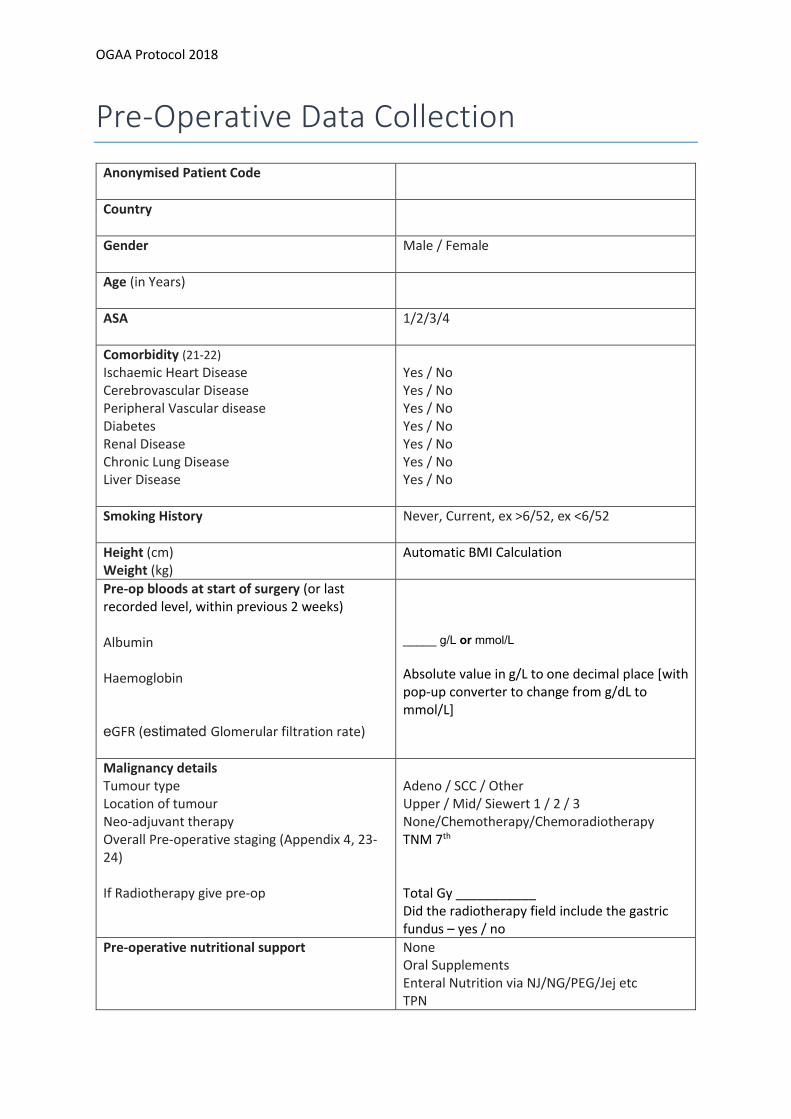
Pre-Operative Data Collection
Anonymised Patient Code
Country
Gender
Male / Female
Age (in Years)
ASA
1/2/3/4
Comorbidity (21-22) Ischaemic Heart Disease Cerebrovascular Disease Peripheral Vascular disease Diabetes Renal Disease Chronic Lung Disease Liver Disease
Yes / No Yes / No Yes / No Yes / No Yes / No Yes / No Yes / No
Smoking History Never, Current, ex >6/52, ex <6/52
Height (cm) Weight (kg)
Automatic BMI Calculation
Pre-op bloods at start of surgery (or last recorded level, within previous 2 weeks) Albumin Haemoglobin eGFR (estimated Glomerular filtration rate)
_____ g/L or mmol/L Absolute value in g/L to one decimal place [with pop-up converter to change from g/dL to mmol/L]
Malignancy details Tumour type Location of tumour Neo-adjuvant therapy Overall Pre-operative staging (Appendix 4, 23-24) If Radiotherapy give pre-op
Adeno / SCC / Other Upper / Mid/ Siewert 1 / 2 / 3 None/Chemotherapy/Chemoradiotherapy TNM 7th Total Gy ___________ Did the radiotherapy field include the gastric fundus – yes / no
Pre-operative nutritional support None Oral Supplements Enteral Nutrition via NJ/NG/PEG/Jej etc TPN

OGAA Protocol 2018
Pre-operative gastric ischaemic preconditioning performed *
Yes / No
* This is when laparoscopy and division of the left gastric vessels +- short gastric vessels are
performed prior to oesophagectomy under a separate anaesthetic
OGAA Protocol 2018
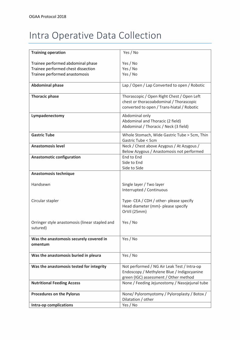
Intra Operative Data Collection
Training operation Trainee performed abdominal phase Trainee performed chest dissection Trainee performed anastomosis
Yes / No Yes / No Yes / No Yes / No
Abdominal phase
Lap / Open / Lap Converted to open / Robotic
Thoracic phase Thorascopic / Open Right Chest / Open Left chest or thoracoabdominal / Thorascopic converted to open / Trans-hiatal / Robotic
Lympadenectomy Abdominal only Abdominal and Thoracic (2 field) Abdominal / Thoracic / Neck (3 field)
Gastric Tube Whole Stomach, Wide Gastric Tube > 5cm, Thin Gastric Tube < 5cm
Anastomosis level Neck / Chest above Azygous / At Azygous / Below Azygous / Anastomosis not performed
Anastomotic configuration
End to End Side to End Side to Side
Anastomosis technique Handsewn
Circular stapler Orringer style anastomosis (linear stapled and sutured)
Single layer / Two layer Interrupted / Continuous Type- CEA / CDH / other- please specify Head diameter (mm)- please specify OrVil (25mm) Yes / No
Was the anastomosis securely covered in omentum
Yes / No
Was the anastomosis buried in pleura Yes / No
Was the anastomosis tested for integrity Not performed / NG Air Leak Test / Intra-op Endoscopy / Methylene Blue / Indigocyanine green (IGC) assessment / Other method
Nutritional Feeding Access
None / Feeding Jejunostomy / Nasojejunal tube
Procedures on the Pylorus
None/ Pyloromyotomy / Pyloroplasty / Botox / Dilatation / other
Intra-op complications Yes / No

OGAA Protocol 2018
Please specify
Operative duration (mins)
OGAA Protocol 2018
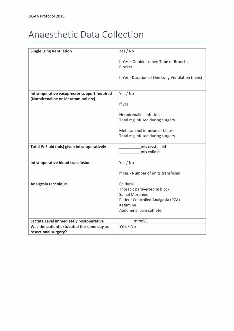
Anaesthetic Data Collection
Single Lung Ventilation Yes / No If Yes – Double Lumen Tube or Bronchial Blocker If Yes - Duration of One Lung Ventilation (mins)
Intra-operative vasopressor support required (Noradrenaline or Metaraminol etc)
Yes / No If yes Noradrenaline infusion Total mg infused during surgery Metaraminol infusion or bolus Total mg infused during surgery
Total IV Fluid (mls) given intra-operatively __________mls crystalloid __________mls colloid
Intra-operative blood transfusion
Yes / No If Yes - Number of units transfused
Analgesia technique Epidural Thoracic paravertebral block Spinal Morphine Patient Controlled Analgesia (PCA) Ketamine Abdominal pain catheter
Lactate Level immediately postoperative ______mmol/L
Was the patient extubated the same day as resectional surgery?
Yes / No
OGAA Protocol 2018
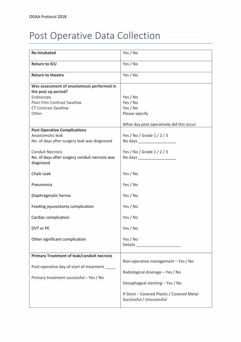
Post Operative Data Collection
Re-intubated
Yes / No
Return to ICU
Yes / No
Return to theatre
Yes / No
Was assessment of anastomosis performed in the post op period? Endoscopy Plain Film Contrast Swallow CT Contrast Swallow Other
Yes / No Yes / No Yes / No Please specify What day post operatively did this occur
Post Operative Complications Anastomotic leak No. of days after surgery leak was diagnosed Conduit Necrosis No. of days after surgery conduit necrosis was diagnosed Chyle Leak Pneumonia Diaphragmatic hernia Feeding jejunostomy complication Cardiac complication DVT or PE Other significant complication
Yes / No / Grade 1 / 2 / 3 No days _________________ Yes / No / Grade 1 / 2 / 3 No days _________________ Yes / No Yes / No Yes / No Yes / No Yes / No Yes / No Yes / No Details ____________________
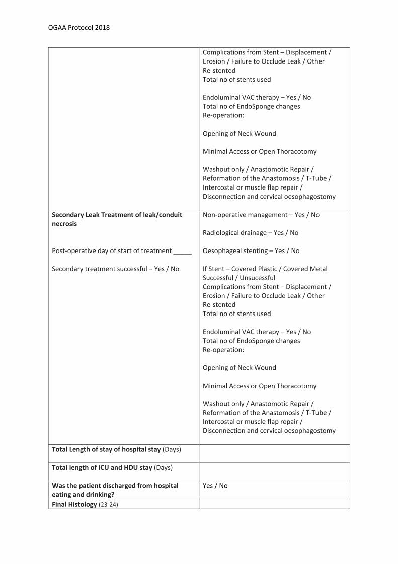
Primary Treatment of leak/conduit necrosis Post-operative day of start of treatment _____ Primary treatment successful – Yes / No
Non-operative management – Yes / No Radiological drainage – Yes / No Oesophageal stenting – Yes / No If Stent – Covered Plastic / Covered Metal Successful / Unsucessful
OGAA Protocol 2018
Complications from Stent – Displacement / Erosion / Failure to Occlude Leak / Other Re-stented Total no of stents used Endoluminal VAC therapy – Yes / No Total no of EndoSponge changes Re-operation: Opening of Neck Wound Minimal Access or Open Thoracotomy Washout only / Anastomotic Repair / Reformation of the Anastomosis / T-Tube / Intercostal or muscle flap repair / Disconnection and cervical oesophagostomy
Secondary Leak Treatment of leak/conduit necrosis Post-operative day of start of treatment _____ Secondary treatment successful – Yes / No
Non-operative management – Yes / No Radiological drainage – Yes / No Oesophageal stenting – Yes / No If Stent – Covered Plastic / Covered Metal Successful / Unsucessful Complications from Stent – Displacement / Erosion / Failure to Occlude Leak / Other Re-stented Total no of stents used Endoluminal VAC therapy – Yes / No Total no of EndoSponge changes Re-operation: Opening of Neck Wound Minimal Access or Open Thoracotomy Washout only / Anastomotic Repair / Reformation of the Anastomosis / T-Tube / Intercostal or muscle flap repair / Disconnection and cervical oesophagostomy
Total Length of stay of hospital stay (Days)
Total length of ICU and HDU stay (Days)
Was the patient discharged from hospital eating and drinking?
Yes / No
Final Histology (23-24)
OGAA Protocol 2018
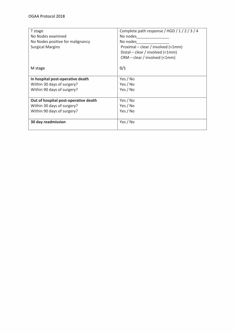
T stage No Nodes examined No Nodes positive for malignancy Surgical Margins
M stage
Complete path response / HGD / 1 / 2 / 3 / 4 No nodes_______________ No nodes_______________ Proximal – clear / involved (<1mm) Distal – clear / involved (<1mm) CRM – clear / involved (<1mm) 0/1
In hospital post-operative death Within 30 days of surgery? Within 90 days of surgery?
Yes / No Yes / No Yes / No
Out of hospital post-operative death Within 30 days of surgery? Within 90 days of surgery?
Yes / No Yes / No Yes / No
30 day readmission
Yes / No

OGAA Protocol 2018
Unit Questionnaire
Number of consultant surgeons performing oesophagectomy
Total No.
Number of oesophagectomies performed between Jan 2015 and Dec 2016
Speciality of Surgeons Thoracic / Oesophagogastric / General Surgeon / Surgical Oncologist
Size of institution Total number of beds Total number of ICU beds
24 hour on call rota for oesophageal emergencies
24hour / 9-5 / none
24 hour on call availability for interventional radiology
24hour / 9-5 / none
24 hour access to emergency theatre
24hour / 9-5 / none
Where do oesophagectomy patients routinely go post-operatively
Ward HDU ICU Dedicated GI HDU
ERAS protocol for oesophagectomy patients ERAS nurse Physio input
Yes / No Yes / No Nil dedicated / Daily / Twice daily
Does your unit perform gastric ischaemic preconditioning?
Yes – Routinely Yes – Selectively No If Yes – how many days prior to surgery
Does your unit have an agreed approach to oesophagectomy for lower 1/3 adenocarcinoma?
No Yes Open Right Transthoracic Oesophagectomy Open Left thoracoabdominal oesophagectomy Open Transhiatal Oesophagectomy Hybrid Transthoracic Oesophagectomy (Lap abdominal mobilisation) 2 stage Minimal Access Oesophagectomy 3 stage Minimal Access Oesophagectomy Robotic Oesophagectomy Other
Does you unit have an agreed technique to perform intra-thoracic anastomosis?
No Yes Handsew Circular Stapled OrVil
OGAA Protocol 2018
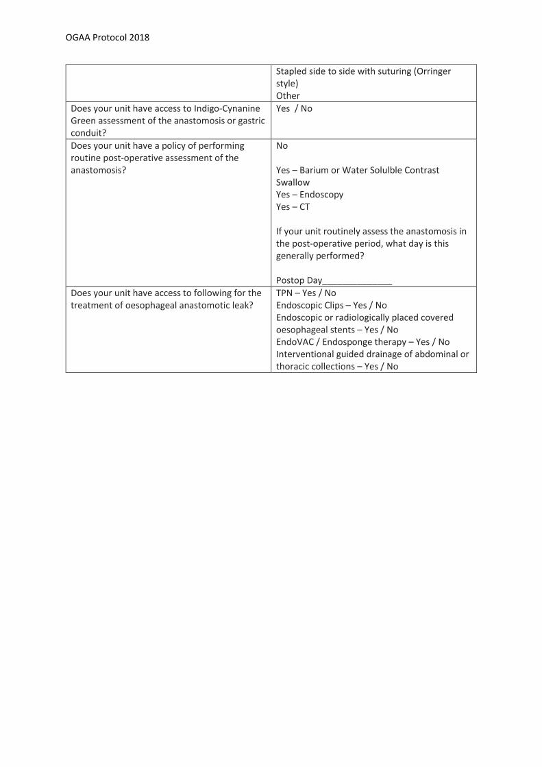
Stapled side to side with suturing (Orringer style) Other
Does your unit have access to Indigo-Cynanine Green assessment of the anastomosis or gastric conduit?
Yes / No
Does your unit have a policy of performing routine post-operative assessment of the anastomosis?
No Yes – Barium or Water Solulble Contrast Swallow Yes – Endoscopy Yes – CT If your unit routinely assess the anastomosis in the post-operative period, what day is this generally performed? Postop Day______________
Does your unit have access to following for the treatment of oesophageal anastomotic leak?
TPN – Yes / No Endoscopic Clips – Yes / No Endoscopic or radiologically placed covered oesophageal stents – Yes / No EndoVAC / Endosponge therapy – Yes / No Interventional guided drainage of abdominal or thoracic collections – Yes / No

OGAA Protocol 2018
Appendix 1 - How to register this audit
Every hospital has an audit department which should be able to advise on the information required to
register the project. Please contact them well in advance to ensure all the paper work is correct (we
would recommend at least one month prior to the study commencing).
At Trust level:
1. Identify a PI (Primary Investigator) at each trust – this is a Consultant who agrees to support the
study.
2. Create a team of Consultants/ surgical registrars.
3. Contact your hospital’s Clinical Audit Department preferably by email
a. They will provide you with a standard audit form to complete, via email or from the intranet
b. You can copy and paste from this protocol
c. Ensure that the audit department know that this is part of a larger project and that you will
send anonymised data for central collation via secure nhs.net email addresses. This will
involve gaining permission from the Trust’s Caldicott Guardian if based in the UK.
4. Once the form is completed, you may need to ask your supervising consultant to sign it.
5. Form submission
a. Submit the form and protocol to the Audit Department as soon as possible.
6. Email form to [email protected] to register your interest.
OGAA Protocol 2018
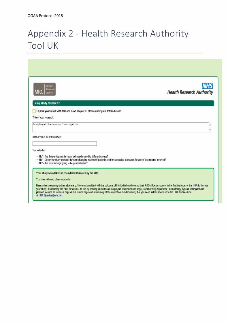
Appendix 2 - Health Research Authority Tool UK
OGAA Protocol 2018
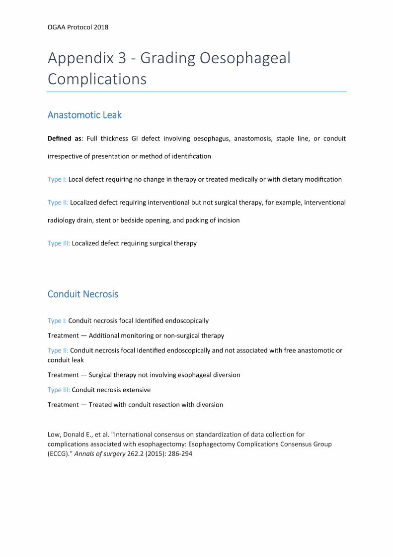
Appendix 3 - Grading Oesophageal Complications
Anastomotic Leak
Defined as: Full thickness GI defect involving oesophagus, anastomosis, staple line, or conduit
irrespective of presentation or method of identification
Type I: Local defect requiring no change in therapy or treated medically or with dietary modification
Type II: Localized defect requiring interventional but not surgical therapy, for example, interventional
radiology drain, stent or bedside opening, and packing of incision
Type III: Localized defect requiring surgical therapy
Conduit Necrosis
Type I: Conduit necrosis focal Identified endoscopically
Treatment — Additional monitoring or non-surgical therapy
Type II: Conduit necrosis focal Identified endoscopically and not associated with free anastomotic or
conduit leak
Treatment — Surgical therapy not involving esophageal diversion
Type III: Conduit necrosis extensive
Treatment — Treated with conduit resection with diversion
Low, Donald E., et al. "International consensus on standardization of data collection for
complications associated with esophagectomy: Esophagectomy Complications Consensus Group
(ECCG)." Annals of surgery 262.2 (2015): 286-294
OGAA Protocol 2018
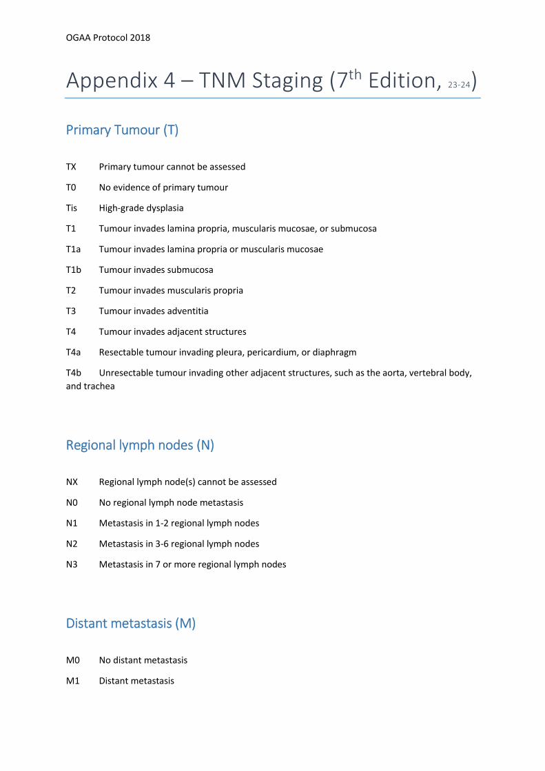
Appendix 4 – TNM Staging (7th Edition, 23-24)
Primary Tumour (T)
TX Primary tumour cannot be assessed
T0 No evidence of primary tumour
Tis High-grade dysplasia
T1 Tumour invades lamina propria, muscularis mucosae, or submucosa
T1a Tumour invades lamina propria or muscularis mucosae
T1b Tumour invades submucosa
T2 Tumour invades muscularis propria
T3 Tumour invades adventitia
T4 Tumour invades adjacent structures
T4a Resectable tumour invading pleura, pericardium, or diaphragm
T4b Unresectable tumour invading other adjacent structures, such as the aorta, vertebral body,
and trachea
Regional lymph nodes (N)
NX Regional lymph node(s) cannot be assessed
N0 No regional lymph node metastasis
N1 Metastasis in 1-2 regional lymph nodes
N2 Metastasis in 3-6 regional lymph nodes
N3 Metastasis in 7 or more regional lymph nodes
Distant metastasis (M)
M0 No distant metastasis
M1 Distant metastasis


