106
| Date post: | 18-Dec-2016 |
| Category: |
Documents |
| Upload: | michael-doherty |
| View: | 220 times |
| Download: | 2 times |


An Atlas of Investigation and Diagnosis
OSTEOARTHRITIS
Adrian JonesNottingham City Hospital
Nottingham, UK
Michael DohertyUniversity of Nottingham
Nottingham, UK
CLINICAL PUBLISHINGOXFORD
Distributed worldwide byCRC Press
Boca Raton London New York Washington D.C.

Clinical PublishingAn imprint of Atlas Medical Publishing LtdOxford Centre for InnovationMill Street, Oxford OX2 0JX, UK
Tel: +44 1865 811116Fax: +44 1865 251550Web: www.clinicalpublishing.co.uk
Distributed by:
CRC Press LLC2000 NW Corporate BlvdBoca Raton, FL 33431, USAE-mail: [email protected]
CRC Press UK23—25 Blades CourtDeodar RoadLondon SW15 2NU, UKE-mail: [email protected]
' Atlas Medical Publishing Ltd 2005
First published 2005
All rights reserved. No part of this publication may be reproduced, stored in a retrievalsystem, or transmitted, in any form or by any means, without the prior permission inwriting of Clinical Publishing or Atlas Medical Publishing Ltd.
Although every effort has been made to ensure that all owners of copyright materialhave been acknowledged in this publication, we would be glad to acknowledge insubsequent reprints or editions any omissions brought to our attention.
A catalogue record for this book is available from the British Library
ISBN 1 904392 16 4
Electronic ISBN 978 1 84692 525 2
The publisher makes no representation, express or implied, that the dosages in this book are correct. Readers must therefore always check the productinformation and clinical procedures with the most up-to-date published productinformation and data sheets provided by the manufacturers and the most recentcodes of conduct and safety regulations. The authors and the publisher do notaccept any liability for any errors in the text or for the misuse or misapplicationof material in this work.
Printed in Spain by Fisa - Escudo de Oro SA, Barcelona

Contents
Abbreviations
Preface
Acknowledgements
1 Introduction
2 General features of osteoarthritis
3 Subsets of osteoarthritis
4 Features of osteoarthritis at specific sites
5 Principles of management
Appendices
Index
vi
vii
vii
1
31
41
53
95
101
103

vi
Abbreviations
AADA apatite associated destructive arthritis
ACR American College of Rheumatology
ANKH ankylosis human (gene)
BCP basic calcium phosphate (crystals)
BMI body mass index
CMC carpometacarpal (joint)
CT computed tomography
COX cyclooxygenase
CPPD calcium pyrophosphate dihydrate (crystals)
DIP distal interphalangeal (joint)
DISH diffuse idiopathic skeletal hyperostosis
MCP metacarpophalangeal (joint)
MRI magnetic resonance imaging
NSAIDs non-steroidal anti-inflammatory drugs
OA osteoarthritis
PIP proximal interphalangeal (joint)
WOMAC Western Ontario and McMasters Universities(Index)

Preface
Osteoarthritis is the commonest joint disorder, being more prevalent than all other forms of arthritis addedtogether. No book is fully comprehensive, but we hope that this Atlas of Investigation and Diagnosis will proveof interest to doctors and allied health professionals who manage people with osteoarthritis, and will act asa catalyst to encourage interest in this, the most common single cause of lower limb disability in the elderly.
Adrian JonesMichael Doherty
vii
AcknowledgementsWe are very grateful to Jonathan Gregory for his support and perseverance in commissioning this book. Weare also indebted to the Arthritis Research Campaign for infrastructure funding (Integrated Clinical ArthritisCentre [ICAC] grant), and for substantial funding of osteoarthritis research in Nottingham. Our academicco-ordinator, Helen Richardson (ICAC funded), as always played an essential role in organizing the authors.

Introduction
Chapter 1
What is osteoarthritis?
The answer to this apparently simple question is an ongoingproblem for a condition that is so common. Everyone seemsto be able to recognize ‘osteoarthritis’ when they see it, but,as with many rheumatological conditions, providing clear-cut diagnostic criteria has proved more difficult. Notwith-standing problems of definition, it is widely accepted thatosteoarthritis is the most common condition to affectsynovial joints, and is responsible for a great deal of pain and
1
1.1 X-ray of a hip showing focal cartilage loss (jointspace narrowing) (arrow) in the superior aspect ofthe joint. Note also the prominent acetabular cyst(arrowhead).
disability. Although a universally accepted definition hasproved elusive, there is general agreement on some of thehallmark features of osteoarthritis. Cartilage loss isuniversally observed in all patients with osteoarthritis and,as will be discussed further below, is a sine qua non fordiagnosis. Cartilage loss tends to be focal rather thanwidespread throughout the joint, particularly in the earlystages of osteoarthritis (1.1).

Bone response with increased bone formation adjacent tothe joint is also commonly observed although, as discussedlater, this varies in prominence at different joint sites andbetween patients (1.2–1.4).
Bone response may manifest itself as either asosteophytosis or subchondral bone sclerosis. These twofeatures (i.e. cartilage loss and bone response) have often
been considered the main features of osteoarthritis, but, aswill be discussed later, it is now clear that many other tissuesare involved in osteoarthritis. Indeed, it is likely that theseother tissues are more important in determining thesymptomatic and functional consequences of osteoarthritis.(Table 1.1, 1.5).
Introduction2
1.2 Slab radiograph of femoral head section from anosteoarthritic hip joint, showing marginal new bone(osteophyte) (arrows); thickening and increasedwhiteness (sclerosis) of the superficial subchondralbone (short arrow); and thickening of the trabeculararcades in response to altered stress loading(arrowhead). 1.3 Hypertrophic patellofemoral osteoarthritis. Note
florid new bone (osteophyte) formation at the jointmargins (arrows) and sclerosis (increased bonedensity on x-ray) in the subchondral bone(arrowhead).

Introduction 3
1.4 Atrophic patellofemoral osteoarthritis. Note loss ofbone stock and relatively little osteophyte formation. Alsonote the apparent ‘pressure erosion’ (arrow) in theanterior aspect of the distal femur, as if the patella hasscalloped out the bone and worn it away.
1.5 Diagram showing some ofthe major changes that occur inosteoarthritis.
Cartilage Focal softening and lossBone Osteophyte, sclerosis, but
subchondral osteopeniaCapsule ThickeningSynovium Thickening and modest inflammationMuscle Atrophy and weaknessLigaments DegenerationBursae Secondary bursitisVessels Angiogenesis, avascular necrosis,
venous hypertension
Table 1.1 Tissues involved in osteoarthritis
Bursitis
Muscle atrophy
Osteophyte
Cyst
Cartilageloss
Synovialthickening
Osteochondralbody
Synovialeffusion

Introduction4
1.6 Nineteenth century drawing of a hand affectedby nodal osteoarthritis prior to formal description ofthe condition (Charcot, Paris).
The history of osteoarthritis
In order to understand the concept of any disease, it isnecessary to look at its historical context. It is likely thatosteoarthritis has been present throughout human history.Indeed, it may be of significant evolutionary importance, andthis is discussed further below (page 29). The first attemptsto describe ‘arthritis’ generally did not distinguish inflam-matory from non-inflammatory disease (1.6). Gouty arthritiswas probably the first specific arthropathy to becharacterized, followed by infective arthritis and rheumatoiddisease.
The reduction in infectious diseases as a result of publichealth measures, and the introduction of effective therapiesfor inflammatory arthritis, have resulted in an increasedawareness and interest in non-fatal, but disabling diseases,such as osteoarthritis. In the 1950s, the introduction of thewidespread use of radiography enabled ready study ofdiseases (such as osteoarthritis) which principally affectbony structures.
The combination of these factors resulted in an increasedawareness of the impact of osteoarthritis, and enabled theconduct of large-scale epidemiological studies of thisdisease.
The next boost to the study of osteoarthritis came withthe development of animal models of osteoarthritis and, inparticular, the Pond–Nuki model of osteoarthritis – theanterior cruciate-deficient dog. The importance of thismodel is that it allowed study of the factors involved in thedevelopment of osteoarthritis. This, and other animalmodels, led to the realization that osteoarthritis is an activemetabolic process, rather than a simple eburnation anderosion of cartilage. It also ultimately led to the realizationthat many other tissues, especially the neuromuscularsystem, are crucially important in the development ofosteoarthritis.
Improved biochemical and cellular techniques furtherdeveloped interest in the metabolic features of osteoarthritis,although arguably it may have led to an initial undueemphasis on cartilage.
In the 1980s and 1990s, a re-exploration of theepidemiology of osteoarthritis resulted in a renewedappreciation of the fact that not all patients withosteoarthritis are symptomatic. This is an obvious fact toanyone who looks at community samples of osteoarthritis,but can easily be overlooked in an outpatient setting whereall patients have been referred because of symptoms. Theimplications of this has important consequences forunderstanding the epidemiology of osteoarthritis, and isdiscussed further below (page 29).

More recent developments in osteoarthritis have followedfrom the observations of the metabolic activity ofosteoarthritis tissues and the realization that these processesmay be amenable to modification by pharmaceutical means.This is more fully discussed in the section on diseasemodification (page 99). It has meant, however, that thepessimistic view that osteoarthritis is inevitably progressivehas been challenged, and there is increasing pharmaceuticalinterest in manipulating this process.
The realization that osteoarthritis is the cause ofsignificant health care expenditure, and that use of theseresources may not always be rational, has led to increasedhealth service research in this area, and the development ofcare pathways and guidelines for the management ofosteoarthritis. The principles that have emerged from suchendeavours are discussed in Chapter 5.
Finally, it is important not to forget that, in parallel withthis, there has been immense interest in surgical interven-tions for osteoarthritis. These have included osteotomy,arthroplasty with increasingly complex prostheses, and,more recently, arthroscopic approaches, including tissuetransplantation.
Introduction 5
The epidemiology of osteoarthritis
The epidemiology of osteoarthritis has been elucidated by anumber of major cross-sectional studies (pathological,clinical, and/or radiographic). There have also been anumber of prospective studies of varying duration whichhave also illuminated our knowledge. Due to this, a numberof risk factors and associations have been identified.
Other speciesOsteoarthritis appears to have been present throughout ourevolutionary history and, indeed, in many current non-human species. Looking across species, it is clear that allanimals that fuse epiphyses of synovial joints are capable ofdeveloping osteoarthritis. The few often quoted exceptions(e.g. bats and sloths) probably simply reflect lack of studyrather than any specific species difference (1.7). This hasseveral important implications. Firstly, it allows potentialstudy of mechanisms of osteoarthritis in non-human, non-primate species which can thus enhance understanding.Secondly, it probably implies that the biological processesthat underlie osteoarthritis are most likely of greatevolutionary value to the host organism. This has led someto speculate that osteoarthritis is an aspect of the inherentrepair process of the joint, and has led to the coining of theterm ‘regenerative’ joint disease.
1.7 Although only minimally studied, osteoarthritis hasbeen said not to occur in sloths and bats because theyhang upside down and place distraction forces, ratherthan impact forces, through their joints. However, whalescan develop osteoarthritis even though their weight issupported in water. It seems that osteoarthritis affectsmost species that have synovial joints and which fusetheir epiphyses in the adult.

AgeAll studies clearly define that the prevalence of osteoarthritisincreases with age at nearly all joint sites (1.8). Obviously,since osteoarthritis, once established, demonstratespermanent changes in the joint, some of this could beregarded as the simple accrual of new joint involvement.However, there is a rapid rise in the prevalence ofosteoarthritis after the age of 40. This occurs at all synovialjoints, although the absolute prevalence varies at differentjoint sites.
Some forms of osteoarthritis are more likely to develop atspecific ages. For example, osteoarthritis of the distalinterphalangeal joint is uncommon before the age of 40, butpolyarticular onset around the time of the menopause iscommon with a period of rapid accrual in the decade eitherside of 50 years of age.
There is a suggestion that the prevalence of osteoarthritismay even decline in the very elderly. These data derive fromcross-sectional surveys and, of course, may reflect a survivaleffect (censureship) in that osteoarthritis may associate withpremature cardiovascular mortality.
GenderA number of studies have demonstrated that the relativeprevalence of osteoarthritis at different joint sites differsbetween genders, usually being more prevalent in women.Both polyarticular osteoarthritis of the distal interphalangealjoints (‘nodal generalized osteoarthritis’) and kneeosteoarthritis are more common in women than in men. Atother joint sites, this sex difference is less dramatic and maydiffer with age. For example, hip osteoarthritis is morecommon in men before retirement age, but becomes moreprevalent in women in older age.
Animal studies have confirmed that sex hormones canhave a major effect on the development of osteoarthritis.Studies in humans have, however, only hinted at an effect onosteoarthritis, and a therapeutic use for sex hormones hasnot been demonstrated.
Introduction6
1.8 Prevalence of osteoarthritis at the hip and knee and in men andwomen by age. (Data derived from van Saase JL, van Romunde LK, Cats A, et al. [1989]. Epidemiology of osteoarthritis: Zoetermeer survey.comparison of radiological osteoarthritis in a Dutch population with that in10 other populations. Ann Rheum Dis, 48:271–280.)
40–49 50–59 60–69 70+
Age band (years)
50454035302520151050
Hip (male)
Knee (male)
Hip (female)
Knee (female)
Pre
vale
nce
%

ObesityAt most joint sites, but particularly the knee, weight has animportant impact on the development and severity ofosteoarthritis (1.9). More importantly reduction of weight inobese and overweight adults has been shown in aprospective observational study to reduce the subsequentdevelopment of knee osteoarthritis (1.10).
How obesity and overweight lead to the development ofosteoarthritis is not entirely clear. The most likelyexplanation is mechanical overloading. The support for thiscomes from a study that has demonstrated that obesityinteracts with malalignment at the knee (either varus orvalgus) to increase the risk of developing radiographicosteoarthritis.
A counter to the simple mechanical argument is thatobesity is acting through a metabolic mechanism. The mainsupport for this proposal relates to hand osteoarthritis. It isargued that hand joints are ‘non-weight bearing’. Therefore,the mechanism of the observed increase in hand osteo-arthritis seen in the obese must relate to an associated meta-bolic imbalance. A number of possible mediating factorshave been suggested including insulin-derived growthfactor. However, it may not be that straightforward sincemechanical studies have shown that forces through the handjoints in the obese are substantially increased.
Introduction 7
1.9 Standing x-ray of an obese woman withsevere medial tibiofemoral osteoarthritis andvarus malalignment. The soft-tissue shadowsclearly illustrate this important risk factor forknee osteoarthritis.
1.10 Effect of obesity on the subsequent development of kneeosteoarthritis, and the additional effect of losing or gaining weightin the preceding 10 years. (Data derived from Felson DT, Zhang Y,Anthony JM, et al. [1992]. Weight loss reduces the risk forsymptomatic knee osteoarthritis in women. The Framinghamstudy. Arthritis Rheum, 116:535–539.)
10
1
0.1 Baseline BMI
Weight gain
Weight loss
Odds ratio/2 unit increasein BMI occuring:
4 years before
6 years before
8 years before
10 years before
12 years before
Odd
s ra
tio f
or d
evel
opin
gos
teoa
rthr
itis/
2 un
its o
f B
MI

TraumaIt has long been suspected that severe trauma to a synovialjoint might predispose to subsequent osteoarthritis. Supportfor this comes from clinical observation of patients who havesuffered fractures and who subsequently develop osteo-arthritis of an adjacent joint (1.11). The risk of such‘secondary’ osteoarthritis is particularly high when a fractureinvolves the articular surface of a joint.
Additional evidence comes from follow-up of patientswho have undergone meniscectomy at the knee (1.12).Removal of a meniscus is a major mechanical insult to theknee and post-meniscectomy subjects are at increased risk of
premature osteoarthritis on the side of the meniscectomy(1.13). The lifetime risk of developing osteoarthritis may notbe increased, but the time of onset certainly does seem to bebrought forward.
The degree of trauma required to increase the risk ofsubsequent osteoarthritis is unclear. However, a recentprospective study of college students suggests that evenrelatively minor trauma, insufficient to lead to hospital-ization, is still associated with an increased risk of kneeosteoarthritis in young adult life.
Introduction8
1.11 Osteoarthritis isolated to the left knee in a 64-year-old man who, 25 years before, had suffered a severeupper tibial fracture (arrow), resulting in leg lengthshortening and altered biomechanics. Note also bilateralchondrocalcinosis.
1.12 A longitudinal ‘bucket-handle’ tear of themedial knee meniscus. Tears of the medialmeniscus are three times more common thanlateral tears.

GeneticAn important genetic contribution to development ofosteoarthritis has been suggested by classic twin and familystudies. Strong heritability was first noticed for generalizednodal osteoarthritis, characterized by the presence ofmultiple Heberden’s nodes. These nodes were described byWilliam Heberden in 1802 in his Commentary on the Historyand Cure of Disease:
‘What are those little hard knobs, about the size of asmall pea, which are frequently seen upon the fingers,particularly a little below the top, near the joint? Theyhave no connection with the gout, being found inpersons who never had it; they continue for life; andbeing hardly ever attended with pain, or disposed tobecome sores, are rather unsightly, than inconvenient,though they must be some little hindrance to the freeuse of the fingers.’ (Heberden W [1802]. Commentarii deMorborum Historia et Curatione, London.)
As will be discussed later, this form of osteoarthritisgenerally has its onset in perimenopausal women, hence itsother common name of ‘menopausal arthritis’. It showsstrong inheritance, particularly in women, and behavesalmost as a sex-linked, autosomal-dominant condition.
Other rare forms of atypical, young-onset osteoarthritis,often with minor degrees of dysplasia, have been describedwhich transmit as monogenic disorders. In some families,the precise genetic association has been identified asmutations of the gene COL2A1 that encodes Type IIcollagen – the principal structural collagen of hyalinearticular cartilage. However, investigation of patients withmore common ‘sporadic’ osteoarthritis has failed to findsuch mutations as a common cause. Such reports in familieshave, however, fuelled interest in the genetics of morecommon forms of osteoarthritis.
Introduction 9
1.13 Effect of meniscal injury on the 16-year likelihood of developingosteoarthritis. (Data from a case-control study by Englund M, Roos EM,Lohmander LS [2003]. Impact of type of meniscal tear on radiographic andsymptomatic knee osteoarthritis: a sixteen-year follow up of meniscectomywith matched controls. Arthritis Rheum, 48:2178–2187.)
Radiographic OA
Symptomatic OA
128
64
32
16
8
4
2
1Rel
ativ
e ris
k of
dev
elop
ing
oste
oart
hriti
s
Degenerative Traumatic Partial Subtotaltear tear resection resection

More recent genetic epidemiology studies, using a varietyof strategies (1.14), have confirmed an important geneticcomponent to the development of osteoarthritis at a numberof sites including the hip, knee, hand, and spine. Theheritability of osteoarthritis at these sites (that is, the degreeof variance for osteoarthritis in the population that isexplained by genetic factors) is estimated to be between40–60%. Linkage and association studies continue to try toidentify the responsible genes (1.15). Although a number of
findings are reported, most have not been replicated insubsequent studies. It is clear, however, that osteoarthritis isa common complex disorder, and that several, possibly evenmultiple genes, will contribute to susceptibility. These arelikely to be common polymorphisms rather than mutations;they may vary according to joint site, and they may need tointeract with other genes, or with other constitutional orenvironmental risk factors to express the phenotype ofosteoarthritis (1.16).
Introduction10
1.14 Strategies to determine the genetic contribution to osteoarthritis. Classic twin studies (left) compare theconcordance for osteoarthritis in monozygotic (MZ) (identical) and dizygotic (DZ) (non-identical) twins – a higherconcordance in monozygotic twins suggests genetic predisposition.
A higher prevalence of osteoarthritis (middle), compared to controls (C), in siblings (Sib) of patients with hip or kneeosteoarthritis severe enough to lead to joint replacement (TJR) also suggests genetic predisposition to osteoarthritis.
Examination of the way osteoarthritis transmits vertically and horizontally through families (right), compared to theexpected population prevalence of osteoarthritis, can show familial clustering.
MZ MZ
DZ DZ
Versus
TJR
Versus
C
Twin Exposed sib Segregation, aggregation
Sib

Introduction 11
1.15 Two sisters affected by nodal generalizedosteoarthritis affecting hands and knees. A sibling ofsomeone who has required joint replacement for kneeosteoarthritis is at more than twice the risk of developingknee osteoarthritis than someone without such a ‘geneticexposure’; the risk for a sibling of someone who hasundergone joint replacement for hip osteoarthritis is evenhigher (3–9 fold depending on age and gender). Geneticstudies undertaken on multiples families with suchaffected sibling pairs is the main way of determininglinkage between osteoarthritis and specific chromosomalregions. Examination of possible candidate genes withinthose regions is a common strategy to determine thegenes responsible for genetic susceptibility.
1.16 Diagram showing osteoarthritis to bea ‘common complex disorder’. Multiple(currently unknown) polymorphisms needto interact with other constitutional or localrisk factors to permit expression of theosteoarthritis phenotype. These risk factorsmay vary according to joint site. Riskfactors for development of osteoarthritismay also differ from risk factors for a goodor bad clinical outcome. Blue shadingrepresents commonly affected joints.
ConstitutionGenderAgeObesityNutritionBone densityMuscle strength
MechanicsJoint shapeAlignmentStabilityJoint usageTrauma

OccupationA role for occupation in the development of osteoarthritis isoften cited by patients, and can be an important discussionpoint in medico–legal claims. Early studies of mill andcotton workers from the UK and USA suggested thatdiffering occupational hand usage could influence thepattern and distribution of osteoarthritis in the hand. Whatwas not clear from these studies was whether there was anabsolute increase in the prevalence of osteoarthritis orsimply a different localization of disease. A similar argumentis often debated for trauma – does trauma increase the riskor does it just bring forward the development ofosteoarthritis in patients at risk?
In at least one occupation, that is, farming, an increasedrisk of hip osteoarthritis has been demonstrated comparedto appropriate controls. This risk appears greatest for arablefarmers and for farmers brought up as children on a farm,but the precise mechanism is unclear. Because the risk ismore than twofold, UK farmers who develop hiposteoarthritis are entitled to industrial compensation.
In many cases, simple job descriptions do not adequatelydescribe the actual locomotor stresses that are involved.Accordingly, attempts have been made to examine andquantify the actual amount of a particular activity that anoccupation involves, and to examine these as risk factors forosteoarthritis. Using such an approach, it has beendemonstrated that repeated occupational knee bending,particularly if this involves heavy lifting, associates with anincreased risk of knee osteoarthritis.
SportThe role of sport in the development of osteoarthritis is notentirely clear. Moderate levels of sporting activity appear toimprove functional outcome in osteoarthritis. This may bemediated by the benefits to neuromuscular functioning,improved aerobic fitness and well-being, easier weightcontrol, and improved self-efficacy. Even at elite levels,overall, participation in sport appears to be beneficial.However, pitted against this is the risk of trauma from directinjury or from more minor trauma that is repetitive and stillbad for joint health. Therefore, it seems likely thatexercise/activity and osteoarthritis will show a U-shaped,rather than linear, dose–response relationship (1.17).
Despite the overall benefits of exercise, moderate–elitesporting activity appears to increase osteophyte formationthough this may not be true osteophyte (that associates withcartilage loss) but rather a stress reaction at enthesealinsertions. Power lifting has been associated with kneeosteoarthritis, presumably sharing the same biomechanicalmechanism as with occupational knee bending. Othersports, such as football, are associated with osteoarthritis,but it is suspected that much of this is related to directtrauma and a high prevalence of internal derangement andinstability. More minor but repetitive trauma may beinjurious, and different patterns of osteoarthritis have beenreported in different types of sports, leading to specific termssuch as: mid-tarsal arthritis – footballers; ‘pitchers elbow’ –baseball; and ballerina’s foot.
Introduction12
1.17 Joints are designed to move. Under-usageleads to tissue atrophy and is bad for joint health.Excessive activity may lead to tissue injury andpredispose to osteoarthritis. Moderate, regularactivity that provides physiological loading withouttissue injury seems the ideal between these twoextremes.
Tissue atrophy
Health
Tissue damage
Disuse
Physiologicalloading
Extreme loading

Introduction 13
1.18 Muscles are essential for normal joint function (fromthe second book of De Humani Corporis Fabrica [1543],by Andreus Vesalius [1514–1568]).
MuscleThe synovial joint does not simply involve cartilage, synoviallining, fluid, and bone: it is a complex organ that requiresmany other tissues for successful function. Amongst these ismuscle (1.18). Often overlooked in early studies, a renewedinterest occurred when it was demonstrated that muscleweakness is very prominent in patients with kneeosteoarthritis. Indeed, studies from Bristol and elsewheredemonstrated very clearly that muscle weakness was a moreimportant associate of disability than the degree of structuralchange as evidenced by radiography. Apart from its role as aprime mover of joints, muscle is an important pro-
prioceptive organ, and muscle weakness related to kneeosteoarthritis also associates with reduced knee propriocep-tion, increased sway when standing with eyes shut, anincreased risk of falls, and an abnormal gait pattern.
Experimental models confirm that arthropathy canrapidly produce marked weakness of adjacent muscles. Thisresults from a combination of neural inhibition of muscleactivation and an increased, but imbalanced, turnover ofmuscle tissue resulting in muscle atrophy. This observationhas important therapeutic implications, as will bediscussed later (page 97).

Neurological factorsIf muscles are important in osteoarthritis, then it is likelythat neurological factors may also be important. It is clearthat the muscle weakness and atrophy observed are criticallydependent on neurological function. In animals withexperimental acute knee damage, muscle atrophy is criticallydependent upon on an intact dorsal root ganglion and reflexarc. Similarly, in a number of different forms of osteo-arthritis, the presence of pre-existent or subsequentneurological disease can markedly affect the phenotypicexpression of arthritis (1.19).
This clinical observation has had clinical correlates invarious animal models of osteoarthritis, including in theguinea pig and the dog. More recently, it has been suggestedthat neurological dysfunction may play a role in theinitiation of osteoarthritis in man. In a study of young,asymptomatic adults suspected to be at high risk of thesubsequent development of knee osteoarthritis, subtleabnormalities in neuromuscular ‘bracing’ have beenmeasured, and referred to as ‘micro-klutziness’. This maysimply reflect a protopathic bias, that is, the presence of pre-clinical disease. However, examination of the relatives ofthese affected individuals suggests that this phenomenonmight predate development of any pathology.
Other joint and bone diseaseOther disorders of joints may predispose to subsequentosteoarthritis. These include other defined arthropathies,such as rheumatoid arthritis, psoriatic arthritis, sepsis,juvenile idiopathic arthritis, and gout. This does not usuallypresent a diagnostic difficulty, but it is important to considersince supervening osteoarthritis may require a very differenttherapeutic approach to the underlying inflammatory jointdisease. With primary inflammatory arthritis such asrheumatoid, it is usually only when the inflammatory,damaging synovitis is controlled that any attempts at tissuerepair can occur and ‘secondary’ osteoarthritis can develop.
Childhood disorders, such as Perthe’s disease, slippedfemoral epiphysis, or mild acetabular dysplasia (1.20) at thehip may compromise the joint, and if untreated predisposeto osteoarthritis in the adult.
Similarly, disorders of adjacent bone (Paget’s disease, forexample) may also be associated with osteoarthritis. Thismay occur because of adverse effects on cartilage and bonethat accompany bone remodelling and altered bonevascularity (1.21).
Introduction14
1.19 Effect of a stroke on the subsequent development of osteoarthritis.(Data derived from Segal R, Avrahami E, Lebdinski E, et al. [1998]. Theimpact of hemiparalysis on the expression of osteoarthritis. Arthritis Rheum,41:2249–2256.)
Non-paretic hand
Paretic hand
All CVA Moderate Moderate Mildpatients to severe to severe paralysis
paralysis paralysis>3 years
2.5
2.0
1.5
1.0
0.5
0
Ost
eoar
thrit
is s
cale

Introduction 15
1.20 A shallow acetabular roof, as seen here in thecontext of minor acetabular dysplasia, can allow thefemoral head to migrate upwards and outwards andlead to osteoarthritis.
1.21 Paget’s disease causing bowing of the left tibia (A). The x-ray (B) shows theabnormal Pagetic bone abutting up to the knee (arrow), widening of the tibia which nolonger matches the femoral width, and associated cartilage loss (arrowhead) in bothtibiofemoral compartments. Cartilage loss is often more extensive and diffuse with such‘Pagetic arthropathy’ than with common osteoarthritis.
A B

Genetically determined conditions that affect bone orcartilage may result in premature ‘osteoarthritis’ at multiplesites, sometimes in association with short stature, bodydisproportion, and other clinical features. The mostcommon of these conditions are the many forms of multipleepiphyseal dysplasia (1.22–1.24). When the spine isinvolved, it is called spondyloepiphyseal dysplasia. A familyhistory may sometimes be present, but in many cases itappears to result from an apparently spontaneous mutation.Radiographic appearances are often characteristic.
Introduction16
1.22 Hand of a 24-year-old woman withspondyloepiphyseal dysplasia showing what clinicallyappears to be osteoarthritis of multiple interphalangealjoints characterized by bony swelling.
1.23 The standing knee radiograph of the same patientas 1.22. Note the abnormal tilt of the tibiofemoral joint lineand the sclerosis (arrow) and osteophyte (arrowhead) thatmainly involves the lateral compartment.
1.24 The lumbar spine radiograph of the same patientshowing anterior-posterior elongation of the vertebralbodies, the abnormal contours to the endplates, and thewidening of the anterior disc spaces.

Introduction 17
iron in tissues, including the joint. There is a clearly-definedarthropathy of haemochromatosis which is very similar toosteoarthritis, notably the subset known as pyrophosphatearthropathy (page 44). The mechanism by whicharthropathy is produced is not completely defined, but thereis an accumulation of iron within cartilage and synovium,which may cause direct toxicity to chondrocytes. Theobserved arthropathy often involves aggressive loss of jointspace at sites (such as the metacarpophalangeal joints, andradiocarpal joint), that are less commonly affected insporadic osteoarthritis (1.25–1.27). Multiple subchondralcysts are a prominent feature. In addition, iron interfereswith inorganic pyrophosphate metabolism, and leads toincreased production and crystallization of calciumpyrophosphate crystals. This may be observed radio-graphically as chondrocalcinosis, and clinically as acutecrystal synovitis.
Acromegaly also associates with an arthropathy thatclinically and radiographically resembles osteoarthritis. Themechanism is unclear, but interestingly there is often initialwidening of the joint space (1.28). Pathologically, there is anexuberant thickening of the hyaline cartilage, but thisoutgrows the nutrient supply provided below by the bone
1.25 Hand radiograph of a 56-year-old man withhaemochromatosis showing eccentric joint space lossand hook osteophytes (arrows) on the radial aspect ofthe metacarpal heads. Symptomatic arthritis was thepresenting feature of his haemochromatosis.
Toxins and metabolic factorsGeographical ‘hot-spots’ of endemic osteoarthritis havebeen recognized for some time. Perhaps the bestcharacterized of these is Kashin–Beck disease. This presentsas a premature form of polyarticular osteoarthritis inMongolia, eastern Russia and northern China. Other similarforms are often named according to the area or populationaffected, e.g. Mselini disease in southern Africa, andMalmad disease in India. Early studies involving migrationof people into and out of the affected regions suggestedenvironmental, rather than genetic factors in itspathogenesis. Recent studies have started to identify some ofthose factors. For example, Kashin–Beck disease almostcertainly results from an interaction between seleniumdeficiency and hypothyroidism due to iodine deficiency, thatappears to encourage chondrocyte death in hyaline cartilageof children and young adults. While these endemicarthropathies are clearly dissimilar from sporadicosteoarthritis and present at a very young age (2nd and 3rd
decades), they suggest the possibility that other toxins andmetabolic factors may be important in the development ofmore common ‘sporadic’ osteoarthritis.
Haemochromatosis is an increasingly well-understoodgenetic disorder where there is a marked accumulation of

Introduction18
1.26 The wrist radiograph of the samepatient as in 1.25, showing cartilage loss inthe 1st carpometacarpal and trapezio-scaphoid joints (short arrow) (common sitesfor osteoarthritis), but also in the radiocarpal(arrows) and midcarpal rows (sites notusually affected by osteoarthritis). Multiplesubchondral cysts are a prominent feature inthe radiocarpal joint (arrowheads).
1.27 The ankle radiograph of the samepatient as 1.26, showing marked cartilageloss and again multiple cysts (arrows).
blood vessels and superficially by the synovial fluid.Subsequently, there is erosion and loss of the cartilage andeventual joint space narrowing. In addition, there may bealteration of the bones with periosteal, cortical thickening.
Avascular necrosis (osteonecrosis) can result in collapseand involution of a segment of bone and its overlyingcartilage. The subsequent distortion of the anatomical shapeof the joint then results in osteoarthritis. Avascular necrosismay be secondary to conditions that cause externalcompression of blood vessels in bone (e.g. steroid therapy,and alcohol excess – mainly due to increased fat depositionin bone) or reduced intravascular flow (e.g. sickle cellanaemia, and Caisson’s disease – the ‘bends’). In thesesituations, it mainly targets the hip (femoral head, 1.29)shoulder (humeral head), knee (distal femur), or elbow(distal humerus). Primary osteonecrosis of large bonesusually targets the medial femoral condyle (1.30).Osteochondritis, resulting from trauma during childhood ordevelopmental abnormality, affects small bones and may bea predisposing factor to subsequent osteoarthritis (e.g.osteonecrosis of the lunate – Kienbock’s disease).
Although relatively uncommon, presentation of whatclinically appears to be osteoarthritis at a young age (under55), or in an atypical pattern, or both, should lead toconsideration of an underlying disease. The usual cause ofyoung onset osteoarthritis at a single joint site is precedingtrauma, and this is often apparent in the patient’s history oron the x-ray. However, for young onset osteoarthritisaffecting several or multiple joints, several conditions mayrequire consideration (Table 1.2).

Introduction 19
1.28 Standing knee radiographsof a 52-year-old man withacromegaly who presented witharthralgia, and symptoms andsigns of osteoarthritis in hishands. His knees show obviouswidening of the tibiofemoral jointspaces and bony enlargement(‘squaring’) of the femoralcondyles.
1.30 Idiopathic medial femoral necrosis showinglocalized segmental collapse, sclerosis, and alteredcontour of the distal femur (arrow).
1.29 Late osteonecrosis of the left femoral headin a 39-year-old patient who had received highdose steroids for asthma, showing segmentalcollapse and increased sclerosis of the superiorpole (arrows).

Introduction20
Monoarticular• Prior trauma• Localized instability• Avascular necrosis
Pauci/polyarticular• Prior joint disease (e.g. juvenile idiopathic arthritis)• Spondyloepiphyseal dysplasia• Metabolic disease – haemochromatosis• Endocrine disease – acromegaly• Avascular necrosis• Neuropathic (Charcot) joint• Endemic osteoarthritis
Table 1.2 Causes of young-onset osteoarthritis orosteoarthritis with atypical distribution
Mortality and co-morbidity inosteoarthritis
Osteoarthritis is not an entirely benign disease, in terms ofgeneral health. A number of studies have demonstrated anincreased mortality in osteoarthritis, although the cause forthis unclear. Although this a small relative risk, theattributable population risk is high because of the highprevalence of osteoarthritis.
A number of co-morbidities principally associated withobesity and insulin resistance (‘metabolic syndrome’) arealso associated with osteoarthritis. These include: diabetes,hypertension, cardiovascular disease, and gout. In dealingwith patients with osteoarthritis, it is important to considerthese issues.
Towards a definition of osteoarthritis
One of the problems that has bedevilled the study ofosteoarthritis is that while clinicians instinctively recognizewhen a patient has osteoarthritis, defining it in a form whichis easily communicated to others has proved more difficult.Clearly, the reason for defining osteoarthritis may affect howit is defined. For example, if the purpose is to look at
determinants of structural change, then a radiographic orpathologic definition may be required. However, if thepurpose is to define patients who may be suitable for aclinical trial, then the only appropriate ones are those withsymptoms which also may demonstrate structural(radiographic) change. Even if structural change andsymptoms can be agreed upon, some form of grading ofseverity may be important and, indeed, individual aspectsmay require separate evaluation.
Pathological features
Pathological changes are central to many definitions ofosteoarthritis. In essence, an osteoarthritic joint has changesin many, if not all, of its tissue components.
The most obvious change is in the articular hyalinecartilage. The earliest change is thought to be increasedhydration followed by softening and fissuring of the surface.Eventually, this results in thinning of the cartilage andexposure of the subchondral bone (1.31). These changes arefocal, and generally occur mainly at sites of maximummechanical stress within the joint.
Changes in bone are also prominent (1.32, 1.33). At themacroscopic level, this involves thickening of thesubchondral bone (apparent as sclerosis) and formation ofosteophyte. The formation of osteophyte is initiated by newfibrocartilage that subsequently undergoes endochondralossification, usually at the joint margins at sites of capsuleand ligament insertions. Small areas of pressure damage andosteonecrosis may also result in loss of bone. At themicroscopic level, avascular necrosis is probably a morecommon mechanism than is generally appreciated. Holes or‘cysts’ may also occur in bone (1.32) some of whichcommunicate with the joint cavity. These most likely reflectliquefaction following localized osteonecrosis, or possiblyarise at sites where synovial fluid is forced under pressurethrough clefts in the cartilage and subchondral bone.
But osteoarthritis is not just about hyaline cartilage andbone. The synovium in osteoarthritis may show markedinflammation, though this is patchy rather than diffuse, as ininflammatory rheumatoid disease. The mechanism under-lying this is far from being understood. It may representsecondary ‘debris synovitis’, with the synovium beinginflamed by small shards of cartilage and bone that becomefree within the damaged joint (1.33). Another possible causeof inflammation could be due to calcium pyrophosphate and

Introduction 21
1.31 Macroscopic appearance of tibia removed during jointreplacement for osteoarthritis. Note the unequal, and in this case,severe loss of articular cartilage with antero-posterior grooves inthe exposed subchondral bone of the medial tibial plateau (arrow).Note also the florid rolling osteophyte at the joint margins.
1.32 Slab radiograph of an osteoarthritic femoralhead showing florid marginal osteophyte (arrow)and multiple small and large ‘cysts’ with scleroticborders (arrowheads).
1.33 Small fragments of cartilage within synovial fluidfrom an osteoarthritic knee (x400 magnification). Thesefragments may be taken up by synoviocytes and causelocal inflammation.

apatite crystals released from osteoarthritic cartilage (1.34).A low-grade primary inflammatory process may, however,underlie some cases. As a result osteoarthritic synovium canbe thickened, have an infiltrate of inflammatory cells, andshow a marked degree of vascularity.
Intra-articular fibrocartilage (e.g. in the knee menisci), iscommonly affected in osteoarthritis, showing the samehistological changes, thinning, and fibrillation as hyalinecartilage (1.35). It may reflect the generalized nature of theosteoarthritis process, or the altered biomechanics withinthe joint. Alternatively, meniscal injury may predate theosteoarthritis, and be a predisposing risk factor. Althoughpost-meniscectomy osteoarthritis has long been recognized,recent MRI studies suggest that meniscal abnormality maybe a very common feature of early osteoarthritis.
Ligaments and tendons also demonstrate degenerativechanges with thinning and weakening. Again, and quiteinterestingly, MRI studies have suggested that this may bean early feature of osteoarthritis and, perhaps in somesituations, predate cartilage and bone disease.
Neuromuscular changes are also commonly seen withatrophy and increased muscle turnover, as well as increasedinnervation in conjunction with angiogenesis.
Definition of presence of osteoarthritisFor many studies and, indeed, often in the clinical setting,the establishment of the presence or absence ofosteoarthritis is all that is required. However, not allstructural osteoarthritis is symptomatic. The correlationbetween presence of osteoarthritis and the occurrence ofassociated pain and disability varies between joint sites, andin general is not strong (1.36). Most certainly, at a numberof sites asymptomatic osteoarthritis is far more commonthan symptomatic change.
The most common symptom of osteoarthritis, and theone that most concerns patients, is pain. While it wouldseem that the presence or absence of pain at a specificlocation would be easy to establish, in fact, even subtledifferences in the wording of the question(s) used can resultin major differences in the prevalence of osteoarthritis.Furthermore, the localization of pain at different joint sitescan sometimes prove problematic. For example, in the spinepain can commonly radiate widely and may arise from anumber of other structures and sites. Sometimes it is very todifficult to establish whether apparent radiographicosteoarthritis is symptomatic or not.
Introduction22
1.34 Large collections of calcium pyrophosphate crystals withinosteoarthritic synovium (x200 magnification). These inflammatory crystalsform in hyaline and fibro-cartilage, but may then be shed into the jointcavity and be taken up by synovium, resulting in synovitis.

Introduction 23
1.35 Degeneration and tearing of a meniscus in anosteoarthric knee removed at joint replacement. Note thesevere fibrillation and thinning of the hyaline cartilage ofthe underlying tibial plateau.
1.36 Diagram showing the imprecise inter-relationship between the presence of pain,disability and structural change of osteoarthritis.The correlation is best at the hip, but is generallypoor at other sites (especially the spine and hand).
Pain
Disability Structural change

Regional pain in relation to osteoarthritis can arise for anumber of reasons (1.37). There are no clear differences inthe characteristics of osteoarthritic pain that distinguish itfrom other causes of pain. Thus whether symptoms can beused as a reliable surrogate for osteoarthritis in the absenceof some form of structural assessment would seem unlikely.
Conversely, whether radiographic or other structuraldefinitions alone can be used to define osteoarthritis isequally problematic if the purpose of the definition is todefine the clinical problem.
Finally, clinical signs can be used in various combinationswith structural and symptomatic change to defineosteoarthritis. Unfortunately, the major problem with this isthat all studies that have examined the reliability andreproducibility of clinical signs have suggested that many ofthese, with the possible exception of Heberden’s nodes, canbe very unreliable. In addition, their relationship tounderlying radiographic change can be problematic.
Currently, radiographic osteoarthritis is usually definedby the presence of a definite osteophyte on an appropriateradiograph, especially for knee and hand osteoarthritis, or bydefinite focal joint space narrowing (the main feature usedfor hip osteoarthritis).
Symptomatic osteoarthritis can be defined in a number ofways. One example of this is the American College ofRheumatology (ACR) definition of knee osteoarthritis(Table 1.3). Using this system the best definition requires thepresence of pain and either a radiographic, clinical feature
(crepitus), or demographic feature (age >50 years).However, this demonstrates the problem with adopting auniversal definition. The ACR definition was created anddeveloped in order to differentiate between patients withsymptomatic knee osteoarthritis and those withinflammatory arthritis in a rheumatology service setting. Assuch, it is appropriate in defining a population drawn froma hospital base suitable for clinical trials in osteoarthritis, butperhaps not in defining osteoarthritis for epidemiologicalpurposes or community service settings where radiographsmay be impractical. It also does not address the issue ofasymptomatic radiographic change, nor the fact that thedetection of clinical signs is insensitive and unreliable, andthe defined age cut-off may miss early disease. Definitionthus remains problematic.
Assessing symptoms in osteoarthritisThere are various symptoms that are associated withosteoarthritis (Table 1.4). Of these, pain is the mostclinically relevant. Pain itself can be subdivided into variouscategories according to its timing (usage pain, rest pain,and night pain). There is some evidence that this may haveclinical relevance with, for example, pain at rest, and atnight being more responsive to NSAIDs than toacetaminophen (paracetamol). Pain in osteoarthritis tendsto be worse towards the end of the day. It also shows aweekly (circaseptan) variation in being worse around theweekend, even in people who are not working (1.38).
Introduction24
Knee pain (majority of previous month)
Plus three of the following:
• Age >50 years
• Morning stiffness <30 minutes
• Crepitus
• Bony enlargement on palpation
• Bony tenderness
• No palpable warmth
Knee pain (majority of previous month)
X-ray with osteophyte
Plus one of the following:
• Age >50 years
• Morning stiffness <30 minutes
• Crepitus
Table 1.3 ACR criteria for knee osteoarthritis

Introduction 25
1.37 Osteoarthritis may give rise to pain fromintraosseous hypertension and microfracture (yellow),intracapsular hypertension (synovial thickening, increasedfluid production, capsular tightening) (green), or fromperiarticular syndromes, such as bursitis, tendinitis, andenthesopathy (red), secondary to remodelling and alteredjoint mechanics.
Pain:
• on movement
• at night
• at rest
Stiffness
Deformity
Loss of range of movement
Crepitus
Locking
Giving way
Muscle weakness and wasting
Nerve compression (spine)
Table 1.4 Symptoms associatedwith osteoarthritis
1.38 Circadian and circaseptan variation in pain intensity in osteoarthritis.(Adapted from Bellamy N, Sothern RB, Campbell J [1990]. Rhythmicvariations in pain perception in osteoarthritis of the knee. J Rheumatol,17:364–372.)
Circadian Circaseptan
6 12 18 24 M Tu W Th F Sa Su Hours Days

Stiffness – a subjective feeling of difficulty in moving -also occurs in osteoarthritis. Usually it is relatively brief(5–10 minutes) and rarely lasts more than 30 minutes eitherin the morning (morning stiffness) or after rest (‘gelling’).
Some patients complain of crepitus, which is a roughpalpable or occasionally audible vibration on movement.Although this is sometimes assessed by examination, thissign is unreliable and grading systems are not used.
Deformity, either in terms of bony swelling ormalalignment across the joint, is common, but is rarelysubject to measurement or grading.
Muscle wasting around affected joints is common.Measurement of both muscle size (girth and imaging) andmuscle function (strength and power) can be performed,and may be used to assess disease progress.
Various composite measures have been developed inorder to assess and score pain and functional impairment inosteoarthritis. Perhaps the two best developed and usedmeasures are the Western Ontario and McMastersUniversities (WOMAC) Osteoarthritis Index and theLesquesne Algofunctional Index. The scores derived bythese disease-specific instruments can be separated intotheir specific domains (pain, stiffness, and function), or usedas a single composite score.
Assessing structural (radiographic) change in osteoarthritis Radiographs can be used to detect the presence or absenceof osteoarthritis, and to assess progression of structuralchange. Plain radiographs are the traditional imagingmeasure, and they are easy to obtain and readily available.Radiographic features of osteoarthritis include focal jointspace narrowing (from cartilage thinning), marginalosteophyte, sclerosis (increased whiteness of subchondralbone), bone cysts and osteochondral ‘loose’ bodies, andeventually bone attrition, deformity of bone ends, andmalalignment (1.39).
The most widely used system to analyse and graderadiographic osteoarthritis is the Kellgren and Lawrencecomposite system (Table 1.5). This employs combinedassessment of individual features, and can be scored usingthe verbal descriptions or by comparison with a standardradiographic atlas. There are problems with the system, asthe hierarchy of change may not be as described,standardization may be problematic, and radiographs are bydefinition two-dimensional and may thus underestimate thedegree of involvement. It does, however, provide a methodfor categorizing joint involvement into four or fivesimple grades.
Introduction26
1.39 Diagram showing a normal jointoutline (A) and the major features ofosteoarthritis (B). These include focaljoint space narrowing, marginalosteophyte, subchondral sclerosis,cysts, osteochondral bodies andeventual deformity of bone ends, and malalignment.
A B

Introduction 27
Grade 0 Normal
Grade 1 Doubtful narrowing of joint space, possible osteophyte
Grade 2 Definite osteophyte, possible narrowing
Grade 3 Moderate multiple osteophytes, definite narrowing, some sclerosis, possible deformity of bone ends
Grade 4 Large osteophytes, marked narrowing, severe sclerosis, definite deformity of bone ends
(Adapted from The Epidemiology of Chronic Rheumatism. Volume II Atlas of Standard Radiographs of Arthritis,Blackwell Scientific Publications [1963].)
Table 1.5 Kellgren and Lawrence grading system for osteoarthritis
The problems with the system have led to attempts todevelop other ways of assessing radiographs for osteo-arthritic changes. Generally, these have involved thedevelopment of atlases of standard images and the separategrading of individual features (osteophyte, joint spacenarrowing, etc.). One system for knee osteoarthritis thatuses line drawings rather than photographs has a number ofadvantages, including: an interval rather than ordinal scalefor narrowing and osteophyte; a value for normal joint spacewidth that is based on a population sample; separate imagesfor narrowing and osteophyte (thus avoiding the distractionand bias when several features occur together); uniformmagnification of images; and separate atlases for joint spacenarrowing for men and women (men have wider joint spacesat the knee) (1.40).
Since cartilage loss is often considered the major featureof osteoarthritis and joint space loss the major surrogate forthis, then it makes sense to try to quantify this accurately.Standardization of radiographic technique is clearlyimportant as an initial step, and several methods areavailable to increase reproducibility and precision ofradiographs. This has been combined with variousmeasurement techniques (both manual and semi- or fully-automated) to achieve this.
Several important consequences of these approaches haveemerged. The joint space widths in women and men at
different ages have been defined. For the hip, at least, adefinition has been established of hip osteoarthritis based onthe level of joint space width which best correlates with thepresence or absence of symptoms.
As regards the specific features of osteoarthritis, theirindividual association with symptoms varies and, thus, theirusefulness in defining osteoarthritis. For example, at the hipit is joint space narrowing, not osteophyte, that most closelyassociates with hip pain. Conversely, at the knee it isosteophyte, not narrowing, that associates best with kneepain, such that even quite minor osteophytosis is areasonable correlate of symptomatic osteoarthritis.However, in assessing disease progression at the knee, jointspace measurement is more accurate and sensitive to changethan grading of osteophyte. One possible problem at theknee is that pain can affect loading of the joint and hencemeasured joint space width. This has been postulated as amechanism to explain how symptom-modifying drugs mayfalsely appear to associate with slowing or even reversal ofjoint space narrowing. The maximally-thinned cartilage inthe tibiofemoral compartments is brought into the weight-bearing position when the knee is semi-flexed. If pain isrelieved, the knees often straighten more, thus bringingwider, less-affected cartilage into the weight-bearingposition, and making the radiographic joint space appearwider than before.

Introduction28
1.40 Extract from an atlas that uses line drawings rather than photographs, showing 0–3 grades for narrowing of themedial tibiofemoral compartment in women (top row) and 0–3 grades for osteophyte at all tibiofemoral sites. (FromNagaosa Y, Mateus M, Hassan B, et al. [2000]. Development of a logically devised line drawing atlas for grading ofknee osteoarthritis. Ann Rheum Dis, 59:587–595.)
Grade 0 Grade 1 Grade 2 Grade 3
Women
Men
Grade 0 Grade 1 Grade 2 Grade 3

Introduction 29
1.41 The proposed scenario of osteoarthritis as the inherent repair process of synovial joints. It isonly if this slow, efficient repair process cannot compensate for the triggering insults that the jointcontinues to remodel and more commonly associates with symptoms and disability.
Traumatic Inflammatory
Metabolic
Cartilage Bone Capsule Synovium
Insults
Repair
Osteoarthritis process
Compensation
Decompensation
Outcome
Is osteoarthritis useful?
The fact that osteoarthritis has been observed in manyspecies and throughout evolutionary time has led tospeculation that it has been conserved for a reason, that is,it may have a selection advantage. Of course, this maysimply reflect the fact that osteoarthritis in humans is adisease of older, non-reproductive age so there is no negativeselection pressure which would confer a survivaldisadvantage, but it does not explain its presence in otherspecies. However, one perspective is that this so-called‘degenerative disease’ is the inherent repair process ofsynovial joints that in some circumstances may confersurvival advantage. As has been alluded to before, it couldbe argued that osteoarthritis is essentially a reparativeprocess, characterized by new tissue production (especiallybone) that is brought into action by a variety of joint insults(1.41). In general, it is an efficient repair process, and oftencompensates for the triggering insult without associatingwith symptoms or disability. Osteophytosis and boneremodelling may increase the surface area of thecompromised joint and beneficially redistribute loadtransmission, and vertically growing osteophyes may splintthe joint and counter instability. In this sense, theosteoarthritic response can be seen as potentially beneficial.
It is only if osteoarthritis cannot compensate due tooverwhelming insult or a poor repair response that‘decompensated’ osteoarthritis may associate with pain anddisability and become a clinical problem. Such a scenarioexplains the marked clinical heterogeneity of osteoarthritis,the high prevalence of asymptomatic osteoarthritis, and theoften good outcome of symptomatic cases. Decompensatedsymptomatic osteoarthritis may be seen as a form of end-stage ‘joint failure’ with a common phenotype irrespective ofthe triggering insult.
Further reading
Brandt KD, Doherty M, Lohmander LS (2003).Osteoarthritis, 2nd edn, Oxford University Press, Oxford.
Felson DT (Conference Chair) (2000). Osteoarthritis: NewInsights Part 1. The disease and its risk factors. AnnIntern Med, 133:635–649.
Moskowitz RW, Howell DS, Altman RD, et al. (2001).Osteoarthritis: Diagnosis and Medical/Surgical Management.WB Saunders, London.

General features ofosteoarthritis
Chapter 2
What are the clinical features ofosteoarthritis?
The clinical features of osteoarthritis relate to bothsymptoms and signs. As has been discussed in Chapter 1,the principle symptom of osteoarthritis is pain. Typicallythis occurs during movement and/or loading of the joint. Itmay vary greatly in severity from mild to very severe.Indeed, as already discussed, structural change may beentirely asymptomatic. Usually pain from osteoarthritischanges only slowly and undergoes variation with ‘goodweeks’ and ‘bad weeks’. Some authors consider thatosteoarthritis undergoes ‘flares’ with temporary markedincreases in pain and swelling, although the mechanismunderlying this is unclear.
Nocturnal pain may be a prominent feature. Althoughtaken as a late, poor prognostic feature by some, it may bepresent early in the clinical course, and possibly may beparticularly helped by NSAIDs. The mechanism in thissituation is thought to be vascular in nature with increasedintraosseous pressure. As was discussed earlier, this can beassociated with localized areas of avascular necrosis andmore rapid deterioration in joint architecture with morerapid clinical progression.
Osteophyte growth and bony remodelling can result inaltered joint shape and, at superficial sites, this can readilybe observed clinically (2.1). Similarly, loss of bone stock,often as a result of avascular necrosis, may result in mal-alignment with resultant varus, valgus, flexion, or otherdeformities (2.2). Again these deformities can be readilyobserved, and may be a risk factor for future progression anddeterioration.
31
2.1 Femoral osteophyte is visible and palpable inthis flexed knee as prominent bony ridging (arrows)along the anterior border of each condyle giving aninverted ‘V’ appearance.

Swelling that is due to soft-tissue rather than bonyenlargement may be observed. Although synovial thickeningmay be a factor, it is more commonly due to increasedsynovial fluid and distension of the joint capsule. Jointswellings due to fluid tend to occur in natural weak areas ofthe capsule, and thus in characteristic locations. Acuteswellings may sometimes occur in a fashion similar to thatseen in the ‘flares’ of pain. The mechanism is again unclear,although in some cases a crystal synovitis may occur, most
commonly related to calcium pyrophosphate crystals(‘pseudogout’) (2.3).
In association with crystal synovitis, an overlyingerythema may occur, but in general cutaneous featuresincluding local heat are unusual.
Stiffness in osteoarthritis is usually relatively short-livedand often more related to inactivity, such as sitting, ratherthan being the more typical and very prolonged earlymorning stiffness seen in, for example, rheumatoid arthritis.
General features of osteoarthritis32
2.2 Right hindfoot varus deformity in a patientwith chronic pyrophosphate arthropathy.
2.3 Acute pseudogout of the right knee.Note swelling and a degree of erythema.

General features of osteoarthritis 33
2.4 Global wasting of muscles – especially visiblefor deltoid, supraspinatus, infraspinatus, and teresminor – in association with right glenohumeralosteoarthritis.
2.5 Osteoarthritis of the elbow showing largeosteochondral bodies (arrows). This patient hadrecurrent locking that required surgical exploration.It was probably the smaller osteochondral bodies,not apparent on this radiograph, that were causingthe problem.
Muscle wasting may be a prominent feature ofosteoarthritis. Although general immobility may producegeneralized wasting, the most marked wasting is seen in themuscles that act across the affected joint. Location willdetermine whether this is readily appreciated with some jointinvolvement: the hip and hand, for example, less likely toproduce obvious atrophy than others, such as the knee andshoulder (2.4). Although atrophy may be hard to appreciate,the consequence of this may be more easily observed in termsof reduced strength and function. Associated proprioceptive
impairment can also result in functional impairment with, forexample, impaired gait and balance.
Ligamentous involvement and joint space loss may resultin instability and further functional impairment which maymanifest as a lack of confidence, falling, and fear of falling.Associated meniscal damage and intra-articular ‘loose-bodies’ may result in symptoms of ‘locking’, especially at theknee and elbow (2.5) and of ‘giving way’ (mainly at theknee). Locking is a temporary, painful inability to move thejoint in one plane, usually extension, and this often lasts a

matter of minutes. It is a mechanical problem that resultsfrom an interpolation of tissue between the articular surfacesand, if recurrent, may require surgical removal.
‘Giving way’ is a more difficult concept and has beenconsidered variously as a feeling of instability and lack ofconfidence in a limb, usually the leg, to a more transient,sudden weakness in the muscles causing a partial but notcomplete loss of ability to weight-bear, lasting for just afraction of a second.
Systemic upset is not a feature of osteoarthritis since it isa non-inflammatory condition that does not trigger theacute phase response. An exception is those patients whoexperience associated calcium crystal synovitis. In thissituation, the marked release of inflammatory mediatorsmay result not only in local pain and inflammation, but alsofever, myalgia, sweating, and confusion.
What are the radiological features ofosteoarthritis?
Joint space narrowing can be appreciated by looking at theintercortical distance between bone separated by hyalinecartilage. The situation is more complex in those joints thatcontain a fibrocartilage meniscus, such as the tibiofemoraljoints of the knee. In this situation, meniscal damage, orindeed surgical removal, will result in a reduced joint spacewithout this necessarily reflecting hyaline cartilage loss.Conversely, at certain sites, such as the knee, a normal jointspace width does not exclude significant cartilage loss. Thiscan arise if a non-weight-bearing view is taken (2.6) or if theknee is fully extended – a semi-flexed weight-bearing view isoptimal and more sensitive at detecting focal cartilage loss.Similarly, the alignment of the x-ray beam relative to thejoint may also make a major difference to the appreciation ofjoint space loss (2.7).
General features of osteoarthritis34
2.6 A non-weight bearing view (A) and a weight-bearing view (B) of the same knee. Theweight-bearing view shows marked medial joint space narrowing that is not apparent onthe non-weight bearing view.
A B

The early stages of osteophyte formation involve chondralmetaplasia and growth at the joint margins. These earlycartilaginous osteophytes are radiolucent and will not beapparent on a plain radiograph. Pathologically, they may beexuberant and, indeed, other imaging modalities (such asMRI) may demonstrate them. These osteophytes tend todevelop at the site of capsular and ligamentous insertions.Subsequent ossification occurs and, of course, theassociated calcification will cause them to become radio-opaque. In nearly all cases, radiographs will underestimatethe size of osteophytes.
Bone growth may occur in other locations. Subchondralbone may demonstrate sclerosis and the trabeculararchitecture may alter in response to load bearing. This canoccur internally leading to thickened trabeculae alignedparallel to stressing forces and externally leading to corticalthickening and periosteal osteophyte or ‘buttressing’. Thebest examples of this are usually seen at the hip (2.8).
Bone loss may also be a feature. Typically, this involvessubchondral bone leading to altered contour, collapse, and
reduced bone height, often as a late feature in associationwith marked cartilage loss (2.9). As previously discussed,localized areas of osteonecrosis may play a role in this. Thesubchondral loss may also be associated with cyst formation(2.10). Prominent multiple cyst formation is more commonin patients with haemochromatosis and pyrophosphatearthropathy.
Major focal loss can occur resulting in massive alterationsin the cortical outline of a joint. This is most commonly seenat the knee and hip with marked attrition and loss of normalbone outlines (2.11).
Finally, appropriate views can identify ossified loosebodies. These are not, as the name implies, floating freewithin the joint, but are usually embedded in the synovium.They may arise from synoviocytes that undergo metaplasiato cartilage that then ossifies, in which case they may bemultiple (2.12). Alternatively, they may result from abroken-off fragment of articular cartilage that is then takenup by the synovium and grows and ossifies there, in whichcase, they may be single or less numerous.
General features of osteoarthritis 35
A
2.7 (A) An anteroposterior radiograph of a shoulder which, although showing definite osteophytosis (arrow),appears to demonstrate relatively preserved joint space. On the axial view (B) the marked loss of joint spaceand, by implication, cartilage is readily apparent (arrowhead).
B

General features of osteoarthritis36
2.8 Radiograph showing marked superior polehip osteoarthritis with periosteal osteophyte(‘buttressing’) of the inferior aspect of thefemoral neck (arrow).
2.9 Skyline view of the patellofemoral joint showing markedcartilage loss of the lateral facet with lateral patellasubluxation, sclerosis of subchondral bone, a ‘saw-tooth’deformity (ridging of both sides of the joint), and reducedheight of the patella.
2.10 Skyline view of the patellofemoralcompartment showing marked cartilageloss and large well-corticated cysts inthe patella (arrows).

General features of osteoarthritis 37
2.11 Hip radiograph showing severe superior pole osteoarthritiswith complete loss of joint space (‘bone-on-bone’), sclerosis,cysts (arrows), and marked loss of bone in the femoral headand acetabulum (arrowhead).
2.12 Hip radiograph showing superiorpole osteoarthritis with multipleosteochondral ‘loose bodies’ (arrows).

What do other imaging techniquesdemonstrate in osteoarthritis?
Other imaging techniques can demonstrate the facets ofosteoarthritis seen in radiographs to greater or lesser extents.MRI is more sensitive than radiographs in demonstratingcartilage loss, and can also delineate changes in fibro-cartilaginous menisci, synovium, bone, and periarticularstructures that are completely invisible on a plain radio-graph (2.13).
Bone marrow ‘oedema’ or ‘bruising’ on MRI, both at theknee (2.13) and spine (where it is called Modic change), ismore likely to be found in a patient with symptoms, and may
predict future progression (2.14). The precise pathologicalnature of this abnormal MRI signal is unclear, but it hasbeen described in vertebral bodies, facet joints, femur, andtibia, and at all these sites a relationship with pain has beendescribed. Other features appreciated on MRI scanninginclude ligamentous and capsular/synovial change – againnot visible on plain radiographs. Cartilage can also bedirectly measured and quantified and, indeed, measuredcartilage volume can be used as a surrogate for joint spacenarrowing.
General features of osteoarthritis38
2.13 MRI of an osteoarthritic knee showing osteophyteand cartilage thinning in the medial compartment, but also‘bone oedema’ of the upper medial tibia (arrowhead) andmedially a very large anserine bursa (arrow) (the sourceof much of this patient’s current pain) – features that arenot seen on plain radiographs.
2.14 Relationship of bone marrow oedema tosubsequent radiographic progression. (Data derived fromFelson DT, McLaughlin S, Goggins J, et al. [2003]. Bonemarrow edema and its relation to progression of kneeosteoarthritis. Ann Int Med, 139:330–336.)
40
35
30
25
20
15
10
5
0Medial
progressionLateral
progression
Bone marrow oedema
No oedema
Kne
es d
emon
stra
ting
prog
ress
ion
in t
he r
elev
ant
com
part
men
t (%
)

General features of osteoarthritis 39
Ultrasound scanning equipment is generally cheaper thanMRI, and newer high-resolution scanners can be used toassess cartilage, osteophytes, and ligaments. In general,however, this is predominantly a research tool at present andis not widely used clinically to assess osteoarthritis.
Radioisotope scanning involves injecting radio-labelledtechnetium into the circulation and measuring subsequentradioactive emission, both with time and by site. Byscanning at appropriate times, this can be used to examinevascularity and perfusion, tissue uptake, and new boneformation. Although not widely used to assess osteoarthritis,it is apparent that bone scan changes related to osteoarthritis
are commonly detected in scans performed for otherreasons. This requires careful evaluation and differentiationfrom osteoblastic metastases, inflammatory synovitis, orinfection.
At the knee and the hand, radioisotope scanning has beensuggested to predict those patients with osteoarthritis whoare likely to demonstrate radiographic progression. Thepredictive value is not high, but certainly patients with‘hotter scans’ and with certain scan patterns (such as ‘diffusesubchondral bone change’ at the knee) (2.15, 2.16) aremore likely to demonstrate future progression. The clinicalusefulness of this finding is not yet clear.
2.15 Radioisotope bone scan (delayed phase) of a patient withknee osteoarthritis showing bilateral diffuse increased uptake insubchondral bone either side of the medial joint lines. This is apattern that is a risk factor for progressive structural change.
2.16 Radioisotope scan (delayed phase) of anosteoarthritic knee showing a ‘tramline’ patternof focal uptake in subchondral bone either sideof the medial joint line. This pattern is less likelyto progress than that in 2.15.

General features of osteoarthritis40
2.17 Commonly described subsets of osteoarthritis.
Are there subsets of osteoarthritis?
Since the 1960s, there have been suggestions that osteo-arthritis may encompass a number of different conditions.The first notion was that in some cases a cause could beidentified, usually trauma but occasionally other inflam-matory arthritides, leading to division into ‘primary’ and‘secondary’ forms of osteoarthritis. However, such divisionhas been challenged as it is suggested that overt trauma isjust one factor in a common complex disorder that merelybrings forward the onset of osteoarthritis in otherwisegenetically and constitutionally predisposed individuals.The best example of this comes from the study of patientswho have undergone meniscectomy in early adult life.Those people who subsequently develop hand osteoarthritisin middle life (i.e. those with a generalized predisposition toosteoarthritis) are more likely to develop post-meniscectomyosteoarthritis at the knee and to have higher x-ray scores,suggesting interaction between extrinsic (trauma) andintrinsic (individual) risk factors.
One way to differentiate subsets of osteoarthritis is simplyby the joint involved. Over and above this clinicalobservation, and epidemiological studies then began todescribe patients with particular patterns of involvement ofosteoarthritis, although whether these are true distinctentities or simply extreme examples of a continuousspectrum remains unclear. More recent studies haveconfirmed some of these patterns and have even begun todescribe differing genetic and biochemical bases for these.These are now described in more detail (2.17).
Further reading
Brandt KD, Doherty M, Lohmander LS (2003).Osteoarthritis, 2nd edn, Oxford University Press, Oxford.
Moskowitz RW, Howell DS, Altman RD, et al. (2001).Osteoarthritis: Diagnosis and Medical/Surgical Management.WB Saunders, London.
Osteoarthritis
Pyrophosphatearthropathy
Generalizednodal
osteoarthritis
Erosiveosteoarthritis
Trauma
Apatite associatedarthropathy
Haemochromatosis
Prematureand endemicosteoarthritis

Subsets of osteoarthritis
Chapter 3
Generalized osteoarthritis
Early epidemiological studies using radiological surveysdetailed the fact that many patients had osteoarthritis ofmultiple joints, suggesting a generalized, in-builtpredisposition (3.1).
Furthermore, the pattern of joint involvement did notappear random, in that certain joints were commonlyaffected together. In particular, distal interphalangeal joints,
41
3.1 Accrual of multiple joint involvement with osteoarthritis, and the effectof age.
medial tibiofemoral compartments, thumb bases, andintervertebral discs and apophyseal joints of the lumbar andcervical spine seemed to be affected quite commonly in thesame patients (3.2). In addition, many patients were notedto have Heberden’s nodes. These occur at the distalinterphalangeal joints, and were originally described as ‘pea-like swellings’ on the posterolateral (radial and ulnar)
45
40
35
30
25
20
15
10
5
0
0 1 2 3 4 5 6 7 8 9 10+Number of joint groups involved
Women 45–54
Women 55–64
Women 65+
%
3.2 Distribution of joint involvement in generalized nodal osteoarthritis.Major target joints are shown in dark blue, moderately targeted joints inpale blue.

aspects of the joint, either side of the extensor tendon. Theirearly development appears to involve a cystic swellingarising from the synovial cavity of the joint. The cystcontains a gelatinous material which subsequently appearsto undergo chondroid metaplasia, and then ossification.Originally, it was unclear if Heberden’s nodes always relateto osteoarthritis, but recent studies suggest that the two areclosely related. Similar changes can occur at the proximalinterphalangeal joints where they are commonly known asBouchard’s nodes (3.3).
While multiple Heberden’s nodes commonly associate
with osteoarthritis at other sites, they may also occur in afew fingers without osteoarthritis elsewhere, perhapsreflecting localized hand trauma. However, multiple nodescommonly associate with polyarticular osteoarthritis inwomen(3.4). The appearance of multiple nodes oftenoccurs around the time of the menopause, and was onceknown as ‘menopausal arthritis’. It is now more commonlytermed ‘generalized nodal osteoarthritis’. It has a stronggenetic component with a marked female preponderance,and a high degree of penetrance, behaving almost like aMendelian recessive trait.
Subsets of osteoarthritis42
3.3 Nodal osteoarthritis of the handsdemonstrating Heberden’s nodes at thedistal interphalangeal joints, andBouchard’s nodes at the proximalinterphalangeal joints. Note additionalsideways (radial or ulnar) deviation atsome joints – a characteristic deformityof interphalangeal osteoarthritis.
3.4 Numbers of joints involved in bothmen and women at age >65 years, andthe effect of the presence of Heberden’snodes.
30
25
20
15
10
5
0
%
0 1 2 3 4 5 6 7 8 9 10+Number of joint groups involved
Women, nodes
Women, no nodes
Men, nodes
Men, no nodes

Subsets of osteoarthritis 43
Erosive osteoarthritis
Some patients with polyarticular hand osteoarthritis appearto have a worse prognosis. Clinically, this includes a highdegree of interphalangeal joint instability (3.5), a restrictedrange of movement, and even joint ankylosis. In addition,there is an almost equal involvement of proximal and distalinterphalangeal joints, compared to generalized nodalosteoarthritis where the involvement is predominantly distal.
Radiographically, erosive osteoarthritis is characterized bysubchondral erosion of bone. This can give rise to acharacteristic ‘sea-gull wing’ appearance (3.6). Bone losscan be marked and lead to finger shortening and, hence,joint laxity. In some cases, there can be late bony fusion(3.7). Instability and bony ankylosis are not features ofcommon nodal osteoarthritis.
3.5 Marked lateral instability of a distalinterphalangeal joint in a patient with‘erosive’ osteoarthritis. Such instabilityis not a feature of common nodalosteoarthritis.
3.6 Erosiveosteoarthritis.Marked subchondralerosion leads toscalloping of thebone ends and a‘gull’s wing’appearance.
3.7 Erosive osteoarthritis with bony fusion (arrow) of a distalinterphalangeal joint.

Subsets of osteoarthritis44
3.8 Calcium pyrophosphate crystals (arrow) in knee synovial fluid(x400 magnification) aspirated during an acute attack of synovitis.The crystals are few in number, small, weakly birefringent, andlargely rhomboid in shape.
Pyrophosphate arthropathy
After the nature of gout as a crystal-based arthropathy waselucidated, a group of patients were described in the 1960swith acute self-limiting synovitis of the knee, which clinicallyresembled gout. However, examination of their aspiratedsynovial fluid under polarizing light did not reveal thebright, negatively birefringent, needle-shaped crystals ofmonosodium urate (the gout culprit), but other muchsmaller, positively birefringent, rhomboid crystals that werechemically identified as calcium pyrophosphate dihydrate(CPPD) crystals (3.8, 3.9). The attacks of acute self-limiting inflammation caused by CPPD crystals were called‘pseudogout’.
Plain radiographs of some patients can also demonstratecalcification of cartilage (chondrocalcinosis) affecting fibro-cartilage and less commonly hyaline cartilage (3.10).Cadaveric and synovial fluid studies have confirmed thatusual cause of chondrocalcinosis is CPPD crystaldeposition, with other less common causes being apatite andother basic calcium phosphate crystals. Chondrocalcinosis isa common, age-associated radiographic finding that canoccur in structurally normal joints. However, there is anincreased prevalence of chondrocalcinosis and CPPDcrystal deposition in osteoarthritic joints, especially knees,and when they co-exist, the term ‘chronic pyrophosphatearthropathy’ is often used.

Subsets of osteoarthritis 45
3.9 A higher magnification of the same fluid as in 3.8, showing anintracellular calcium pyrophosphate crystal that has beenphagocytosed by a neutrophil (arrow).
3.10 Chondrocalcinosis in the lateral compartment of aknee. The elongated triangle of calcification in the central,outer half of the joint (arrow) is in the fibrocartilaginousmeniscus, whereas the lines of calcification parallel andclose to the cortical line (arrowhead) are in hyalinearticular cartilage.

While ordinary osteoarthritis and pyrophosphatearthropathy appear very similar, there are some qualitativedifferences. Apart from the presence of chondrocalcinosis,joints with pyrophosphate arthropathy tend to demonstratemore marked osteophyte, cysts and bone remodelling,resulting in a ‘hypertrophic’ radiographic appearance (3.11).
The pattern of joint involvement also tends to differ(3.12). In particular, there tends to be more glenohumeral,metacarpophalangeal, midtarsal, and radiocarpal jointinvolvement – sites that are not commonly targeted forgeneralized osteoarthritis.
Clinically, apart from possible superimposed acutepseudogout attacks, there is a tendency for moreinflammation (stiffness, chronic knee effusions, and soft-tissue swelling) in chronic pyrophosphate arthropathy thanuncomplicated osteoarthritis. Chondrocalcinosis and CPPDcrystal deposition have also been suggested to be risk factors
Subsets of osteoarthritis46
3.11 Lateral knee radiograph of pyrophosphate arthropathyshowing a ‘hypertrophic’ appearance with florid osteophyteformation.
3.12 Distribution of joint involvement in chronic pyrophosphatearthropathy. Major target joints are shown in dark blue,moderately targeted joints in pale blue.
for more rapid radiographic progression of osteoarthriticstructural change at the knee and hip.
Ageing and osteoarthritis are the two commonpredisposing factors to CPPD crystal deposition andchondrocalcinosis. However, occasionally metabolicdiseases that interfere with pyrophosphate metabolism maypredispose to chondrocalcinosis (Table 3.1) and there arerare kindreds with familial predisposition to floridpolyarticular chondrocalcinosis. Such metabolic or familialpredisposition should be considered and screened for inpatients with polyarticular chondrocalcinosis or ifunexplained chondrocalcinosis occurs under age 55. Oftenthere are additional clinical or radiographic clues thatsuggest metabolic disease (3.13).
Of the metabolic diseases, only haemochromatosis isassociated with structural changes resembling osteoarthritis.Haemochromatosis is a disorder of iron storage caused by a

Subsets of osteoarthritis 47
Condition Chondrocalcinosis Pseudogout Pyrophosphate arthropathy
Hyperparathyroidism + + –Haemochromatosis + + +Hypomagnesaemia + + –Hypophosphatasia + + –
Table 3.1 Metabolic diseases that predispose to CPPD crystal deposition
3.13 Knee radiograph of a 50-year-old patient with polyarticularchondrocalcinosis and recurrent pseudogout attacks due tohypophosphatasia. He had also suffered multiple stressfractures, including both femora (hence the surgical rod), from the osteoporosis that results from hypophosphatasia. Note chondrocalcinosis of hyaline cartilage (arrow) andfibrocalcinosis (arrowhead).
genetic defect in the iron transfer mechanism in the bowel.This results in an accumulation of total body iron withpreferential deposition in certain locations, such asendocrine organs, heart, liver, skin, and joints. This can
result in diabetes, hypogonadism, hypopituitarism, cardiacfailure, liver failure, and diffuse bronzed-skin pigmentation.However, arthritis is very common, and arthropathy can bethe presentation of the disease. Haemochromatotic

arthopathy has many similarities to pyrophosphatearthropathy, but may be suspected particularly if there isinvolvement of metacarpophalangeal joints, and if multiplesubchondral cysts are a prominent feature.
Familial forms of chondrocalcinosis behave as autosomaldominant monogenic disorders, and in some families
mutations of the ANKH (ankylosis human) gene that resultin excessive extracellular pyrophosphate levels have beenidentified. Some families develop chondrocalcinosis alone(3.14, 3.15), whereas others develop structural changes ofpyrophosphate arthropathy.
Subsets of osteoarthritis48
3.14 Elbow radiograph of a 45-year-oldpatient with familial chondrocalcinosis whohad suffered recurrent pseudogout attacksat multiple joint sites. He had floridchondrocalcinosis of many joints (arrows).
3.15 Shoulder radiograph of the samepatient as in 3.14 showing chondrocalcinosisof hyaline cartilage over the humeral head(arrow).

Subsets of osteoarthritis 49
Apatite associated destructive arthritis (AADA)
Apatite and other basic calcium phosphate (BCP) crystalsare too small to be seen using conventional light microscopy.However, aggregates of BCP crystals in synovial fluid orsynovium can be seen using calcium stains, such as Alizarinred S (3.16). The relationship of these crystals toosteoarthritis is not totally clear, but they are commonlyfound in synovial fluid of osteoarthritic knees and may co-exist with CPPD crystals (‘mixed crystal deposition’).
Intra-articular apatite and BCP crystals were describedinitially in association with a rapidly progressive form ofosteoarthritis affecting large joints (hips, knees, andshoulders) of elderly people, especially women (3.17).
At the shoulder this is commonly known as ‘Milwaukeeshoulder’. AADA is often very painful and associates, at theknee and shoulder, with large cool effusions and oftenmarked instability. It has a poor prognosis and usuallyrequires joint replacement. The radiographs show ‘atrophic’changes with marked bone attrition and very little if anyosteophyte (3.18, 3.19). At the hip and knee, there may bewidening of the joint space, reflecting the marked loss ofcartilage and subchondral bone (3.20), though stressed,weight-bearing views usually bring the bone ends together.The differential diagnosis is septic arthritis, late osteo-necrosis or neuropathic joint.
3.16 Alizarin red S stained synovial fluid showing aggregates ofbasic calcium phosphate crystals.
3.17 Target joints affected by AADA.

Subsets of osteoarthritis50
3.18 An elderly woman with AADA of the right shoulder. Note the right subdeltoidswelling due to a large effusion that communicates with the subacromial space andglenohumeral joint.
3.19 Shoulder radiographs of the same patient as in 3.18. The first radiograph (A) wasobtained at onset of her symptoms and shows narrowing, sclerosis and osteophyte atthe glenohumeral joint. The second radiograph (B), taken just 4 months later, showsrapid progression with marked bone loss either side of the joint.
A B

Subsets of osteoarthritis 51
3.20A, B Two examples of AADA of the hip. Both patients came to joint surgery.Note the marked ‘atrophic’ appearance with marked bone attrition (arrows) andapparent joint space widening.
BA
Premature onset osteoarthritis
Osteoarthritis is principally a condition of the middle-agedand elderly and, in general, is ubiquitous throughout theworld. If osteoarthritis occurs at a younger age, or if anatypical distribution is seen, then a search for a predisposingcause should be made (Table 1.2, page 20). Since thecommunities affected by endemic osteoarthritis are oftendisadvantaged, it is unusual to see subjects with this outsideof endemic areas. Nevertheless, the family history may berelevant, and a full general medical enquiry and examinationshould be undertaken. Radiographs may give additionalclues (e.g. dysplasia, chondrocalcinosis) and further targetedtests for metabolic or endocrine disease might be required.
Having excluded known predisposing causes, most causesof ‘premature’ osteoarthritis are limited to one or a fewjoints, and are explained by prior trauma or arthropathy.
Nevertheless, there remain some patients with apparentlysporadic, often symmetrical, osteoarthritis that presents intheir 4th decade. It is unclear whether this represents a trulydistinctive form of arthritis, or merely represents the ‘tail’ ofthe distribution of age of onset.
Further reading
Brandt KD, Doherty M, Lohmander LS (2003).Osteoarthritis, 2nd edn, Oxford University Press, Oxford.
Moskowitz RW, Howell DS, Altman RD, et al. (2001).Osteoarthritis: Diagnosis and Medical/Surgical Management.WB Saunders, London.

Features of osteoarthritisat specific sites
Chapter 4
Features of osteoarthritis of the knee
The knee is comprised of one synovial cavity with threeprinciple compartments: the medial and lateral tibiofemoraljoints, and the patellofemoral joint. Any or all of thesecompartments may be affected by osteoarthritis, althoughdifferent patterns of involvement occur and may beassociated with different epidemiological risk factors.
Since the knee is a large and relatively superficial joint,the signs of osteoarthritis can be easily appreciated clinically.Modest synovitis results in local warmth and joint effusion.
53
Small effusions tend to fill in the sulci on the medial andlateral aspects of patellae. Larger effusions tend to open upthe large suprapatellar pouch, presenting a swelling aboveand to either side of the patella.
There is a natural weak point in the posterior aspect ofthe knee capsule, and with any synovial effusion there is apotential for a cyst-like protrusion to form known as a‘popliteal’ or Baker’s cyst (4.1). This may cause localizedposterior pain, and may expand into the calf as a calf cyst.
4.1 A left popliteal cyst in association with knee osteoarthritis.

Course crepitus is a common feature of osteoarthritis,and is readily appreciated by holding the front of the knee asit moves. It probably reflects fibrillation of the articularcartilage, or eventually bone rubbing on bone, and hencefriction on movement. Bony swelling, i.e. osteophyte, can bepalpated and often seen, particularly on the medial andlateral aspects of the knee.
Varus (4.2) and valgus (4.3) deformities should beassessed standing, since they are then maximized. Varus is
the more common since osteoarthritis targets the medialmore than the lateral tibiofemoral compartment. Fixedflexion deformity is also common. It may be obvious withthe patient standing, but mild degrees are best assessed withthe patient lying down and pushing both knees backwardsonto the couch. This manoeuvre also allows detection ofquadriceps wasting, which is common and often exag-gerated by the extent of the bony swelling.
The pattern of compartmental involvement has, for many
Features of osteoarthritis at specific sites54
4.2 Severe bilateral varus deformity, withaccompanying fixed flexion, in an elderlywoman with knee osteoarthritis.
4.3 Gross right valgus and mild left varus deformityin a patient with osteoarthritis. This asymmetricalcombination is sometimes called ‘windswept’ or‘skier’s’ knees.
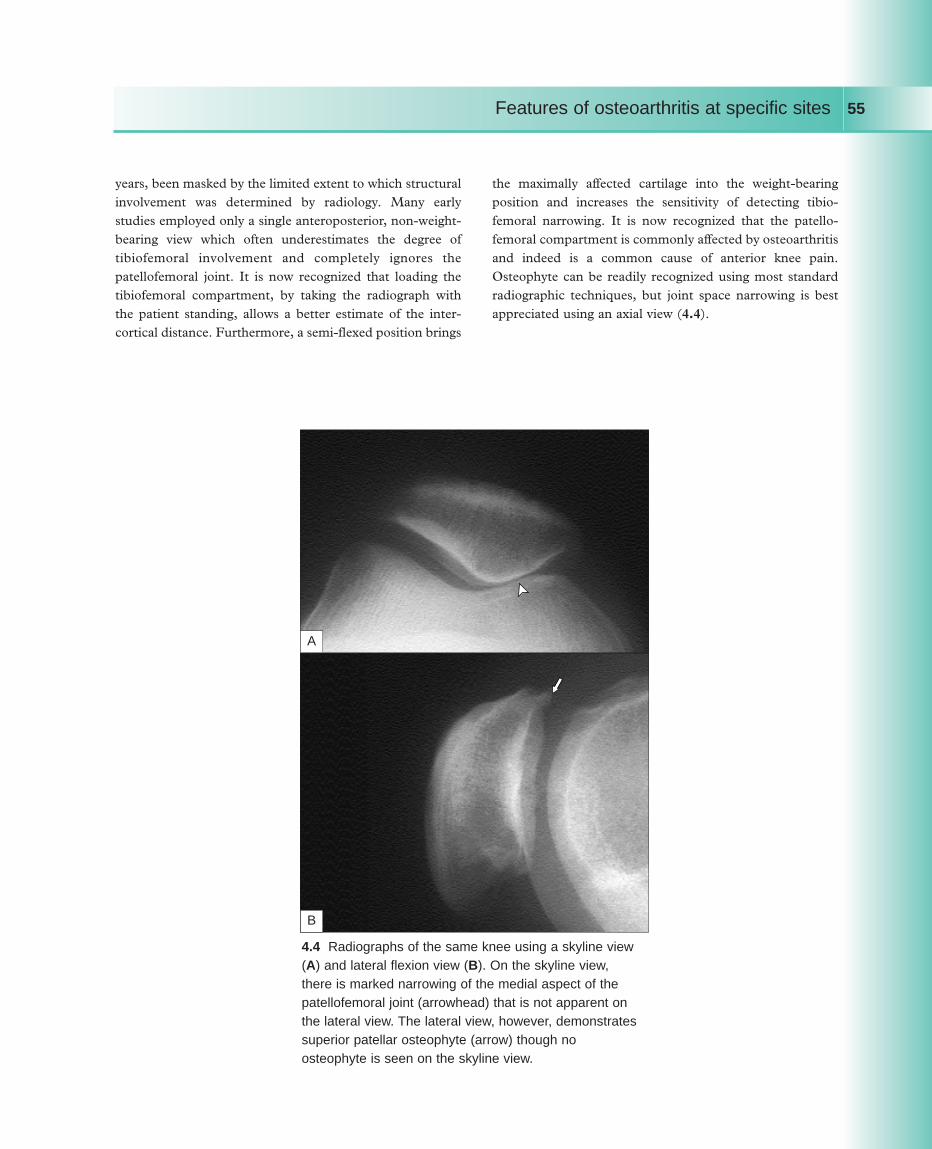
years, been masked by the limited extent to which structuralinvolvement was determined by radiology. Many earlystudies employed only a single anteroposterior, non-weight-bearing view which often underestimates the degree oftibiofemoral involvement and completely ignores thepatellofemoral joint. It is now recognized that loading thetibiofemoral compartment, by taking the radiograph withthe patient standing, allows a better estimate of the inter-cortical distance. Furthermore, a semi-flexed position brings
the maximally affected cartilage into the weight-bearingposition and increases the sensitivity of detecting tibio-femoral narrowing. It is now recognized that the patello-femoral compartment is commonly affected by osteoarthritisand indeed is a common cause of anterior knee pain.Osteophyte can be readily recognized using most standardradiographic techniques, but joint space narrowing is bestappreciated using an axial view (4.4).
Features of osteoarthritis at specific sites 55
B
A
4.4 Radiographs of the same knee using a skyline view(A) and lateral flexion view (B). On the skyline view,there is marked narrowing of the medial aspect of thepatellofemoral joint (arrowhead) that is not apparent onthe lateral view. The lateral view, however, demonstratessuperior patellar osteophyte (arrow) though noosteophyte is seen on the skyline view.

Using sensitive imaging techniques, it is apparent thatmost cases of knee osteoarthritis involve all threecompartments to some extent, but cartilage narrowingusually predominates in just one or two compartments.Osteophytes are often more widespread at many sites withinthe knee, and tend to show characteristic directions ofgrowth. For example, in the medial tibiofemoral com-partment, they tend to grow horizontally and then awayfrom the joint line (4.5); whereas in the lateral tibiofemoralcompartment, the femoral osteophyte grows proximallyaway from the joint line, but the tibial osteophyte oftengrows vertically towards the femur (4.6). In thepatellofemoral compartment, lateral subluxation is common(readily seen on the skyline view) and osteophyte tends togrow laterally, hugging the femoral contour (4.7). Lateralviews show osteophyte superiorly and inferiorly, and oftenenthesophyte at the quadriceps and patella tendon insertionsites (4.8). Presumably, these differences in osteophytedirection are driven by biomechanical forces. Surgical
removal of tibiofemoral osteophytes has been shown toincrease joint movement, so the osteophytes appear to be astabilizing factor that help splint the osteoarthritic joint as itloses cartilage and remodels its shape.
Osteochondral bodies are not infrequent within thecapsule and ligaments of the joints (4.9), and may relate tolocking, if close to the joint line. Chondrocalcinosis is readilyappreciated in the knee, and 80% of cases of chondro-calcinosis are apparent on anteroposterior radiographs.Calcification occurs in the fibro- and hyaline cartilage andmay also be apparent in the capsule of the joint (4.10).
Predominant medial tibiofemoral osteoarthritis appearsto be particularly common in men. Associated meniscaldegeneration is usually a feature, and is more oftenappreciated if other imaging modalities (such as MRI) areused. Varus deformity, often with fixed flexion if severe, isthe most common deformity in men with kneeosteoarthritis. Varus deformity is a risk factor for more rapidX-ray progression.
Features of osteoarthritis at specific sites56
4.5 Medial tibiofemoral osteophytes, growing horizontally (arrow) or away(arrowhead) from each other.

Features of osteoarthritis at specific sites 57
4.6 Lateral tibiofemoral osteophyte. The femoral osteophyte (arrow)is growing proximally away from the joint, but the tibial osteophye(arrowhead) can be seen coming upwards towards the joint.
4.7 Skyline view showing marked lateral narrowing, lateral patellasubluxation, and osteophytes on both the femoral and patellaaspects (arrows), following a similar direction of growth.

Features of osteoarthritis at specific sites58
4.8 Lateral view showing superior and inferior patellar andfemoral osteophytes (arrowheads), and enthesophyte (arrow) atthe quadriceps tendon insertion into the anterior/superior aspectof the patella.
4.9 Anteroposterior radiograph of the knee demonstrating markedosteochondral body formation.

Features of osteoarthritis at specific sites 59
4.10 Knee radiograph showing medial tibiofemoral osteoarthritis andchondrocalcinosis of fibrocartilage and hyaline cartilage(‘pyrophosphate arthropathy’).
Predominant patellofemoral osteoarthritis is alsocommon, particularly in women. Since the patella isintimately involved in the quadriceps mechanism, musclewasting and disability appear to be a prominent feature ofpatellofemoral osteoarthritis. Characteristics of patello-femoral pain include well-localized anterior knee pain,particularly bad when going up or down stairs or an incline,and progressive aching anteriorly when sitting, relieved bygetting up and ‘stretching the legs.’
The third most common pattern is a combination of bothpatellofemoral and medial tibiofemoral osteoarthritis. Themore knee compartments involved, and the more severe thestructural changes, the more likely it is to detect calciumpyrophosphate or apatite crystals in aspirated knee synovialfluid.
Isolated lateral tibiofemoral osteoarthritis is a veryuncommon pattern. However, pathological involvement isoften present, and it may be that the natural varus deformity
(common in medial tibiofemoral osteoarthritis) tends toopen up the lateral tibiofemoral joint. If this is the case,cartilage loss in this compartment is not readily appreciatedradiographically. Predominant involvement of the lateraltibiofemoral joint may be associated with preceding trauma,previous lateral meniscectomy, or apatite-associateddestructive arthritis (4.11). A valgus deformity is the usualconsequence of severe involvement.
In contrast to osteoarthritis, chronic inflammatorysynovitis, such as occurs with rheumatoid arthritis, usuallyresults in a diffuse tricompartmental pattern of joint spaceloss and little, if any, bone response (4.12).
The superior tibiofibular joint is the final joint of theknee. Although radiographic osteoarthritis and oftenmarked cystic change (4.13) can occur at this joint, it israrely a cause of symptoms. Cystic change at this site ismore common in patients with knee chondrocalcinosis.

Features of osteoarthritis at specific sites60
4.11 Apatite-associated destructivearthritis targeting the lateral tibiofemoralcompartment of an elderly woman.Note the severe bone attrition, valgusdeformity, and soft-tissue outline of alarge effusion.
4.12 Knee radiographs of a patient with rheumatoid arthritis showingdiffuse joint space narrowing with osteopenia, and without osteophyteor sclerosis.
4.13 Marked cysticchange (arrows) each sideof the superior tibiofibularjoint in a patient withpyrophosphate arthropathyof the knee.

Features of osteoarthritis at specific sites 61
Features of osteoarthritis of the hip
Osteoarthritis of the hip is a common clinical problem, andis responsible for a great deal of pain and suffering, as wellas being the main indication for hip arthroplasty. Unlikeosteoarthritis elsewhere, it shows an overall equal sexdistribution, but it predominates in younger men (pre-retirement age) and in elderly women. The principle clinicalfeature is pain. This is usually felt anteriorly, deep in thegroin. It may, however, radiate widely and can be felt in theanteromedial thigh, buttock or the anteromedial aspect ofthe knee, and may extend as far as the ankle. It can presentas knee pain alone, and this may lead to clinical confusion
until the knee and hip are both examined.Restriction of movement commonly occurs and may be
relatively painless. The restriction initially affects internalrotation of the hip, particularly in flexion (4.14). Later aflexion deformity of the hip occurs, and the patient may walkwith a flexed knee to compensate and a degree of equinus.Leg length shortening, due to both the flexion deformity andpossibly loss of joint space, can result in a compensatoryscoliosis. At an even later stage, a fixed adduction andexternal rotation of the hip can occur (4.15, 4.16).
4.14 Internal rotation with the hip inflexion. This is first movement tobecome painful and restricted with hiparthropathy, and is the most sensitiveclinical test for early hip osteoarthritis.
4.15 Right hip osteoarthritisin a 59-year-old man,showing the typical deformityof hip flexion (withcompensatory knee flexion)and external rotation.
4.16 Advanced right hip osteoarthritisin an elderly woman, showing typicallate deformity of hip (and knee)flexion, external rotation, andadduction at the hip.

Muscle wasting is not readily appreciated at the hip,especially in elderly patients, although gluteal andquadriceps wasting is often present. Acute synovitis canoccur, including pseudogout, but appears to be unusual inclinical practice. Functional limitation is often a majorproblem through interference with daily activities, such asputting on socks, getting in and out of cars, and walking.
Radiographically, osteoarthritis of the hip tends to be afocal disease with regard to joint space loss. Superior-
narrowing is the most common pattern, especially in men,though focal-narrowing may also be axial or medial (4.17).The superior pattern may be predominantly superolateral(4.18), and associate eventually with upwards and outwardssubluxation of the femoral head, or superomedial. Themedial (4.19) or axial patterns are less common (4.20), butan axial pattern, in particular, may result in a protrusioacetabulae abnormality (4.21).
Features of osteoarthritis at specific sites62
4.17 Diagram showing the different sites for focalnarrowing at the hip. Superior
Axial
Medial
4.18 Superolateral narrowing in hiposteoarthritis. There is also osteophyte onthe femoral head (arrows), and sclerosis atthe site of maximal narrowing (arrowhead).

Features of osteoarthritis at specific sites 63
4.19 Medial narrowing in hip osteoarthritis (arrow).
100
90
80
70
60
50
40
30
20
10
0
%
Women Men
Indeterminate
Medial
Axial
Superior
4.20 Different patterns of osteoarthritis of the hip. (Dataderived from a sibling study of hip osteoarthritis byLanyon P, Muir K, Doherty S, et al. [2004]. Influence ofradiographic phenotype on risk of hip osteoarthritis withinfamilies. ARD, 63:259–263.)
4.21 Protrusio acetabulae abnormality – the femoral headhas migrated axially, narrowing the acetabular bone andpushing it inwards into the pelvis (arrowheads).

Sometimes, especially with late, marked osteoarthritis, adiscrete pattern is hard to discern (an ‘indeterminate’pattern). A uniform concentric pattern of cartilage loss isvery rare and, especially if there is paucity of accompanyingosteophyte, should always suggest an underlying inflam-matory or metabolic disease.
Unless there is a specific localizing factor in operation,such as prior trauma, the pattern of involvement is usuallyidentical if osteoarthritis affects both hips.
Osteophyte occurs at specific locations which can includethe acetabular margin, and the site of capsular insertion onthe femur where it may appear as a line across the femoralhead, which is in reality a two-dimensional view of a ring ofosteophyte (4.22). Cortical buttressing may occur, as well as
Features of osteoarthritis at specific sites64
4.22 Superolateral hip osteoarthritis. Note the osteophyte (arrows) that isforming a collar around the margins of the femoral head, appearing as awavy line crossing the femoral neck in this two-dimensional radiograph.
trabecular thickening. The latter tends to occur parallel tothe forces acting on the femoral neck, and thus buttress themedial aspect of the femoral neck. Chondrocalcinosis maybe seen occasionally in the hyaline cartilage of the femur(4.23), and in the acetabular labrum, though it is moreprevalent in the symphysis pubis (4.24). Osteochondralbodies may be seen around the joint, with some appearingto form as extensions to acetabular osteophyte.
Cyst formation can occur in the acetabulum, and is parti-cularly common in patients with pyrophosphate arthropathy,especially in association with haemochromatosis (4.25).Avascular necrosis of the femoral head can sometimes beappreciated as a focal-marked loss of bone stock.

Features of osteoarthritis at specific sites 65
4.23 Hip chondrocalcinosis affecting the femoral headhyaline cartilage, just visible superolaterally (arrow).
4.24 Chondrocalcinosis (arrow) of thesymphysis pubis (the most common sitefor chondrocalcinosis in the pelvis).
4.25 Hip radiograph in a patient with haemochromatosis.Note the diffuse narrowing and multiple small cysts. Anadditional clue to underlying metabolic disease ischondrocalcinosis of hyaline cartilage, seen laterally onthe femoral head (arrow).

Features of osteoarthritis at specific sites66
4.26 Paget’s disease affecting the left hemipelvis and resulting inosteoarthritis. Note the diffuse, rather than focal narrowing at the hip.
There has been discussion as to how much dysplasiamight predispose to osteoarthritis. It appears to be anunusual cause of sporadic osteoarthritis, but occasionallypatients presenting with osteoarthritis show clear evidence ofacetabular dysplasia, or even possible congenital dislocationof the hip. Prior trauma leading to leg length shortening ormalalignment may be appreciated. All these factors havebeen implicated in determining speed of progression in hiposteoarthritis, but recent meta-analyses suggest that onlysome of these may be important (Table 4.1).
Paget’s disease is common in the pelvis, and may affectthe acetabulum with resultant hip osteoarthritis, often withdiffuse joint space narrowing (4.26).
Good evidence for a positive effect• Superolateral migration of the femoral head• Atrophic bone response
Good evidence for no effect• Body mass index• Hip dysplasia
Lievense AM, Bierma-Zeinstra SMA, Verhagen AP, et al. (2002). Prognostic factors of progress of hiposteoarthritis: a systematic review. Arthritis Care Res,47:556–562.
Table 4.1 Hip osteoarthritis: factors associated with progression

Features of osteoarthritis at specific sites 67
Features of osteoarthritis of the hand and wrist
All joints of the hand and wrist can potentially be affected byosteoarthritis (4.27, 4.28), although certain patterns ofinvolvement are characteristic. Distal interphalangeal jointinvolvement is particularly common, often as part ofgeneralized nodal osteoarthritis. Clinically, this usually startswith development of painful Heberden’s nodes around thetime of the menopause. These often appear cystic or soft inthe early stages of development (4.29) and occur at thesuperolateral aspects of the joint. Once established, the twosuperolateral swellings either side of the extensor tendonmay fuse to form a hard bony posterior bar (4.30). Naildystrophy with ridging (‘Heberden’s nodes nails’) is anoccasional associated feature (4.29). Varying degrees ofinflammation can be appreciated while the nodes evolve totheir final form. The onset tends to be stuttering withsequential involvement of new joints, often in a symmetricalfashion, with earlier involvement of the dominant hand.
4.27 Osteoarthritis of the pisiform bone of the wrist. Notesclerosis (arrows), cyst formation (arrowheads) and jointspace loss.
4.28 Accrual of joint involvement by ageand gender in a Finnish population.(Data derived from Haara MM, KrogerH, Arokoski JP, et al. [2003].Osteoarthritis of finger joints in Finnsaged 30 or over. Prevalence,determinants and association withmortality. ARD, 62:151–158.)
80
70
60
50
40
30
20
10
030–44 45–54 55–64 65–74 75+
Age (years)
Male DIP
Male PIP
Male MCP
Male CMC
Symmetrical DIP
Female DIP
Female PIP
Female MCP
Female CMC
%

Features of osteoarthritis at specific sites68
4.29 Superolateral cystic change in anearly Heberden’s node. Note theaccompanying nail dystrophy.
4.30 Established Heberden’s node forming a single hard bar over thesuperior aspect of the joint.
4.31 Radiograph showing distalinterphalangeal osteoarthritis inassociation with a Heberden’s node,showing narrowing, osteophyte,sclerosis, and cysts.
Radiographically, joint space narrowing, sclerosis,osteophytes, and cysts may all occur (4.31). Similarinvolvement of the proximal interphalangeal joints formingBouchard’s nodes may occur, although this is less common.Although usually symptomatic for several years whileevolving, Heberden’s and Bouchard’s nodes often becomeasymptomatic once fully developed and the prognosis, withrespect to subsequent hand function, is usually good.
Gout has a tendency to develop in previously-damagedjoints and in post-menopausal women, particularly those onlong-term diuretics (thiazides or loop diuretics). Goutytophi may form at the site of distal interphalangealosteoarthritis (4.32). In severe cases, acute inflammationmay occur; and with chronic deposition, the tophi may growvery large, and may even ulcerate and discharge whitetophaceous material (4.33).

Features of osteoarthritis at specific sites 69
4.32 Chronic diuretic-induced gouty tophi selectively depositing aroundHeberden’s nodes. The white colour of the monosodium urate crystaldeposits can easily be seen.
4.33 An ulcerating tophus on a Heberden’s node.

In the variant known as ‘erosive osteoarthritis,’ theinvolvement of distal and proximal interphalangeal joints ismore equal. Clinically, ligamentous instability seems morecommon, and lateral and medial deviations of the joint maybe seen either at rest or on stressing the joint (3.5). Inaddition, bony fusion may lead to stiffness and rigidity of thejoints. Therefore, the functional impairment that resultsfrom this form of finger osteoarthritis is often high.Subchondral erosion is the hallmark of this condition, and
may give rise to a typical ‘gull’s wing’ appearance, andsometimes resultant bony ankylosis (3.6, 3.7).
Involvement of the ‘thumb base’ is common. Pain fromthis site is felt mainly in the snuff box area of the radial sideof the wrist, although it often radiates proximally into thelateral border of the forearm, and distally up the thumbmetacarpal. The pain may be provoked on examination byextending and adducting the thumb, and is often madeworse by gripping. Bony swelling and subluxation may
Features of osteoarthritis at specific sites70
4.34 Squaring of the thumb base due to osteoarthritis.Note the accompanying wasting of the thenar eminence.

Features of osteoarthritis at specific sites 71
4.35 Radiograph showing osteoarthritisof the scaphotrapezoid (arrow) andtrapeziometacarpophalangeal joints(arrowhead) with sclerosis andnarrowing the main features.
4.36 Prominent bony swelling of theindex and middle metacarpophalangealjoints due to osteoarthritis in a 62-year-old man who had a physicallydemanding job as a machinist.
result in squaring of the thumb base, and wasting andweakness of the thenar muscles may occur (4.34).
Radiographically there may be involvement of either thescaphotrapezoid or the trapeziometacarpophalangeal joint,or both (4.35). Joint space narrowing and sclerosis are oftenthe main features, though osteophyte and cysts may alsooccur.
Metacarpophalangeal joint involvement, mainly affectingthe index and middle finger joints, is relatively uncommon
and should lead to consideration of pyrophosphatearthropathy and haemochromatosis. However, it may occurin more elderly patients with nodal generalized osteo-arthritis, or in men who have physically demandingoccupations (‘Missouri arthropathy’). Clinically bonyswelling of the joint is seen (4.36) together with crepitus andoccasional soft-tissue swelling. The appearance can be somarked as to lead to possible confusion with the changesseen in rheumatoid arthritis. Radiographically there may be

Features of osteoarthritis at specific sites72
4.37 Hand radiograph of the patient in4.36. Note the narrowing of bothaffected metacarpophalangeal joints andthe large ‘hook’ osteophyte (arrow) onthe radial aspect of the middle fingermetacarpal head.
4.38 Haemachromatotic arthropathy. Note involvement ofthe metacarpophalangeal joints with ‘hook’ osteophytes onthe ulnar border (arrow). Note additional involvement of theradiocarpal joint and multiple small cysts at that site(arrowhead).

Features of osteoarthritis at specific sites 73
joint space narrowing, sclerosis, cysts, and osteophyte.Large ‘hook’ osteophytes, mainly on the radial side of thedistal metacarpal, may be striking (4.37). In haemo-chromatosis there may be more widespread involvement ofall metacarpophalangeal joints, additional involvement ofthe radiocarpal and midcarpal joints, multiple subchondralbone cysts and chondrocalcinosis (4.38).
Involvement of the radiocarpal joint is relativelyuncommon, but more likely to occur in the context ofpyrophosphate arthropathy or previous trauma, particularlyscaphoid fracture. Missed scaphoid fracture with
displacement may result in avascular necrosis of theproximal portion of the scaphoid, due to its retrogradevascularization via the neck of the scaphoid. Clinically, thereis usually restriction of flexion and extension of the jointwith varying degrees of bony swelling, crepitus, and possiblysynovitis. Radiographically, the features are the same as seenelsewhere, although there may be so-called scapholunatedissociation presumably as a result of damage to the inter-osseous ligaments by synovitis (4.39). Although unusual,other synovial joints in the hand and wrist may be affected.
4.39 Wrist radiograph in pyrophosphate arthropathy showingchondrocalcinosis of the triangular ligament (arrow) and osteoarthritis ofthe radiocarpal, midcarpal, and scaphotrapezioid joints. Note the V-shapeddepression in the distal radius (arrowhead) (associated with scapho-lunatedissociation) and marked bone attrition also in the proximal carpal row.

Features of osteoarthritis of the spine
Osteoarthritis of spine is often neglected in many studies ofosteoarthritis. All the spinal synovial joints can be affectedalthough some are targeted more than others. The mostfrequently affected are the apophyseal or facet joints,particularly in the lower cervical and lumbar spine. Theclinical features of osteoarthritis of the facet joints can bedifficult to correlate with the structural and radiographicabnormalities. Pain from spinal structures is often diffuseand hard to localize. Asymptomatic radiographic change iscommon and so attribution of symptoms to structuralchange is problematic. Indeed, provocation studies suggestthat the pattern of pain produced by different spinalstructures (joints, ligaments, discs, and muscles) can besimilar if not identical. Previously, it was taught that pain onspinal extension was typical of lumbar facet jointosteoarthritis, but recent studies suggest that this is not thecase, and there is no specific pain syndrome associated withfacet joint arthritis.
Osteophyte formation can, however, be associated withencroachment of the neural foramina and subsequentimpingement of the nerve root (4.40). This may produce aspecific radiculopathy which may be provoked or aggravatedby posture. Encroachment of the central canal may result incord compression or symptoms of spinal claudication. Thismay be associated with ligamentum flavum hypertrophy andosteophyte formation at the site of the intervertebral discinsertion. This is discussed below when consideringspondylosis.
Radiographically, facet joint arthropathy may be difficultto appreciate. On anteroposterior radiographs, sclerosis maybe seen around the facet joints, and the presence ofosteophyte may result in an apparent narrowing of theinterpedicular distance as one looks caudally. On the lateralview sclerosis may be apparent, but the multiple overlappingbones and joints may make this difficult to evaluate.Foraminal encroachment may be observed, particularly if
Features of osteoarthritis at specific sites74
4.40 Foraminal encroachment resulting in nerve root compressionin a patient with osteoarthritis of the cervical spine (arrow). Notedifference in foraminal diameter between the involved superiorforamen and the uninvolved lower foramen.

Features of osteoarthritis at specific sites 75
4.41 Lateral MRI scan of the neck. Loss of disc heightand hydration is easily appreciated as blackening of thedisc as well as posterior bony encroachment on the spinalcord (arrows).
4.42 CT scan showing narrowing (arrow) andexuberant osteophytosis (arrowheads) ofapophyseal joints.
oblique views are taken centred on the foramen in question.There is no doubt that cross-sectional imaging, such as CTscanning and MRI scanning, is clearly superior in terms ofevaluating structural change in osteoarthritis (4.41, 4.42).Although MRI scanning may detect oedema of the adjacent
bone which may correlate with symptoms, these techniquesessentially evaluate structure. Radioisotope scanning maydetect increased bone turnover which, in turn, may correlatebetter with symptoms and response to local injection.
Other synovial joints in the spine, such as the atlantoaxial

joint and the atlanto-occiptal joints, may demonstratefeatures of osteoarthritis, but these are rarely clinicallysymptomatic. The joints of Lushka are the evolutionaryremnants of the costotransverse joints of the cervical spine.The clinical consequences of involvement of these joints canbe hard to determine, but osteophyte formation may causeimpingement symptoms, such as dysphagia.
Occasionally, failure of segmentation of the spine maylead to additional joints, and these may develop features ofosteoarthritis. A good example of this is the joint that maysometimes develop between the ilium of the pelvis and thetransverse process of a lumbar vertebra.
While not a synovial joint, the discovertebral joint of thespine can develop features that have some similarities withosteoarthritis. Loss of hydration of the gelatinous nucleosuspulposus of the disc may result in loss of intervertebral height(4.43). Osteophytes may develop at the insertions of theannulus fibrosus and the longitudinal ligaments. These maysimply produce a radiographic appearance which may beflorid although often asymptomatic (4.44). It is likely thatsome disc changes, particularly if associated with bonemarrow oedema, may cause pain. Large osteophytes maycause impingement on other structures. Anteriorly this mayinclude the oesophagus, laterally the exiting nerve roots, and
Features of osteoarthritis at specific sites76
4.43 Lateral cervical spine radiograph showingintervertebral narrowing between the 5th and 6th cervicalvertebra, with sclerosis and remodelling of the affectedvertebrae (arrows).
4.44 Lumbar spine radiograph showing narrowing atL2/L3 and L3/L4 and associated large lateral osteophytesarising from the vertebral end plates (arrows).

Features of osteoarthritis at specific sites 77
posteriorly it may encroach on the central canal. Theshortening that results from loss of disc height may producebuckling of the longitudinal ligaments which further narrowthe bony canal. Although frank cord compression may beproduced with typical long-tract signs (in the lumbar regionparticularly), the syndrome of spinal claudication mayalso result.
Changes in the disc are often associated with facet jointarthropathy at the same level and this complex is oftentermed ‘spondylosis’, although this is a somewhat impreciseterm and may be used differently by different clinicians. Theissue as to whether facet joint osteoarthritis or disc
degeneration is the initiating event is difficult, but moststudies suggest that usually disc degeneration/dehydrationoccurs first. It is possible that the relative instabilityproduced by the lost disc height increases stress on the facetjoint and produces secondary osteoarthritic changes.Indeed, anteroposterior slippage may occur resulting in aspondylolisthesis (4.45).
Occasionally, there is loss of bone and a markeddegenerative scoliosis may develop (4.46). Although thismay cause major deformity, it may be surprisingasymptomatic.
4.45 Lateral lumbar spine view demonstratingloss of disc height and a subsequentdegenerative spondylolisthesis (arrows).
4.46 Degenerative scoliosis of the spine. Notethe wedging of the lateral vertebral body andassociated disc degeneration.

Features of osteoarthritis at specific sites78
4.47 MRI scan of the cervical spine (A) showing osteophytosis discdegeneration and subsequent cordcompression of the cervical cord at C2/3 and C3/4 (vertebral bodies arenumberered). Indentation of the cord is clearly visible with loss of thesurrounding high signal from thecerebrospinal fluid (normal anatomyshown: spinal cord [arrow head];cerebrospinal fluid [short arrow]). In addition, there is high signal within the cord suggestive of cord oedemaand/or ischaemia (arrows). The plainanteroposterior radiograph (B)demonstrates florid facet jointosteophytosis, narrowing, and sclerosis.
4.48 CT scan showing calcificationdue around the odontoid peg(‘crowned dens syndrome’) (arrow).
A
B
6
5
4
3
1
2

Features of osteoarthritis at specific sites 79
Spinal claudication is a clinical syndrome when exertionand/or spinal extension results in neurogenic pain, usually inthe buttocks and perineum, and in more severe casesneurological dysfunction (4.47). On resting, this showsreversibility in the same way that claudication due toperipheral vascular disease does, though the time-scale forrecovery is usually longer: 30 minutes compared to a fewminutes. Clinically, it can be provoked by exercising thepatient and evaluating neurological signs and symptomsbefore and after exercise. An alternative, and possiblysimpler approach, is to hold the patient in lumbar extensionfor 2 minutes.
CPPD deposition can occur in the fibrocartilaginousdiscs and spinal ligaments. Occasionally, it occurs aroundthe dens (4.48). Acute pseudogout episodes in relation tothese deposits can cause self-limiting episodes of
meningism, fever, neck and head ache, and sometimesconfusion.
No discussion of osteoarthritis of the spine would becomplete without a discussion of diffuse idiopathic skeletalhyperostosis (DISH or Forestier’s disease). In thiscondition, there is flowing anterolateral paraspinal new boneformation that can bridge vertebrae and superficiallyresemble osteophytes or the syndesmophytes of ankylosingspondylitis. It particularly affects the right side of thethoracic spine (4.49) and both sides of the lumbar spine. Incontrast to osteophyte, the underlying disc is usuallystructurally normal and the bridging new bone initiallyprojects vertically rather than horizontally. It differs fromsyndesmophyte in that the new bone is almost layered ontothe vertebrae (4.50), rather than arising from the annulusfibre insertions.
4.49 DISH. Flowing new boneformation bridging between vertebraeon the right side of the thoracic spine.
4.50 DISH. Lateral radiograph showing anteriornew bone separate and ‘layered’ onto the anteriorvertebral bodies. Note the initial projection of theosteophyte is away from the vertebral body ratherthan parallel to the annulus as is seen inankylosing spondylitis. Note also ‘cleft’ betweenvertebral body and new bone (arrows). Theintervertebral disc spaces are normal.

Features of osteoarthritis at specific sites80
4.51 DISH. Lateral radiograph of the hindfootshowing exuberant new bone ‘layered’ overthe back of the calcaneum and growing intothe Achilles tendon (arrow) and plantar fasciainsertion (arrowhead) sites. This patient hadsymptoms at both enthesis sites.
4.52 Radiograph showing osteoarthritis of thelower synovial part of the sacroiliac joint, withfocal narrowing, sclerosis, and minor inferiorosteophyte.
This diffuse hyperostosis may also occur at peripheralenthesis sites, particularly around the pelvis, knee, heel, andelbow (4.51). Symptoms that have been attributed to DISHincluded spinal stiffness and peripheral entheseal pain. Themechanism of the hyperostosis is unclear though DISH onlyoccurs in middle-aged and elderly subjects and shows a
strong association with obesity and type II diabetes. The sacroiliac joint is part fibrous and part synovial.
Although not a commonly appreciated problem, osteo-arthritis of the sacroiliac joint does occur and may bedifficult to distinguish radiographically from sacroiliitis seenin ankylosing spondylitis and the seronegative arthropathies.

Features of osteoarthritis at specific sites 81
4.53 The first three diagramsare of sacroliliitis showing: (A) erosion of the cortical linesand initial apparent widening ofthe joint space; (B) progressivesclerosis and narrowing of thejoint, still with ill-defined corticallines; and (C) eventualobliteration of joint space and bony ankylosis. In contrast,the final diagram (D) showsosteoarthritis with focalnarrowing, sclerosis, and inferior osteophyte with distinct cortical lines.
A B
C D
Irregularity of joint space width with sclerosis and inferiorosteophyte are the typical x-ray features (4.52), and the earlyjoint space widening, cortical erosion, and late ankylosisseen with sacroiliitis do not occur (4.53). Whetherosteoarthritis of the sacroiliac joint is a common cause ofsymptoms is unclear.
Similar changes to that seen in the discovertebral jointsmay be seen in the symphysis pubis, another non-synovial,fibrous articulation. Sclerosis and apparent osteophyteformation are commonly seen, but it is unclear how muchthis is a normal developmental variation and how much areaction to stress.

Features of osteoarthritis of the shoulder girdle
The shoulder girdle comprises the glenohumeral, acromio-clavicular, and sternoclavicular joints, all of which may beaffected by osteoarthritis.
Acromioclavicular joint osteoarthritis is a common causeof pain. This is localized over the joint and notedparticularly on abduction of the shoulder. On examination,a superior arc of pain may be observed (4.54), and thesimplest way to provoke the pain is with a scarf test – theshoulder is flexed to 90 degrees, and the arm adducted andinternally rotated so the hand moves towards the oppositeshoulder – with pain being felt principally over the apex ofthe shoulder. Crepitus can be felt on palpation duringmovement. Osteophyte can form superiorly where it can be
palpated and may form a visible prominence (4.55).Chondrocalcinosis is another occasional cause of palpableswelling (4.56). Inferior osteophyte may impinge on thesubacromial space and cause pain on abduction or stress ofthe supraspinatus muscle. Osteophyte formation and bonysclerosis may be difficult to appreciate on routine shoulderradiographs (which are usually designed to image theglenohumeral joint), but may be more apparent whendedicated acromioclavicular views are obtained. Associateddegeneration of the underlying supraspinatus tendon maylead to rupture of the tendon and subsequent weakness ofshoulder abduction.
Features of osteoarthritis at specific sites82
4.54 Two classic painful arcs. A painful middle arc (A) causespain in the upper arm and may occur with either asupraspinatus lesion or subacromial bursitis. A painful superiorarc (B) causes pain at the acromioclavicular joint site.
B
A

Features of osteoarthritis at specific sites 83
4.55 Prominent acromioclavicular joint swelling due tosuperior osteophyte.
4.56 Exuberant chondrocalcinosis forminga palpable CPPD ‘tophus’ over theacromioclavicular joint (arrow).

The glenohumeral joint is not a typical target site forosteoarthritis, but this may occur in the elderly, particularlyin association with pyrophosphate arthropathy. This joint isalso a target site for AADA or ‘Milwaukee shoulder’. Gleno-humeral pain is felt mainly in the dorsal aspect of the upperarm (‘sergeant’s stripes region’, C5 dermatome), oftenoccurring throughout the whole range of movement of theshoulder. There may be restriction of movement, especiallyabduction and external rotation and global muscle wasting.In addition, there may be associated rotator cuff disease withpain and possible weakness on resisted active movement(abduction – supraspinatus; external rotation – infraspinatus,teres minor; internal rotation – subscapularis (4.57). Acutecrystal synovitis, due to CPPD (glenohumeral pseudogout)or apatite (acute calcific periarthritis), may lead to largeglenohumeral and subacromial effusions, which may rupture
and cause extensive bruising that tracks down the arm (4.58).Radiographically, osteophytes may be appreciated most
commonly on the inferior border of the glenoid, but alsoaround the humeral head (4.59). However, joint space andsubsequent flattening of the humeral head with sclerosis isoften more readily appreciated, with an occasionaladditional finding of osteochondral bodies. Chondro-calcinosis of the hyaline cartilage and glenoid labrum maybe seen radiographically, and apatite deposition in thesuperior rotator cuff (‘calcific periarthritis’) may also occur(4.60). If there is marked rotator cuff disease, the humeralhead often migrates upwards (4.61).
Although the sternoclavicular joint commonly developsosteoarthritis, this is rarely a clinical problem. Ligamentousdegeneration may result in subluxation of the joint and abony prominence (4.62).
Features of osteoarthritis at specific sites84
4.57 Anteroposterior radiograph of the shoulder. Notesclerosis (arrows) and encroachment laterally on thesubacromial space which may result in symptoms ofimpingement on abduction.

Features of osteoarthritis at specific sites 85
4.58 Acute pseudogout that has led to rupture of the glenohumeralcapsule and subsequent bleeding and bruising down the arm. Thiscan also occur with calcific arthritis.
4.59 Glenohumeral osteoarthritis, showing narrowing,sclerosis, and a cuff of osteophyte around the marginsof the humeral head (arrowheads).

Features of osteoarthritis at specific sites86
4.60 Chondrocalcinosis of hyaline cartilageforming a thin line close to, and parallel tothe cortex of the humeral head (arrow). Inaddition, there is more dense deposits ofapatite in the fibrous superior rotator cuff(arrowhead).
4.61 Glenohumeral osteoarthritis withassociated rotator cuff degeneration and someassociated widening of the acromioclavicularjoint (arrow). Note joint space loss of theglenohumeral joint (arrowhead).
4.62 An asymptomatic sternoclavicular joint swelling due toosteoarthritis. Examination revealed bony swelling and crepituson shrugging the right shoulder.

Features of osteoarthritis at specific sites 87
Features of osteoarthritis of the foot and ankle
Any joint of the foot and ankle may be affected, but againthere is a predilection for certain joints.
Probably the most common joint affected is the 1st
metatarsophalangeal joint of the hallux or big toe (4.63).This may occur as an isolated feature, but often is associatedwith so-called bunion formation (4.64). The principlefeatures of a bunion are a marked valgus deformity of theproximal phalanx and formation of an adventitial bursa over
4.63 Radiograph showing osteoarthritis of the1st metatarsophalangeal joint with focalnarrowing, sclerosis and mild hallux valgusdeformity.
the medial aspect of the joint. The major aetiological factorin bunion formation is probably varus alignment of the firstmetatarsal bone, usually as an inherited variation (4.65).
Clinically, there is pain over the joint, particularly ondorsiflexion of the hallux. Since dorsiflexion is necessary inthe toe-off phase of gait, painful or poor mobility oftenresults. On examination, there is restricted dorsiflexion,crepitus on movement, stress pain on dorsiflexing the toe,
4.64 Bunion formation in a patient withhallux valgus due to osteoarthritis of thefirst metatarsophlangeal joint.

and bony swelling. If a bunion is present, the valgusalignment of the toe will be seen, and varying degrees ofcystic bursal swelling or bony osteophytosis may bepalpable. Occasionally, the bursa can become markedlyinflamed with local heat and erythema. Gout has apredilection for damaged joints and a superimposed goutyarthritis may be developing (4.66).
The ankle mortise joint (between the tibia, fibula, andtalus) may develop osteoarthritis, especially in the context ofpyrophosphate arthropathy, and repetitive impact loading(parachuting) or trauma may be an overt aetiological factor.Pain in and around the ankle, particularly felt anteriorly,
occurs on dorsiflexion. Involvement of the lateral or medialmalleoli may result in pain on lateral or medial stress.Restriction of movement, particularly dorsiflexion, occurs inlater disease and there may be varus or valgus hindfootdeformity. Radiographically, joint space narrowing can beappreciated; notably on anteroposterior views (4.67),although the loss of cartilage can be extremely focal.Osteophytes are more readily appreciated on the lateral view(4.68), and tend to occur on the anterior margin of the tibiaand sometimes the talus. Other osteophytes may developinferior to the medial and lateral malleoli.
Features of osteoarthritis at specific sites88
4.65 Radiograph showing metatarsusprimus varus.
4.66 Acute gout affecting the 1st
metatarsophalangeal joint – ‘podagra’.

Features of osteoarthritis at specific sites 89
4.67 Anteroposterior views of right ankle joint of a man in his 60s with pyrophosphate arthopathy. The twoviews were taken just 3 years apart. Note the marked narrowing, sclerosis, deformity and osteophyte, andprogressive bone attrition over this relatively short time.
A B
4.68 Lateral views of same patient as in 4.67, again showing marked bone attrition over the 3-year period.
A B

Subtalar involvement may be hard to establish, but canresult in reduced eversion and inversion of the hindfoot withpain, especially when walking on uneven surfaces (4.69).Radiographically, sclerosis may be seen and occasionalosteophytosis (4.70), although the complex shape of the
joint can make assessment difficult. Midtarsal joint involvement is not uncommon, especially
in the context of pyrophosphate arthropathy. Althoughpronation and supination of the forefoot can be reduced, themajor clinical feature is often osteophytes that develop over
Features of osteoarthritis at specific sites90
4.69 A 70-year-old man withsubtalar and midfoot osteoarthritisand pyrophosphate arthritis of bothknees. Note the eversion of bothhindfeet, midfoot bony prominenceand flat-footedness. He also has 1st
metatarsophalangeal osteoarthritisand a marked left hallux valgus.
4.70 Radiograph showingsubtalar osteoarthritis withnarrowing, sclerosis(arrowhead), and superiorosteophyte (arrow).

Features of osteoarthritis at specific sites 91
4.71 Midfoot osteophye causing prominentdorsal swelling.
4.72 Radiograph showing osteoarthritis ofthe midtarsal (arrow) and tarsometatarsaljoints (arrowhead) with narrowing, irregularity,sclerosis, and prominent dorsal osteophyte.
the dorsal aspect of the foot (4.71, 4.72). This can makefitting of footwear problematic and may lead toimpingement of overlying tendons with pain on walking andmovement of the toes. Radiographically, the osteophytosiscan be readily appreciated, especially on lateral views, as
well as some joint space narrowing (4.72), although againdue to the complex nature of the joint specialized views orcross-sectional imaging may be required for full assessment.
Although other joints may be affected, these are not oftena clinical problem.

Features of osteoarthritis of the elbow
Osteoarthritis of the elbow is not commonly encountered,but this may be because it is often relatively asymptomatic.Trauma, including repetitive impact loading, may be aprecipitating factor, especially in men, and an associationwith metacarpophalangeal osteoarthritis in men has beendescribed. All three components of the joint (humeroulnar,humeroradial, and radioulnar) may be affected. Althoughpain may be a feature, for example, on extension orpronation/supination of the elbow, a relatively painless fixed
flexion deformity is common. Crepitus may be seen,especially on palpation over the radioulnar joint duringpronation/supination (4.73).
Radiographically, there is usually focal joint spacenarrowing and sclerosis (4.74, 4.75). Osteophyte is generallymost apparent medially and laterally. Occasionally,anterior or posterior osteophyte may act as a physical blockto extension, and flexion loose bodies may associatewith locking.
Features of osteoarthritis at specific sites92
4.73 Palpation for crepitus over the proximalradioulnar joint during passive pronation/supination.
4.74 Lateral radiograph showing humeroulnar(arrows) and humeroradial osteoarthritis(arrowheads) with osteophyte, narrowing,sclerosis, and several anterior and posteriorosteochondral bodies.

Features of osteoarthritis of the jaw
The temporomandibular joint is a complex gliding joint, con-taining a fibrocartilaginous meniscus. Although commonlyoverlooked by rheumatologists, it is frequently affected byosteoarthritis. Associated meniscal degeneration is common.
Clinically there is pain, usually felt anterior to the ear,although it may radiate widely to the jaw, side of head, andeven the neck and shoulder region. The range of movementmay be affected with reduced mouth opening and lateralmovement. Meniscal degeneration may result in locking,usually with the mouth open.
Dedicated imaging techniques are required to evaluate thejoint and these may demonstrate joint space narrowing andsclerosis. Arthrography (and now more recently MRI scan-ning) may demonstrate meniscal degeneration and tears.
Further reading
Brandt KD, Doherty M, Lohmander LS (2003).Osteoarthritis, 2nd edn, Oxford University Press, Oxford.
Moskowitz RW, Howell DS, Altman RD, et al. (2001).Osteoarthritis: Diagnosis and Medical/Surgical Management.WB Saunders, London.
Resnich D, Niyama G (2002). Diagnosis of the Bone JointDisorders, WB Saunders, London.
Features of osteoarthritis at specific sites 93
4.75 Anteroposterior view of elbow showing humeroulnar(arrow) and humeroradial (arrowhead) osteoarthritis withfocal narrowing, sclerosis, and osteophyte.

Principles ofmanagement
Chapter 5
Introduction
In the management of osteoarthritis, there are several toolsavailable (5.1). There are a wide number of options,including non-pharmacological, pharmacological, andsurgical options, which may be selected and tried for theindividual patient. The management plan has to beindividualized, taking into account factors such as: severityof pain and disability; constitutional factors (e.g. obesity,muscle weakness); psychosocial factors; co-morbidity;
95
concurrent drug therapy; and patient beliefs. With respect tochoice of additional interventions, the evidence for efficacyis clearly important, although other factors that will guidedecision-making include: the safety profile; the availabilityand practicality of the intervention; its mode of delivery; thecost of treatment; previous experience of the patient; andphysician and patient preference.
5.1 The core elements and the optional ‘toolkit’ for managing osteoarthritis.
All patients should receive:
Education
Advice on exercise
Advice on adverse mechanics/obesity
Simple analgesia
Topical agents
NSAIDs, coxibs
Opioids
Nutripharmaceuticals
Intra-articular steroid
Intra-articularhyaluronan
Other physicaltherapies
Complex analgesics
Coping strategies
Surgery
Plus, select from the following options:

Education
As with any chronic disease, patient education isparamount, particularly as evidence suggests that patientswith high degrees of self-efficacy have better outcomes interms of pain and disability. Ideally, education should take acognitive approach, as enhanced problem-solving strategiesand behaviours are the goal, rather than simple knowledge.A variety of approaches have been used, but essentially thefundamental principles are:
• Messages need to be given that are consistent.• There needs to be an exploration of the patients beliefs
and logical constructs so that the educational message iseither not dissonant with this, or the underlying beliefsare challenged when required.
• Educational materials need to be culturally relevant andappropriate to the educational level of the recipient.
While these principles are easy to elucidate, putting theminto practice is a considerable challenge within most healthand social care services. Doctors and allied health-careprofessionals clearly have a role in this, but patients mayreceive a lot of their information and, indeed, possiblemisinformation from a number of sources (5.2, 5.3). Thereare a wide variety of sources that provide information onosteoarthritis (e.g. patient literature, interactive program-mes, websites, and books). Many patients receive much oftheir education from non-professionals, such as friends andfamily. Education, therefore, may need to take place at asocietal, as well as personal level. This is again a majorpublic health challenge.
Principles of management96
5.2 Possible sources of information that a patient may access.
Primary carephysicians
Allied healthprofessionals Alternative
therapists
Family
Friends
Television
Radio
SchoolMagazines
Pharmacists
Other doctors
Orthopaedics
Rheumatologists
Patient

Principles of management 97
5.3 Patient education and informationaccess should be given to everyone withosteoarthritis.
5.4 Patients should be encouraged to performregular daily exercises to maintain movement andincrease strength of the muscles that act over anosteoarthritic joint.
Exercise
Many recent studies have demonstrated that ‘exercisetherapies’ can improve pain and disability in osteoarthritis,particularly that of the knee. There is still uncertainty as tothe most appropriate exercise programme, but elements thatemphasize aerobic fitness, proprioception, strengthening,and confidence all seem to be helpful. A major componentmay be the increase in self-belief and self-efficacy thatfollows, helping patients to view their osteoarthritis as amanageable process rather than a passive, degenerative, andinevitably progressive process (5.4).

Reduction of adverse mechanical factors
Pacing of activities – breaking up activities into shorterepisodes – can reduce strain on osteoarthritic joints, andallows tasks to be completed successfully. Appropriatefootwear, with thick soft soles and no raised heels, reducesimpact-loading on the feet, knees, hip, and spine (5.5).Adaptation of footwear can also alter mild valgus/varusmalalignment and correction of leg length discrepancy. Ifthe patient is overweight or obese, weight loss can improvesymptoms and reduce risk of progression of knee and hiposteoarthritis. A walking stick, used correctly, cansuccessfully reduce loading across a compromised knee orhip, and there are a wide range of orthoses that similarly canreduce adverse stress and improve symptoms and function.
Many other therapies have been suggested to be helpfulfor osteoarthritis at some sites, including: acupuncture;transcutaneous nerve stimulation; balneotherapy; passivephysiotherapy modalities; osteopathy; chiropraxy; manipu-lative therapies; magnet therapy; homeopathy; dietarysupplements; and herbal remedies. Although each has itsdevotees, the evidence for these various approaches is oftennot very strong. Nevertheless, a large number of patients willtry one or more of these therapies, and so it behoovesprofessionals to enquire and advise regarding them.
Pharmacological therapy
Most pharmacological approaches to therapy in osteo-arthritis take a symptomatic approach, and all currentanalgesics have been employed in management. Paracetamol(acetaminophen) is the cornerstone of most guidelinesregarding initial management because of its efficacy, lowcost, and excellent safety in recommended dosage. If this isinsufficient, other agents, particularly the non-steroidal anti-inflammatory drugs (NSAIDs) are used. Other alternativesfor more severe pain include opioids, such as codeine,dihydrocodeine (alone, or with paracetamol), nefopam,tramadol, and meptazanol. For all patients withosteoarthritis, the important consideration is balancingefficacy of symptom control against the potential toxicity ofthe agent employed.
Surgery
There is no doubt that surgery, particularly prosthetic jointarthroplasty, has transformed the management of severeosteoarthritis (5.6). Problems remain, however. Not all jointsites are amenable to arthroplasty and, in essence, the hipand knee are really the only joints that are commonly
Principles of management98
5.5 Many training shoes have the fourqualities of a good shoe for arthritis: athick, soft (air-filled) sole; no raised heel;broad forefoot; and soft, deep uppers.Increasingly, other fashion shoes withthe same qualities are becomingavailable.

addressed by this approach. Even when a prosthesis isavailable, there are issues regarding the optimal design andmethod of fixation. The core of the problem is that long-term follow-up over many years is required before the truesuccess of a prosthesis can be determined. No amount of invitro testing can truly obviate the need for decades of clinicalfollow-up and assessment. The results can be criticallyconfounded by surgical experience, perioperative manage-ment, and patient selection, and major questions remain asto the optimal approach.
Other surgical approaches are possible at some joint sites.For example, at the 1st metatarsophalangeal joint, non-prosthetic arthroplasty and excision arthroplasty iscommonly used. At some painful joints, arthrodesis may beappropriate, and this is often employed at the thumb base,and even more so in the spine, where spinal fusion has longbeen used in an attempt to reduce pain.
Osteophytes can cause problems, in some circumstances,and examples of this include sub-acromial impingement inacromioclavicular osteoarthritis and neural compression, inand around the spine. Operations to decompress impinge-ment and remove osteophyte can be successful in thesecircumstances.
Meniscal and ligamentous damage can be intimatelyassociated with osteoarthritis. These clearly may needdealing with in their own right, with the added issue thatinterference with these structures may have an impact on thesubsequent development of osteoarthritis. Minimally,invasive techniques have had a major impact on approach-ing these problems.
In recent years, the idea of ‘joint repair’ has gainedground. In essence this involves transplantation of tissuesand tissue-engineering approaches in order to try to replacedamaged tissues, especially cartilage. At present, theindications for this remain limited, and the process is stillexperimental.
Disease modification in osteoarthritis
The appreciation that osteoarthritis is a metabolically activeprocess has lead to many attempts to develop strategies thatpromote the reparative aspects of the process and/orameliorate the deleterious pathways. While a number ofagents, some currently available for human use, have shownpromise in this respect, it is probably fair to say that thisapproach still requires validation in humans.
Further reading
Brandt KD, Doherty M, Lohmander LS (2003).Osteoarthritis, 2nd edn, Oxford University Press, Oxford.
Felson DT (Conference Chair) (2000). Osteoarthritis: NewInsights Part 1. The disease and its risk factors. AnnIntern Med, 133:635–649.
Hosie DT, Dickson J (2000). Managing Osteoarthritis inPrimary Care. Blackwell Science, Oxford.
Resnick D, Niyama G (2002). Diagnosis of the Bone JointDisorders, WB Saunders, London.
Zhang W, Doherty M, Arden N, et al. EULAR evidencebased recommendations for the management of hiposeoarthritis. Ann Rheum Dis, (in press).
Principles of management 99
5.6 Joint arthroplasty at the knee with replacementof both the tibial and femoral joint components.

Appendices101
Appendix 1: EULAR guidelines for the management of knee osteoarthritis
Final 10 key recommendations
• Use a combination of pharmacological and non-pharmacological measures.
• Tailor treatment according to symptoms, structural damage, location, and underlying risk factors.
• Use patient education, orthoses, and aim to reduce obesity.
• Use paracetamol (acetaminophen) as the mainstay of pain relief.
• Use topical agents (non-steroidal anti-inflammatory drugs (NSAIDs)/capsaicin).
• Use oral NSAIDs in unresponsive patients and select on the basis of safety: use COX 2 selective drugs ‘coxibs’ or co-prescription of gastroprotective agents where required.
• Use opiod analgesia if required or where NSAIDs are contra-indicated or not tolerated.
• Slow-acting osteoarthritis drugs, including glucosamine sulphate and hyaluronans, may aid symptoms and may bestructure modifying.
• Intra-articular corticosteroids may have short-term benefits.
• Joint replacement surgery should be considered in patients with pain and disability in spite of the above measures.
Adapted from: Jordan KM, Arden NK, Doherty M, et al. (2003). EULAR Recommendations 2003: an evidence basedapproach to the management of knee osteoarthritis: Report of a Task Force of the Standing Committee forInternational Clinical Studies Including Therapeutic Trials (ESCISIT). Ann Rheum Dis, 62: 1145–1155.

Appendices102
Appendix 2: American College of Rheumatology guidelines for the management ofhip and knee osteoarthritis
• Non-pharmacological measures should include education, weight loss, exercise therapy, orthoses, social support, andadvice on joint protection.
• Pharmacological therapies should include NSAIDs, paracetamol (acetaminophen), opioids, and topical agents.
• The choice of pharmacological agent should be individualized to the patient according to preference, severity ofsymptoms and the risk of toxicity.
• Gastroprotective agents and safer agents (coxibs) should be considered.
• Intra-articular hyaluronan injections should be considered, particularly if NSAIDs are contra-indicated.
• Intra-articular corticosteroids may be indicated for acute exacerbations of pain particularly if there is associatedclinical evidence of inflammation.
• Opioid agents should be considered.
• Tidal irrigation of the knee joint may be effective in some patients but the results of larger trials are awaited.
• Surgery should be considered in patients who fail to respond to conservative measures.
Adapted from: Altman RD, Hochberg MC, et al. (2000). Recommendations for the medical management ofosteoarthrits of the hip and knee. Arth Rheum, 43: 1905–1915

Index
Note: page numbers in bold refer toillustrations and those in italics to tables or boxes
acetabular dysplasia 14, 15, 66acetaminophen (paracetamol) 98acromegaly 17–18, 19acromioclavicular joint 82, 86age 6American College of Rheumatology (ACR)
24, 102analgesics 98animal models 4animals, non-human 5ankle joint 18, 88–91ankylosis, bony 70, 81ankylosis human (ANKH) gene 48apatite associated destructive arthritis
(AADA) 49, 50–1, 59, 60, 84arthritis, primary inflammatory 14, 59, 60arthrography 93arthroplasty 99asymptomatic osteoarthritis 22avascular necrosis (osteonecrosis) 18, 19,
31, 64, 73
Baker’s (popliteal) cyst 53basic calcium phosphate (BCP) crystals 49blood vessels 3body weight 7bone loss 3, 20, 31, 35, 37
in AADA 49, 51bone marrow oedema 38, 76bone response 2
see also osteophyte formationBouchard’s nodes 42, 68bruising 84, 85bunion formation 87‘buttressing’ 35, 36
calcific periarthritis 84calcium pyrophosphate dihydrate (CPPD)
deposition 20–1, 32, 44–8ankle 88, 89knee 59, 60metabolic diseases predisposing to 46–7patterns of joint involvement 46shoulder 83, 84spine 79
cartilage loss 1, 20, 21assessment and grading 26–7, 28, 34, 35see also joint space narrowing
childhood disorders 14chondrocalcinosis 44–8
familial 48hip 64, 65knee 45, 56, 59metabolic diseases predisposing to 46–7shoulder 82, 83, 86wrist 73
co-morbidity 20computed tomography (CT) 75, 78cortical buttressing 64crepitus 26, 54, 82, 92‘crowned dens syndrome’ 78, 79cyst, popliteal 53cyst formation 17, 18, 20, 21, 35, 36, 37
hip osteoarthritis 6, 63, 65knee 59wrist 18, 72
‘decompensated’ osteoarthritis 29definitions of osteoarthritis 1–3, 20, 24deformity 26
foot 87, 88hip 61knee 54, 56, 58
‘degenerative disease’ 29diffuse idiopathic skeletal hyperostosis
(DISH) 79, 80discovertebral joint 76disease modification 5, 99distal interphalangeal joints 6, 41–2, 67–8diuretic therapy 68dysplasia
acetabular 14, 15, 66epiphyseal/spondyloepiphyseal 16
education, patient 96, 97elbow 33, 92, 93endemic osteoarthritis 17enthesophyte 56, 58epidemiology 4, 5–19, 20erosive osteoarthritis 43, 70European League Against Rheumatism
(EULAR) guidelines 101evolutionary perspectives 29
exercise therapies 97
facet joint osteoarthritis 74, 77femoral condyle, medial 18, 19femoral head 2, 18, 19, 21fibrocartilage, intra-articular 22fixed flexion deformity, knee 54flexion deformity, hip 61foot 80, 87–91footwear 98Forestier’s disease 79
gender 6generalized nodal osteoarthritis 4, 6, 9, 41–2genetic factors 9–10, 11, 46–8‘giving way’ 33glenohumeral joint 33, 84, 85, 86gout 44, 68, 69, 88‘gull’s wing’ sign 70
haemochromatosis 17, 18, 35, 46–8, 64, 65,72, 73
hallux valgus 87, 90hand osteoarthritis 7, 12, 17, 67–73Heberden, William 9Heberden’s nodes 9, 41–2, 67–8, 69hip dysplasia 14, 15, 66hip osteoarthritis 2, 61–6
AADA 51cyst formation 6, 63, 65factors in progression 66joint space narrowing 1, 37, 62, 63osteonecrosis 18, 19osteophyte formation 2, 35, 36, 62, 64pain 61patterns of 62, 63risk factors 6, 12, 15
history of osteoarthritis 4–5‘hook’ osteophytes 72, 73hyperostosis, diffuse idiopathic skeletal
(DISH) 79, 80hyperparathyroidism 47‘hypertrophic’ osteoarthritis 2, 46hypomagnesaemia 47hypophosphatasia 47
imaging techniques 38–9jaw 93knee 56
103

imaging techniques (continued)spine 74–5, 79
information, patient 96
jaw osteoarthritis 93joint capsule 3joint effusions
glenohumeral joint 84, 85knee 53shoulder 49, 50
joint repair 99joint space narrowing 1
ankle joint 88, 89elbow 92glenohumeral joint 86grading 26–7, 28hand and wrist 68, 71hip 1, 37, 62, 63knee 55midfoot 91radiographic assessment 34, 35, 37spine 76, 80–1
joint swelling 32, 82, 83, 86joint usage 12
Kashin–Beck disease 17Kellgren and Lawrence grading system 26,
27knee, joint replacement 99knee osteoarthritis 2, 3, 36, 45, 53–60
ACR definition 24bone marrow oedema 38factors in progression 39imaging 39, 56pyrophosphate deposition 44–5risk factors 6, 7, 8, 11, 12, 13, 14
Lesquesne Algofunctional Index 26ligaments 22ligamentum flavum hypertrophy 74‘locking’ 33–4, 56loose bodies 33, 37, 92Lushka, joints of 76
magnetic resonance imaging (MRI) 38, 75,78, 93
management 95–7guidelines 101–2pharmacological 98surgical 99
mechanical factors, reduction of adverse 98meniscal injury 8, 9, 22, 23, 99menisectomy 22menopause 6, 9, 67metabolic disease 7, 17–19, 35, 46–8, 64,
65, 72, 73metabolic syndrome 20metacarpophalangeal joints 71, 72metatarsus primus varus 87, 88‘micro-klutziness’ 14midfoot 90–1midtarsal joint 90, 91‘Milwaukee’ shoulder 49, 84‘Missouri arthropathy’ 71Modic change 38monosodium urate crystals 69
mortality 20muscle strength 13muscle wastage 26, 33, 62
nail dystrophy 67neurological risk factors 14nodal generalized osteoarthritis 4, 6, 9, 41–2nodes, see Bouchard’s nodes; Heberden’s
nodesnon-steroidal anti-inflammatory drugs
(NSAIDs) 98
obesity 7occupation 12, 71opioids 98, 101osteochondral bodies 18, 33, 37, 56, 58, 64,
92osteonecrosis 19, 31, 35, 64, 73osteophyte formation 2, 20, 21, 31
ankle 88, 89early, cartilaginous 35elbow 92, 93hand and wrist 72, 73hip 2, 35, 36, 62, 64imaging 35, 36knee 2, 21, 55, 56, 57, 58midfoot 90–1as repair process 29shoulder girdle 82, 83, 84, 85spine 74, 75, 76–7, 80–1surgical management 99
Paget’s disease 14, 15, 66pain 22–4, 31
causes 25hand and wrist 70hip 61jaw 93nocturnal 31pharmacological management 98shoulder 82spinal osteoarthritis 74, 79variation in intensity 24, 25
paracetamol (acetaminophen) 98patellofemoral joint 2, 3, 36Perthe’s disease 14pharmacological therapy 98‘podagra’ 88Pond–Nuki model 4popliteal cyst 53progression, factors in 38, 39, 46protrusio acetabulae 62, 63pseudogout 32, 44, 47, 84, 85pyrophosphate arthropathy, see calcium
pyrophosphate dihydrate (CPPD)deposition
quadriceps tendon, patella insertion 56, 58
radiographic features 34–5, 36–7assessment and grading 34–5, 36–7
radioisotope scanning 39, 75‘regenerative joint disease’ 5, 29rheumatoid arthritis 14, 59, 60risk factors 6–19, 11rotator cuff disease 84, 86
sacroiliac joint 80, 81‘saw-tooth’ deformity 36scapholunate dissociation 73scaphotrapezoid joint 71sclerosis 2, 35, 36, 68, 80–1, 90, 92, 93scoliosis 61, 77sex hormones 6shoes 98shoulder girdle 33, 49, 50, 82–6sibling studies 10, 11‘skier’s knees’ 54spinal claudication 77, 79spinal cord compression 74, 77spinal osteoarthritis 74–81
diffuse idiopathic skeletal hyperostosis79, 80sacroiliac joint 80, 81
spondylolisthesis, degenerative 77spondylosis, spinal 77sport 12sternoclavicular joint 84, 85, 86steroid therapy 18, 19stiffness 26, 32stroke 14subsets of osteoarthritis 40, 41
erosive osteoarthritis 43, 70generalized nodal osteoarthritis 41–2pyrophosphate arthropathy 44–8
subtalar osteoarthritis 90surgery 98–9symphysis pubis 64, 65, 81symptoms 22–4, 25
assessment 24–6synovitis 20–2, 53, 59
temporomandibular joint 93tendons 22, 56, 58‘thumb base’ squaring 70tissues, involved in osteoarthritis 2, 3tophi 68, 69, 83toxins 17–18, 19trapeziometacarpophalangeal joints 71trauma 8, 9, 12twin studies 10
ultrasound scanning 39
‘vacuum’ phenomenon 77valgus deformity
foot 87knee 54, 59, 60
varus deformityfirst metatarsal 87, 88knee 54, 56, 59
Western Ontario and McMastersUniversities (WOMAC) OsteoarthritisIndex 26
whales 5‘windswept’ knees 54wrist 17, 18, 67–73
young onset osteoarthritis 9, 16, 18, 20, 51
Index104


