J. clin. Path., 1977, 30, 800-811 Histopathological occurrence and characterisation of calcium oxalate: a review A. J. CHAPLIN From the Histology Department, Gibson Laboratories, Radcliffe Infirmary, Oxford OX2 6HE, UK SUMMARY Oxalosis is the histological manifestation of a number of diverse clinicopathological states involving abnormalities of both endogenous and exogenous oxalate. Crystalline deposits of calcium oxalate, usually first detected by their birefringence, may be characterised by a combination of their physical and tinctorial properties. 'Oxalic acid is an example of a toxic substance that is consumed with impunity in small amounts in the daily food, yet when ingested in large amounts in pure form causes serious illness or death' (Jeghers and Murphy, 1945). Since that statement was made, a number of distinct clinical states involving the oxalate ion have been well defined, and it has been shown further that raised 'toxic' levels of oxalate may occur as a result of endogenous as well as exogenous processes. In such situations the solubility product of calcium oxalate may be exceeded, with consequent precipitation in tissues of this highly insoluble salt, a phenomenon known as oxalosis (Zarembski and Hodgkinson, 1967). Calcium oxalate is a major constituent of most stones occurring in the urinary tract (Watts, 1973) and, as such, its presence has been unknowingly recognised since the time of Hippocrates (Smith, 1968), though only in the last century has it been identified histologically, initially in the thyroid (Zeiss, 1877) and kidney (Kobert and Kussner, 1879). In the light of subsequent knowledge, it is reasonable to assume that other reports of non- silicous, acid soluble, doubly refractile material occurring histologically refer most probably to calcium oxalate, and indeed many of these reports have been confirmed in recent years. Much is now known of the metabolism of oxalic acid in health and disease, though organ selective deposition of calcium oxalate remains in many instances only poorly understood (Hodgkinson and Zarembski, 1968; Williams and Smith, 1972). Despite these overall advances, more widespread knowledge of the histopathological occurrence of calcium oxalate is frequently scant, and few text- Received for publication 28 March 1977 books of pathology make even passing reference to such crystals. The object of this article is to review the known examples of oxalosis and to discuss methods for the histological characterisation of the crystals. (A) Histopathological occurrence of calcium oxalate An initial division of the known states of oxalosis into 'hereditary' and 'acquired' may be made. It is those occurring in the latter group that are more likely to be encountered in routine pathology. All types are summarised in Table 1. 1 HEREDITARY OXALOSIS Primary hyperoxaluria is a general term for at least two rare genetic disorders of glyoxylate metabolism characterised by recurrent calcium oxalate nephro- lithiasis, chronic renal failure, and usually death in uraemia at an early age. In 12% of cases symptoms occur before the age of 1 year, in 65% before the age of 5 years. About 80% die by the age of 20, and Table 1 Classification of main forms of oxalosis with underlying clinical states 1 Hereditary Type I primary hyperoxaluria Type II primary hyperoxaluria ? Others 2 Acquired (a) Exogenous Oxalate poisoning Ethylene glycol poisoning Xylitol infusion Methoxyflurane anaesthesia (b) Enteric Hyperabsorption of normal dietary oxalate in enteric disease (c) Uraemic Hyperoxaluria in renal insufficiency (d) Dystrophic Various. No apparent abnormality in body oxalate levels (e) Deficiency ? Hyperoxaluria in vitamin deficiencies 800 group.bmj.com on March 25, 2018 - Published by http://jcp.bmj.com/ Downloaded from
Transcript
J. clin. Path., 1977, 30, 800-811
Histopathological occurrence and characterisationof calcium oxalate: a reviewA. J. CHAPLIN
From the Histology Department, Gibson Laboratories, Radcliffe Infirmary, Oxford OX2 6HE, UK
SUMMARY Oxalosis is the histological manifestation of a number of diverse clinicopathologicalstates involving abnormalities of both endogenous and exogenous oxalate. Crystalline deposits ofcalcium oxalate, usually first detected by their birefringence, may be characterised by a combinationof their physical and tinctorial properties.
'Oxalic acid is an example of a toxic substance thatis consumed with impunity in small amounts in thedaily food, yet when ingested in large amounts inpure form causes serious illness or death' (Jeghersand Murphy, 1945). Since that statement was made,a number of distinct clinical states involving theoxalate ion have been well defined, and it has beenshown further that raised 'toxic' levels of oxalatemay occur as a result of endogenous as well as
exogenous processes. In such situations the solubilityproduct of calcium oxalate may be exceeded, withconsequent precipitation in tissues of this highlyinsoluble salt, a phenomenon known as oxalosis(Zarembski and Hodgkinson, 1967).Calcium oxalate is a major constituent of most
stones occurring in the urinary tract (Watts, 1973)and, as such, its presence has been unknowinglyrecognised since the time of Hippocrates (Smith,1968), though only in the last century has it beenidentified histologically, initially in the thyroid(Zeiss, 1877) and kidney (Kobert and Kussner,1879). In the light of subsequent knowledge, it isreasonable to assume that other reports of non-silicous, acid soluble, doubly refractile materialoccurring histologically refer most probably tocalcium oxalate, and indeed many of these reportshave been confirmed in recent years. Much is nowknown of the metabolism of oxalic acid in healthand disease, though organ selective deposition ofcalcium oxalate remains in many instances onlypoorly understood (Hodgkinson and Zarembski,1968; Williams and Smith, 1972).
Despite these overall advances, more widespreadknowledge of the histopathological occurrence ofcalcium oxalate is frequently scant, and few text-
Received for publication 28 March 1977
books of pathology make even passing reference tosuch crystals. The object of this article is to reviewthe known examples of oxalosis and to discussmethods for the histological characterisation of thecrystals.
(A) Histopathological occurrence of calcium oxalate
An initial division of the known states of oxalosisinto 'hereditary' and 'acquired' may be made. It isthose occurring in the latter group that are morelikely to be encountered in routine pathology. Alltypes are summarised in Table 1.
1 HEREDITARY OXALOSISPrimary hyperoxaluria is a general term for at leasttwo rare genetic disorders of glyoxylate metabolismcharacterised by recurrent calcium oxalate nephro-lithiasis, chronic renal failure, and usually death inuraemia at an early age. In 12% of cases symptomsoccur before the age of 1 year, in 65% before theage of 5 years. About 80% die by the age of 20, and
Table 1 Classification ofmain forms of oxalosis withunderlying clinical states
1 Hereditary Type I primary hyperoxaluriaType II primary hyperoxaluria? Others
Histopathological occurrence and characterisation of calcium oxalate: a review
90% of fatal cases display symptoms for less than10 years (Williams and Smith, 1972).In type I primary hyperoxaluria, a defective
metabolism of glyoxylic acid leads to increasedurinary oxalic and glycolic acids (glycolic acidosis),and in type II primary hyperoxaluria, a defectprobably in hydroxypyruvate metabolism leads toincreased urinary oxalic and glyceric acids (L-glyceric acidosis) (Williams and Smith, 1972). Aform of hyperoxaluria occurring in adults differsfrom the classic type only in being rather less severe
so that patients survive until later in life (Cochranet al., 1968). It seems possible also that there are
further variants which are not yet fully characterised(Bourke and Costello, 1975).
Nephrocalcinosis and extrarenal oxalosis, whichcharacterise the pathological findings, have beenregarded by some as being pathognomonic ofprimary hyperoxaluria though this is not necessarilyso. It has also been suggested that primary hyper-oxaluria and generalised oxalosis are two distinctentities, the former being a manifestation of renalmalfunction and the latter being the true metabolicdisturbance comparable to cystinuria and cystinosis(Daniels et al., 1960). Most, however, agree withArcher et al. (1958) that both conditions are reflec-tions of the same underlying defect, and that wide-spread oxalosis, the extreme manifestation ofprimary hyperoxaluria, is a tissue storage complica-tion of excessive oxalate synthesis, comparable tourate deposition in tophaceous gout (Williams andSmith, 1972).
It has been pointed out that, strictly speaking,the primary hyperoxalurias are in fact secondarystates resultant upon the primary genetic defect(Williams and Smith, 1968). Thus hereditary oxalosisseems an appropriate term for the occurrence ofsystemic crystalline deposits in these metabolicdisorders.The chief site of oxalate deposition in hereditary
oxalosis is the kidney with up to 5% of the dry weightascribable to calcium oxalate (Scowen et al., 1959).The crystals are found chiefly in tubular lumen,sometimes in interstitial tissue, but only rarelywithin glomeruli, and are often accompanied bypyelonephritic changes (Hockaday et al., 1964;Williams and Smith, 1968). Another important siteof deposition is the myocardium, crystals occurringin both myocardial and conducting fibres. Suchdeposits may give rise to a variety of conductiondefects and even to complete heart block (Stauffer,1960; Deodhar et al., 1969; Coltart and Hudson,1971).
Further deposits may be found in the media ofarteries. This is the most common site of precipita-tion in many major organs and accounts for the
majority of systemic deposits (Scowen et al., 1959).Arterial involvement may be associated withsubintimal fibrosis, sometimes leading to ischaemic,even grangrenous lesions of the extremities (Boquistet al., 1973; Arbus and Sniderman, 1974). Furthersites of predilection are the rete testis and bone, withcrystals found within the Haversian systems ofyounger patients and the marrow of patients of anyage (Williams and Smith, 1968). Less commonareas of involvement include the central nervoussystem (Hughes, 1959), thymus, skeletal muscle andadipose tissue (Lindholm, 1965), synovial tissue(Mohr and Hey, 1969), lymph nodes (Klauwers etal., 1969), and skin (Jansen et al., 1974).
2 ACQUIRED OXALOSISThis group includes cases of oxalosis which are moreor less the direct result of the ingestion of substanceswhich either contain the oxalate ion or which arereadily metabolised to oxalate, and cases which maybe attributed to substances quite unrelated to oxalicacid which are introduced for legitimate medicalpurposes but which undoubtedly give rise to toxiclevels of oxalate by often obscure metabolic path-ways. The frequency alone of enteric and uraemicoxalosis justifies their individual classification, whileremaining examples may conveniently be groupedtogether as dystrophic oxalosis.
(a) Exogenous oxalosis(i) Acute or chronic illness and even death mayfollow the ingestion of oxalic acid or one of thesoluble oxalates (Jeghers and Murphy, 1945). Theoccurrence of such illness, particularly in children,as a result of eating rhubarb has been generallyascribed to oxalate toxicity (Crampton and Charles-worth, 1975) and, though most cases in Englandoccurred in the first world war, when rhubarbleaves were recommended as a green vegetablesubstitute, there have been more recent instances(Tallqvist and Vaananen, 1960). Similarly, manygrasses, such as sorrel, eaten by domestic grazinganimals contain high levels of oxalate, leading tosimilar illness (James, 1972; Franco and Krinitz,1973; Roughan and Slack, 1973). Renal dysfunctionis a feature common to all these instances, and renaloxalosis has long been recognised as a furthercomplication (Dunn et al., 1924). Thus excessivedietary intake of oxalate must always be consideredas a possible causal agent of renal oxalate deposition.
(ii) Ethylene glycol, diethylene glycol, and propy-lene glycol (glycerin) are aliphatic straight chainedsaturated poly-alcohols used industrially as solventsand freezing-point depressants. Although glycerinis relatively innocuous, the increasing availability ofthe first two mentioned has been paralleled by an
801
group.bmj.com on March 25, 2018 - Published by http://jcp.bmj.com/Downloaded from
increase in reports of accidental ingestion, andingestion either as an ethanol substitute or as a means
of suicide (Friedman et al., 1962). It was the 'Massen-gill disaster' of 1937, in which over 75 people diedafter taking an elixir made up in 72% diethyleneglycol, that forced recognition of the toxicity of theglycols (Geiling and Cannon, 1938). Soldiersdrinking antifreeze as an alcohol substitute andsuicidal ingestion result in similar illness andusually death (Pons and Custer, 1946; Friedman etal., 1962; Roscher, 1971). Renal and sometimesmeningeal oxalosis are the characteristic pathologicalfindings in many of these cases.Only about 3% of ingested glycol is converted to
oxalate, most of the remainder being metabolisedto respiratory C02. The intermediate metabolicproducts, glycolaldehyde and glycolic and glyoxylicacids, appear to be more toxic in large amountsthan either the parent glycol or the end productoxalate, which is probably only a histologicallysignificant phenomenon (Gessner et al., 1961; Bove,1966).
(iii) Xylitol is a compound used parenterally as acarbohydrate energy source. Recently, a clinicalsyndrome associated with the use of xylitol has beendescribed (Thomas et al., 1972). Acidosis is followedby deteriorating renal function and death. Oxalatecrystals have been demonstrated in the kidney andbrain of patients dying in this way (Evans et al.,1973; Schroder et al., 1974). More recent studieshave suggested that patients with renal failure whoare infused rapidly with large doses of xylitol areunable to excrete the large amounts of glycolic acidformed. This state could inhibit the reduction ofglyoxylate to glycolate and so increase the simul-taneous oxidation of glyoxylate to oxalate (Haus-childt et al., 1976). This may be due to the clinicalstate of the patient, or to a genetic abnormality ofone of the enzymes involved in the metabolism ofglyoxylate (Hauschildt and Watts, 1976).
(iv) Methoxyflurane (2,2-dichloro-1,1-fluoro-methyl ether; Penthrane; MOF) is a volatile anaes-thetic sometimes associated with postoperativerenal complications including hyperoxaluria andintratubular oxalosis (Aufderheide, 1971). Mostcases occur in the elderly, and the administration oftetracycline has been said to predispose to thecomplication (Kuzucu, 1970; Watts, 1973). Thepathophysiological mechanism is not clear, and thenephrotoxicity has been ascribed to both possiblemetabolic products of MOF, ie, fluoride and oxalate(Mazze et al., 1971). Bullock et al. (1974) describeda unique case of generalised oxalosis in a patientdying in renal failure some three and a half yearsafter MOF anaesthesia. Oxalate deposits were
found in the kidney, bronchus, epididymus, peri-
A. J. Chaplin
cardium, thyroid, and retinal pigment epithelium.Bergstrand et al. (1972) described an interestingseries of cases of oxalate deposition in kidneystransplanted under MOF anaesthesia and suggestedthat ischaemia and tubular necrosis may particularlypredispose to oxalosis after MOF anaesthesia. Arecent isolated report has implicated halothaneanaesthesia in a similar way (Cotton et al., 1976).
(b) Enteric oxalosisThere is much interest currently in an acquired formof hyperoxaluria related to intestinal disease.Dowling et al. (1971) first reported hyperoxaluria andoxalate urolithiasis in patients with extensive smallbowel disease or undergoing resection of the smallbowel. Later, diverticular disease, pancreatic in-sufficiency, and coeliac sprue were shown to giverise to a similar clinical state (Gelfand and Taranto,1973; Saunders et al., 1975). Children are equallyas prone as adults (Valman et al., 1974), and inchildren, at least, hepatic dysfunction may beassociated with this 'enteric hyperoxaluria' (Mc-Collum et al., 1974).
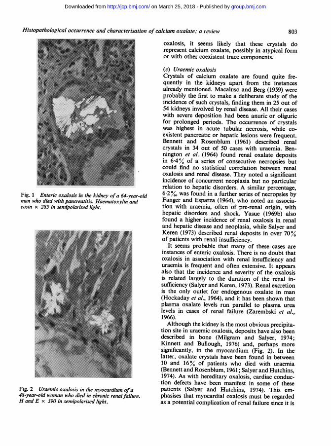
Since preformed crystal aggregates are essentialfor the genesis and growth of calculi (Vermeulenand Lyon, 1968) it is not surprising that in thesecases with a high tendency to urolithiasis, oxalateshave been demonstrated in renal material (Fig. 1)(Cryer et al., 1975; Vainder and Kelly, 1976). Morewidespread oxalosis has been described in suchpatients coming to necropsy (Lewis et al., 1974;Beppu et al., 1975).These patients generally have an excessive excre-
tion of bile salts in faeces, and it was originallythought that bacterial degradation of glycocholateliberates glycine which is oxidatively deaminated tooxalate (Watts, 1973). It has been shown subsequent-ly that the hyperoxaluria is in fact due to an increasein the uptake of normal dietary oxalate, possiblydue to the excessive colonic luminal concentration ofbile salts and long chain fatty acids (Earnest et al.,1974; Saunders et al., 1975).There has been mild controversy over the nature
of certain spheroidal crystals found in renal tubulesin cases of severe hepatic disturbances. These havebeen called 'leucine like' by Allen (1962) andcalcium oxalate by Fanger and Esparza (1964).Allen (1976) doubts the validity of the latter,chiefly on a morphological basis, though in-vitrostudies have shown that calcium oxalate willcrystallise in an identical spheroidal form as well asthe more common sheaf-like forms (Chaplin andGrace, 1975). Johnson and Pani (1962) identifiedbirefringent renal crystals in severe liver disease ascalcium oxalate rather than amino acids. In view ofthese facts, and the general pattern of enteric
group.bmj.com on March 25, 2018 - Published by http://jcp.bmj.com/Downloaded from
Histopathological occurrence and characterisation of calcium oxalate: a review
Fig. 1 Enteric oxalosis in the kidney ofa 64-year-oldman who died with pancreatitis. Haematoxylin andeosin x 285 in semipolarised light.
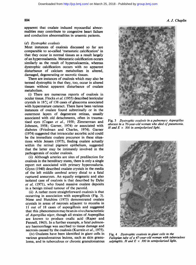
Fig. 2 Uraemic oxalosis in the myocardium ofa48-year-old woman who died in chronic renal failure.H and E x 390 in semipolarised light.
oxalosis, it seems likely that these crystals dorepresent calcium oxalate, possibly in atypical formor with other coexistent trace components.
(c) Uraemic oxalosisCrystals of calcium oxalate are found quite fre-quently in the kidneys apart from the instancesalready mentioned. Macaluso and Berg (1959) wereprobably the first to make a deliberate study of theincidence of such crystals, finding them in 25 out of54 kidneys involved by renal disease. All their caseswith severe deposition had been anuric or oliguricfor prolonged periods. The occurrence of crystalswas highest in acute tubular necrosis, while co-existent pancreatic or hepatic lesions were frequent.Bennett and Rosenblum (1961) described renalcrystals in 34 out of 50 cases with uraemia. Ben-nington et al. (1964) found renal oxalate depositsin 6-4% of a series of consecutive necropsies butcould find no statistical correlation between renaloxalosis and renal disease. They noted a significantincidence of concurrent neoplasia but no particularrelation to hepatic disorders. A similar percentage,6-2 %, was found in a further series of necropsies byFanger and Esparza (1964), who noted an associa-tion with uraemia, often of pre-renal origin, withhepatic disorders and shock. Yasue (1969b) alsofound a higher incidence of renal oxalosis in renaland hepatic disease and neoplasia, while Salyer andKeren (1973) described renal deposits in over 70%of patients with renal insufficiency.
It seems probable that many of these cases areinstances of enteric oxalosis. There is no doubt thatoxalosis in association with renal insufficiency anduraemia is frequent and often extensive. It appearsalso that the incidence and severity of the oxalosisis related largely to the duration of the renal in-sufficiency (Salyer and Keren, 1973). Renal excretionis the only outlet for endogenous oxalate in man(Hockaday et al., 1964), and it has been shown thatplasma oxalate levels run parallel to plasma urealevels in cases of renal failure (Zarembski et al.,1966).Although the kidney is the most obvious precipita-
tion site in uraemic oxalosis, deposits have also beendescribed in bone (Milgram and Salyer, 1974;Kinnett and Bullough, 1976) and, perhaps moresignificantly, in the myocardium (Fig. 2). In thelatter, oxalate crystals have been found in between10 and 16% of patients who died with uraemia(Bennett and Rosenblum, 1961; Salyer and Hutchins,1974). As with hereditary oxalosis, cardiac conduc-tion defects have been manifest in some of thesepatients (Salyer and Hutchins, 1974). This em-phasises that myocardial oxalosis must be regardedas a potential complication of renal failure since it is
803
group.bmj.com on March 25, 2018 - Published by http://jcp.bmj.com/Downloaded from
apparent that oxalate induced myocardial abnor-malities may contribute to congestive heart failureand conduction abnormalities in uraemic patients.
(d) Dystrophic oxalosisMost instances of oxalosis discussed so far arecomparable to so-called 'metastatic calcification' inthat they occur in normal tissues as a result largelyof an hyperoxalaemia. Metastatic calcification occurssimilarly as the result of hypercalcaemia, whereasdystrophic calcification occurs with no apparentdisturbance of calcium metabolism in altered,damaged, degenerating or necrotic tissues.There are instances of oxalosis which may also be
termed dystrophic in that they, too, occur in alteredtissues without apparent disturbance of oxalatemetabolism.
(i) There are numerous reports of oxalosis inocular tissue. Flocks et al. (1955) described lenticularcrystals in 18% of 138 cases of glaucoma associatedwith hypermature cataract. There have been variousinstances of oxalate found subretinally or in theoutermost layers of degenerate retinas, usuallyassociated with old detachments, often in trauma-tised eyes (Cogan et al., 1958; Zimmerman andJohnson, 1958; Garner, 1974) or associated withdiabetes (Friedman and Charles, 1974). Garner(1974) suggested that intraocular ascorbic acid couldbe the immediate oxalate precursor in these situa-tions while Jensen (1975), finding oxalate actuallywithin the retinal pigment epithelium, suggestedthat the latter may be intimately involved in thepathogenesis of ocular oxalosis.
(ii) Although arteries are sites of predilection foroxalosis in the hereditary states, there is only a singlereport not associated with primary hyperoxaluria.Glynn (1940) described oxalate crystals in the mediaof the left middle cerebral artery distal to a fatalruptured aneurysm. An equally enigmatic and alsoisolated case of oxalosis is that described by Dykeet al. (1971), who found massive oxalate depositsin a benign mixed tumour of the parotid.
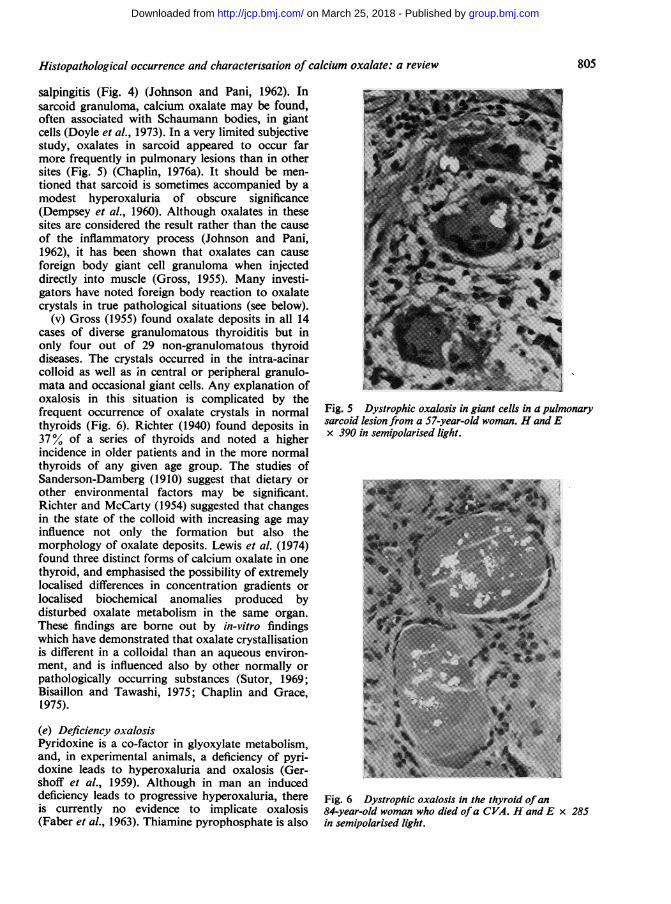
(ii) A rather more straightforward oxalosis is thatoccurring in association with aspergillosis (Fig. 3).Nime and Hutchins (1973) demonstrated oxalatecrystals in areas of necrosis adjacent to mycelia in11 out of 18 cases of aspergillosis and suggestedthat this phenomenon may bean in-vivo characteristicof Aspergillus niger, though all strains of Aspergillusare known to produce oxalic acid (Raper andFennell, 1965). In a further example, a fatal pulmon-ary haemorrhage was ascribed to tissue damage andnecrosis caused by the oxalosis (Kurrein et al., 1975).
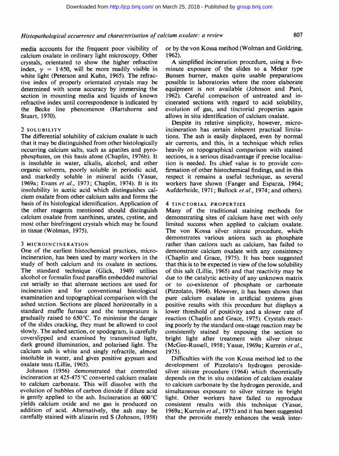
(iv) Oxalates have been identified in giant cells invarious granulomatous lesions, such as skin granu-loma, and in tuberculous or chronic granulomatous
Fig. 3 Dystrophic oxalosis in a pulmonary Aspergillusabscess in a 70-year-old woman who died ofpneumonia.H and E x 300 in semipolarised light.
Fig. 4 Dystrophic oxalosis in giant cells in theFallopian tube ofa 47-year-old woman with tuberculoussalpingitis. H and E x 300 in semipolarised light.
804
group.bmj.com on March 25, 2018 - Published by http://jcp.bmj.com/Downloaded from
Histopathological occurrence and characterisarion of calcium oxalate: areview8
salpingitis (Fig. 4) (Johnson and Pani, 1962). Insarcoid granuloma, calcium oxalate may be found,often associated with Schaumann bodies, in giantcells (Doyle et al., 1973). In a very limited subjectivestudy, oxalates in sarcoid appeared to occur farmore frequently in pulmonary lesions than in othersites (Fig. 5) (Chaplin, 1976a). It should be men-tioned that sarcoid is sometimes accompanied by amodest hyperoxaluria of obscure significance(Dempsey et al., 1960). Although oxalates in thesesites are considered the result rather than the causeof the inflammatory process (Johnson and Pani,1962), it has been shown that oxalates can causeforeign body giant cell granuloma when injecteddirectly into muscle (Gross, 1955). Many investi-gators have noted foreign body reaction to oxalatecrystals in true pathological situations (see below).
(v) Gross (1955) found oxalate deposits in all 14cases of diverse granulomatous thyroiditis but inonly four out of 29 non-granulomatous thyroiddiseases. The crystals occurred in the intra-acinarcolloid as well as in central or peripheral granulo-mata and occasional giant cells. Any explanation ofoxalosis in this situation is complicated by thefrequent occurrence of oxalate crystals in normalthyroids (Fig. 6). Richter (1940) found deposits in37% of a series of thyroids and noted a higherincidence in older patients and in the more normalthyroids of any given age group. The studies ofSanderson-Damberg (1910) suggest that dietary orother environmental factors may be significant.Richter and McCarty (1954) suggested that changesin the state of the colloid with increasing age mayinfluence not only the formation but also themorphology of oxalate deposits. Lewis et al. (1974)found three distinct forms of calcium oxalate in onethyroid, and emphasised the possibility of extremelylocalised differences in concentration gradients orlocalised biochemical anomalies produced bydisturbed oxalate metabolism in the same organ.These findings are borne out by in-vitro findingswhich have demonstrated that oxalate crystallisationis different in a colloidal than an aqueous environ-ment, and is influenced also by other normally orpathologically occurring substances (Sutor, 1969;Bisaillon and Tawashi, 1975; Chaplin and Grace,1975).
(e) Deficiency oxalosisPyridoxine is a co-factor in glyoxylate metabolism,and, in experimental animals, a deficiency of pyri-doxine leads to hyperoxaluria and oxalosis (Ger-shoff et al., 1959). Although in man an induceddeficiency leads to progressive hyperoxaluria, thereis currently no evidence to implicate oxalosis(Faber et al., 1963). Thiamine pyrophosphate is also
>~~~~~~~~~~--E:-- -------',
Fig. 5 Dystrophic oxalosis in giant cells in a pulmonarysarcoid lesion from a 57-year-old woman. H and Ex 390 in semipolarised light.
Fig. 6 Dystrophic oxalosis in the thyroid ofan84-year-old woman who died ofa CVA. H and E x 285in semipolarised light.
805
group.bmj.com on March 25, 2018 - Published by http://jcp.bmj.com/Downloaded from
essential in the normal metabolism of glyoxylateand, in theory, a thiamine deficiency could lead toincreased formation and excretion of oxalate andglycolate (Williams and Smith, 1968). There havebeen no reports of this, and in a recent study ofthiamine deficiency cases there was no greaterincidence of renal oxalosis than in control groups(Salyer and Salyer, 1974). It is assumed that theseverity of the deficiency required for hyperoxaluriato develop is greater than that required for theclinical manifestations. Recently, hyperoxaluriaassociated with raised vitamin C intake has beendescribed (Briggs, 1976; Harris, 1976). Each ofthese states involving vitamin abnormalities mustalways be considered in oxalosis of otherwiseobscure pathogenesis.
3 TISSUE REACTION TO OXALATE DEPOSITS
Reference has already been made to the fact thatoxalates may cause some reaction in tissues. Calciumoxalate is generally extremely inert, but tissuechanges are presumably due to its local dissociationand liberation of toxic oxalate ions (Hodgkinsonand Zarembski, 1968). It has indeed been shownthat, despite the low solubility of calcium oxalate,oxalate deposition in tissues is not necessarily anirreversible process (Bergstrand et al., 1972).A fibroblastic reaction around crystals as well as
frank fibrosis has been observed in renal tissue(Bennett and Rosenblum, 1961; Boquist et al., 1973)while in the myocardium slight chronic inflammation,fibrosis, and focal necrosis have all been seen(Bennett and Rosenblum, 1961; Koten et al., 1965;Salyer and Keren, 1973; Salyer and Hutchins, 1974).Apart from the instances already mentioned,foreign body reactions to oxalate crystals have beendescribed in bone marrow (Deodhar et al., 1969;Milgram and Salyer, 1974) and ocular tissue(Jensen, 1975) while Hughes (1959) describedrosettes of microglia around crystals in brain.
(B) Histological characterisation of calcium oxalate
Various sophisticated techniques such as x-raydiffraction, electron microprobe, and chroma-tography have all been used to identify calciumoxalate derived from histological material (Bennettand Rosenblum, 1961; Bennington et al., 1964;Evans et al., 1973). Facilities for such techniques arenot always readily available, while the randomorientation and fragmentation of the crystals createdifficulties in their application. Thus more simplemethods are preferable and indeed are quite ade-quate for the histological characterisation of oxalates.The more pertinent of these, summarised in Table2, serve to distinguish calcium oxalate from a wide
A. J. Chaplin
variety of potentially confusing substances of bothendogenous and exogenous origin (Wolman, 1975).
Table 2 Summary of histological properties ofcalcium oxalate
Insolubility in acetic acid von Kossa's silver nitrate methodSolubility in mineral acids Pizzolato's mercurous nitrate methodConversion to CaCO3 by Naphthalhydroxamic acid method
microincineration
1 OPTICAL PROPERTIESThe identification of crystals by their opticalproperties is well known, and detailed descriptionsof the theory and practice of quantitative polarisa-tion microscopy are available (Hartshorne andStuart, 1970). In such studies, various opticalcharacteristics are determined using whole crystalswhose orientation is known and controlled, whereasin histological studies the crystals are randomlyorientated and often fragmented, creating difficultiesin interpretation. However, at least four opticalproperties may be useful to the present problem.
(a) HabitIn histological material, calcium oxalate occurs inmany forms, though basically there are three majorforms-rosettes of rods, dipyramids or diamond-shaped crystals, and large overlapping plates. It isnot uncommon for all major forms to occur simul-taneously in a single organ (Lewis et al., 1974).
(b) BirefringenceCrystals of calcium oxalate may be readily detectedby virtue of their positive form birefringence inpolarised light. Most crystals display white or higherfirst-order polarisation colours, while the plates mayshow second or even third order colours (Petersonand Kuhn, 1965).
(c) ExtinctionExtinction angles are only significant if theorientation of the crystal is known, but, ingeneral terms, calcium oxalate may display parallel,symmetrical or indeterminate extinction (Petersonand Kuhn, 1965).
(d) Refractive indexCalcium oxalate has three refractive indices due toits biaxial form. The most frequent orientation inhistological material presents the two lower indices,a = 1 490 and , = 1-555. The proximity of theseto the refractive indices of most common mounting
group.bmj.com on March 25, 2018 - Published by http://jcp.bmj.com/Downloaded from
Histopathological occurrence and characterisation of calcium oxalate: a review
media accounts for the frequent poor visibility ofcalcium oxalate in ordinary light microscopy. Othercrystals, orientated to show the higher refractiveindex, y = 1 650, will be more readily visible inwhite light (Peterson and Kuhn, 1965). The refrac-tive index of properly orientated crystals may bedetermined with some accuracy by immersing thesection in mounting media and liquids of knownrefractive index until correspondence is indicated bythe Becke line phenomenon (Hartshorne andStuart, 1970).
2 SOLUBILITYThe differential solubility of calcium oxalate is suchthat it may be distinguished from other histologicallyoccurring calcium salts, such as apatites and pyro-phosphates, on this basis alone (Chaplin, 1976b). Itis insoluble in water, alkalis, alcohol, and otherorganic solvents, poorly soluble in periodic acid,and markedly soluble in mineral acids (Yasue,1969a; Evans et al.., 1973; Chaplin, 1974). It is itsinsolubility in acetic acid which distinguishes cal-cium oxalate from other calcium salts and forms thebasis of its histological identification. Application ofthe other reagents mentioned should distinguishcalcium oxalate from xanthines, urates, cystine, andmost other birefringent crystals which may be foundin tissue (Wolman, 1975).
3 MICROINCINERATIONOne of the earliest histochemical practices, micro-incineration, has been used by many workers in thestudy of both calcium and its oxalate in sections.The standard technique (Glick, 1949) utilisesalcohol or formalin fixed paraffin embedded materialcut serially so that alternate sections are used forincineration and for conventional histologicalexamination and topographical comparison with theashed section. Sections are placed horizontally in astandard muffle furnace and the temperature isgradually raised to 650°C. To minimise the dangerof the slides cracking, they must be allowed to coolslowly. The ashed section, or spodogram, is carefullycoverslipped and examined by transmitted light,dark ground illumination, and polarised light. Thecalcium ash is white and singly refractile, almostinsoluble in water, and gives positive gypsum andoxalate tests (Lillie, 1965).Johnson (1956) demonstrated that controlled
incineration at 425-475°C converted calcium oxalateto calcium carbonate. This will dissolve with theevolution of bubbles of carbon dioxide if dilute acidis gently applied to the ash. Incineration at 600°Cyields calcium oxide and no gas is produced onaddition of acid. Alternatively, the ash may becarefully stained with alizarin red S (Johnson, 1958)
or by the von Kossa method (Wolman and Goldring,1962).A simplified incineration procedure, using a five-
minute exposure of the slides to a Meker typeBunsen burner, makes quite usable preparationspossible in laboratories where the more elaborateequipment is not available (Johnson and Pani,1962). Careful comparison of untreated and in-cinerated sections with regard to acid solubility,evolution of gas, and tinctorial properties againallows in situ identification of calcium oxalate.
Despite its relative simplicity, however, micro-incineration has certain inherent practical limita-tions. The ash is easily displaced, even by normalair currents, and this, in a technique which reliesheavily on topographical comparison with stainedsections, is a serious disadvantage if precise localisa-tion is needed. Its chief value is to provide con-firmation of other histochemical findings, and in thisrespect it remains a useful technique, as severalworkers have shown (Fanger and Esparza, 1964;Aufderheide, 1971; Bullock et al., 1974; and others).
4 TINCTORIAL PROPERTIESMany of the traditional staining methods fordemonstrating sites of calcium have met with onlylimited success when applied to calcium oxalate.The von Kossa silver nitrate procedure, whichdemonstrates various anions such as phosphaterather than cations such as calcium, has failed todemonstrate calcium oxalate with any consistency(Chaplin and Grace, 1975). It has been suggestedthat this is to be expected in view of the low solubilityof this salt (Lillie, 1965) and that reactivity may bedue to the catalytic activity of any unknown matrixor to co-existence of phosphate or carbonate(Pizzolato, 1964). However, it has been shown thatpure calcium oxalate in artificial systems givespositive results with this procedure but displays alower threshold of positivity and a slower rate ofreaction (Chaplin and Grace, 1975). Crystals react-ing poorly by the standard one-stage reaction may beconsistently stained by exposing the section tobright light after treatment with silver nitrate(McGee-Russell, 1958; Yasue, 1969a; Kurrein et al.,1975).
Difficulties with the von Kossa method led to thedevelopment of Pizzolato's hydrogen peroxide-silver nitrate procedure (1964) which theoreticallydepends on the in situ oxidation of calcium oxalateto calcium carbonate by the hydrogen peroxide, andsimultaneous exposure to silver nitrate in brightlight. Other workers have failed to reproduceconsistent results with this technique (Yasue,1969a; Kurrein et al., 1975) and it has been suggestedthat the peroxide merely enhances the weak inter-
807
group.bmj.com on March 25, 2018 - Published by http://jcp.bmj.com/Downloaded from
reaction of silver nitrate and calcium oxalate(Yasue, 1969a). It is interesting to note that Macalusoand Berg (1959) described 'no modification' ofoxalates by hydrogen peroxide in their studies onthe solubility of calcium oxalate.The silver nitrate-rubeanic acid method described
by Yasue (1969a) has proved to be a reliableprocedure and is regarded as histologically selectivefor calcium oxalate when used after acetic acid treat-ment to remove any possible phosphate or carbonatecontamination (Chaplin, 1974). It has given con-sistent positive results, regardless of illuminatingconditions, with material which is shown to becalcium oxalate by other techniques but which givesequivocal staining reactions with other methods(Chaplin, 1974; Jensen, 1975; Kurrein et al., 1975).A recent study of metal substitution by various
tissue components led to the development of amercurous nitrate procedure for calcium salts, atechnique which gives particularly good visualisationof calcium oxalate (Pizzolato and Lillie, 1968;Pizzolato, 1971).Most of the complexing methods used in the past
to demonstrate calcium are unsatisfactory whenapplied to calcium oxalate. Alkaline quinalizarin(McGee-Russell, 1958) and alizarin red S (Kurreinet al., 1975) have given clear-cut positive staining,but other anthraquinone type dyes generally staincalcium oxalate only peripherally or not at all(Yasue, 1969a; Meloan et al., 1972; Chaplin, 1974).Of more recently introduced complexing methodsfor calcium, only chloranilic acid and naphthal-hydroxamic acid at pH 8&5 proved capable of visual-ising known oxalate deposits (Voigt, 1957; Chaplinand Grace, 1976). Using naphthalhydroxamic acid onmaterial from a case of ethylene glycol intoxication,Roscher (1971) obtained positive staining in hepaticnuclei, as well as in renal tubules and meninges,providing histological correlation of the bio-chemically elevated hepatic oxalate levels found insimilar cases by Zarembski and Hodgkinson (1967).They suggested that oxalic acid may be deposited asa non-crystalline complex of calcium oxalate andlipid in the liver in such cases. It is possible that suchorganic deposits are unreactive to other tinctorialtechniques due to their unusual chemical combina-tion.
I should like to thank Professor J. O'D. McGee, inwhose department the practical aspects of this studywere carried out, Dr M. S. Dunnill for his adviceand criticism, Mr T. Reed for the photomicrographs,and Mrs R. Hunt for the typescript. Dr R. D. Lewis,of the Huntingdon Memorial Hospital, Pasadena,provided the myocardial tissue used for Figure 2.
References
Allen, A. C. (1962). The Kidney, 2nd edition. Grune andStratton, New York.
Allen, A. C. (1976). Acute lobular (membranoprolifera-tive) glomerulonephritis with hyperuricemia andobstructive uric acid nephropathy. American Journal ofClinical Pathology, 65, 109-120.
Arbus, G. S., and Sniderman, S. (1974). Oxalosis withperipheral gangrene. Archives ofPathology, 97,107-110.
Archer, H. E., Dormer, A. E., Scowen, E. F., and Watts,R. W. E. (1958). The aetiology of primary hyperoxalu-ria. British Medical Journal, 1, 175-181.
Aufderheide, A. C. (1971). Renal tubular calcium oxalatecrystal deposition. Archives ofPathology, 92, 162-166.
Bennett, B., and Rosenblum, C. (1961). Identification ofcalcium oxalate crystals in the myocardium of patientswith uraemia. Laboratory Investigation, 10, 947-955.
Bennington, J. L., Haber, S. L., Smith, J. V., and Warner,N. E. (1964). Crystals of calcium oxalate in the humankidney. American Journal of Clinical Pathology, 41,8-14.
Beppu, T., Mori, S., Omoto, R., Nishi, T., and Oya, G.(1975). Secondary oxalosis after ileocaecal resection.Japanese Journal of Gastroenterology, 72, 520-526.
Bergstrand, A., Collste, L. G., Franksson, C., Glass, J. E.,Lofstrom, B., Magnusson, G., Nordenstam, H., andWerner, B. (1972). Oxalosis in renal transplantsfollowing methoxyflurane anaesthesia. British JournalofAnaesthesia, 44, 569-574.
Bisaillon, S., and Tawashi, R. (1975). Growth of calciumoxalate in gel systems. Journal of PharmaceuticalSciences, 64, 458-460.
Boquist, L., Lindqvist, B., Ostberg, Y., and Steen, L.(1973). Primary oxalosis. American Journal ofMedicine,54, 673-681.
Bourke, E., and Costello, J. (1975). The clinical import-ance of oxalic acid. Journal of the Irish MedicalAssociation, 68, 93-96.
Bove, K. E. (1966). Ethylene glycol toxicity. AmericanJournal of Clinical Pathology, 45, 46-50.
Briggs, M. (1976). Vitamin-C-induced hyperoxaluria(Letter). Lancet, 1 (7951), 154.
Bullock, J. D., Albert, D. M., Skinner, C. W., Miller,W. H., and Galla, J. H. (1974). Calcium oxalateretinopathy associated with generalised oxalosis.Investigative Ophthalmology, 13, 256-265.
Chaplin, A. J. (1974). Some observations on the demon-stration of calcium oxalate in tissue sections. StainTechnology, 49, 165-173.
Chaplin, A. J. (1976a). Unpublished observations.Chaplin, A. J. (1976b). Calcium pyrophosphate. Histo-
logical characterisation of crystals in pseudogout.Archives of Pathology and Laboratory Medicine, 100,12-15.
Chaplin, A. J., and Grace, S. R. (1975). Calcium oxalateand the von Kossa method with reference to theinfluence of citric acid. Histochemical Journal, 7, 451-458.
Chaplin, A. J., and Grace, S. R. (1976). An evaluation ofsome complexing methods for the histochemistry ofcalcium. Histochemistry, 47, 263-269.
808
group.bmj.com on March 25, 2018 - Published by http://jcp.bmj.com/Downloaded from
Histopathological occurrence and characterisation of calcium oxalate: a review
Cochran, M., Hodgkinson, A., Zarembski, P. M., andAnderson, C. K. (1968). Hyperoxaluria in adults.British Journal of Surgery, 55, 121-128.
Cogan, D. G., Kuwabara, T., Silbert, J., Kern, H.,McMurray, V., and Hurlbut, C. (1958). Calciumoxalate and calcium phosphate crystals in detachedretinas. Archives of Ophthalmology, 60, 366-371.
Coltart, D. J., and Hudson, R. E. B. (1971). Primaryoxalosis of the heart: a cause of heart block. BritishHeart Journal, 33, 315-319.
Cotton, J. R., Jr., Schwartz, M. M., Lindley, J. D., andHunsicker, L. G. (1976). Acute renal failure followingHalothane anesthesia. Archives of Pathology andLaboratory Medicine, 100, 628-629.
Crampton, R. F., and Charlesworth, F. A. (1975).Occurrence of natural toxins in food. British MedicalBulletin, 31, 209-213.
Cryer, P. E., Garber, A. J., Hoffsten, P., Lucas, B., andWise, L. (1975). Renal failure after small intestinalbypass for obesity. Archives of Internal Medicine, 135,1610-1612.
Daniels, R. A., Michels, R., Aisen, P., and Goldstein, G.(1960). Familial hyperoxaluria. American Journal ofMedicine, 29, 820-831.
Dempsey, E. F., Forbes, A. P., Melick, R. A., and Henne-man, P. H. (1960). Urinary oxalate excretion. Metab-olism, 9, 52-58.
Deodhar, S. D., Tung, K. S. K., Ziihlke, V., and Naka-moto, S. (1969). Renal homotransplantation in apatient with primary familial oxalosis. Archives ofPathology, 87, 118-124.
Dowling, R. H., Rose, G. A., and Sutor, D. J. (1971).Hyperoxaluria and renal calculi in ileal disease. Lancet,1, 1103-1106.
Doyle, W. F., Brahman, H. D., and Burgess, J. H. (1973).The nature of yellow-brown bodies in peritoneallymph nodes. Archives ofPathology, 96, 320-326.
Dunn, J. S., Haworth, A., and Jones, N. A. (1924). Thepathology of oxalate nephritis. Journal of Pathologyand Bacteriology, 27, 299-318.
Dyke, P. C., Hajdu, S. I., Strong, E. W., Erlandson,R. A., and Fleisher, M. (1971). Mixed tumour ofparotid containing calcium oxalate crystals. ArchivesofPathology, 91, 89-92.
Earnest, D. L., Johnson, G., Williams, H. E., andAdmirand, W. H. (1974). Hyperoxaluria in patientswith ileal resection: an abnormality in dietary oxalateabsorption. Gastroenterology, 66, 1114-1122.
Evans, G. W., Phillips, G., Mukherjee, T. M., Snow,M. R., Lawrence, J. R., and Thomas, D. W. (1973).Identification of crystals deposited in brain and kidneyafter xylitol administration by biochemical, histo-chemical, and electron diffraction methods. Journal ofClinical Pathology, 26, 32-36.
Faber, S. R., Feitler, W. W., Bleiler, R. E., Ohlson,M. A., and Hodges, R. E. (1963). The effects of aninduced pyridoxine and pantothenic acid deficiency onexcretions of oxalic and xanthurenic acids in the urine.American Journal of Clinical Nutrition, 12, 406-412.
Fanger, H., and Esparza, A. (1964). Crystals of calciumoxalate in kidneys in uremia. American Journal ofClinical Pathology, 41, 597-603.
Flocks, M., Littwin, C. S., and Zimmerman, L. E. (1955).Phacolytic glaucoma. A clinco-pathologic study of onehundred and thirty-eight cases of glaucoma associatedwith hypermature cataract. Archives of Ophthalmology,54, 37-45.
Franco, V., and Krinitz, B. (1973). Determination ofoxalic acid in foods. Journal of the Association ofOfficial Analytical Chemists, 56, 164-166.
Friedman, A. H., and Charles, N. C. (1974). Retinaloxalosis in two diabetic patients. American Journal ofOphthalmology, 78, 189-195.
Friedman, E. A., Greenberg, J. B., Merrill, J. P., andDammin, G. J. (1962). Consequences of ethylene glycolpoisoning. American Journal of Medicine, 32, 891-902.
Garner, A. (1974). Retinal oxalosis. British Journal ofOphthalmology, 58, 613-619.
Geiling, E. M. K., and Cannon, P. R. (1938). Pathologiceffects of elixir of sulfanilamide (diethylene glycol)poisoning; clinical and experimental correlation: finalreport. Journal of the American Medical Association,111, 919-926.
Gelfand, S. G., and Taranto, A. I. (1973). Hyperoxaluriaand Meckel's Diverticulum (Letter). Annals of InternalMedicine, 79, 748-749.
Gershoff, S. N., Faragalla, F. F., Nelson, D. A., andAndrus, S. B. (1959). Vitamin B6 deficiency and oxalatenephrocalcinosis in the cat. American Journal ofMedicine, 27, 72-80.
Gessner, P. K., Parke, D. V., and Williams, R. T. (1961).Studies in detoxication 86. The metabolism of 14C-labelled ethylene glycol. Biochemical Journal, 79,482489.
Glick, D. (1949). Techniques ofHisto- and Cytochemistry.Interscience, New York.
Glynn, L. E. (1940). Crystalline bodies in the tunicamedia of a middle cerebral artery. Journal ofPathologyand Bacteriology, 51, 445-446.
Gross, S. (1955). Granulomatous thyroiditis with aniso-tropic crystalline material. Archives of Pathology, 59,412-418.
Harris, A. B. (1976). Vitamin-C-induced hyperoxaluria(Letter). Lancet, 1, 366.
Hartshorne, N. H., and Stuart, A. (1970). Crystals andthe Polarising Microscope, 4th edition. Arnold,London.
Hauschildt, S., Chalmers, R. A., Lawson, A. M.,Schultis, K., and Watts, R. W. E. (1976). Metabolicinvestigations after xylitol infusion in human subjects.American Journal of Clinical Nutrition, 29, 258-273.
Hauschildt, S., and Watts, R. W. E. (1976). Studies onthe effect of xylitol on oxalate formation. BiochemicalPharmacology, 25, 27-29.
Hockaday, T. D. R., Clayton, J. E., Frederick, E. W.,and Smith, L. H., Jr. (1964). Primary hyperoxaluria.Medicine, 43, 315-345.
Hodgkinson, A., and Zarembski, P. M. (1968). Oxalicacid metabolism in man: a review. Calcified TissueResearch, 2, 115-132.
Hughes, D. T. D. (1959). The clinical and pathologicalbackground on two cases of oxalosis. Journal ofClinical Pathology, 12, 498-509.
James, L. F. (1972). Oxalate toxicosis. Clinical Toxi-
809
group.bmj.com on March 25, 2018 - Published by http://jcp.bmj.com/Downloaded from
Jansen, L. H., Groeneveld, J. L., and van der Meer, J. B.(1974). Deposition of calcium oxalate in the skin in twopatients suffering from oxalosis caused by primaryhyperoxaluria. Archiv fir Dermatologische Forschung,250, 323-350.
Jeghers, H., and Murphy, R. (1945). Practical aspects ofoxalate metabolism. New England Journal of Medicine,233, 208-215.
Johnson, F. B. (1956). A method for demonstratingcalcium oxalate in tissue sections (Abstract). Journalof Histochemistry and Cytochemistry, 4, 404-405.
Johnson, F. B. (1958). Further observations on theidentification of calcium oxalate in tissue sections(Abstract). Journal of Histochemistry and Cyto-chemistry, 6, 405.
Johnson, F. B., and Pani, K. (1962). Histochemicalidentification of calcium oxalate. Archives ofPathology,74, 347-351.
Kinnett, J. G., and Bullough, P. G. (1976). Identificationof calcium oxalate deposits in bone by electrondiffraction. Archives of Pathology and LaboratoryMedicine, 100, 656-658.
Klauwers, J., Wolf, P. L., and Cohn, R. (1969). Failureof renal transplantation in primary oxalosis. Journalof the American Medical Association, 209, 551.
Kobert, R., and Kiissner, B. (1879). Die experimentellenWirkungen der Oxalsaure. Virchows Archiv furpathologische Anatomie und Physiologie, 78, 209-244.
Koten, J. W., van Gastel, C., Dorhout Mees, E. J.,Holleman, L. W. J., and Schuiling, R. D. (1965). Twocases of primary oxalosis. Journal of Clinical Pathology,18, 223-229.
Kurrein, F., Green, G. H., and Rowles, S. L. (1975).Localised deposition of calcium oxalate around apulmonary Aspergillus niger fungus ball. AmericanJournal of Clinical Pathology, 64, 556-563.
Kuzucu, E. Y. (1970). Methoxyflurane, tetracycline, andrenal failure. Journal of the American Medical Associa-tion, 211, 1162-1164.
Lewis, R. D., Lowenstam, H. A., and Rossman, G. R.(1974). Oxalate nephrosis and cystalline myocarditis.Archives ofPathology, 98, 149-155.
Lillie, R. D. (1965). Histopathological Technic andPractical Histochemistry, 3rd edition. McGraw-Hill,New York.
Lindholm, J. (1965). Intra-vitam diagnosis of oxalosis.Acta Medica Scandinavica, 178, 155-159.
Macaluso, M. P., and Berg, N. 0. (1959). Calcium oxalatecrystals in kidneys in acute tubular nephrosis and otherrenal diseases with functional failure. Acta PathologicaMicrobiologica Scandinavica, 46, 197-205.
McCollum, J. P. K., Packer, S., Manning, J., and Harries,J. T. (1974). Hyperoxaluria in children with hepaticand intestinal dysfunction (Abstract). Archives ofDiseases in Childhood, 49, 749.
McGee-Russell, S. M. (1958). Histochemical methods forcalcium. Journal of Histochemistry and Cytochemistry,6, 22-42.
Mazze, R. I., TrudeAl, J. R., and Cousins, M. J. (1971).
A. J. Chaplin
Methoxyflurane metabolism and renal dysfunction:clinical correlation in man. Anesthesiology, 35,247-252.
Meloan, S. N., Puchtler, H., and Valentine, L. S. k1972).Alkaline and acid alizarin red S stains. Archives ofPathology, 93, 190-197.
Milgram, J. W., and Salyer, W. R. (1974). Secondaryoxalosis of bone in chronic renal failure. Journal ofBone and Joint Surgery, 56A, 387-395.
Mohr, W., and Hey, D. (1969). Endogene Oxalose mitManifestation im Erwachsenenalter. Virchows ArchivAbt A, Pathologische Anatomie, 347, 185-196.
Nime, F. A., and Hutchins, G. M. (1973). Oxalosiscaused by Aspergillus infection. Johns HopkinsMedical Journal, 133, 183-194.
Peterson, B. J., and Kuhn, R. J. (1965). Optical character-istics of crystals in tissue. Cystine and calcium oxalatemonohydrate. American Journal of Clinical Pathology,43, 401-408.
Pizzolato, P. (1964). Histochemical recognition of calciumoxalate. Journal of Histochemistry and Cytochemistry,12, 333-336.
Pizzolato, P. (1971). Mercurous nitrate as a histochemicalreagent for calcium phosphate in bone and patho-logical calcification and for calcium oxalate. Histo-chemical Journal, 3, 463-469.
Pizzolato, P., and Lillie, R. D. (1968). The impregnationof bone and pathologic calcification by metal salts andtheir recognition by unoxidised haematoxylin. Histo-chemie, 16, 333-338.
Pons, C. A., and Custer, R. P. (1946). Acute ethyleneglycol poisoning: a clinico-pathologic report ofeighteen fatal cases. American Journal of MedicalScience, 211, 544-552.
Raper, K. B., and Fennell, D. I. (1965). The GenusAspergillus. Williams and Wilkins, Baltimore.
Richter, M. N. (1940). Anisotropic crystalloids in thehuman thyroid gland (Abstract). American Journal ofPathology, 16, 654-655.
Richter, M. N., and McCarty, K. S. (1954). Anisotropiccrystals in the human thyroid gland. American JournalofPathology, 30, 545-552.
Roscher, A. A. (1971). A new histochemical method forthe demonstration of calcium oxalate in tissuesfollowing ethylene glycol poisoning. American Journalof Clinical Pathology, 55, 99-104.
Roughan, P. G., and Slack, C. R. (1973). Simple methodsfor routine screening and quantitative estimation ofoxalate content of tropical grasses. Journal of theScience ofFood and Agriculiure, 24, 803-811.
Salyer, W. R., and Hutchins, G. M. (1974). Cardiaclesions in secondary oxalosis. Archives of InternalMedicine, 134, 250-252.
Salyer, W. R., and Keren, D. (1973). Oxalosis as acomplication of chronic renal failure. Kidney Inter-national, 4, 61-66.
Salyer, W. R., and Salyer, D. C. (1974). Thiaminedeficiency and oxalosis. Journal of Clinical Pathology,27, 558-559.
Sanderson-Damberg, E. Die(1910). Schildriisenvom 15-25Lebensjahr aus der norddeutschen Ebene und Kusten-gegend, sowie aus Bern. Frankfurter Zeitschrift furPathologie, 6, 312-334. Cited by Richter and McCatty
group.bmj.com on March 25, 2018 - Published by http://jcp.bmj.com/Downloaded from
Histopathological occurrence and characterisation of calcium oxalate: a review
(1954).Saunders, D. R., Sillery, J., and McDonald, G. B. (1975).
Regional differences in oxalate absorption by ratintestine: evidence for excessive absorption by thecolon in steatorrhoea. Gut, 16, 543-554.
Schroder, R., De Lacroix, W. F., and Franzen, U., et al.(1974). Therapie-bedingte Form einer reno-cerebralenOxalose. Acta Neuropathologica (Berlin), 27, 181-184
Scowen, E. F., Stansfeld, A. G., and Watts, R. W. E.(1959). Oxalosis and primary hyperoxaluria. Journal ofPathology and Bacteriology, 77, 195-205.
Smith, L. H., Jr. (1968). Symposium on stones. Intro-duction. American Journal of Medicine, 45, 649-653.
Stauffer, M. (1960). Oxalosis. Report of a case, with areview of the literature and discussion of the patho-genesis. New EnglandJournal ofMedicine, 263, 386-390.
Sutor, D. J. (1969). Growth studies of calcium oxalate inthe presence of various ions and compounds. BritishJournal of Urology, 41, 171-178.
Tallqvist, H., and Vaananen, I. (1960). Death of a childfrom oxalic acid poisoning due to eating rhubarbleaves. Annales Paediatriae Fenniae, 6, 144-147.
Thomas, D. W., Edwards, J. B., Gilligan, J. E., Lawrence,J. R., and Edwards, R. G. (1972). Complicationsfollowing intravenous administration of solutioncontaining xylitol. Medical Journal of Australia, 1,1238-1246.
Vainder, M., and Kelly, J. (1976). Renal tubular dys-function secondary to jejunoileal bypass. Journal of theAmerican Medical Association, 235, 1257-1258.
Valman, H. B., Oberholzer, V. G., and Palmer, T. (1974).Hyperoxaluria after resection of ileum in children.Archives of Diseases in Childhood, 49, 171-173.
Vermeulen, C. W., and Lyon, E. S. (1968). Mechanismsof genesis and growth of calculi. American Journal ofMedicine, 45, 684-692.
Voigt, G. E. (1957). Der histochemische Nachweis des
Calciums im Calciumoxalate bei der Athylenglykol-vergiftung. Acta Pathologica Microbiologica Scandin-avica, 41, 89-95.
Watts, R. W. E. (1973). Oxaluria. Journal of the RoyalCollege ofPhysicians ofLondon, 7, 161-174.
Williams, H. E., and Smith, L. H., Jr. (1968). Disordersof oxalate metabolism. American Journal of Medicine,45, 715-735.
Williams, H. E., and Smith, L. H., Jr. (1972). Primaryhyperoxaluria. In The Metabolic Basis of InheritedDiseases, 3rd edition, edited by OJ. B. Stanbury, J. B.Wyngaarden, and D. S. Frederickson, pp. 196-219.McGraw-Hill Book Company, New York.
Wolman, M. (1975). Polarised light microscopy as a toolof diagnostic pathology. Journal ofHistochemistry andCytochemistry, 23, 21-50.
Wolman, M., and Goldring, D. (1962). Histochemicaldemonstration of calcium oxalate crystals. Journal ofHistochemistry and Cytochemistry, 10, 505-506.
Yasue, T. (1969a). Histochemical identification ofcalcium oxalate. Acta Histochemica et Cytochemica, 2,83-95.
Yasue, T. (1969b). Renal crystalline deposition and itspathogenesis. Acta Histochemica et Cytochemica, 2,96-111.
Zarembski, P. M., and Hodgkinson, A. (1967). Plasmaoxalic acid and calcium levels in oxalate poisoning.Journal of Clinical Pathology, 20, 283-285.
Zarembski, P. M., Hodgkinson, A., and Parsons, F. M.(1966). Elevation of the concentration of plasma oxalicacid in renal failure. Nature, 212, 511-512.
Zeiss, 0. (1877). Mikroskopische Untersuchungen uberden Bau der Schilddruse. Inaugural dissertation,Strassburg. Cited by Richter and McCarty (1954).
Zimmerman, L. E., and Johnson, F. B. (1958). Calciumoxalate crystals within ocular tissues. Archives ofOphthalmology, 60, 372-383.
811
group.bmj.com on March 25, 2018 - Published by http://jcp.bmj.com/Downloaded from