EXPERIMENT STATION OF THE KANSAS STATE AGRICULTURAL COLLEGE, MANHATTAN. Bulletin No. 79—April, 1898. VETERINARY DEPARTMENT. PAUL FISCHER, B. AGR., M V. D., Veterinarian. Bovine Tuberculosis. INTRODUCTION. The recent public post-mortem examination of the fifteen head of tuberculous cattle belonging to the college herd seems to have awakened an unusual interest in the question of tuberculosis among animals, especially dairy herds, and its relation to human health. There has sprung up a sudden demand among farmers and stockowners for more minute information regarding the cause, nature and treatment of tuberculosis in dairy herds, as well as the all important question of “disinfection.” The object of this bulletin is to meet this demand; but since the Veterinary Department has recently issued a short report on this same subject, a few wholesome repetitions will have to be in- dulged in. It has been thought well to append, with comments, a copy of the temperatures taken while testing the fifty-nine animals of the college herd for tuberculosis; and also the results of the test on the recently acquired dairy herd, which consisted of twenty grade cows of different breeds, bought up in one of our western counties and selected for their milk producing qual- ities and general appearance of perfect health. The fact that ten per cent of this little herd was tuberculous, as shown by the tuberculin reaction and the following post-mortem examination, furnishes material for reflection to those who think there is no such disease as tuberculosis in Kansas cattle.
Transcript
EXPERIMENT STATION OF THE
KANSAS STATE AGRICULTURAL COLLEGE, MANHATTAN.
Bulletin No. 79—April, 1898.
VETERINARY DEPARTMENT.
PAUL FISCHER, B. AGR., M V. D.,Veterinarian.
Bovine Tuberculosis.
INTRODUCTION.The recent public post-mortem examination of the fifteen head
of tuberculous cattle belonging to the college herd seems to haveawakened an unusual interest in the question of tuberculosisamong animals, especially dairy herds, and its relation to humanhealth.
There has sprung up a sudden demand among farmers andstockowners for more minute information regarding the cause,nature and treatment of tuberculosis in dairy herds, as well as theall important question of “disinfection.”
The object of this bulletin is to meet this demand; but sincethe Veterinary Department has recently issued a short report onthis same subject, a few wholesome repetitions will have to be in-dulged in. It has been thought well to append, with comments,a copy of the temperatures taken while testing the fifty-nineanimals of the college herd for tuberculosis; and also the resultsof the test on the recently acquired dairy herd, which consistedof twenty grade cows of different breeds, bought up in one ofour western counties and selected for their milk producing qual-ities and general appearance of perfect health. The fact that tenper cent of this little herd was tuberculous, as shown by thetuberculin reaction and the following post-mortem examination,furnishes material for reflection to those who think there is nosuch disease as tuberculosis in Kansas cattle.
Unknown
82 Veterinary Department. [Bulletin 79
BOVINE TUBERCULOSIS.
Definition. Tuberculosis is an infectious disease that iscaused by the tubercle bacillus, a minute micro-organism belong-ing to the lowest order of plants, the bacteria. This micro-organ-ism was discovered by Dr. Robert Koch in 1882. Through itsdiscovery Koch was enabled to prove conclusively that tubercu-losis in animals, and tuberculosis or consumption in man, areidentically the same disease. Through its discovery the disputeas to whether or not tuberculosis is an infectious disease, is alsoended. Wherever we find a case of tuberculosis the presence ofthe tubercle bacillus can be demonstrated. Wherever we find thetubercle bacillus in an animal organ, we have tuberculosis.
No disease can be rationally studied nor successfully treatedwithout a knowledge of its cause. Prevention is today consideredthe greatest factor in the treatment of all infectious diseases. Athorough knowledge of the cause of a disease can throw thegreatest light on methods of prevention.
It will therefore be proper to begin with a study of the tuberclebacillus.
THE TUBERCLE BACILLUS.
As above mentioned, this micro-organism is a bacterium belonging to an order of fungi. Fungi are plants that are destitutof chlorophyll (the green coloring matter of ordinary leaves) andhence connot live on inorganic matter as can ordinary plants, butdepend for their sustenance on matter already organized by otherliving things, plants or animals. Another characteristic of theselow organisms is that they grow in the absence of sunlight; indeed,sunlight in most cases is absolutely injurious to them, so much sothat a very short exposure will often result in their total destruc-tion. This circumstance accounts for an underlying principle ofdisinfection and hygiene, as we shall see later on.
The tubercle bacillus is an inconceivably small, one-celledorganism, that has the shape of a slender, slightly bent rod. It isfrom two to five micro-millimeters in length, or about 2/3 the lengthof the diameter of a human red blood corpuscle. When viewedunder a microscope with a magnifying power of one thousanddiameters the tubercle bacillus looks like a delicate rod, about 1/8 ofan inch in length. Eight thousand of these bacilli placed end toend would make a line about an inch in length. A single layer of
George Brandsberg
April, 1898.] Bovine Tuberculosis. 83
them over a surface one inch square would contain more than fourhundred millions.
When placed in a suitable medium, the tubercle bacillusgrows and multiplies by a process of simple division. A singlecell increases in length, divides at the middle and two new cells, orbacilli, are the result. This process goes on rapidly and indefinitelyas long as suitable food material is present. The tissues of theanimal body are the most favorable medium for the growth of thisgerm. A great many disease germs, indeed most of them, havethe power of living and propagating either in an animal body oroutside of it; in dead organic matter of any kind, vegetable oranimal. Some germs prefer one kind, others prefer another kind.They can remain indefinitely under such conditions when they arenot disturbed by the presence of sunshine. Continued sunshinechecks the growth, just as continued absence of sunlight cheeksor retards the growth of higher plants and finally destroysthem. In the absence of sunlight the disease germs justreferred to can adapt themselves to either a parasitic life in thebody of an animal or they can exist outside of the animal bodywith equal facility. Parasites of this kind are called facultativeparasites. The tubercle bacillus does not belong to this class.This germ is a true parasite; it can grow and multiply, (so far aswe know, under natural conditions) in a living animal body only.Outside of the animal body it can exist or retain its vitality for acertain time, and when again introduced into an animal body itwill again grow and multiply; but otherwise it would finally perish.A parasite of this kind, one that depends entirely on an animalbody for its growth and development, is known as a true or obli-gate parasite. Such a one we have in the tubercle bacillus. Theknowledge of this fact is of the greatest importance; it meansthat diseases that are produced by obligate parasites can be wipedout of existence, if the sacrifices necessary to bring about the properconditions are made.
A great many bacteria, when placed under certain conditions, usually conditions unfavorable for their growth, develop withinthemselves a minute oval or roundish body called a spore. Thisspore corresponds to the seed in higher plants. Like a seed it hasthe power of remaining dormant or inactive for a very long time;and when suitable opportunity is afforded it germinates, developsinto a bacterium, and this latter multiplies by division, as beforeexplained. From a pathological point of view the most important
George Brandsberg
84 Veterinary Department. [Bulletin 79
characteristic of spores is their great power to resist the destruc-tive action of various agents called disinfectants.
To illustrate:The bacillus of Anthrax is killed by continued exposure to a
temperature of 131 degrees Fahr. A one three-hundredth percent solution of corrosive sublimate also destroys it.
The spores, on the other hand, resist the boiling water tem-perature for over an hour. Artificial cold of 166 degrees Fahr.below zero has no effect on their vitality. And it requires tenminutes for a one-tenth per cent solution of corrosive sublimate tokill them.
This is enough to show the importance of so-called spores ofdisease germs.
Although less is known about their properties, it is believedthat tubercle bacilli also develop spores under certain conditions.
THE TUBERCLE BACILLUS AND ITS RELATION TO ANTISEPTICS.
Since the tubercle bacilli form spores, their power of resist-ing the destructive action of disinfectants is very considerable.Outside of the animal body, however, they soon die; the continuedaction, for a few hours, of strong sunlight is capable of destroy-ing them. For this reason the development of these germs out-side of an animal body after the manner of a miasma is impossible,and hence tuberculosis is a distinctly contagious disease.
On the whole, however, and compared with other diseasegerms, tubercle bacilli are quite resistant to the action of externalagencies; e.g., in common water they remain virulent (diseaseproducing) 120 days. Tuberculous sputum of man has been knownto remain virulent 226 days. When diluted with four hundredthousand times their bulk of water, and introduced into theabdominal cavity, they are still capable of producing tuberculosis.
The action of steam at 212 degrees Fahr., must be continuedfor fifteen minutes before the germs are killed in dried sputum.Dry heat of the same temperature requires an hour to producethe same effect.
Tubercle bacilli in milk are killed by heating it to 185 degreesFahr.
One-tenth per cent solutions of corrosive sublimate willdestroy the bacilli in one-twelfth their bulk of sputum in 24 hours.
Five per cent solutions of carbolic acid or creolin have thesame effect.
On the other hand—concentrated solutions of common salt,
George Brandsberg
April, 1898.] Bovine Tuberculosis. 85
drying, freezing, alcohol and putrefaction processes, have noeffect upon the vitality of the tubercle bacillus.
In the production of all contagious and infectious diseases twocauses may be considered as acting, viz., the exciting cause whichwe have just considered somewhat at length (as the tuberclebacillus) and the predisposin g cause, or causes, a discussion ofwhich may now properly follow.
THE PREDISPOSING CAUSES OF TUBERCULOSIS.It has long been observed that tuberculosis or consumption
occurs most frequently in certain families; among human beingsthis is just as noticeable, as, or perhaps more so than, amongthe lower animals. The first and perhaps natural inference wasthat the disease was transmitted by inheritance. For a greatmany years this was the generally accepted theory, and not until.the infectious nature of the disease began to be suspected, diddoubts arise as to the correctness of this view.
Since Koch’s discovery that tuberculosis was invariablycaused by the tubercle bacillus and by nothing else, and that thetubercle bacillus produced tuberculosis and no other disease,the peculiar fact that tuberculosis runs in certain families had tobe re-explained.
It is explained in this way. When infectious diseases likesmallpox, yellow fever, etc., occur in the form of an epidemic it isnoticed that certain individuals are always attacked first, otherslater, and still others not at all. Some experience a light attackand recover; others have a severe attack and die. We explainthis by saying that the latter class of persons is predisposed to takethe disease. Some animals and some men are more predisposed,that is, they have less power to ward off an attack of disease, thanothers. The greater an animal’s predisposition, the easier will bethe attack, and the greater will be its suffering as a consequence.
Just as certain physical characteristics, such as size, formand color, are transmitted from parent to offspring, certain bio-logical characteristics, of which predisposition to disease is one,are also transmitted.
The predisposition to attack by tuberculosis; in other words,the inability to resist an attack by this disease, is inherited.*
*This points to the doubtful advisability of breeding from affected animals.It is wellknown that animals free from tuberculosis can be bred and reared from slightly affecteddams; but this offspring must be considered more liable to contract the disease than off-spring from originalIy healthy animals.There is little if anymore reason in favor of try-ing breed healthy animal from diseased ones than there is for trying to breed a milk cowfrom a beef cow.When we try to raise milk cows we begin with the best milk cow we have.When we breed for health. why not make it a rule to start with the healthiest animal at ourdisposal?Health is just as likely to be inherited as a predisposition to tuberculosis.
George Brandsberg
86 [Bulletin 79
In exceedingly rare cases the disease itself is inherited. Buthow rarely this actually takes place in human beings may beinferred from definite statistics gathered among animals.
Of one million calves slaughtered for veal in Munich from1878-1882 only five were found with congenital tuberculosis. InBerlin, 1885-1886, of 80,000 calves only seven, in 1886-1887 of 90-000 only six were found tuberculous. In Augsburg (1873-1886) of230,000 slaughtered calves only nine were found tuberculous. In1889, in Prussia, of 370,000 calves 73 were tuberculous, or 0.02:per cent. These figures are somewhat lower than they shouldbe because many cases of abortion are due to congenital tubercu-losis, and these of course are not found on slaughter house records.But taking even that into account we see that congenital tubercu-losis is much more rare than is commonly supposed.
To go back to the question of why tuberculosis runs in fam-ilies, one other factor besides the inheritance of predispositionmust not be forgotten. An animal or child born of tuberculousparents inherits one thing more, it inherits the chance (if the ex-pression may be used) to become infected. It constantly asso-ciates with a tuberculous mother, and is exposed to every dangerthat such a tuberculous mother creates by her presence. Stablesthat contain tuberculous animals, or dwellings with consumptivepersons, are a source of danger to any one, but infinitely more soto a young being that is constantly exposed.
Now the question arises, if the tubercle bacillus, together withthe various predisposing causes, produces the disease by enteringthe system and developing there, how does the tubercle bacillusgain entrance into the animal body?
Briefly stated, this germ, like many others, may enter thebody in one or all of three or four ways.
1. It may be inhaled directly into the lungs, in the form ofdust; to the particles of which the germ clings.
The germ itself has no power of moving, nor of flying in the air;but when matter containing it (sputum and other material coughedup from the lungs, feces from animals with intestinal tuberculosis,etc.) dries, it may be reduced to dust; and this dust when carriedinto the air by the wind takes the germs with it. A single particleof dust may contain innumerable germs.
When thus inhaled the germs lodge in the lungs or air pas-sages, and if conditions are favorable they remain there and de-velop, and produce pulmonary tuberculosis. This method ofinfection is common in cattle and in man.
George Brandsberg
April, l898.]
2. This germ may be taken directly into the stomach and di-gestive tract by means of food containing it, most commonly meator milk from tuberculous animals, or meat from healthy animalscontaminated with tuberculous offal. This would produce anintestinal form of tuberculosis, and is common in pigs and poultry.
3. The germ may enter the tissues directly through a woundin the skin and produce either local or general tuberculosis. Thismethod of infection is not common and is most frequently pro-duced experimentally in the lower animals.
4. Infection through the genital organs is also possible.HOW THE TUBERCLE BACILLUS IS DISSEMINATED.
The source of the tubercle bacillus is always, directly or in-directly, animal tissue containing it.
Animals affected with lung tuberculosis cough up dead piecesof lung tissue, sometimes pus and mucus, that contain the bacilli.The feces of animals with intestinal tuberculosis often containthem. These ejecta are scattered about more or less promiscu-ously. They are reduced to dust, carried into the air by a gustof wind, and inhaled. Tuberculous meat and milk are anotherfertile vehicle for the dissemination of this germ. Among humanbeings the bed clothes, handkerchiefs, wearing apparel, carpets inrooms of tuberculous patients, the dust on the floors of publicbuildings, street cars, railroad coaches, etc., are good places tolook for the bacillus tuberculosis
The mangers from which tuberculous animals feed, and intowhich their nasal secretions drop, can readily act as carriersof the contagion. Contact of the animals themselves, by licking,smelling, etc., is another means of contagion. Finally, hay, strawor feed of any kind kept in a barn harboring infected animals;beams, floors, posts, etc., with which such animals have been incontact, can each and all act as carriers of the infectious material.
After dissemination in this manner it can readily be seen howan animal or person may take the germs into its (or his) system.
EXTENT OF TUBERCULOSIS.Tuberculosis is one of the most widely distributed diseases.
It occurs in all countries and in all races of men, and few ofthe domesticated animals are exempt from its attacks.
Among human beings, one-seventh of all recorded deaths aredue to tuberculosis. This does not include the great number ofcases of this disease that are cut short by death from other causes.
George Brandsberg
88 Veterinary Department. [Bulletin 79
Among cattle it is the most common and widely spread disease,and there is no doubt that this is the most frequent source of in-fection for human beings, except, of course, infected human beingsthemselves.
The disease occurs in cattle more frequently than in otheranimals; it will therefore occupy our chief attention. Amonghogs tuberculosis is much more common than is supposed, evenby men that ought to be informed on the subject. In Berlin in1883-5 0.4—0.9 per cent of all slaughtered hogs were found tuber-culous. In Copenhagen 3.0 per cent were found affected. Inchickens tuberculosis is also very frequently met with.
Sheep and goats, horses, asses and mules, dogs and cats, areaffected, but not so commonly as the above named animals.
Animals kept in confinement, under unnatural conditions, asin zoological gardens, often fall victims to tuberculosis. Inmonkeys it is common, and it is observed in lions, tigers, bears,dromedaries, jackals, panthers and rodents of many kinds. Butto return to the ox, the subject of this bulletin:
No breed of the bovine species is exempt from possible infec-tion with tuberculosis. It is true however that certain breedsare attacked more frequently than others. Breeds that originateor inhabit wet lowlands always show a higher percentage of casesof tuberculosis than do those of dry highlands. Stabled cattleand pampered pure breds show a higher percentage than scrubsroaming wild over the plains. The reason for this is evidentenough. The former classes are subjected to more unsanitaryconditions, and greater exposures to infection. Overfeeding withpoorly selected rations and unscientific attempts to develop animalsfor special purposes, inbreeding, etc., reduce the vitality of ananimal or breed and thus lessen its power to resist an attack ofdisease. This applies not only to tuberculosis but to all diseases.
Below are given a few statistics to show the prevalence oftuberculosis among cattle.
The figures given, as will be seen, are mainly from Germanreports. These seem to be more reliable than those of othercountries because there the system of meat inspection is mostperfectly organized; and through the records of the slaughterhouses alone can extensive observations of this kind be gathered,most animals, at slaughter age, showing no external symptoms ofthe disease whatever.
In the whole German Empire, during the year 1888-1889, from
George Brandsberg
April, 1898.] Bovine Tuberculosis. 89
two to eight per cent of all cattle have been estimated as beingtuberculous, and this is supposed to be an under estimate.
The different German states furnish the following figuresPrussia, 5 per cent (6-7 per cent of all cows.)Saxony, 8 per centBavaria, 3 per centBaden and Hesse, 2 per cent.Province of Pommerania, 16 per cent.W. and E. Prussia, 3 per cent.Magdeburg, 12 per cent.The actual slaughter house records show the following per-
centages:Munich, 2-11 per cent.Berlin, 2-5 per cent.Chemnitz, 3-5 per cent.Goldberg, 20 per cent.Leipzig, 4 per cent.Bremen, 2 per cent.Copenhagen, 16 per cent.Paris, 6 per cent.Belgium, 4 per cent.Saxony, 1889—1-16 per cent. 1888—½ to 22 ½ per cent.Reliable statistics showing the prevalence of tuberculosis
throughout the United States cannot be obtained for the simplereason that enough ground has not as yet been worked over.Estimates by the Bureau of Animal Industry place the figurefor some of the eastern states at 2 ½ to 3 ½ per cent of all cattle, butthis is undoubtedly too low. This figure is much lower than thefigures given for Germany, for two reasons perhaps: tuberculosisis undoubtedly of less frequent occurrence in this country, owingto the less crowded and consequently more sanitary conditionsunder which the cattle are kept, and secondly, our statistics uponwhich estimates are based are not nearly so complete as they arefor Germany. Our machinery for obtaining them is more recentlyintroduced and consequently less thorough in its results. Howevermuch too low these figures may be they are high enough as theyare to demand our serious attention.
There exists in this state a wide spread and well rooted be-lief that the climate of Kansas is such that tuberculosis cannotdevelop. While it is true that a dry climate like that of Kansas ismuch less conducive to the development of this disease than the
George Brandsberg
90 [Bulletin 79
humid climate of some of the more eastern states there can hardlybe a doubt but that the time has arrived when something shouldbe done in Kansas to at least determine the extent to which thedisease exists.
The less we have of this disease in our state the more do ourstock interests demand that attention be given to this subject.
Tuberculosis could now be exterminated at a less cost thanwill ever again be possible.
The dairy interests in this state are growing wonderfully.Every dairyman knows that this means more artificial conditionsfor our cattle. Our cows will be housed more in the future, theywill be fed more and richer food and their systems will be taxedmore. This means development of certain organs at the expenseof others, and at the expense of disease resisting power. Farmerscontemplating the introduction of a dairy as part of their farmwork cannot study this question too closely. Science has dis-covered means to select cattle free from tuberculosis and to keepthem free from it. Farmers should take advantage of these facts.But more of this later on.
Of the 59 head of pure bred cattle at the college farm 15 werefound tuberculous at the November post-mortem examinations.
On the strength of the prevailing belief that Kansas cattle arefree from tuberculosis the remainder of this herd was disposedof and a small herd of twenty Kansas cows was bought in one ofour western counties for the purpose of establishing a small dairy,(for instruction purposes) at the college. These animals were fairrepresentatives of the grade cattle found throughout the state.They were selected at random, and each and every one as far asphysical examination and general appearance could indicate, wasin perfect health. As soon as these animals had been stabled fora sufficient length of time to become accustomed to their new en-vironments, they were tested with tuberculin; 10 per cent of thisherd proved to be tuberculous. The record of their temperaturestogether with those of the 59 head constituting the college herdwill be found appended.
The fact that 10 per cent of this little herd proved to be tuber-culous of course proves nothing but the existence of tuberculosisin some of our herds. To what extent this disease actually existsmust be determined by extensive tuberculin tests throughout thestate. This work has been planned to go into effect in the nearfuture.
George Brandsberg
April, 1898.] Bovine Tuberculosis. 91
The extent to which tuberculosis is sometimes found to existis shown in bulletin 51 of the Veterinary Department of the Uni-versity of Minnesota. Of 3,430 animals tested with tuberculin,
7.8 per cent of the natives were found tuberculous,10.8 per cent of the high grades,16.6 per cent of the pure breds,14.2 per cent of farm herds, or 7.8 per cent if 55 animals from
two herds are omitted.10.4 per cent of city dairy herds,10.1 per cent of herds kept under “good” general condition of
stables.
“The data submitted (in this table) was collected in the mostimpartial way that could be devised and was not collected with aview to establishing any theory or to promote any argument; butis merely a showing of cold facts and is offered for what it is worth,not as proving anything but merely as so much circumstantialevidence”*
SYMPTOMS OF TUBERCULOSIS.Tuberculosis is characterized by its slow and chronic course.
The earliest stages of the disease produce no observable symp-toms whatever; even in very advanced stages the affected ani-mals may be in good flesh and in apparently perfect health. Asa rule, however, after the disease has existed for some time andhas made some progress in its development various groups ofsymptoms, that differ according to the seat of the attack, willappear.
The very first symptom that may follow infection is a slightfever, but as this soon passes away it is rarely noticed; the ani-mals accustom themselves to their morbid condition and live onas before, with general health, appetite and spirits little affected.
The symptoms of the advanced stages, when noticeable, maybe grouped under the following heads, depending on the seat ofdisease.
1. PULMONARY TUBERCULOSIS. When the lungs are affected there may develop a short, dry,
*Minnesota Bulletin, No. 51.
George Brandsberg
92 [Bulletin 79
dull cough which gradually becomes more and more distressingto the animal. Later on the cough may be attended with more orless copious discharges, through the nostrils, of purulent mucuscontaining particles of broken down lung tissue swarming withtubercle bacilli (which of course can be detected with a powerfulmicroscope only). These symptoms are usually most prominentearly in the morning after the animals rise, or after drinkingwater, or taking exercise.
If the lungs are examined by placing the ear on the ribs oppo-site them, various kinds of abnormal sounds can be heard; rub-bing, grating, gurgling, etc., depending on the condition andcharacter of the lesions present. Sometimes no sounds whatevercan be heard in certain areas, indicating that the process of breath-ing is entirely interrupted. Abnormal sounds can best be recog-nized as such by comparing them with the sounds heard in ahealthy animal.
Later on the animal begins to “run down in condition.” Themucous membranes of the mouth and eyes become pale, the eyes aresunken, the skin is hard and dry and the coat of hair rough; theanimals get thin and thinner, lose appetite and spirits, breathe ina distressed manner, get weaker and weaker and finally die ofsheer exhaustion. The time when the animal begins to run downand show plain, external symptoms depends to a great extent onthe general care it has been receiving in the form of food, shelterand general sanitary surroundings.
TUBERCULOSIS OF THE INTESTINES AND ABDOMINAL ORGANS.The symptoms of this form of disease are usually much less
pronounced than those of the previous form. Sometimes severediarrhoeas that often become chronic set in; sometimes periodsof diarrhoea and constipation alternate. In advanced stages thesame general symptoms noted under pulmonary tuberculosis willappear. When the ovaries and uterus are affected an abnormallyfrequent desire for the male (nymphomania) may be manifested.Cows come into heat with abnormal frequency and remain in thatcondition an unusual time. They often fail to breed, or when theydo breed, abortion is a common result. Discharges from thegenital organs may also occur. Abortion is often the first indica-tion of tuberculosis in a herd of cattle, although, as a rule, abortionis produced by other causes.
TUBERCULOSIS OF THE BRAIN AND SPINAL CORD.This produces symptoms similar to or identical with many
George Brandsberg
Bovine Tuberculosis. 93
other nervous affections—great excitability, raving madness,cramps and spasms; or the reverse, great dullness, paralysis,peculiar gait, etc.
This form cannot be diagnosed as such and can only be sus-pected when other marked symptoms of other forms of tubercu-losis are present.
TUBERCULOSIS OF THE UDDER.The symptoms of this may attend those of the previously
described forms, or they may occur independently. Usuallythere is a diffuse (scattered) painless, but firm swelling noticeablein one, rarely two, and usually the hind quarters of the udder.
The affected quarters may also take on a knotty appearance;they grow harder and harder as the disease progresses; becomingsometimes almost stonelike.
The character and appearance of the milk is usually unchanged(contrast with milk in acute inflammations and other diseases ofthe udder); and not until the disease is quite advanced are notice-able changes present, the milk then becoming flaky, thin andwatery. The milk often contains tubercle bacilli in enormousnumbers, and when fed to man or other animals, especially pigs(which are popularly supposed to be proof against tuberculosis) itwill produce tuberculosis. This is not theory, but fact demon-strated by numerous experiments.
The lymphatic glands situated above and behind the udder areoften enormously enlarged.
GENERAL TUBERCULOSIS.The symptoms of this form are various combinations of all
those already mentioned, more prominent or less so according tothe importance of the organ or organs most seriously affected.
When bones and joints are attacked the affected parts maybecome enlarged, and stiffness or lameness may result.
HOW TO DIAGNOSE TUBERCULOSIS.None of the symptoms above outlined will serve to diagnose
a case of tuberculosis within a reasonable degree of certainty, atleast no one but an expert could presume to assume such respon-sibility.
The fact that an animal coughs, or is out of condition, or hasa discharge from its genitals, or aborts, or has a hard and knottyudder, or a swollen joint or gland, does not necessarily indicatethat it is affected with tuberculosis. Many other affections of a
George Brandsberg
much less serious nature show these same symptoms. Only whenthese conditions are considered in connection with other things,especially their history, the manner of their occurrence, the con-dition of other animals kept in the same stables, the known pre-vious occurrence of tuberculosis in one or another herd, etc.,—then only can tolerably safe conclusions be drawn. A laymancan not draw reliable conclusions in this case. He can at mostsuspect this disease to be present. In most cases even the skilledveterinarian can not begin to detect the disease by ordinary means-
Fortunately, science, through the medium of Dr. RobertKoch, that eminent investigator, has provided a method by whicheven the slightest cases of tuberculosis can be detected; cases soslight that it often requires no ordinary amount of skill andknowledge to discover the lesions in a slaughtered animal, lesionsthat might not become recognizable in the living animal aftermonths or even years; and still such an animal, apparently inperfect health, might constitute the medium through which otheranimals became affected. This invaluable diagnosticum is tuber-culin.
Tuberculin is a product (an excretion, so to speak) of thetubercle bacillus (the cause of tuberculosis); it is present in thebodies of all tuberculous patients.
It was prepared artificially by Dr. Robert Koch by growingthe tubercle bacillus in a nutrient medium, and then separatingthe tuberculin resulting from this process of growth by means ofheat to kill the germs and filtering processes to remove the deadorganisms.
The first object in preparing this toxin (tuberculin) was touse it as a curative agent in human beings. For this as well asfor diagnostic purposes in man it proved a failure. In cattle,however, it has proved to be a diagnosticum, not infallible to besure, but nevertheless of the greatest value and importance. I thas opened the way for the possible extermination of this diseasefrom our herds of cattle.
The use of tuberculin as a diagnostic agent is based on thecircumstance that when a certain quantity of this substance isinjected into the tissue of an animal affected with tuberculosis, nomatter in what stage of development the disease may be, a distincttemporary fever is the result. That is, the animal’s body whichhas a normal temperature of 102 degrees Fahr., (varying some-
George Brandsberg
April, 1898.]
what with age, sex, individual, time of day and other conditions,pregnancy, etc.) will in the lapse of six to eighteen hours, (averagefifteen hours,) after the injection, indicate a temperature of two ormore degrees Fahr., above the normal or usual temperature. Thisincrease of temperature is spoken of as the reaction, and must, inorder to be of serious significance, continue for several hours; asa rule it should last six, eight or ten hours, or longer, but some-times a shorter duration is observed as shown in cow numberseventy-two of the grade dairy herd. When such cases are observedwe can usually find a cause for the sudden drop in temperature.In this case the fact that the animal drank a large quantity ofcold water furnished the explanation. Sometimes animals react-ing in that way prove to be free from the disease.
Under ordinary conditions all animals that react will be foundto be tuberculous. Single exceptions are found only among largenumbers of cases of disease. As a rule, the animals that do notreact have the disease in such an advanced stage that it can bedetected by physical means.
Sometimes perfectly healthy animals e.g., cows heavily incalf;* animals affected with actinomycosis or lumpy jaw, lungabscesses, liver abscesses, mastitis or garget, caseated and cal-cified echinococci, etc., may react, but many of these cases canoften be diagnosed as such, and serious mistakes need not resultif the examination be made by a competent and experienced man.
Because affected animals sometimes fail to react it is custom-ary to repeat the “tuberculin test” after the lapse of a month orsix weeks on all doubtful animals, and in that way discover theremaining cases. If the test is repeated before the lapse of fourto six weeks the results are not trustworthy.
POST-MORTEM APPEARANCES.The anatomical changes brought about by this disease are
most frequently found in the lungs and on the lining membrances(serous membranes) of the lungs and chest cavity, and of theabdominal cavity. When these organs are attacked the lymphaticglands which are in connection with these places are alwaysattacked also. In generalized tuberculosis every organ in the
body may show some morbid changes, and on the other hand, themorbid changes may be limited to a part of a single organ.
The characteristic change resulting from the presence oftuberculosis is the so-called tubercle. The tubercle is the result
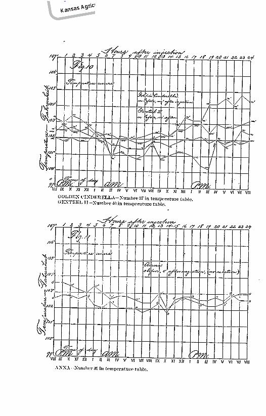
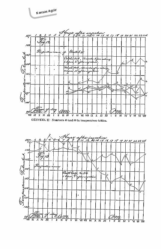
*See record of Genteel II, No. 40 and 80 CollegeHerd: also illustration Fig. 10
George Brandsberg
96 [Bulletin 79
of the colonization and development of the tubercle bacillus in theanimal tissue. At first these tubercles are microscopic in size,they gradually increase in size until they become as large as a mil-let seed or larger. Through processes of further growth andunion with each other, and finally through death and disintegra-tion, these original tubercles give rise to the various sized butalways conspicuous nodules, variously outlined, and containingwithin their centers masses of a cheesy, yellow pus. At first thisis comparatively soft, but as it gets older it becomes firmer, andfinally gritty or even stonelike in texture. There is always a dis-tinct, tough tissue capsule surrounding each focus of pus. Whensuch an abscess is located near a bronchus (large air tube in lung)it may occur that it breaks, discharges its contents into abronchus, is coughed up by the animal, and with the germs itcontains is scattered around to the risk of other victims.
The advanced tubercles are of various sizes and shapes. Onthe serous membranes of the thoracic and abdominal cavitiesthey range from smooth millet seed and pea-like nodules toenormous conglomerations resembling huge bunches of varioussized grapes, hence the name “pearly disesse” and “grapes” thatwas sometimes applied.
The mass of tubercles present on the serous membranesalone, in a case recorded by Spinola, had a weight of fifty-twopounds, while another weighed seventy pounds. Sometimes,however, only a very few are found, and occasionally the morbidchange is limited to a slight adhesion of a portion of the lung tothe thoracic wall.
When in a certain stage of advancement these nodules con-tain cheesy or partly calcifled (gritty) centers.
Other places where these cheesy abscesses are commonlyfound are the lymph glands situated between the two lungs, beforeand behind the heart (anterior and posterior mediastinal glands),which sometimes enlarge to tuberculous tumors eighteen incheslong! The glands behind the upper end of the windpipe (retro-pharyngeal glands), the glands between the kidneys (renal glands),and those under the liver (portal glands) are also commonly affected.The lungs, liver, spleen and kidneys themselves may be literally packed with tuberculous abscesses, or, one of these organs maycontain only a few.
As before stated, any tissue or organ of the body may bemore or less affected, but the process is always most marked and
George Brandsberg
George Brandsberg
George Brandsberg
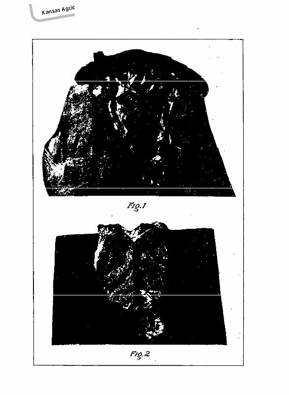
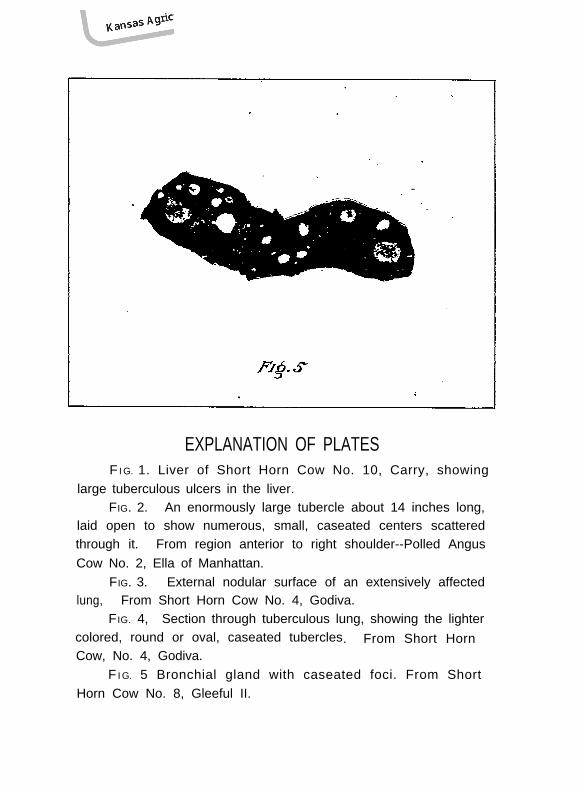
EXPLANATION OF PLATESF I G. 1. Liver of Short Horn Cow No. 10, Carry, showing
large tuberculous ulcers in the liver.FIG. 2. An enormously large tubercle about 14 inches long,
laid open to show numerous, small, caseated centers scatteredthrough it. From region anterior to right shoulder--Polled AngusCow No. 2, Ella of Manhattan.
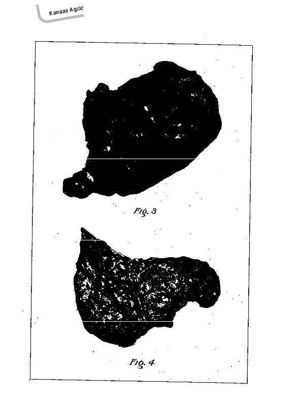
FIG. 3. External nodular surface of an extensively affectedlung, From Short Horn Cow No. 4, Godiva.
FIG. 4, Section through tuberculous lung, showing the lightercolored, round or oval, caseated tubercles. From Short HornCow, No. 4, Godiva.
F I G. 5 Bronchial gland with caseated foci. From ShortHorn Cow No. 8, Gleeful II.
George Brandsberg
George Brandsberg
George Brandsberg
George Brandsberg
George Brandsberg
April, 1898.] Bovine Tuberculosis. 97
characteristic in those organs or clusters and chains of organsknown as lymphatic glands. A chain of these organs, situatedabove the intestines in their supporting fold, is the chief seat ofchanges in intestinal tuberculosis.
Sometimes the lining membranes of the stomach and intes-tines contain tuberculous ulcers. All of these abscesses, butespecially their enveloping tissue capsules, contain tubercle bacilli.
(See plates for illustrations of morbid changes in various organs.)
DISPOSITION OF TUBERCULOUS CATTLE.
The question now arises, what is to be done with the affectedanimals after they have been discovered in an infected herd?This is the most important question in the whole subject of tuber-culosis. As soon as the disease is discovered and the affectedanimals have been singled out they should be separated from therest of the herd and kept under quarantine until the method oftheir final disposition is determined. The treatment of theremaining healthy animals is discussed later on.
As to the diseased animals; those visibly affected should beimmediately killed and their carcasses destroyed by fire or deepburial. Either method should be carried out as near as possibleto the spot where the killing is done and the post-mortem exam-ination is made, because moving affected carcasses after they havebeen opened simply increases the opportunities for spreading theinfectious material.
Cattle that have reacted but are apparently in good healthcould be disposed of in the same way as the above, but it wouldentail a loss that could not or at least would not willingly be borneby many owners, and hence such a suggestion would stand in theway of successfully combating this disease. There is anotherway out of the difficulty; a method involving so small a loss thatif every cattle owner in the state would avail himself of it wecould soon rid our herds of the disease at the smallest possiblecost. Before discussing this question further, a few generalstatements are necessary.
The meat and milk, butter and cheese, from tuberculousanimals may, and in a great many cases do, prove to be the sourceof infection of human beings.
This has been demonstrated by experiments with the loweranimals.
George Brandsberg
98 Veterinary Department. [Bulletin 79
On the other hand it has also been demonstrated that thesesame products from animals with undoubted tuberculosis canoften be fed with impunity to susceptible animals without pro-ducing any serious consequences.
These two apparently conflicting statements rest on the factthat certain forms of tuberculosis have an entirely different natureas far as danger of infection from feeding affected carcasses isconcerned. When the disease is strictly localized, the affectedparts and certain organs in intimate relation with them are alonedangerous. When the disease is generalized or in any degreeextensive, the whole carcass, including of course the milk, isdangerous.
A competent meat inspector can determine when the carcassfrom an affected animal is fit for human consumption and when itis dangerous and consequently unfit to be put on the market.
If, therefore, proper provision were made for their thoroughinspection before being placed on the market, the cattle whichare in apparent health, but have reacted under the tuberculintest, could be fattened and sold for beef. We would then haveplaced on our markets carcasses of animals slightly affected withtuberculosis; but from which all parts that could in any possibleway be dangerous had been removed under the supervision ofmen trained for that business.
As matters now stand, tuberculous cattle in all stages and inall forms of the disease are sold indiscriminately by unscrupulousor ignorant or at the least uninformed butchers, and there is nopossible way for the public to protect itself.
By following the plan here suggested, i.e., by the establish-ment of meat inspection within the state, the placing of such asystem into operation under skilled men, and by using quarantineprecautions against imported cattle to prevent renewed importa-tion of the disease, the state of Kansas would soon solve the tuber-culosis question.
Similar plans have been adopted by Massachusetts; but inthat state the disease is so much more common and all affectedanimals being wholly condemned the expense to that state isenormous compared with what it need be in Kansas.
The sooner this question is taken up by the state of Kansasthe more will be accomplished at the less cost.
The introduction of a compulsory general meat inspecting
George Brandsberg
April, 1898.] Bovine Tuberculosis. 99
system in the state of Kansas would affect not only the existenceof tuberculosis, but it would control the spread and thus preventthe occurrence of a great many parasitic diseases in man, ofwhich tapeworm disease, trichinosis and botulism are only a veryfew. The effect that such a system would have in preventingthese diseases is not a matter of speculation, it is a matter offigures and statistics that are decided enough to convince anyoneinclined to entertain doubts. The hospital records of parasiticdiseases in most meat inspection countries present astoundingcontrasts when periods before and periods after meat-inspectionhas been practiced are compared. Such diseases diminish rapidlyin number under those conditions, and meat inspection alone isresponsible for this.
AFTER TREATMENT OR DISINFECTION.
The after treatment of a tuberculosis infection applies ofcourse only to the remaining healthy animals and the quartersoccupied by them.
As before intimated, rearing healthy calves from slightlyaffected tuberculous dams is a practice that should not be encour-aged; there are enough healthy animals for this purpose.
The treatment for the remaining healthy animals consists inkeeping them in non-infected quarters, giving them pure food andwater, judicious exercise and an abundance of fresh air and sun-shine; all of this is necessary for the permanent maintenance ofhealth. Unscientiftc inbreeding and injudicious breeding forspecial purposes should be avoided.
When new purchases are made into a herd a clear bill ofhealth should be obtained for every newly acquired animal. Thetuberculin test will decide this.
Bulls used for breeding purposes should be known to be freefrom tuberculosis, and from every tendency toward acquiring it.In this manner and in this manner alone can we keep a herd freefrom a disease so insidious and wide-spread as tuberculosis.
One more important thing must not be neglected if all expensein the past and all care and management in the future are not tobe in vain. The barns, sheds, or stables that were used as shel-ter for the affected cattle are infected and constitute a source ofdanger to other cattle that are kept in them for any considerablelength of time, and no less so to the attendants of these animals.
George Brandsberg
100 Veterinary Department. [Bulletin 79
To make these places safe they must be disinfected! In orderto disinfect properly the work must be done thoroughly; disin-fection is one of those things that cannot be done halfway. Half-way disinfection is no disinfection at all.
The direct object of disinfection is to destroy disease germs.We have seen how inconceivably small these things are, and inhow many different ways and through what various channels theycan be spread and become lodged in secret hiding places. In theminutest particles of dirt, the smallest holes and finest cracks,these germs may be hiding in untold numbers.
All these places must be reached with the disinfecting fluid.This may seem a simple and trivial thing, but nothing in thewhole question of tuberculosis is of greater importance.
The disinfecting fluid may be one thing or another, manysolutions of chemicals in water will answer the purpose. Thecheapest and most effective, perhaps, is corrosive sublimate orbichloride of mercury in two per mil solutions (one-fifth of one percent) or approximately one pound of corrosive sublimate dissolvedin sixty (60) gallons of water; (accurately, 62 1/2 gal.) It is veryimportant that the chemical is thoroughly dissolved in thewater; in order to accomplish this it is best first to dissolveall in a small quantity of hot water and then to mix this solutionwith sufficient additional cold water to make the proper bulk.This solution should be made in wooden vessels as it corrodesmetal, and being highly poisonous it should be kept out of reachof children and animals.
A good spray pump is the best instrument for applying adisinfecting solution in a barn or stable: by its use all corners,holes and cracks can be penetrated. In old buildings containingpartially rotted floors, sills, walls, partitions etc., it may often bebest to do some tearing down and rebuilding, but this question isone to be settled for each individual case. Loose boards mustalways be removed before spraying.
The one object of disinfection is to reach every speck or spotof surface that has had any possible chance to have the contagionadhere to it. Contact with this solution is sure death to thetubercle bacillus.
It may be needless to state that before the floor of a stable isdisinfected all litter should be removed from it and firmly adher-ing solids scraped off with a sharp hoe, and removed to a safeplace.
George Brandsberg
April, 1898.] Bovine Tuberculosis. 101
Although cases of poisoning are no doubt rare it might bewell, after the mangers have received their spray, to wash theman hour later with hot water, thus removing all corrosive subli-mate which by that time will have done its work, and avoiding allrisk of possible poisoning of the animals.
If the procedure above outlined is thoroughly carried out thebuilding may be considered perfectly safe as far as any danger ofcontracting an infectious disease is concerned.
The healthy animals may now be put back into their oldquarters; and the more fresh air and sunlight that is permittedto enter such a building the longer will it be likely to remain pureand free from disease germs.
After this the tuberculin test should be repeated every yearfor several years and every reacting animal properly disposed of.With such treatment disinfection need be repeated in and forthose stalls only in which an affected animal has stood and for theone on each side.
DISPOSITION OF MANURE.
Manure is a dangerous source of infection in cases whereintestinal tuberculosis was present. It is a valuable article andits destruction by burning would be a considerable as well as anunnecessary loss. All animals, especially pigs, should be kept awayfrom a suspicious manure pile. At the first opportunity suchmanure should be hauled out on a level field where no washing orsurface drainage occurs, and spread out in a thin, even layer andexposed to the disinfecting action of the sun’s rays. A Kansassummer’s sun will here easily outdo a two per mil solution ofcorrosive sublimate.
THE TUBERCULIN TEST.
This subject has already been touched upon, and the princi-ple upon which the results are interpreted, explained. The bestpractical method to pursue in testing a herd of cattle is first tohouse or stable them for a sufficient length of time until allunnatural excitement incident to this unaccustomed change haspassed away.
We next begin at sx a. m., and take the temperature* of every*Tho temperatures of animals are taken by inserting the thermometer into the rectum
and allowing it to remain there at least three minutes. A self registering thermometer isthe only reliable kind. The same person should take all the temperatures of the sameanimal and with the same thermometer. Insertion of the thermometer is facilitated bymoistening it with saliva, or oil if preferable.
George Brandsberg
102 [Bulletin 79
animal every two hours untilten p. m., of the, same day. At tenp.m., immediately after taking the last temperature, the animalsshould be injected† with the tuberculin. The amount to be injectedvaries with the brand of the article and the size or weight of theanimal. The tuberculin manufactured by the Bureau of AnimalIndustry** of the United States Department of Agriculture isinjected at the rate of two cubic centimeters for an adult animalof average size. The amount injected in the test shown bythe appended tables was at the rate of two cubic centimeters forevery 1200 lb. wt. of the animal, for about half the cases. For theother half the rate was 2cc., for every 1000 lb. wt. Equally goodresults seem to have been obtained.
At six a.m., on the following morning temperatures should beagain taken, this time every hour, and continued for twenty-fourhours or until definite results are obtained.
The temperatures of the second day are then compared withthose of the first, and if an increase of two degrees Fahr., or morefor six or eight hours or longer, above the corresponding temper-ature of the preceding day is shown, the animal may be set downas being tuberculous; unless, as already stated, other reasons canbe assigned for the abnormal temperature.
Some animals have abnormally high temperatures to beginwith; a temporary fever, pregnancy or other causes may accountfor this. Such animals should be reserved for a second or latertest.
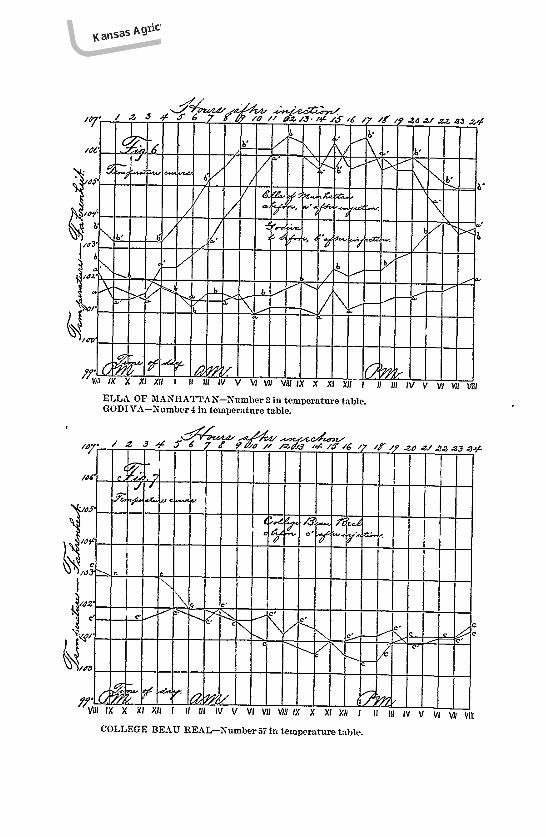
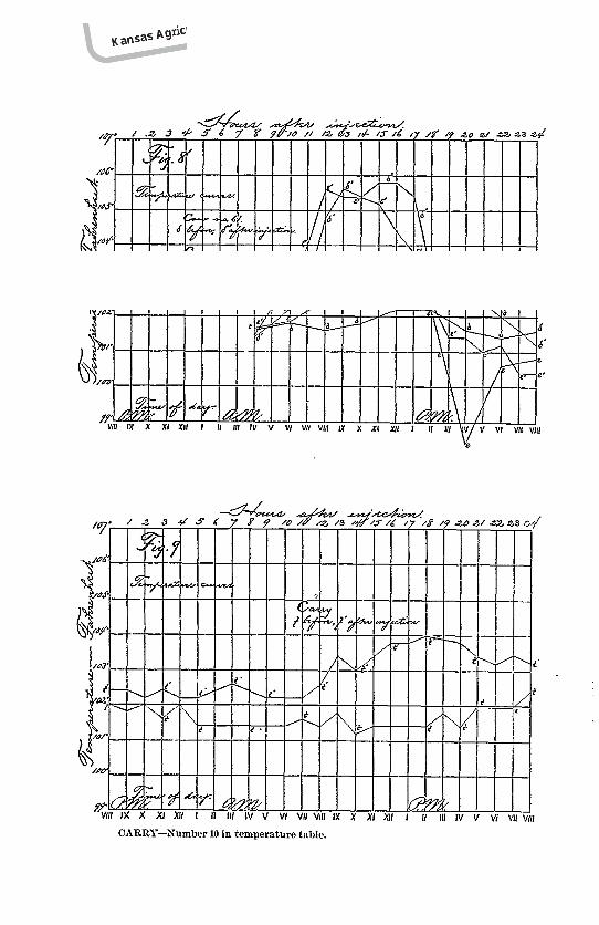
The best way to interpret the results of a tuberculin test is toconstruct a fever or temperature chart as is shown in figures sixto thirteen.
A number of vertical lines are drawn at equal distances fromeach other to represent the time between hours. Twelve or fifteenof these are usually sufficient to answer the purpose. These arethen crossed by another set of similar but horizontal lines, eightin number, representing the degree of temperature, say from100 degrees Fahr., to 107 degrees Fahr. The spaces between thehorizontal lines are divided into fifths or tenths by points or dotsplaced on the vertical lines. At these points the temperature eachhour for two days may be indicated, and the points connected withlines. This forms two broken lines known as temperature curvesthat represent the variation of temperature during a day, and the
†The injection is made with a hypodermic syringe, into the loose tissue under the skin.and preferably in the region of the neck.
**This brand was used in testing the College animals.
George Brandsberg
April, 1898.] Bovine Tuberculosis. 103
difference before and after injection, in a very graphic manner.The higher and longer the second day curve as compared withthe first day curve is found to be, the nearer will it approach atypical tuberculosis reaction curve.
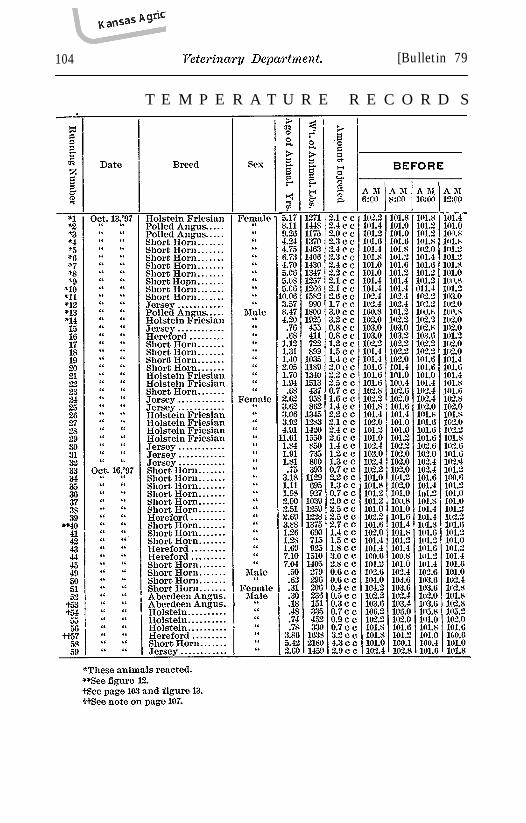
Fig. 6 shows several typical reactions.Fig. 10 shows doubtful reactions.Fig. 11 shows no reaction.Following is the temperature record of the fifty-nine animals
constituting the College herd. Some of these animals had beentested once or even more times before. The details of these testscan be found by referring to bulletin 69, pp. 118-121.
Animals Nos. 46, 47 and 48, whose temperatures do not appearin the record, were young calves affected with diarrhoea at thetime of the test and hence could not be tested.
Animals No. 53 and 54 were unthrifty bull calves; both hadabnormally high temperatures, No. 54 excessively so, both beforeand after injection; but neither showed any reaction. Both werekilled and post-mortem examinations made of their carcasses. Asthe post-mortem notes show, no unequivocal evidence of tubercu-losis was found.
Fig. 13 illustrates the temperature of No. 54.
George Brandsberg
104 [Bulletin 79
T E M P E R A T U R E R E C O R D S
George Brandsberg
April, 1898.] Bovine Tuberculosis.
O F T H E C O L L E G E H E R D .
George Brandsberg
106 [Bulletin 79
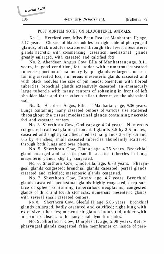
POST MORTEM NOTES ON SLAUGHTERED ANIMALS.No. 1. Hereford cow, Miss Beau Real of Manhattan II; age,
5.17 years. Cluster of black nodules on right side of pharyngealglands; black nodules scattered through the liver; mesentericglands necrotic, with commencing caseation; mediastinal glandsgreatly enlarged, with caseated and calcified foci.
No. 2. Aberdeen Angus Cow, Ella of Manhattan; age, 8.11years, in good condition, fat; udder with numerous caseatedtubercles; portion of mammary lymph glands enlarged and con-taining caseated foci; numerous mesenteric glands caseated andwith black nodules the size of pin heads; omentum with fibroidtubercles; bronchial glands extensively caseated; an enormouslylarge tubercle with many centers of softening in front of leftshoulder blade and three other similar tubercles on the abdominalwall.
No. 3. Aberdeen Angus, Ethel of Manhattan; age, 9.36 years.Lungs containing many caseated centers of various size scatteredthroughout the tissue; mediastinal glands containing necroticfoci and caseated centers.
No. 3. Shorthorn Cow, Godiva; age 4.24 years. Numerouscongested tracheal glands; bronchial glands 3.5 by 2.5 inches,caseated and slightly calcified; mediastinal glands 3.5 by 3.5 and6.5 by 4 inches; small caseated tubercles abundantly scatteredthrough both lungs and over pleura.
No. 5. Shorthorn Cow, Diana; age 4.75 years. Bronchialgland enlarged and caseated; small caseated tubercles in lung;mesenteric glands slightly congested.
No. 7. Shorthorn Cow, Fanny; age, 4.7 years. Bronchialglands caseated; mediastinal glands highly congested; deep sur-face of spleen containing tuberculous neoplasms; congestedglands of third and fourth stomachs; numerous mesenteric glandswith several small caseated centers.
No. 8. Shorthorn Cow, Gleeful II; age, 5.06 years. Bronchialglands enlarged, badly caseated and calcified; right lung withextensive tubercles; mesenteric glands indurated; udder withtuberculous abscess with many small lymph nodules.
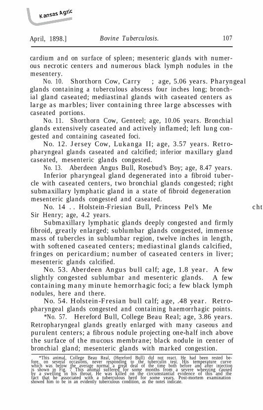
No. 9. Shorthorn Cow, Dimples II; age, 5.08 years. Retro-pharyngeal glands congested, false membranes on inside of peri-
George Brandsberg
April, 1898.] Bovine Tuberculosis. 107
cardium and on surface of spleen; mesenteric glands with numer-ous necrotic centers and numerous black lymph nodules in themesentery.
No. 10. Shorthorn Cow, Carry ; age, 5.06 years. Pharyngealglands containing a tuberculous abscess four inches long; bronch-ial gland caseated; mediastinal glands with caseated centers aslarge as marbles; liver containing three large abscesses withcaseated portions.
No. 11. Shorthorn Cow, Genteel; age, 10.06 years. Bronchialglands extensively caseated and actively inflamed; left lung con-gested and containing caseated foci.
No. 12. Jersey Cow, Lukanga II; age, 3.57 years. Retro-pharyngeal glands caseated and calcified; inferior maxillary glandcaseated, mesenteric glands congested.
No. 13. Aberdeen Angus Bull, Rosebud’s Boy; age, 8.47 years.Inferior pharyngeal gland degenerated into a fibroid tuber-
cle with caseated centers, two bronchial glands congested; rightsubmaxillary lymphatic gland in a state of fibroid degeneration ;mesenteric glands congested and caseated.
No. 14 . . Holstein-Friesian Bull, Princess Pel’s Me chthildesSir Henry; age, 4.2 years.
Submaxillary lymphatic glands deeply congested and firmlyfibroid, greatly enlarged; sublumbar glands congested, immensemass of tubercles in sublumbar region, twelve inches in length,with softened caseated centers; mediastinal glands calcified,fringes on pericardium; number of caseated centers in liver;mesenteric glands calcified.
No. 53. Aberdeen Angus bull calf; age, 1.8 year. A fewslightly congested sublumbar and mesenteric glands. A fewcontaining many minute hemorrhagic foci; a few black lymphnodules, here and there.
*No. 57. Hereford Bull, College Beau Real; age, 3.86 years.Retropharyngeal glands greatly enlarged with many caseous andpurulent centers; a fibrous nodule projecting one-half inch abovethe surface of the mucous membrane; black nodule in center ofbronchial gland; mesenteric glands with marked congestion.
*This animal, College Beau Real, (Hereford Bull) did not react. He had been rested be-fore, on several occasions, never responding to the tuberculin test. His temperature curvewhich was below the average normal a great deal of the time both before and after injectionis shown in Fig. 7 This animal suffered for some months from a severe wheezing causedby a swelling in his throat. He was killed on the circumstantial evidence of this and thefact that he associated with a tuberculous herd for some years. Post-mortem examinationshowed him to be in an evidently tuberculous condition, as the notes indicate.
George Brandsberg
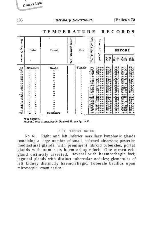
T E M P E R A T U R E R E C O R D S
POST MORTEM NOTES.
No. 61. Right and left inferior maxillary lymphatic glandscontaining a large number of small, softened abscesses; posteriormediastinal glands, with prominent fibroid tubercles, portalglands with numerous haemorrhagic foci. One mesentericgland distinctly caseated; several with haemorrhagic foci;inguinal glands with distinct tubercular nodules; glomerules ofleft kidney distinctly haemorrhagic. Tubercle bacillus uponmicroscopic examination.
George Brandsberg
April, 1898.] Bovine Tuberculosis. 109
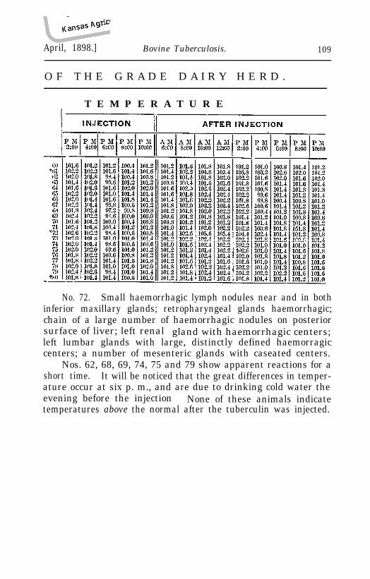
O F T H E G R A D E D A I R Y H E R D .
T E M P E R A T U R E
No. 72. Small haemorrhagic lymph nodules near and in bothinferior maxillary glands; retropharyngeal glands haemorrhagic;chain of a large number of haemorrhagic nodules on posteriorsurface of liver; left renal gland with haemorrhagic centers;left lumbar glands with large, distinctly defined haemorragiccenters; a number of mesenteric glands with caseated centers.
Nos. 62, 68, 69, 74, 75 and 79 show apparent reactions for ashort time. It will be noticed that the great differences in temper-ature occur at six p. m., and are due to drinking cold water theevening before the injection None of these animals indicatetemperatures above the normal after the tuberculin was injected.