OF THE A Professional Guide for the Management of Patients Undergoing Chemotherapy and Head and Neck Radiation Therapy Authors Gerry J. Barker, R.D.H., M.A. Bruce F. Barker, D.D.S. Ronald E. Gier, D.M.D., M.S. Editorial Review by Peter Stevenson-Moore, B.D.S., M.S.D. Ernest G. Glass, D.D.S., M.S., M.S.D. Loretta S. Loftus, M.D. and the Executive Committee of the International Society for Oral Oncology ' Joel B. Epstein, D.M.D., M.S.D. Philip Fox, D.D.S., Ph.D. Loree K. Oberle-Edwards, R.D.H., M.S. Douglas E. Peterson, D.M.D., Ph.D. Fred K.L Spijkervet, D.D.S., Ph.D.
Transcript
OF THE
A Professional Guide for the
Management of Patients
Undergoing Chemotherapy and
Head and Neck Radiation Therapy
Authors
Gerry J. Barker, R.D.H., M.A. Bruce F. Barker, D.D.S.
Ronald E. Gier, D.M.D., M.S.
Editorial Review by
Peter Stevenson-Moore, B.D.S., M.S.D. Ernest G. Glass, D.D.S., M.S., M.S.D.
Loretta S. Loftus, M.D.
and the Executive Committee of the International Society for Oral Oncology
' Joel B. Epstein, D.M.D., M.S.D. Philip Fox, D.D.S., Ph.D.
Loree K. Oberle-Edwards, R.D.H., M.S. Douglas E. Peterson, D.M.D., Ph.D. Fred K.L Spijkervet, D.D.S., Ph.D.
Produced by:
w- SCHOOL OF C
[/c Biomedical Communications L University of Missouri-Kansas City
)ENTISTRY School of Dentistry 650 East 25th Street Kansas City, MO 64108-2784
UMKC is an equal opportunity institution.
Development initially funded by a grant from the Department of Health and Human Services, Public Health Service Grant Number CA27688-03.
The University of Missouri-Kansas City School of Dentistry and the International Society for Oral Oncology gratefully acknowledge the unrestricted professional educational grant from Colgate Oral Pharmaceuticals in support of the production of this manual.
Copyright 0 1981 by THE CURATORS OF THE UNIVERSITY OF MISSOURI
All rights reserved. Except for use in a review, the reproduction or utilization of this work in any form or by any electronic, mechanical, or other means now known or hereafter invented, including photocopying and recording, or in any information storage and retrieval system is forbidden without the written permis- sion of the publisher.
Sixth edition, January 2000
Gerry J. Barker, RD.H., M.A. Associate Professor, Department of Dental Public Health and Behavioral Science Coordinator of Oncology Education and the Oncology Dental Support Clinic University of Missouri-Kansas City School of Dentistry Kansas City, Missouri
Bruce F. Barker, D.D.S. Professor, Department of Oral and Maxillofacial Pathology Director of the Oncology Dental Support Clinic University of Missouri-Kansas City School of Dentistry Kansas City, Missouri
Ronald E. Gier, D.M.D., M.S.D. Professor, Department of Diagnostic Sciences University of Missouri-Kansas City School of Dentistry Kansas City, Missouri
Joel B. Epstein, D.M.D., M.S.D. Douglas E. Peterson, D.M.D., Ph.D. Clinical Professor, Faculty of Dentistry Professor and Head, Department of Oral Diagnosis University of British Columbia, Vancouver, Canada School of Dental Medicine Head, Division of Oral Medicine and University of Connecticut Health Center Clinical Dentistry, Vancouver General Hospital Farmington, CT Medical-Dental Staff, British Columbia Cancer Agency Research Associate, Department of Oral Medicine University of Washington, Seattle WA
Philip Fox, D.D.S., Ph.D. Director, Research and Development Amarillo Biosciences, Inc. 6509 Seven Locks Road Cabin John, MD
Loree K. Oberle-Edwards, R.D.H., M.S. Dental Hygiene/Oral Medicine Scripps Center for Dental Care LaJolla, CA
Ernest G. Glass, D.D.S., M.S., M.S.D. Fred K. L. Spijkervet, D.D.S., Ph.D. Associate Professor Associate Professor Director of Special Patient Care Center Oral and Maxillofacial Surgeon University of Missouri-Kansas City School of Dentistry University Hospital Kansas City, MO Groningen, The Netherlands
Loretta S. Loftus, M.D. Peter Stevenson-Moore, B.D.S. Chief, Hematology/Oncology L.D.S.R.C.S., M.S.D., M.R.C.D.(C) Professor of Medicine and Assistant Dean Chairman, Division of Dentistry University of Missouri-Kansas City School of Medicine Head, Department of Dentistry
and Truman Medical Center British Columbia Cancer Agency, Vancouver Canada Kansas City, MO
This manual provides a general outline for the oral management of the cancer patient undergoing chemotherapy, bone marrow transplantation and/or radiation therapy to the oral cavity and/or salivary glands. It is neither intended to serve as a comprehensive academic review nor to cover all of the pos- sible complications that develop in the treatment of cancer patients or the morbidity associated with cancer surgery. Further information may be obtained by reviewing related literature.
For Patients Receiving Chemotherapy for any Malignancy Approximately 40 percent of patients receiving chemotherapy will experi-
ence oral complications. The majority of patients with leukemia and those who receive a bone marrow transplant will develop oral complications. Research shows, however, that fewer problems develop when oral disease is eliminated, when an oral prophylaxis is performed prior to the initiation of chemotherapy and when excellent oral hygiene is maintained throughout therapy.
For Patients Receiving Radiation Therapy to the Oral Cavity and/or Salivary Glands
Radiation therapy to the head and neck, which includes the salivary glands and/or the oral and pharyngeal tissues, may result in acute side effects that include taste loss, mucositis, infection and decreased salivary flow. Long-term, permanent side effects may include salivary gland dysfunction, dental demineralization, radiation caries, trismus, soft tissue breakdown and failure to heal, and osteoradionecrosis (ORN).
The obiectives of an oralldental program for the cancer patient are to: Improve oral function and quality of life.
Improve and maintain oral hygiene in order to reduce the risk and severity of oral complications.
Eliminate oral infection and prevent potentially fatal systemic infections of dental origin.
Prevent, eliminate or control oropharyngeal pain.
Prevent or control salivary gland dysfunction and the destruction of the dentition.
Assist with maintaining adequate nutrition.
Prevent or reduce the incidence of bone necrosis.
Pages
Introduction and Objectives ........................................................................................................ i
Chemotherapy Oral Manifestations of Chemotherapy .................................................................. 1 . 2
OraVDental Management Prior to Chemotherapy ................................................ 3 . 5
OraVDental Management During Chemotherapy .................................................. 6 . 8
OraVDerital Management Following Chemotherapy .................................................. 8
Bone Marrow and Stem Cell Transplantation .................................................... 9 . l o
Radiation Therapy Oral Manifestations of Radiation Therapy to the Head and Neck .................................. 11
Chemotherapeutic drugs are administered systemically over several weeks or months in a sequence of "treatment rounds or courses." This schedule allows some recovery of healthy tissues between each treatment of the toxic drugs. Complications arise from the direct cytotoxic effects of chemotherapeutic agents on oral tissues and/or from the indirect effects of myelosuppression. Oral manifestations are related to the drug protocol (type of drugs, dose and duration), the patient's mucosal integrity, and oral and systemic status. The reactions are often hi-shlv individualized.
Mucositis and ulceration The gastrointestinal (GI) mucosa, because of its high cellular turnover rate, is highly susceptible to the toxic effects of many chemotherapeutic agents. Inflammation and ulceration of the mucosal lining of the mouth, pharynx, esophagus and the entire GI tract may occur. The patient may experience pain, nausea, vomiting and diarrhea.
Pain Oropharyngeal pain is a prominent and frequent sequela of chemo- therapy-induced mucositis. Descriptions of pain by patients do not always correlate with severity of tissue injury as assessed upon clinical examination.
Infection Many drugs and some malignancies can suppress bone marrow produc- tion and induce leukopenia, which can result in increased risk of infec- tions. The usual clinical signs of inflammation (redness, pain, swelling, heat) may not be present during periods of significant immunosuppres- sion. If pain is present, the symptomatic areas of possible infection (operculum, periodontal pockets or mucosal ulcerations) should be cul- tured if the patient develops a fever of unknown origin. Infection may be caused by organisms usually found in the mouth such as Candida species, herpes viruses, streptococci and staphylococci. Candidiasis may have the typical appearance of soft white plaques or present as general- ized erythematous painful tissue. Infections may also be caused by opportunistic organisms not commonly found in the mouth such as aspergillus, other fungae, gram-negative bacilli and coliform bacteria. Angular cheilosis is a common candida-related oral manifestation. Oral infections may lead to systemic infection or sepsis and can be life- threatening in the neutropenic patient.
I CHEMOTHERAPY I
Oral Manifestations of Chemotherapy
Bleeding Reduction of platelets (thrombocytopenia) and other clotting factors dur- ing periods of bone marrow suppression are the major causes of bleed- ing. Transfusion of platelets and/or clotting factors in conjunction with topical agents may be necessary for control.
Xerostomia/Salivary gland dysfunction
Patients may complain of decreased or thickened saliva. The duration of xerostomia is associated with the length of therapy, other prescribed medications and the health of the patient. Xerostomia may result in a lowered pH, alterations in the constituents of the saliva, and it may lead to rampant dental caries. A dry mucosa is more susceptible to pain, infections and irritation.
Taste alteration Transient alteration in taste is common after the administration of some chemotherapeutic drugs.
Neurotoxicity The patient may present with numbness or constant, deep pain that is often bilateral and frequently mimics toothache (odontalgia), but no odontogenic or mucosal source can be found. This phenomenon may be present after the administration of drugs such as vincristine and vin- blastine.
Dental developmental abnormalities Chemotherapy administered during dental development in childhood may cause shortened or malformed roots, enamel defects, disturbance in crown development and eruption.
OrallDental Evaluation Prior to Chemotherapy
Consultation with the medical oncologist
Consultation with the medical oncologist prior to the dental evaluation is essential to ensure appropriate treatment planning and coordination of dental care with the proposed schedule of cancer therapy. The information on the "referral form" below may be requested from the medical oncologist to guide dental treatment planning, as well as aid in seeking reimbursement from medical insurance for "medically-necessary oral health care."
Oncology Dental Support Clinic
is referred for an orddental evaluation and treatment. The need for this medically necessary oral health care is a direct result of and can directly impact the underlying medical condition and/or its resulting oncology therapy.
physician signature date
Patient's diagnosis: ICD-9 code
Proposed therapy:
Anticipated degree of myelosuppression: Anticipated days to nadir:
Current blood counts: Absolute neutrophil count: Platelet count:
Does the patient have a central venous catheter? Yes No
Are antibiotics or special precautions necessary prior to dental treatment? Yes No
Additional important medical information:
OrallDental Evaluation Prior to Chemotherapy
Important information includes:
Indwelling central venous catheter: Some patients are given chemotherapy through a catheter placed in a major vein. Catheters may become colonized with bacteria that enter the blood during dental pro- cedures. Although there are no clinical studies to document the need to premedicate these patients, it is clinical practice in many settings for these patients to receive the American Heart Association endocarditis prophylactic antibiotic regimen prior to an invasive dental procedure, including dental prophylaxis. Physician consultation is recommended.
Clotting factors: Hemorrhage may be a complication when the platelet count is < 50,000/mm3 or with abnormal clotting factors (PT, PTT, fib- rinogen).
Absolute neutrophil count: * Risk of infection and septicemia is high when the absolute neutrophil count is < l,000/mm3. Patients receiving immunosuppressive chemotherapy will usually reach their "nadir" (the lowest blood counts) 7-14 days after a round or course of therapy.
Dental treatment can be performed after the neutrophil count has begun to rise from the nadir and has reached a level of l,000/mm3 or above. If a dental procedure is necessary and the neutrophil count is < l,OOO/mm3, the oncologist must be consulted concerning antibiotic coverage. Extensive invasive oral procedures should not be performed if the absolute neutrophil count will be <l,000/mm3 prior to adequate healing.
Pre-chemotherapy oral/dental treatment Eliminate any area of infection or irritation, such as teeth with fractures,
fractured restorations, advanced carious lesions, pulpal or periapical involvement, periodontal inflammation, pericoronitis, or ill-fitting pros- theses.
Remove orthodontic bands if highly stomatotoxic chemotherapy is sched- uled. The decision should be made in consultation with the oncologist.
Institute periodontal disease control measures that include plaque control, and if possible, a full dental prophylaxis. Patients who continue to maintain excellent oral hygiene throughout therapy may have fewer complications.
Provide oral hygiene instruction, including use of an extra or ultra-soft toothbrush and dental floss, and arrange for ongoing supervision of hygiene during cancer therapy.
Patients should be advised to avoid commercial mouthrinses with alcohol and/or a high sugar content.
Review dietary recommendations to limit highly cariogenic foods without compromising adequate caloric intake.
Additional needs of children Evaluate the dentition and estimate exfoliation time of primary teeth.
Remove mobile primary teeth as well as those expected to be lost dur- ing the chemotherapy.
Consider removal of gingival opercula if there is a clinical risk for entrap- ment of food debris and/or nidus for infection, particularly if the area has previously been symptomatic.
OrallDental Management During Chemotherapy
Dental treatment Once chemotherapy has been initiated, oral prophylaxis and restorative
dental treatment can usually be scheduled within a few days of the next pro- posed round or course of therapy. Generally the patient's blood counts will have recovered from the toxicity of the previous course of drugs. Blood counts, however, should be ordered the day before dental treatment to docu- ment hematologic status. If oral surgery is required, it should be scheduled to allow at least 7-10 days of healing prior to the anticipated date of bone mar- row suppression. It b imperative that the dentist seek consultation with the oncologist prior to any invasive dental procedure, including pro- phylaxis.
Infection control Culture all lesions for infection (bacterial, fungal and/or viral). Prescribe
treatment in cooperation with the oncologist. Exfoliative cytology or ELISA may be performed for rapid identification of the herpes virus. If positive, an antiviral medication may be administered to prevent progression of the lesions. For fungal infections, patients should be aware that topical antifun- gal agents are efficacious only when in contact with the lesions and are used for the prescribed time period. Use of a sugar-free antifungal should be con- sidered if extended use is necessary or if the patient is caries-prone.
Orthodontic bands If bands are not removed prior to chemotherapy, soft wax or a plastic
mouthguard may be used to protect the oral tissues from injury during peri- ods of oral inflammation or ulceration.
Prevention of dental caries and demineralization Measures for the prevention of tooth demineralization are required only
when xerostomia persists for longer than six weeks. It is recommended that a 1.1% neutral pH sodium fluoride or 0.4% unflavored stannous fluoride be brushed on the teeth or applied in custom-made gel applicator trays. A neu- tral pH fluoride gel should be used by patients with porcelain crowns. Acidulated fluoride should not be used. (Refer to page 18 for gel application directions.)
Palliation of xerostomia and pain Appropriate pain management is important to preserving quality of life, as
well as enhancing compliance with oral hygiene interventions and infection prevention. Suggestions for symptomatic relief of pain and xerostomia should be offered to the patient. (see pages 23-24, 26)
Continued on page 7-8
OrallDental Management During Chemotherapy
Oral hygiene and patient self-care procedures Excellent oral hvgiene must be maintained. An oral hygiene program
must be individualized for each patient and modified throughout therapy according to his or her medical status. To foster complia~ce the patient must understand the risks of septicemia from poor oral hygiene.
toothbrushing
The patient should be taught a very gentle but effective toothbrushing technique with a soft nylon-bristle brush. Toothbrushes should be changed frequently and/or disinfected in chlorhexidine.
During periods of neutropenia and/or thrombocytopenia (ANC < 1000/mm3 and/or platelet count of <50,000/mm3) optimum plaque control measures may necessitate gentle oral lavage, increased baking soda-saline rinses, toothbrush bristles softened in hot water or the use of an ultrasoft toothbrush.
Sponge toothettes, gauze and/or cotton-tipped applicators do not ade- quately remove plaque and should be supplemented with other mea- sures. Dipping a sponge toothette in chlorhexidine rinse may increase effectiveness. Regular toothbrushing should resume as soon as possible.
flossing
Flossing can continue during chemotherapy if the patient has previously flossed regularly and has good manual dexterity. During periods of severe neutropenia, the patient may need to discontinue flossing if bleeding is excessive. Toothpicks should not be used and caution should be taken when eating crunchy or sharp foods that may damage friable oral tissues.
denture care
Edentulous patients must not wear dentures while they sleep or when their dentures irritate ulcerated mucosal tissues. Dentures must be brushed daily with a denture brush and soaked in an antimicrobial cleanser or mild detergent. After brushing and soaking, the dentures should be rinsed well and stored in clean water or a fresh chlorhexidine solution. Edentulous patients should cleanse their tongue and oral tis- sues with gauze or a soft toothbrush.
I LHEMOTHERAPY
OrallDental Management During Chemotherapy
mouth rinses Alcohol-based mouthwashes and full-strength peroxide solutions or gels
should not be used due to their drying and irritating effects. Use of all diluted peroxide solutions should be limited to the removal of
adherent debris; its use may disrupt the normal oral flora. Peroxide solutions are acidic and, if used, should be followed by a neutralizing rinse, such as a bicarbonate water solution.
The prophylactic use of chlorhexidine gluconate rinse may be helpful in suppressing bacterial colonization but should not replace mechanical removal of plaque with a toothbrush. The patient should be monitored closely and evaluated regularly to determine the benefit of the rinse ver- sus the risk for irritation and/or drying of tissues.
The mouth may be rinsed with a baking soda-saline solution and followed by a plain water rinse several times a day. The solution is prepared by mixing 1-2 tsp(s) of baking soda and 1/2 tsp of salt with one quart of water. The salt may be eliminated according to patient preference. This solution may be put in a disposable irrigation bag and hung overhead to allow the solution to flow through the mouth. The solution must not be swallowed.
Patients who experience frequent emesis should be encouraged to rinse thoroughly with a baking soda and water solution. Brushing the teeth without first neutralizing the gastric acids in the mouth may result in etching of the enamel.
Dietary counseling Guidance in food selection should be offered in order to maintain the
patient's nutritional status and to control caries.
At completion of all planned courses of chemotherapy, closely monitor the patient until all side effects of therapy have resolved, including immunosuppression. The patient may then be placed on a normal dental recall schedule. Since these patients may need to undergo additional myelosuppressive therapy if they relapse in the future, it is very important to maintain optimal oral health.
Children should receive close lifetime follow-up, with specific attention to growth and development patterns.
Blood/bone marrow transplantation (BMT) for the treatment of leukemias, aplastic anemias, lym- phomas and some solid tumors is becoming more common. Intensive chemotherapy with or without total body irradiation is administered to eliminate malignant cells and immunosuppress the patient. Following the intensive therapy, the bone marrow is restored by infusing bone marrow or peripheral blood stem cells (PBSC) into the patient through a central venous catheter. The dental team plays a vital role in the management of these patients because preexisting dental disease may lead to signifi- cant infections during and after the transplant, thus compromising the success of the procedure and possibly resulting in sepsis and death.
Types of transplants Autologous: The patient's own bone marrow and/or PBSC are removed and
preserved.
Allogeneic: Bone marrow and/or PBSC are donated by a family member or an individual identified through a computer data bank (matched-unrelat- ed donor: "MUD".) Techniques are used to match the cell-surface markers to reduce the risk of complications and graft-versus-host dis- ease.
Oral sequelae
Syngeneic: Bone marrow and/or PBSC are taken from an identical twin.
During initial phases of the transplant, the patient is at risk for develop- ing significant oral sequelae, including oral mucositis and ulcerations, hemor- rhage, infections (fungal, viral and bacterial) and xerostomia. The acute oral complications begin to resolve when engraftment is achieved and the hema- tologic status begins to improve. The initial intensive therapy may result in immunosuppression for up to one year post-BMT.
Graft-Versus-Host Disease (GVHD) Allogeneic bone marrow transplant patients are at risk for developing
acute and chronic GVHD. Associated GVHD oral complications may include mucositis, mucosal atrophy, ulcerations, oral infections including candidiasis; immune-related complications, including lichenoid reactions, scleroderma and lupus-like changes (limited oral opening and tongue mobility); and xerosto- mia with secondary rampant dental breakdown. GVHD and its medical man- agement may cause significant immunosuppression.
Oralldental management prior to BMT Elimination of all oral sources of infection and irritation is the goal of
pre-BMT dental evaluation and treatment. Dental treatment should be scheduled in consultation with the oncologist. AU symptomatic infec- tions must be managed before medical therapy. If the patient has a cen- tral venous catheter, prophylactic antibiotics should be prescribed in consul- tation with the physician prior to the dental appointment.
If oral surgery is required, at least 7-10 days of healing should be allowed before the anticipated date of bone marrow suppression (eg: ANC of < 1000/mm3 and/or platelet count of < 50,000/mm3).
Dental prophylaxis and oral hygiene instruction may have a significant positive impact on the severity of the oral complications. Patients scheduled for total body irradiation and/or allogeneic transplant should be taught an effective toothbrushing technique and should apply fluoride gel on their teeth. If necessary, study models should be made for fabrication of custom fluoride gel-applicator trays.
It should be remembered that many of these patients will remain immunosuppressed for up to a year and will be at risk during selected post- transplant dental treatment.
Oralldental management following BMT Intensive oral hygiene measures must continue to ensure adequate plaque
control and reduce the risk of rampant dental breakdown and oral infections. Daily fluoride (rinse or gel) is necessary for dentate patients until salivary function recovers. Most patients conditioned with total body irradiation who do not develop chronic GVHD will recover normal salivary function 3-12 months post-therapy.
Invasive dental procedures should only be planned following con- sultation with the patient's oncologist and/or the bone marrow trans- plant center's dentist. Elective surgery and invasive procedures should be delayed approximately one year post-transplant due to the possible delay of healing and a theoretical risk of pneumonia until counts recover. Patients with chronic GVHD may experience long-term oral and systemic complica- tions and present with difficult dental management problems. Children may experience altered craniofacial growth and tooth development. Post-bone marrow transplant patients and patients on long-term immunosuppressive drugs are at risk for second malignancies.
Patients that receive radiation therapy to the oropharyngeal area for malignant tumors, lymphomas or leukemia may undergo radiation therapy for 3-7 weeks with a total dose ranging from 3,000 to >7,000 centigray (cGy), depending on tumor type and location. Hodgkin's and non-Hodgkin's lym- phomas require less radiation than other solid tumors of the head and neck. Patients that undergo radiation therapy to areas other than the oral cavity and/or salivary glands will not experience oral complications because the radiation only affects those tissues directly within the therapy field.
Radiation therapy is associated with side effects that vary in intensity and duration, and are depen- dent upon several factors. Not all patients will experience all of these complications but they should be aware of the potential risks.
Potential Oral Manifestations of Radiation Therapy to the Oropharyngeal and Salivary Gland Region
Factors that influence intensity and duration of the oral manifestations total dosage rate of radiation delivery fraction size field of radiation radiation source previous surgical intervention oral hygiene and dental status medical and nutritional status of patient tobacco and alcohol use
Acute Oral Manifestations of Radiation Therapy to the Oral Cavity andlor Salivary Glands
Taste alterations Alteration and loss of taste may begin with the first 200-400 cGy. After three weeks of therapy, it takes 500-8,000 times normal concentrations of taste stimulant to elicit a normal taste response. Taste acuity levels usually return to normal 2-4 months following completion of therapy, if adequate saliva is available.
Xerostomia/salivary gland dysfunction Saliva glands within the field of radiation may be permanently destroyed during therapy. Saliva is reduced in volume and altered in consistency. Reduction is dependent upon the total dose of radiation, degree of salivary gland involvement in the field of radiation, and indi- vidual patient variables. Flow may be reduced 50% by the end of the first week, and further reduction in volume (up to 100%) may occur. The saliva produced is more mucinous. Salivary gland tissue does not recover from high doses of radiation, although some patients may per- ceive an improvement in salivary output over time.
Mucositis/ulceration/pain
Infection
The mucosa exposed to the radiation becomes edematous, erythema- tous, pseudomembranous, and may ulcerate. Pain varies considerably in severity and may be intensified by certain foods. The patient may develop problems in swallowing and speaking. Mucositis usually occurs after the second week of radiation therapy, but will vary according to the dose and fractionation. The lips, buccal mucosa, soft palate, borders of tongue and floor of mouth are at greater risk of mucositis. Severe symptoms usually resolve within six weeks following completion of therapy.
Secondary infections are common. While candidiasis is most common, oral infections may be caused by a wide range of bacterial, mycotic and viral pathogens.
Nutritional deficiency Eating difficulty caused by xerostomia, mucositis and dysphagia may lead to nutritional compromise and dehydration.
Chronic Oral Manifestations of Radiation Therapy to the Oral Cavity andlor Salivary Glands
Salivary gland dysfunction -
Salivary gland tissue does not recover from high doses of radiation. The quality and quantity of saliva is permanently changed.
Radiation cariesldental demineralization Rapid demineralization, breakdown of tooth structure and caries may occur following radiation therapy. The process may be recognized early after treatment. The teeth need not be in the direct field of radiation therapy. Demineralization and caries result when the parotid and/or submandibular/sublingual glands are included in the field of radiation. The saliva becomes more acidic and there is a shift to more cariogenic bacteria. Decreased saliva, particularly of resting flow from the sub- mandibular/sublingual glands, deprives the oral cavity of the protective components of saliva and the tooth structure of the calcium and phos- phate ions necessary to maintain the hydroxyapatite content of enamel and dentin. Although some patients do not clinically appear to be xerostomic after radiation therapy, they may experience a change in the quality of their saliva, leading to rapid dental demineralization. Even a 25 percent decrease in saliva may result in dental breakdown.
Increased periodontal disease Following radiation therapy, teeth within the field of high-dose radiation therapy are at risk for increased attachment loss. The risk may be fur- ther exacerbated in patients with poor oral hygiene or that have other contributing factors such as tobacco use, systemic disease or significant salivary gland dysfunction. Radiographs of previously irradiated bone may demonstrate widening of the periodontal ligament space and distur- bance in the trabecular bone.
Trismus Spasms and/or fibrosis of the muscles of mastication and temporo- mandibular joint may develop within the field of radiation. Limited opening may interfere with oral hygiene, dietary intake, use of prosthe- ses and restrict access for dental care and general anesthesia.
Chronic Oral Manifestations of Radiation Therapy to the Oral Cavity andlor Salivary Glands
Soft tissue necrosis/osteoradionecrosis (ORN) Soft tissue and bone necrosis may develop because tissues within the field of radiation become hypovascular, hypoxic and hypocellular. The threat of this sequela persists indefinitely, although the risk is minimal when the total dose of radiation is < 5000 cGy. Factors that may con- tribute to an increased risk for necrosis include compromised vascularity from previous surgery, poor nutritional or m e d i d status, uncontrolled diabetes, and heavy tobacco or alcohol use.
The necrosis process may result spontaneously or result from trau- ma, leading to non-healing soft tissue, bone lesions, and necrosis. Trauma may result from tooth extraction, invasive periodontal proce- dures and intraoral prosthetic appliances. The mandible is much more susceptible to ORN than the maxilla. The non-healing bone may become secondarily infected, cause chronic pain, bad breath, prevent use of oral prostheses and progress to pathologic fracture.
Developmental maxillofacial deformity Children who receive radiation therapy to facial bones and developing dental structures may experience altered craniofacial growth and tooth development. The degree of deformity depends on the dose of radia- tion therapy and the age of the child at the time of therapy.
Long-term effects of radiation therapy Although patients receiving radiation therapy will experience dramatic resolution of the acute effects following the completion of treatment, the long-term effects are progressive and significant. Fibrosis of tissues, decreased cellularity and hyalinization of blood vessels contribute to decreased perfusion of tissues that intensifies with time.
OrallDental Management Prior to Radiation Therapy
Consultation with radiation oncologist Consultation with the radiation oncologist prior to the initiation of radia-
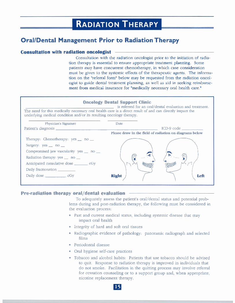
tion therapy is essential to ensure appropriate treatment planning. Some patients may have concurrent chemotherapy, in which case consideration must be given to the systemic .effects of the therapeutic agents. The informa- tion on the "referral form" below may be requested from the radiation oncol- ogist to guide dental treatment planning, as well as aid in seeking reimburse- ment from medical insurance for "medically necessary oral health care."
Oncology Dental Support Clinic is referred for an oravdental evaluation and treatment.
The need for this medically necessary oral health care is a direct result of and can directly impact the underlying medical condition andlor its resulting oncology therapy.
Physician's Signature Date
Patient's diagnosis ICD-9 code Please draw in the field of radiation on dhgmms below
Therapy: Chemotherapy: yes - no - Surgery: yes - no - Compromised jaw vascularity yes - no - Radiation therapy: yes - no - Anticipated cumulative dose cGy
Daily fractionation
Daily dose CGY
Pre-radiation therapy oralldental evaluation To adequately assess the patient's oral/dental status and potential prob-
lems during and post-radiation therapy, the following must be considered in the evaluation process:
Past and current medical status, including systemic disease that may impact oral health
Integrity of hard and soft oral tissues
Radiographic evidence of pathology: panoramic radiograph and selected films
Periodontal disease
Oral hygiene self-care practices
Tobacco and alcohol habits: Patients that use tobacco should be advised to quit. Response to radiation therapy is improved in individuals that do not smoke. Facilitation in the quitting process may involve referral for cessation counseling or to a support group and, when appropriate, nicotine replacement therapy.
Dietary analysis: Analyze and modify the patient's daily dietary habits, including frequency of meals and snacks to eliminate highly cariogenic foods and drinks, and sodas with phosphoric or citric acid without compromising adequate caloric intake.
Medication analysis, including over-the-counter products : Aggressive use of medications that cause xerostomia or those that are high in sugar and/or acid should be discouraged.
Psychosocial profile: The financial commitment of the preventive measures and the costly reality and complications of non-compliance should be strongly emphasized.
Patient's previous interest in oral health, as well as current motivation to comply with a rigorous and lifelong preventive oral health program.
Prosthodontic evaluation: Proper evaluation of existing removable pros- theses is essential. The patient should leave appliances out of the mouth as much as possible during the period of therapy, especially if mucositis develops. New prostheses should not be constructed for at least 3-6 months following radiation, depending upon the integrity of the mucosa, severity of xerostomia and surgical scarring. * Dental implants must be well-integrated and precaution must be taken to avoid increased damage to bone and soft tissue from radiation backscatter. If implants are not removed prior to radiation, a radiation stent with cerrobend alloy may be fabricated to help to protect the soft and hard tissues.
Temporomandibular joint: Patients with temporomandibular disorders can experience increased complications during and after radiation therapy to the TMJ. Conservative management should be planned at the time of the pre-therapy evaluation.
Additional needs of children Evaluate the dentition and estimate the exfoliation of primary teeth.
Remove mobile teeth. Remove gingival operculum if there is a risk for entrapment of food debris or infection.
Continued on page 17- 18
OrallDental Management Prior to Radiation Therapy
Pre-radiation dental treatment plan Treatment should establish a dentition that the patient will be able to
maintain for the rest of his or her life. While all teeth within the proposed treatment field and all abscessed teeth must be treated before radiation thera- py, teeth that are outside of the high dose field can be treated following completion of therapy.
Pre-radiation oral surgery considerations Since osteoradionecrosis has been reported to develop in irradiated jaws
as late as 25 years following radiation therapy, serious consideration must be given to the extraction of any teeth that may be a problem in the future.
All hopeless and questionable teeth - teeth with advanced periodontal disease (especially teeth with
furcation involvement) - partially-impacted or soft-tissue impacted teeth - nonessential, unopposed or nonrestorable teeth - implants with questionable prognosis - root fragments - other pathology (cysts, tumors, etc.)
Total odontectomy, followed by alveoloplasty or alveolectomy should be performed on patients with minimal potential for maintaining adequate oral hygiene, a significant percentage of non-restorable teeth and/or severe periodontitis.
Pre-prosthetic surgery, including removal of interfering tori and exostoses, should be performed at this time since additional surgical procedures are contraindicated on irradiated bone. Additionally, removal of implant superstructures and skin and/or mucosa closure over the implant fix- tures should be considered.
Extractions and surgery, with tension-free primary tissue closure and antibiotic coverage, should be performed to dew at least 14 day& heallne prior to initiation of radiation therapy. The precise time interval depends upon the extent of surgical insult and the philosophy of the treatment center.
Treatment and maintenance of the teeth Provide periodontal care, including oral prophylaxis
Home care instruction: toothbrushing, flossing, implant care
Instructions may need to be adapted to the special needs of the patient, especially those patients with limited opening due to disease and/or surgical defects Perform high priority restorations and eliminate sites of irritation.
Remove orthodontic bands if they are within the field of radiation.
OrallDental Management Prior to Radiation Therapy
Maintenance of the teeth To prevent demineralization of tooth structure, "radiation caries," the miner-
als normally provided by saliva must be replaced on a daily basis for the rest of the patient's life. The presence of fluoride ions enhances the teeth's ability to uptake calcium and phosphate ions; therefore, fluoride gel, and on occasion a calcium phosphate remineralizing gel, should be applied to the teeth in custom gel-applicator trays.
If possible, several days before the initiation of radiation therapy, patients should begin their daily five-minute application of a fluoride gel. Acceptable fluoride gels include a 1.1% neutral pH sodium fluoride or a 0.4% stannous fluoride (unflavored). A neutral pH fluoride should be used by patients with porcelain crowns or patients sensitive to the acidity of other fluoride prepara- tions. Fluoride rinses do not provide adequate fluoride coverage of teeth to prevent deminerdhation.
Custom gel-applicator trays -
Custom gel-applicator trays are fabricated on a vacu-form machine using a flexible vinyl mouthguard material. The trays should completely cover all tooth structure. The edges should be tapered to reduce bulkiness and should be smoothed with either a rag wheel, felt cone, or flamed. To prevent the risk of soft and/or hard tissue necrosis, the trays must not irritate gin- gival or mucosal tissues.
The adaptation of the trays to cervical margins of the teeth should be checked and modified from time to time, as this thermoplastic material will gradually lose intimacy of fit.
Patient Instruction for Gel Application The patient should be instructed to perform the following:*
1. Brush and floss teeth thoroughly.
2. Place a thin ribbon of fluoride gel (or calcium-phosphate remineralizing gel) in each gel tray.
3. Place the gel trays on teeth and leave in place for approximately 5 min- utes. If the gel oozes from the tray, too much gel has been used.
4. Remove the trays from the mouth and expectorate excess gel. Do not rinse mouth. Rinse trays thoroughly with water.
5. Do not eat or drink for 30 minutes following applications.
'Many people find it convenient to apply fluoride while showering or bathing.
OrallDental Management During Radiation Therapy
Dental treatment Restorative treatment not accomplished prior to radiation therapy may be
performed during the first two weeks of radiation or until the patient begins to experience mucositis.
Infection control Ulcerations and dry, friable tissues may easily become infected. Culture
suspected infections, and prescribe treatment in cooperation with the radia- tion oncologist. Fungal infections should be treated with a topical antifungal agent, preferably one without sugar.
Dietary counseling Guidance in food selection should be offered in order to maintain the
patient's nutritional status and to control caries.
Trismus When the muscles of mastication are in the direct field of radiation,
instruct the patient to exercise the muscles three times daily by opening and closing the mouth 20 times as far as possible without causing pain. Opening against gentle pressure generated by placing the hand against the midline mandible may also be helpful. This exercise may lessen the degree of tris- mus experienced by the patient.
OrallDental Management During Radiation Therapy
Oral hygiene and patient self-care procedures The teeth and tongue should be brushed with an extra or ultra-soft nylon
bristle toothbrush after each meal and at bedtime. If the oral tissues become painful, the mouth may be rinsed with a topical anesthetic before brushing. Softening the toothbrush in hot water before use may be helpful.
The mouth should be rinsed with a baking soda-saline solution frequently throughout the day, followed by a plain water rinse. The solution may be prepared by mixing 1-2 tsp(s) of baking soda and 1/2 tsp of salt with one quart of water. Salt may be eliminated according to patient preference. This solution may be put in a disposable irrigation bag and hung overhead to allow the solution to flow through the mouth. The solution must not be swallowed.
Plaque may be wiped from the oral tissues with gauze moistened in bak- ing soda-saline solution.
Dental floss should be used daily.
Water irrigating devices should be used only on the lowest setting.
Sharp objects should not be put in the mouth.
The mouth must be kept as moist as possible to reduce development of infection and pain.
Over-the-counter alcohol-based mouthwashes and full-strength peroxide should not be used due to their drying and irritating effects. Long-term use of diluted peroxide solutions may disrupt the normal oral flora.
Daily fluoride gel applications in custom gel-applicator trays should con- tinue unless pain from mucositis becomes significant. As soon as the mucositis resolves, the patient must resume daily gel application.
Patients should not wear removable prostheses if any irritation, mucositis or ulceration develops. Some radiation oncologists request that the patient not wear dentures throughout the entire therapy period. Dentures should be cleansed daily and soaked in an antimicrobial den- ture-soaking solution. When out of the mouth, they should be stored in clean water that is changed daily. Denture adhesives should not be used.
Palliation of xerostomia and pain Appropriate pain management is important to preserving quality of life, as
well as enhancing compliance with oral interventions including oral hygiene procedures and infection prevention. Suggestions for symptomatic relief of xerostomia and mucositis pain should be offered to the patient. (See pages 23-24, 26)
OrallDental Management Following Radiation Therapy
Screen for cancer Patients with oropharyngeal squamous cell carcinoma are at risk for a
recurrence of cancer or a new primary tumor. Carefully evaluate all intraoral tissues and perform a thorough extra-oral examination at every recall appointment.
Dental recall/restorative treatment Following completion of all radiation therapy and resolution of the acute
oral side effects, the patient should be placed on a dental recall schedule at intervals that will ensure excellent oral hygiene and early intervention in den- tal breakdown. A typical recall frequency for prophylaxis and home care evaluation may be every four to eight weeks for the first six months follow- ing radiation therapy. The frequency of recall visits should be adjusted according to the needs of the individual patient. Patients with implants should be on the same recall system as the dentate patient.
Perform restorative dental procedures as needed. Consideration should be given to the use of glass ionomer and resin-bonded restoration of rem- ineralized tooth structure. The prophylactic bonding of sealant resins to rem- ineralized tooth structure may be beneficial. For the pediatric patient, con- sideration should be given to restoration with stainless steel crowns.
Prosthodontics Prosthodontic appliances may be constructed after mucositis has resolved
and integrity of the oral tissues has been reestablished. This may be six to eight weeks, or longer, after the completion of all radiation therapy.
Apoliances must be carefully adjusted to prevent tissue irritation and the on of soft or hard tissue necrosis.
Some patients may never reacquire the ability to tolerate a tissue- borne prostheses because of friable tissues and xerostomia.
For the comprehensive management of major dental breakdown, or of significant prosthetic need, referral to a maxillofacial prosthodontist with experience in the treatment of cancer patients is indicated when possible.
Oral surgery Invasive surgical procedures involving exposure of irradiated bone
should be avoided if at all possible, due to risk for osteoradionecrosis. If tooth extraction is unavoidable, extreme caution must be exercised. Conservative surgical technique, antibiotic coverage for at least two weeks post-operatively, and the use of hyperbaric oxygen therapy for tissue prepa- ration may all be essential to assure complete healing. Alternatives to tooth extraction include coronal amputation and root canal therapy.
OrallDental Management Following Radiation Therapy
Salivary Gland Dysfunction Patients that suffer from severe xerostomia (dry mouth) may benefit from
a systemic sialogogue if some functional salivary gland tissue remains. Patients unable to benefit from the systemic stimulation of the glands may benefit from the palliative products and suggestions on pages 23-24, 26. Continue to monitor for the identification and treatment of oral infections.
Control of demineralization Patients may believe that, over time, saliva levels have recovered.
However, it is well documented that the quantity and/or quality of sali- va is typically permanently compromised and never recovers to normal values. Therefore fluoride gel applications must be continued at a frequency sufficient to maintain tooth mineralization. This may require lifelong daily application(s) of either a 1.1% neutral sodi- um fluoride or a 0.4% stannous fluoride. A neutral pH fluoride should be used by patients with porcelain crowns.
Patients with enamel breakdown, but who demonstrate compliance with oral hygiene procedures and gel applications, may need assessment of cariogenic flora and a dietary analysis to assist with the elimination of cariogenic foods or oral medications. Chlorhexidine products may help control cariogenic bacterial plaque, and may enhance remineralization.
Additionally, regular application of an in-office fluoride varnish, especially to exposed root surfaces, may be beneficial.
For those patiens unable to achieve remineralization it may be necessary for the patient to regularly apply a calcium-phosphate remineralizing gel in gel-applicator trays. Applications are made in addition to the fluoride gel and should be done after tooth cleansing procedures have been completed.
Patients that are non-compliant with the use of gel applicator trays may be able to control caries/demineralization with a high potency brush-on fluoride dentrifice (1.1% sodium fluoride)
Trismus and Temporomandibular disorder Patients that undergo radiation therapy to the muscles of mastication and
the temporomandibular joint may experience severe pain and limited open- ing. Conservative, non-surgical treatment and referral for physical therapy is indicated.
Children Following radiation therapy to the craniofacial and dental structures, chil-
dren should be closely monitored by a dental specialist in growth and devel- opment.
FOR
There is no one product that has demonstrated complete effectiveness in the relief of xerostomia and pain. A clean, well-hydrated mouth may prevent exacerbation bf the complications associated with cancer therapies and may be the most important suggestion for easing these complaints. The following empirical suggestions may be helpful, and an empathetic ear may greatly enhance the patient's comfort.
Palliation of Xerostomia The following products and practices may increase dryness and should be avoided:
Commercial mouthwashes: Most over-the-counter mouthwashes should not be used because they have a high alcohol content and can dry and irri- tate the oral tissues. Flavoring and coloring agents also may be irritat- ing. Alcohol-free mouthwashes are available.
Peroxide: Hydrogen peroxide 3% and carbamide peroxide 10% are acidic and excessive use may be irritating to the oral tissues and disrupt the normal oral flora. When used, hydrogen peroxide 3% should be diluted (one part peroxide to four parts of water or saline) and should be limit- ed to short-term use.
Alcohol and tobacco products: Use should be discouraged due to the irritat- ing and carcinogenic effects. Passive smoke may be filtered from rooms with an electronic filtering appliance.
Measures to assist the xerostomic patient include: Dietary counselin.: To aid in swallowing, foods may be softened or thinned
with liquids such as skim milk, broth or water, In addition, melted mar- garine or gravy may be added to foods if fat consumption is not a prob- lem. Foods with some bulk may be easier to swallow than liquids. Dry foods may be dunked in liquids. Alcohol and drinks with caffeine may cause additional dryness. Carbonated beverages with sugar and diet drinks with phosphoric and citric acids should be discouraged.
Saliva stimulation; The use of a sugarless gum or candy containing xylitol as a sweetening agent or a wax bolus may help stimulate salivary flow. It may also be helpful to keep a cherry pit or small glass bead in the mouth. Sialogogues such as pilocarpine (and anetholetrithione, which is available in Canada and Europe) may benefit some patients with resid- ual salivary gland function.
Saliva substitutes: A trial of a commercial oral lubricant may be suggested for the patient with a dry mouth. Water alone remains a frequently used mouth-wetting agent, although a small amount of glycerine (1/4 tsp) may be added to 8 oz of water to offer longer-lasting relief from dry- ness. A burnidifiey in the room, especially at night and in dry environ- ments may be helpful.
VALLIATIVE MEASURES FOR I
Palliation of Mucosal Pain It is imperative that the etiology of pain is determined prior to suggesting palliative measures.
Topical preparations; A variety of topical anesthetic and coating agents are available to palliate painful~mucositis,
Systemic pain relief; Systemic analgesics, such as acetaminophen or ibupro- fen, may be taken according to prod.^.^^ direcdians. Mare poteat a~.aJ- gesics may be needed.
Dietary counselin(l; Patients should be aware that irritating foods such as acidic citrus fruits and juices, hot and spicy products and rough-textured foods may cause additional discomfort. Straws may be used to drink liquids. Temporary comfort may be achieved by sucking on ice chips or popsicles. The patient's diet may consist of foods that are easy to chew and swallow such as milk shakes, cooked cereals and scrambled eggs; soft and pureed fruits and vegetables such as apple sauce and mashed potatoes; custards, puddings and gelatins; and high-moisture foods such as sorbets and ices.
Infection cantrol; Early identification and treatment of infections will dimin- ish the severity of mucositis and help control pain.
PRODUCTS AND
The following products are recommended for the prevention or palliation of many of the oral prob- lems associated with cancer therapies. Use of all products should be evaluated for individual patient benefit and should be closely monitored for efficacy. Products that prove to be inef- fective or result in additional morbidity should be discontinued and alternative methods sought. Compliance will be greatly impacted by the patient's perception of need and by the cost and availability of the product.
Bacterial plaque control Ideally patients should use an extra-soft nylon bristle toothbrush and den-
tal floss for mechanical removal of plaque. Sponge toothettes/foam sticks and lemon-glycerine swabs available to hospitalized patients do not adequately remove bacterial plaque. If used, a sponge toothette soaked in chlorhexidine rinse may enhance plaque removal. Patients who suffer from severe oral pain or significant neutropenia may soften their toothbrush in hot water before use, or they may switch to a super-soft, multi-tufted toothbrush. These brushes should be disinfected in chlorhexidine and air dried before reuse.
Chlorhexidine rinses or gels may be used to assist with bacterial plaque control when mechanical methods are inadequate. Chlorhexidine rinses with alcohol that irritate or dry friable tissue should be discontinued, and they should be diluted with water for pediatric patients.
Fluoride gels A 1.1% neutral pH sodium fluoride gel or a 0.4% stannous fluoride gel is
used for the prevention of caries and/or demineralization of the tooth struc- ture secondary to xerostomia. For patients with long-term or permanent xerostomia, daily application is accomplished using custom gel-applicator trays. Patients with a transient xerostomia may brush the fluoride gel on their teeth daily. Acidulated fluorides should not be used. Fluoride rinses do not provide adequate protection. Patients with porcelain crowns should use a neutral pH fluoride.
Remineralizing gel A gel with calcium, phosphate and fluoride can be used in gel-applicator
trays in addition to fluoride gel to remineralize early enamel breakdown in severely xerostomic patients.
FOR THE CANCER PATIENT -
Palliation of pain All palliative pain preparations should be closely monitored for efficacy
and reevaluated if pain persists. Topical anesthetic and protective prepara- tions may be used for isolated ulcerations. The patient should be cautioned that some preparations can anesthetize the gag reflex and lead to aspiration of food. Lack of sensation may result in damage to intact mucosa.
Temporary palliation of pain also may be accomplished with the use of a patient-prepared mixture of over-the-counter products: magnesium aluminum hydroxide antacid mixed with an OTC alcohol-free benadryl in a 1:l ratio. The patient is instructed to swish and hold one teaspoon in the mouth to coat and palliate the oral tissues. A small amount may be swallowed.
Commercially-prepared sucralfate suspension and a pharmacist-prepared topical anesthetic-coating solution also may be beneficial.
Saliva substitutes/o~al lubricants A variety of OTC sprays and gels are available for temporary relief from
xerostomia and dry lips. Occlusive lip balms, such as petrolatum, may pro- mote microbial growth. During radiation therapy the patient should follow the advice of the radiation oncologist.
Saliva stimulants A prescription for pilocarpine (or anetholetrithione, available in Canada
and Europe) may benefit patients with residual salivary gland function.
Tobacco cessation/nutritionaI information Materials are available free of charge from:
Agency on Health Care Policy and Research (1-800-358 9295) AHCPR publication #96-0694, "Clinical Practice Guideline, Smoking
Cessation: Information for Specialists" National Cancer Institute (1-800-4-CANCER)
"How to Help Your Patients Stop Using Tobacco ... A Manual for the Oral Health Team"
"Clearing the Air. How to Quit Smoking and Quit for Keeps" (a patient publication)
"Eating Hints - Tips and Recipes for Better Nutrition During Cancer Treatment" Pub. # 91-2079
Patient Resources National Cancer Institute Cancer Information Service: 1-800-4-CANCER American Cancer Society: 1-800-ACS 2345 Oral Cancer Awareness Project: www.oralcancer.org National Oral Health Information Clearinghouse: Request Oral Health,
Cancer Care, and You, Fitting the Pieces Together. Call 1-877-216-1019 or www.aerie.com/nohicweb
Support for People with Oral and Head and Neck Cancer, Inc. (SPOHNC): (516) 759 5333
I - - AEIMBURSEMENT FOR MEDICALLY-NECESSARY I
Dental treatment associated with cancer therapy is considered "medically-necessary oral health care" and may be covered by some medical insurance companies. As a service to patients, a medical insur- ance claim with a copy of a physician referral for consultation can be submitted with the following information:
A review of this claim by the medical review board is requested This claim is for medically-necessary oral health mre, not routine dental services
This patient: Federal ID# was referred by his or her physician, for consultation, an oral evaluation and necessary treatment. This emhation and treatment is considered by the physidan to be rnedhlly-necessary because this patient has been diagnosed with and treated with The need for this medically necessary oral health care Is a direct result of and can directly impact the underIying medical condition andlor its resulting oncology therapy. The evaluation revealed:
The recommended treatment plan includes:
Rationale for Procedures Related to Cancer Therapy M t e d Consultation: A full oral examination:
to identlfy foci of infection and soft tissue or bony pathology with potential to lead to sepsis dur- ing periods of immunosuppression during cancer chemotherapy or bone marrow transplantation; or to identify foci of infection and soft tissue or bony pathology with potential to lead to oral infections, osteoradionecrosis, and rampant dental caries as a result of radiation therapy to the head and neck. An oravdental treatment plan is established and a report sent to the referring physician and other dental providers.
Radiology or tho pant om^ a radiograph of the jaw (maxilla and mandible) to identrfy infec- tions and other pathology. Bitewing and/or PA films: radiographs to identify infections and dental caries.
Oral Prophylaxis, Scaling, Root Planning, Periodontal Therapy: Procedures to debride oravdental structures of microbial plaque and hardened accretions and to eliminate foci of infection.
Custom Gel Carrier and Fluoride: Custom-fit vinyl trays used for daily fluoride gel application to prevent and control rampant dental caries and subsequent infections in patients with xerostomia sec- ondary to radiation therapy or chemotherapy.
Restorative (Amalgam, Composite, or 0 4% R,Pall treatment): Restoration of large carious lesions that have caused or have the potential to cause dental andlor systemic infections.
Oral Surgery: Extracdon or Odontectomy: Surgical procedures to remove teeth associated with infection.
Abeolectomy: Surgical removal of bone spicules and/or mandibular tori to prevent radiation-therapy induced soft tissue necrosis.