All topics are updated as new evidence becomes available and our peer review process is complete.Literature review current through: Apr 2014. | This topic last updated: Apr 15, 2014.
INTRODUCTION — Trauma is regularly encountered in the emergency department (ED). While injuries can range fromisolated extremity wounds to complex injuries involving multiple organ systems, all trauma patients require a systematicapproach to management in order to maximize outcomes and reduce the risk of undiscovered injuries.
This review will discuss the initial management of adult trauma patients. The management of pediatric trauma patientsand specific injuries are reviewed separately. (See "Trauma management: Approach to the unstable child" and "Traumamanagement: Unique pediatric considerations" and "Initial evaluation and management of shock in adult trauma".)
EPIDEMIOLOGY — Trauma is a leading cause of mortality globally [1]. In the United States, it is the leading cause ofdeath for those under the age of 35 and accounts for 10 percent of all deaths among men and women [2]. In addition,injuries from accidental trauma worldwide leave over 45 million people each year with moderate to severe disability [1].In the United States, more than 50 million patients receive medical care for trauma annually, and trauma accounts forapproximately 30 percent of all intensive care unit admissions [3,4].
According to the World Health Organization (WHO), motor vehicle collisions account for 1.3 million deaths annually,were the ninth leading cause of disability in 2004, and will rise to the third leading cause of disability worldwide by 2030[1]. Outside areas of armed conflict, penetrating injuries are responsible for fewer than 15 percent of traumatic deathsworldwide [5], but these rates vary by country. As examples, while homicide accounts for as many as 45 percent ofdeaths in Los Angeles, penetrating injuries account for only 13 percent of deaths in Norway [6]. Approximately half oftraumatic deaths result from central nervous system (CNS) injury, while another third stem from exsanguination [7].
Patients with traumatic injuries have a significantly lower likelihood of mortality or morbidity (10.4 versus 13.8 percent;relative risk [RR] 0.75, 95% CI 0.60-0.95) when treated at a designated trauma center [8]. Older age, obesity, and majorcomorbidities are associated with worse outcomes following trauma [9-15]. In trauma patients with significanthemorrhage, a lower Glasgow coma score and older age are both independently associated with increased mortality,according to multivariable logistic regression analysis of two large databases [16]. In addition, according to a largeretrospective study from the United States National Trauma Databank, warfarin use is associated with an approximately70 percent increased risk of mortality following trauma, after adjusting for other important risk factors (odds ratio [OR]1.72; 95% CI 1.63-1.81) [17].
While the most common preventable causes of mortality from trauma are hemorrhage, multiple organ dysfunctionsyndrome, and cardiopulmonary arrest [18], the most common preventable causes of morbidity are unintendedextubation, technical surgical failures, missed injuries, and intravascular catheter-related complications [19].
Early studies of trauma described a trimodal distribution of mortality: death at the scene; death one to four hoursfollowing injury; and death weeks after injury—typically in an intensive care setting [20]. However, subsequent studiesreport that relatively few patients die after the first 24 hours following injury and suggest that a bimodal mortality
15/05/14 07:25Initial management of trauma in adults
Página 2 de 30http://www.uptodate.com/contents/initial-management-of-trauma-in-…chTerm=trauma&selectedTitle=1%7E150&view=print&displayedView=full
distribution is more accurate [21,22]. According to these later studies, the majority of deaths occurs either at the scene orwithin the first four hours after the patient reaches a trauma center.
The "golden hour" concept, which emphasizes the increased risk of death and the need for rapid intervention during thefirst hour of care following major trauma, was described in early trauma studies and has been promulgated in textbooksand instructional courses [23]. Undoubtedly there are instances when rapid intervention improves the outcome of injuredpatients (eg, obstructed airway, tension pneumothorax, severe hemorrhage). However, the relationship between timingand mortality may be more complex than once thought. In a large study using registries from multiple trauma centersacross North America, no association between emergency medical services (EMS) intervals (eg, on scene and transporttimes) and trauma patient mortality was found [24].
MECHANISM — Particular mechanisms predispose patients to specific injuries. Common blunt trauma mechanisms andtheir most frequently associated injuries are described in the accompanying table (table 1). In addition, certain high-riskblunt mechanisms, including pedestrians struck by automobiles, motorcycle accidents, severe motor vehicle accidents(eg, extensive damage leading to prolonged extrication time), and falls greater than 20 feet, have been associated withgreater morbidity and mortality [25-28].
PREPARATION TIME
Prearrival preparation — Whenever possible, emergency medical services (EMS) should notify the receiving hospitalthat a trauma patient is en route. This provides the receiving hospital with information and time that can be crucial to themanagement of the severely injured patient.
Ideally, the information provided by EMS includes:
Early notification enables emergency department (ED) staff to perform the following:
Information provided by EMS prior to arrival can help hospital-based clinicians focus on more likely injuries (table 1). Asan example, a description of a feet first fall from great height raises suspicion for fractures of the calcaneus, lowerextremity, and lumbar spine; similarly, report of a prolonged extrication due to collapse of the driver's side compartmentraises concern for such injuries as rib fractures, pulmonary contusion, and lacerations of the spleen and kidney.
Universal precautions against blood and fluid borne diseases should be part of the trauma team's preparation. Theseinclude gloves, gowns, masks, and eye protection for all members of the team involved in the resuscitation.
Trauma team — In rural hospitals, the trauma team may be limited to one physician and a nurse. In such settings, theteam might enlist help from EMS personnel or other clinicians to manage critically ill or multiple patients. On the otherhand, teams at major trauma centers may include emergency physicians, trauma surgeons, subspecialist surgeons,emergency nurses, respiratory therapists, technicians, and social workers. Regardless of the setting, all teams musthave a clearly designated leader who determines the overall management plan and assigns specific tasks. While leadersof smaller teams might find themselves having to perform procedures in order to care effectively for their patients,leaders of larger teams should avoid performing procedures. This allows the leader to remain focused on their
Patient age and sex●Mechanism of injury●Vital signs (some clinicians ask for the lowest blood pressure and highest pulse)●Apparent injuries●
Notify additional services (eg, trauma surgery, obstetrics, orthopedics)●Prepare for anticipated procedures (eg, tracheal intubation, chest tube)●Prepare for blood transfusion●
15/05/14 07:25Initial management of trauma in adults
Página 3 de 30http://www.uptodate.com/contents/initial-management-of-trauma-in-…chTerm=trauma&selectedTitle=1%7E150&view=print&displayedView=full
supervisory responsibilities and on the patient and possible changes in their condition.
Regardless of setting or team composition, optimal care of a trauma patient requires effective and efficientcommunication and teamwork among all members [29,30]. Good care begins with a prearrival briefing and theassignment of general roles and specific tasks, and continues throughout the resuscitation as the team practices closedloop communication and maintains a common vision of the plan of care.
Breakdowns in the care plan and medical mismanagement typically occur due to one of four potential problems [30]:
PRIMARY EVALUATION AND MANAGEMENT
Overview — A clear, simple, and organized approach is needed when managing a severely injured patient. The primarysurvey promulgated in Advanced Trauma Life Support™ (ATLS™) provides such an approach [23]. The primary surveyis organized according to the injuries that pose the most immediate threats to life and is performed in the order describedimmediately below. In settings with limited resources, the primary survey simplifies priorities and any problems identifiedshould be managed immediately, in the order they are detected, before moving on to the next step of the survey.However, at major trauma centers, many capable clinicians may be present, allowing the team to address multipleproblems simultaneously.
The primary survey consists of the following steps:
Keep the following points in mind while performing the primary survey:
Communication breakdowns (eg, changes in the patient's physiologic state or critical test results are not effectivelycommunicated, overall management plan is not outlined clearly by the team leader)
●
Failures in situational awareness (eg, failure to recognize shock, failure to anticipate blood transfusion needs,failure to modify standard management for higher risk patients)
●
Staffing or workload distribution problems (eg, insufficiently trained staff conducting a procedure, inadequate stafffor patient volume)
●
Unresolved conflicts (eg, unresolved hostility about other team members perceived to be performing inadequately,disagreement about overall management plan, disagreement among senior clinicians vying for team leadership)
●
Airway assessment and protection (maintain cervical spine stabilization when appropriate)●
Breathing and ventilation assessment (maintain adequate oxygenation)●
Circulation assessment (control hemorrhage and maintain adequate end-organ perfusion)●
Exposure, with environmental control (undress patient and search everywhere for possible injury, while preventinghypothermia)
●
Airway obstruction is a major cause of death immediately following trauma [24,29].●
Definitive guidelines for tracheal intubation in trauma do not exist. When in doubt, it is generally best to intubateearly, particularly in patients with hemodynamic instability or significant injuries to the face or neck, which may leadto swelling and distortion of the airway.
●
Once an airway has been established, it is important to secure it well and to ensure it is not dislodged any time thepatient is moved. Unintended extubation is the most common preventable cause of morbidity in trauma patients[19].
15/05/14 07:25Initial management of trauma in adults
Página 4 de 30http://www.uptodate.com/contents/initial-management-of-trauma-in-…chTerm=trauma&selectedTitle=1%7E150&view=print&displayedView=full
Airway — Severely injured patients can develop airway obstruction or inadequate ventilation leading to hypoxia anddeath within minutes. Observational studies suggest that airway obstruction is a major cause of preventable deathamong trauma patients [35,36]. Therefore, airway evaluation and management remain the critical first steps in thetreatment of any severely injured patient [23].
Assessment — In a conscious patient, initial airway assessment can be performed as follows [37]:
In the unconscious patient, the airway must be protected immediately once any obstructions (eg, foreign body, vomitus,displaced tongue) are removed. Management of the airway generally and in a patient with direct airway trauma isdiscussed separately. (See "Emergency airway management in the adult with direct airway trauma" and "Initialevaluation and management of penetrating neck injuries" and "Advanced emergency airway management in adults" and"Rapid sequence intubation in adults".)
Airway management in a trauma patient unable to protect his or her airway is completed in an expedient yet controlledfashion. When possible, perform a brief preintubation assessment to gauge the potential difficulty of intubation. Methodsand mnemonics to assess airway difficulty are reviewed separately, but the application of the LEMON mnemonic totrauma patients is described here. (See "The difficult airway in adults".)
Unconscious patients with small pneumothoraces that are not visible or missed on the initial chest x-ray maydevelop tension physiology after tracheal intubation from positive pressure ventilation. It is important toreauscultate the lungs of trauma patients who develop hemodynamic instability after being intubated.
●
Hemorrhage is the most common preventable cause of mortality in trauma [18]. Be alert for subtle signs ofhemorrhagic shock, particularly in the elderly and young, healthy adults who may not present with obviousmanifestations. Hypotension generally does not manifest until at least 30 percent of the patient's blood volume hasbeen lost [31]. Such patients are at high risk of death. Elderly patients may be hypotensive relative to their baselineblood pressure but still have blood pressure measurements in the “normal” range. A single episode of hypotensionsubstantially increases the likelihood that a serious injury has occurred [32,33]. (See "Initial evaluation andmanagement of shock in adult trauma", section on 'Recognition' and "Geriatric trauma: Initial evaluation andmanagement".)
●
Brain injuries are common in patients who have sustained severe blunt trauma and even a single episode ofhypotension increases their risk of death [31,34].
●
Begin by asking the patient a simple question (eg, "What is your name?"). A clear accurate response verifies thepatient's ability to mentate, phonate, and to protect their airway, at least temporarily.
●
Observe the face, neck, and chest for signs of respiratory difficulty, including tachypnea, accessory or asymmetricmuscle use, abnormal patterns of respiration, and stridor.
●
Inspect the oropharyngeal cavity for disruption; injuries to the teeth or tongue; blood, vomitus, or poolingsecretions. Note if there are obstacles to the placement of a laryngoscope and endotracheal tube.
●
Inspect and palpate the anterior neck for lacerations, hemorrhage, crepitus, swelling, or other signs of injury.Palpation of the neck also enables identification of the landmarks for cricothyrotomy.
●
L: LOOK: Facial and neck injuries can distort external and internal structures making it difficult to visualize theglottis or insert an endotracheal tube.
●
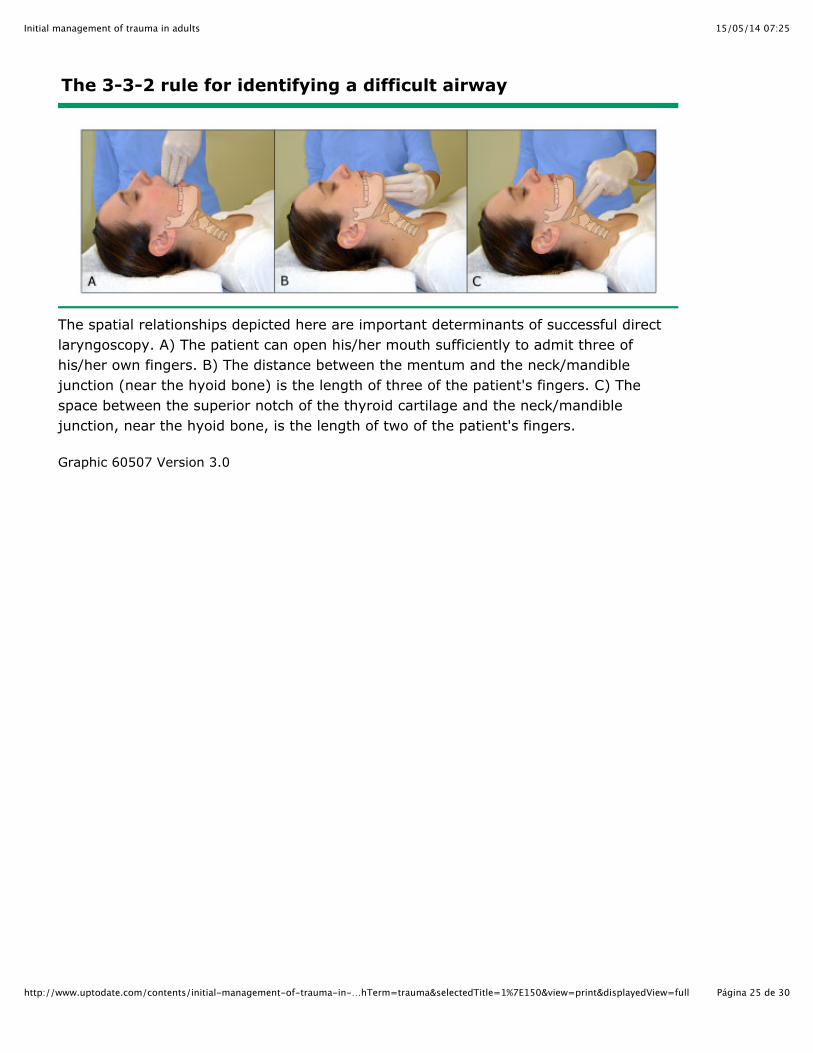
E: EVALUATE 3-3-2: This refers to the intraoral, mandibular, and hyoid-to-thyroid notch distances (picture 1). Thecervical collar must be opened to make these assessments. The distances referred to can be narrowed by fracture,hematoma, or other anatomic distortions (eg, soft tissue swelling).
15/05/14 07:25Initial management of trauma in adults
Página 5 de 30http://www.uptodate.com/contents/initial-management-of-trauma-in-…chTerm=trauma&selectedTitle=1%7E150&view=print&displayedView=full
Difficult airway devices — Devices for difficult airway management are discussed separately. (See "Devices fordifficult emergency airway management in adults".)
A number of airway tools and rescue airways can be helpful when managing a trauma patient. Devices that should beavailable at the bedside include:
Direct laryngoscopy relies on direct visualization of the glottis, which is often difficult in the severely injured patientwhose airway may be obstructed and whose neck cannot be manipulated. In contrast, video laryngoscopes provide anexcellent view of the glottis with minimal movement of the cervical spine and appear to be well suited for airwaymanagement in the trauma patient [38-40]. Larger studies in trauma populations are needed to confirm these initialimpressions.
The endotracheal tube introducer (or gum elastic bougie) is another invaluable tool for airway management in the traumapatient, particularly when the glottic view is limited. Its use is discussed separately. (See "Devices for difficult emergencyairway management in adults", section on 'Endotracheal tube introducers (gum elastic bougie)'.)
Intubation — Tracheal intubation of the injured patient is often complicated by the need to maintain cervicalimmobilization, the presence of obstructions such as blood, vomitus, and debris, and possibly by direct trauma to theairway [41]. Nevertheless, many trauma patients require intubation for immediate airway protection or because of theprojected disease course. Intubation improves oxygenation, thereby helping to meet increased physiologic demands,and allows for testing and procedures to be performed more easily and with less patient discomfort. (See "The decisionto intubate".)
Ideally, airway managers should have a predetermined back-up plan with all necessary tools at the bedside, includingrescue airways and a cricothyrotomy kit, before proceeding with intubation. In crash scenarios, this may not be possible.
M: MALLAMPATI: A standard calculation of the Mallampati score cannot be performed in many trauma patients;injured patients requiring emergent intubation often cannot open their mouths spontaneously (figure 1).Nevertheless, an effort should be made to determine how much of the retropharynx can be seen and whetherinjuries of the oropharynx or pooled blood, vomitus, or secretions are present.
●
O: OBSTRUCTION/OBESITY: Either factor can interfere with visualization and management of the traumatizedairway. Any number of injuries can obstruct the airway including internal or external hematomas or soft tissueedema from smoke inhalation. Obesity complicates performance of cricothyrotomy.
●
N: NECK MOBILITY: In-line stabilization is necessary in most trauma patients. Once the cervical collar is removedby a second skilled provider, that provider should stabilize the spine while orotracheal intubation is performed. It isimportant to note that the risk of neurologic injury from hypoxemia is much greater than the risk of spinal injury dueto neck extension during intubation. Judicious relaxation of immobilization may be necessary in some cases [37].
●
Suction (ie, multiple pumps and tips) may be needed.●Bag-valve mask attached to high flow oxygen●Oral and nasal airways●Rescue airways (eg, Combitube™, Laryngeal mask airway)●Endotracheal tube introducer (ie, gum elastic bougie)●Video laryngoscope if available●Cricothyrotomy kit●Endotracheal tubes in a range of sizes●Laryngoscopes●Preferred adjunct intubating devices (eg, lightwand)●
15/05/14 07:25Initial management of trauma in adults
Página 6 de 30http://www.uptodate.com/contents/initial-management-of-trauma-in-…chTerm=trauma&selectedTitle=1%7E150&view=print&displayedView=full
The performance of rapid sequence intubation and direct laryngoscopy are discussed separately. (See "Rapid sequenceintubation in adults" and "Direct laryngoscopy and tracheal intubation in adults".)
Cricothyrotomy — Clinicians who manage trauma must be prepared to perform a cricothyrotomy when orotrachealintubation cannot be accomplished. The performance of cricothyrotomy and the approach to the failed airway arediscussed separately. (See "The failed airway in adults" and "Emergent surgical cricothyrotomy (cricothyroidotomy)".)
In trauma patients with a potentially difficult airway, a double set-up, in which simultaneous preparation is made toperform orotracheal intubation and cricothyrotomy, may be the best approach. This enables the clinician to transitionimmediately to a cricothyrotomy if attempts at oral intubation are unsuccessful.
Trauma patients may have sustained injuries to the neck that make cricothyrotomy difficult to perform and therefore it isimportant to optimize any attempt at orotracheal intubation.
Cervical spine immobilization — Assume that an injury to the cervical spine has occurred in all blunt trauma patientsuntil proven otherwise. Conversely, patients with isolated penetrating trauma, no secondary blunt injury, and an intactneurologic examination typically do not have an unstable spinal column injury [37]. Spinal immobilization may be harmfulto these patients in some circumstances and is unnecessary when managing their airway [42]. (See "Evaluation andacute management of cervical spinal column injuries in adults".)
The anterior portion of the cervical collar should be temporarily removed and manual in-line stabilization maintained forall patients with blunt traumatic injuries receiving airway interventions, including bag-mask ventilation [43,44].Preintubation airway interventions are associated with as much spinal column subluxation as intubation [43,44].
Tracheal intubation should not be attempted with the anterior portion of the cervical collar in place. Intubationsperformed with the complete cervical collar in place are associated with greater spinal subluxation than those performedwith the anterior portion removed and manual in-line stabilization maintained [45].
The safety of manual in-line stabilization for patients with blunt traumatic injuries needing intubation is well established.Few case reports describe spinal injury during intubation, and in all cases, the spine was not manually stabilized [46-48].
Breathing and ventilation — Once airway patency is ensured, assess the adequacy of oxygenation and ventilation[23]. Chest trauma accounts for 20 to 25 percent of trauma-related deaths, in large part due to its harmful effects onoxygenation and ventilation [22]. The management of blunt chest trauma is discussed separately. (See "Initial evaluationand management of blunt thoracic trauma in adults".)
Inspect the chest wall looking for signs of injury, including asymmetric or paradoxical movement (eg, flail chest),auscultate breath sounds at the apices and axillae, and palpate for crepitus and deformity. In unstable patients, obtain aportable chest x-ray. Tension pneumothorax, massive hemothorax, and cardiac tamponade are immediate threats to lifethat should be identified at this stage of the primary survey.
Presumptively treat patients exhibiting signs of tension pneumothorax, including hypotension, dyspnea, and ipsilateraldecreased breath sounds, with needle decompression before obtaining imaging. Delays to obtain a portable chest x-raycan cause significant morbidity. Needle decompression is performed with a large bore (14 gauge or larger)angiocatheter, either in the second intercostal space in the midclavicular line or in the fifth intercostal space in themidaxillary line.
Of note, a standard 14 gauge angiocatheter cannot penetrate the chest wall and reach the pleural space in 10 to 33percent of trauma patients [49]. A 10 gauge, 7.5 cm (3 inch) armored angiocatheter is able to penetrate the pleuralspace in most instances. Needle decompression is followed immediately by tube thoracostomy. (See "Initial evaluationand management of blunt thoracic trauma in adults", section on 'Initial management' and "Placement and managementof thoracostomy tubes".)
15/05/14 07:25Initial management of trauma in adults
Página 7 de 30http://www.uptodate.com/contents/initial-management-of-trauma-in-…chTerm=trauma&selectedTitle=1%7E150&view=print&displayedView=full
Tube thoracostomy in an unstable trauma patient is placed in anticipation of both hemothorax and pneumothorax using achest tube of at least 32 French in diameter. A generous skin incision should be made in the fifth intercostal space in themidaxillary line allowing for placement of the tube in the inferior portion of the interspace and digital guidance towardsthe posterior-apical portion of the hemithorax.
Circulation — Once the airway and breathing are stabilized, perform an initial evaluation of the patient's circulatorystatus by palpating central pulses. If a carotid or femoral pulse is verified and no obvious exsanguinating external injuryis noted, circulation may momentarily be assumed to be intact; completion of the primary survey should not be delayedby the determination of an exact blood pressure.
While circulation is assessed, two large-bore (16 gauge or larger) intravenous (IV) catheters are placed, most often inthe antecubital fossa of each arm, and blood is drawn for testing, particularly for blood typing and crossmatch.Intraosseous cannulation or central venous catheter placement (ideally under ultrasound guidance) can be performed ifthere is difficulty establishing peripheral IV access. (See "Intraosseous infusion".)
Life-threatening hemorrhage must be controlled. A combination of manual pressure, proximal compression with either atourniquet or a manual blood pressure cuff, and elevation is typically sufficient to control external arterial hemorrhage.When these are unsuccessful, hemostatic agents may be needed, if available. Venous bleeding is controlled with directpressure. (See "Initial evaluation and management of shock in adult trauma", section on 'Hemostatic agents'.)
Emergency thoracotomy may be needed for trauma patients without central pulses. The procedure is most effective forvictims of stab wounds to the chest who have pulses or other witnessed signs of life (eg, voluntary movement) initially. Itis rarely beneficial in patients with blunt trauma or when performed in facilities without ready access to appropriatesurgical care. Emergency thoracotomy is discussed separately. (See "Initial evaluation and management of bluntthoracic trauma in adults", section on 'Emergent thoracotomy'.)
Most trauma patients with hypotension or signs of shock (eg, pale, cool, moist skin) are bleeding, and patients withsevere hemorrhage have significantly higher mortality [50]. Initial fluid resuscitation for these patients often consists of abolus of intravenous crystalloid (eg, 20 mL/kg isotonic saline). Patients with obvious severe or ongoing blood loss shouldbe transfused immediately with type O blood; women of childbearing age are transfused with O negative blood. Mildlyunstable patients may be treated with isotonic crystalloid in lieu of blood, although unnecessary infusion of crystalloidshould be avoided [51]. Fluid resuscitation, including the appropriate use of delayed fluid resuscitation and transfusion ofthe trauma patient in shock are discussed separately. (See "Initial evaluation and management of shock in adulttrauma".)
Patients with persistent hemodynamic instability despite an initial fluid bolus generally require blood transfusion anddefinitive control of the bleeding source. Significant hemorrhage occurs in any of five sites: external, intrathoracic,intraperitoneal, retroperitoneal, and pelvic or long bone fractures. Patients requiring transfusion may benefit fromtreatment with tranexamic acid if it is given within three hours of injury. (See "Initial evaluation and management of shockin adult trauma", section on 'Antifibrinolytic agents'.)
It is important to obtain manual blood pressure measurements in trauma patients with systolic blood pressures below 90mmHg, as automated blood pressure cuffs often overestimate values significantly in these patients [52]. Furthermore,data suggest that the traditional threshold of a systolic blood pressure below 90 mmHg used to define shock isinaccurate [53-56]. A significant proportion of trauma patients with hemorrhagic shock have a higher blood pressure andusing a cut-off of 110 mmHg is likely to be more appropriate, especially in the elderly. (See "Geriatric trauma: Initialevaluation and management".)
Nonhemorrhagic causes of shock include tension pneumothorax and cardiac tamponade. These injuries are bestdetected by physical examination or ultrasound assessment (ie, FAST). (See 'Ultrasound (FAST exam)' below and"Emergency ultrasound in adults with abdominal and thoracic trauma".)
15/05/14 07:25Initial management of trauma in adults
Página 8 de 30http://www.uptodate.com/contents/initial-management-of-trauma-in-…chTerm=trauma&selectedTitle=1%7E150&view=print&displayedView=full
Disability and neurologic evaluation — Once problems related to the airway, breathing, and circulation areaddressed, perform a focused neurologic examination. This should include a description of the patient's level ofconsciousness using the Glasgow Coma Scale (GCS) score, and assessments of pupillary size and reactivity, grossmotor function, and sensation (table 2). Also note any lateralizing signs and the level of sensation if a spinal cord injury ispresent. Acute neurologic injury, including imaging recommendations and medical and surgical management, isdiscussed in detail separately. (See "Management of acute severe traumatic brain injury" and "Acute traumatic spinalcord injury".)
The GCS score is widely used and can be employed by clinicians to follow the patient's neurologic status. Unfortunately,a number of studies suggest that the initial GCS score is not predictive of outcome in patients with severe brain injury,and both intubation and sedatives interfere with its application [57-59].
Maintain spinal precautions for all patients with the potential for spinal cord injury. The presence of a motor deficit or aspinal cord sensory level indicates the need for imaging of the brain and spinal cord.
Exposure and environmental control — Be certain that the trauma patient is completely undressed and that his or herentire body is examined for signs of injury during the primary survey. Missed injuries pose a grave threat [60]. Regionsoften neglected include the scalp, axillary folds, perineum, and in obese patients, abdominal folds. Penetrating woundsmay be present anywhere. While maintaining cervical spine precautions, examine the patient's back; do not neglectexamination of the gluteal fold and posterior scalp.
Hypothermia should be prevented if possible and treated immediately once identified. Hypothermia contributes to bothcoagulopathy [61] and the development of multiple organ dysfunction syndrome [62]. During winter months andwhenever a hypothermic trauma patient is being treated, the resuscitation room should be heated; the United StatesMilitary Joint Theater Trauma System Clinical Practice Guideline on hypothermia prevention recommends emergencydepartment (ED) and operating room (OR) temperatures of at least 29.4°C (85°F) during the treatment of these patients[63]. Make liberal use of warm blankets and active external warming devices. Warm IV fluids and blood. Treatments forhypothermia are discussed separately. (See "Accidental hypothermia in adults".)
Diagnostic studies
Portable x-rays — Plain radiographs play an important role in the primary evaluation of the unstable trauma patient.Screening x-rays should be obtained, either in the emergency department (ED) or the operating room (OR), even inhemodynamically compromised patients who are sent directly to the OR during or after their primary survey. Promptimaging of the lateral cervical spine, chest, and pelvis can detect life threatening injuries that might otherwise be missed.However, the sensitivity of the lateral cervical spine radiograph is only 70 to 80 percent [64-66], and some sacral andiliac fractures can be missed on plain pelvic radiographs.
Patients found to be hemodynamically unstable during the primary survey should be aggressively resuscitated; thedecision of whether to take an unstable patients directly to the OR or to the OR after emergent CT imaging dependsupon their response to resuscitation, probable injuries, and the proximity of the computed tomography (CT) scanner tothe resuscitation bay.
Clinical decision rules (eg, NEXUS) can be used to determine the need for cervical spine imaging in hemodynamicallystable trauma patients. Assessment of the spinal column injuries in trauma, including the selection of imaging studies, isdiscussed separately. (See "Evaluation and acute management of cervical spinal column injuries in adults".)
Plain radiography of the chest and pelvis is often obtained for trauma patients not thought to require CT imaging. Thedecision to obtain these images should be made based upon the injury mechanism and clinical findings. The evaluationof patients with penetrating trauma often includes images of the region of penetration; even in stable patients, theseradiographs can detect retained foreign bodies or fragments. On the other hand, patients with blunt trauma should
15/05/14 07:25Initial management of trauma in adults
Página 9 de 30http://www.uptodate.com/contents/initial-management-of-trauma-in-…chTerm=trauma&selectedTitle=1%7E150&view=print&displayedView=full
undergo imaging with plain radiographs only if clinical findings suggest the presence of injury [67,68]. Plain radiographscan be omitted altogether if there is no clinical suspicion of injury and the studies are unlikely to alter emergentmanagement. (See "Pelvic trauma: Initial evaluation and management", section on 'Plain radiograph'.)
A plain radiograph of the chest should be obtained in patients with penetrating injuries of the chest, back, or abdomenregardless of the need for CT. Plain films may reveal subdiaphragmatic free air, a foreign body, or a pneumothorax orhemothorax.
If the clinician determines that CT imaging is needed based upon the mechanism or clinical suspicion, there is no role foreither a plain radiograph of the chest or pelvis in hemodynamically stable patients with blunt trauma [67,69-71].
Ultrasound (FAST exam) — Focused Abdominal Sonography for Trauma (FAST) is an essential part of the primarycirculation survey for unstable patients, in whom it often determines management [72-76]. FAST is used primarily todetect pericardial and intraperitoneal blood, and it is more accurate than any physical examination finding for detectingintra-abdominal injury. In hemodynamically stable patients, FAST can be delayed until the secondary survey and isideally performed by a second operator while the remainder of the secondary survey is completed. The performance ofthe FAST examination and evidence supporting its use are discussed separately. (See "Emergency ultrasound in adultswith abdominal and thoracic trauma".)
The accuracy and role of FAST may be more limited in patients with significant pelvic fractures because it is lesssensitive for detecting pelvic bleeding and cannot differentiate between blood and urine. The management of suchpatients is discussed separately. (See "Pelvic trauma: Initial evaluation and management", section on 'Initialmanagement'.)
The Extended FAST (E-FAST) includes examinations of the thoracic cavity looking for pneumothoraces. Preliminarystudies suggest the sensitivity of E-FAST is better than plain x-ray for this injury [77].
Diagnostic peritoneal tap or lavage — Diagnostic peritoneal tap or lavage has a role similar to FAST in theunstable patient in whom a source of bleeding has not been found [78]. It can be performed to detect intraperitonealblood when FAST is unavailable, to determine the type of intraperitoneal fluid when it is important to do so (eg, bloodversus urine in the setting of a pelvic fracture), or at physician discretion. (See "Initial evaluation and management ofblunt abdominal trauma in adults", section on 'Diagnostic peritoneal lavage'.)
Electrocardiogram — An electrocardiogram (ECG) should be obtained for all patients injured by mechanisms withthe potential for causing cardiac injury. Signs of blunt cardiac injury can include arrhythmias, significant conductiondelays, or ST segment changes. Findings consistent with pericardial tamponade include tachycardia, low voltage, andelectrical alternans. If ECG findings consistent with cardiac injury are present, formal echocardiography (in addition tothe FAST examination) should be performed. (See "Cardiac injury from blunt trauma" and "Cardiac tamponade".)
Laboratory tests — The practice of obtaining "screening" laboratory tests on trauma patients is neither useful norcost-effective [79,80]. Testing should be performed based upon clinical suspicion and should be limited to those teststhat may alter management. As examples, a pregnancy test (eg, urine hCG) should always be performed on women ofchild-bearing age, and a blood type and screen or crossmatch should be obtained for patients with significant traumawho may reasonably be expected to require transfusion.
Clinical circumstances determine the need for further testing. As examples, patients taking warfarin likely needcoagulation studies (eg, prothrombin time) and patients found on the ground for an undetermined time need studies (eg,creatine kinase) to determine if rhabdomyolysis is present. (See "Clinical features and diagnosis of heme pigment-induced acute kidney injury (acute renal failure)".)
Commonly obtained but rarely helpful tests include the metabolic panel (a fingerstick blood sugar will often sufficeprovided the patient is not exhibiting signs of electrolyte abnormality or acidosis), alcohol level in a patient who is clearly
15/05/14 07:25Initial management of trauma in adults
Página 10 de 30http://www.uptodate.com/contents/initial-management-of-trauma-in-…hTerm=trauma&selectedTitle=1%7E150&view=print&displayedView=full
intoxicated, toxicologic screen when it is not relevant to clinical care, and cardiac biomarkers, unless cardiac contusionor ischemia is suspected [81]. (See "Cardiac injury from blunt trauma", section on 'Diagnostic tests'.)
Elevation of both the serum lactate concentration and base deficit correlates with increased mortality in trauma patients[82-84]. However, the base deficit is essentially a surrogate for lactate and an elevated base deficit in the absence of anelevated lactate is not predictive of increased mortality [85]. Furthermore, while elevated levels should heightensuspicion for severe injury, a normal lactate and base deficit do not ensure the absence of significant injury, especially ingeriatric trauma patients [53]. In addition, laboratory values lag behind clinical improvement after aggressiveresuscitation. Thus, the patient may no longer be in shock despite an elevated lactate suggesting otherwise [86,87].
The white blood cell (WBC) count is nonspecific and of little value during the initial evaluation of the trauma patient [80].The positive and negative predictive value of, respectively, an elevated or normal WBC is poor. Epinephrine releasefrom trauma can cause demargination and may elevate the WBC to 12,000 to 20,000/mm with a moderate left shift.Solid or hollow viscus injury can cause comparable elevations [88].
PATIENT TRANSFER — Clinicians at smaller hospitals should consult the nearest trauma center as soon as it becomesapparent that a patient has sustained injuries beyond the management capacity of their hospital. Patients should bestabilized as well as possible without delaying transfer; delays are associated with increased mortality [89,90]. Criteriafor transfer are based upon the patient’s demographics, mechanism of injury, and clinical findings. It cannot beoveremphasized that a complete workup is not a requirement for transfer; postponing transfer to obtain laboratoryresults or imaging studies only delays definitive treatment. Often such studies must be repeated at the receiving facility.
Computed tomography (CT) imaging should only be obtained in patients who might otherwise be appropriately treated atthe initial facility. If a negative CT would allow the patient to be discharged, it should be performed, but if that patientrequires transfer regardless of the results then transfer should not be delayed. Likewise, procedures and otherinterventions should only be performed to treat emergent conditions or prevent possible patient deterioration duringtransport. Endotracheal intubation, tube thoracostomy, and pelvis fracture stabilization are common examples ofnecessary interventions; laceration repair, unless it is performed to prevent exsanguination, is not.
The decision of when to transfer an unstable patient should ideally be made by the transferring and receiving physiciansin collaboration. Clear communication is critical: the transmission of vital information allows receiving clinicians tomobilize needed resources while the inadvertent omission of such information can delay definitive care. Informationshould be conveyed in both verbal and written (via the patient record) form and should include the patient's identifyinginformation, relevant medical history, prehospital course, and ED evaluation and treatment (including proceduresperformed and imaging obtained) [23]. The use of a transfer checklist can help to ensure that important information is notomitted.
SECONDARY EVALUATION — Definitive management of a hemodynamically unstable trauma patient must not bedelayed to perform a more detailed secondary evaluation. Such patients are taken directly to the operating room (OR) orangiography suite, or transferred to a major trauma center.
A careful, head-to-toe secondary assessment (ie, secondary survey) is performed in all trauma patients determined to bestable upon completion of the primary survey. The secondary survey includes a detailed history, a thorough but efficientphysical examination, and targeted diagnostic studies, and plays a crucial role in avoiding missed injuries. Commonlymissed injuries include [91-93]:
15/05/14 07:25Initial management of trauma in adults
Página 11 de 30http://www.uptodate.com/contents/initial-management-of-trauma-in-…hTerm=trauma&selectedTitle=1%7E150&view=print&displayedView=full
Delayed reevaluation of the trauma patient (ie, tertiary survey) is also useful for preventing missed injuries and fordetecting injuries that present late [91]. It is most helpful if the patient is reevaluated when fully alert. Any member of thetrauma team with advanced assessment skills can perform the tertiary survey; however, it is best if the same clinicianperforms all serial examinations for a given patient in order to detect subtle changes.
History — The mechanism of injury can increase suspicion for certain injuries. Prehospital personnel often knowimportant information and should be queried regarding the mechanism and history of the injury. If this cannot be doneimmediately upon arrival because of the patient's status, ask the prehospital providers to remain in the emergencydepartment (ED) until this can be accomplished. Often the history is conveyed while medics and hospital clinicianstransfer the patient and important information may be forgotten or missed.
While listening to the history, keep in mind that the scenes of accidents can be chaotic and not all information will bereliable. As an example, a patient described as "found down" may have been assaulted or struck by a car.
Mechanism-related information to be obtained from prehospital personnel includes [94]:
Inquire also about the patient's medications, allergies, and medical and surgical history. If this information is unknown, itcan be helpful to assign someone the task of contacting family members to obtain it. The use of anticoagulant andantiplatelet medications is steadily rising and increases the risk of internal bleeding in trauma patients, and thereforethese agents should specifically be discussed [95-97].
As an example of the risks associated with anticoagulants, a retrospective study of 11,374 adult trauma patientsreported that the use of antiplatelet drugs was associated with an increased risk of death (propensity adjusted outcome9.4 versus 8 percent mortality) and major morbidity among the 1327 (11.7 percent) patients taking them at the time of
15/05/14 07:25Initial management of trauma in adults
Página 12 de 30http://www.uptodate.com/contents/initial-management-of-trauma-in-…hTerm=trauma&selectedTitle=1%7E150&view=print&displayedView=full
their injury [95]. Patients taking multiple antiplatelet medications were at greater risk than those taking a single drug.
Physical examination — The goal of the secondary survey is to identify injuries. This includes the performance of athorough but efficient physical examination. Use standard precautions against blood or fluid-borne infection.
Head and face — Inspect and palpate the entire bony structure of the head and face for tenderness, deformity (eg,step off), and bleeding. Scalp lacerations are easily missed visually but often found by palpation.
Note any signs suggesting basilar skull fracture (eg, hemotympanum). Retroauricular (Battle's sign) and periorbitalecchymosis (raccoon's eyes) are also indicative of basilar skull fracture but generally do not appear until at least 24hours after an injury. Look for nasal septal hematomas. (See "Skull fractures in adults" and "Facial trauma in adults".)
Perform an ocular examination including an evaluation of pupillary size, shape, reactivity, and extraocular movement.Look for signs of globe rupture and intraocular hemorrhage. (See "Open globe injuries: Emergent evaluation and initialmanagement" and "Orbital fractures" and "Retinal detachment" and "Traumatic hyphema: Clinical features andmanagement".)
Patients with mild traumatic brain injury may not have external signs of trauma. However, a mechanism consistent withbrain injury may warrant imaging with computed tomography (CT). (See "Concussion and mild traumatic brain injury".)
Neck — Assume that all patients with blunt trauma have sustained an injury to the cervical spine. This assumptioncan be disproved by appropriate application of clinical decision rules, such as NEXUS or the Canadian C-Spine Rule, orby radiologic evaluation using plain radiographs or CT. Assessment of the cervical spine following trauma is discussedseparately. (See "Evaluation and acute management of cervical spinal column injuries in adults".)
Inspect and palpate the entire neck for signs of injury. The management of penetrating neck trauma is discussedseparately. (See "Initial evaluation and management of penetrating neck injuries".)
Chest — Inspect and palpate the entire chest wall. Pay particular attention to the sternum and clavicles. Injuries atthese sites are often missed, and fractures of these bones suggest the presence of further injury, including ofintrathoracic structures. Careful auscultation can detect a previously missed small hemothorax, pneumothorax, orpericardial effusion not yet causing tamponade. (See "Initial evaluation and management of blunt thoracic trauma inadults".)
Abdomen — Perform and document a careful abdominal examination. Inspect the abdomen and flanks forlacerations, contusions (eg, seat belt sign), and ecchymosis; palpate for tenderness and rigidity. The presence of a seatbelt sign, rebound tenderness, abdominal distension, or guarding all suggest intra-abdominal injury. Note that theabsence of abdominal tenderness does not rule out such injury.
Keep in mind that the abdominal examination is often unreliable, particularly in the elderly and patients with distractinginjuries or altered mental status, and can change dramatically over time. (See "Initial evaluation and management ofblunt abdominal trauma in adults".)
Rectum and genitourinary — Inspect the perineum of all patients for signs of injury. (See "Straddle injuries".)
Traditionally, the digital rectal examination (DRE) was considered an essential part of the physical examination for alltrauma patients. However, the sensitivity of the DRE for injuries of the spinal cord, pelvis, and bowel is poor, and falsepositive and negative results are common [94,98-100]. Thus, routine performance is unnecessary and generallyunhelpful. The examination is warranted in cases where urethral injury or penetrating rectal injury is suspected. If theexamination is performed, check for the presence of gross blood (sign of bowel injury), a high-riding prostate (sign ofurethral injury), abnormal sphincter tone (sign of spinal cord injury), and bone fragments (sign of pelvic fracture). (See"Blunt genitourinary trauma" and "Penetrating trauma of the upper and lower genitourinary tract" and "Evaluation and
15/05/14 07:25Initial management of trauma in adults
Página 13 de 30http://www.uptodate.com/contents/initial-management-of-trauma-in-…hTerm=trauma&selectedTitle=1%7E150&view=print&displayedView=full
acute management of cervical spinal column injuries in adults", section on 'Secondary survey' and "Pelvic trauma: Initialevaluation and management".)
Perform a vaginal examination on all patients at risk for vaginal injury (eg, those with lower abdominal pain, pelvicfracture, or perineal laceration) [23]. Take care to avoid injury from bone fragments if a pelvic fracture is known orsuspected.
Musculoskeletal — Inspect and palpate the entire length of all four extremities looking for areas of tenderness,deformity, or decreased range of motion. Also assess and document the neurovascular status of each extremity.Manipulate all joints thought to be uninjured both passively and actively to verify their integrity; immobilize and obtainradiographs of any area with a suspected fracture.
Note all penetrating wounds, especially those overlying suspected fractures, suggesting an open injury. The treatment ofopen fractures includes irrigation and debridement, application of a clean dressing, and prophylactic antibiotics.Preliminary wound irrigation can be performed in the trauma bay, but definitive irrigation and debridement is performedin the operating room (OR). (See "Treatment and prevention of osteomyelitis following trauma".)
Post traumatic compartment syndrome is an important source of patient morbidity. Increasing pain, tense compartments,and pain with passive stretching of the muscles contained within the compartment should prompt immediatemeasurement of intracompartmental pressures. (See "Acute compartment syndrome of the extremities".)
Inspect and palpate the pelvis. Ecchymosis over the pelvis or tenderness along the pelvic ring warrants diagnosticimaging. Examination findings (eg, instability) or imaging studies consistent with pelvic ring disruption indicate the needfor pelvic immobilization and emergent orthopedic evaluation. Repeat examinations to assess pelvic stability areunnecessary and likely to exacerbate bleeding. (See "Pelvic trauma: Initial evaluation and management".)
Neurologic — The trauma patient's neurologic status can change dramatically over time (eg, from the effects of anexpanding subdural hematoma). Serial examinations should be performed and carefully documented. During thesecondary survey, perform a detailed assessment of the sensorimotor function of the extremities and repeat anassessment of the patient's GCS score.
Skin — Examination of the skin may reveal lacerations, abrasions, ecchymosis, hematoma, or seroma formation.Look closely at areas where lesions may be missed, such as the scalp, axillary folds, perineum, and, particularly inobese patients, abdominal folds. Do not neglect examination of the back, gluteal fold, and posterior scalp. Penetratingwounds may be present anywhere. The management of skin wounds is discussed separately. (See "Clinical assessmentof wounds" and "Basic principles of wound management".)
Additional imaging
Plain radiographs — Plain x-rays are used during the secondary survey primarily to evaluate the spine, pelvis, andextremities for fractures, dislocations, and foreign bodies.
Computed tomography — Multidetector computed tomography (MDCT) has become the modality of choice forimaging trauma patients because of its speed and accuracy. However, studies of comprehensive whole body CTscanning ("pan scan") for all patients with significant trauma are methodologically limited and have reached contradictoryconclusions [101-108]. Pending further research, we do not advocate comprehensive CT scanning in patients withoutsignificant alterations in mental status and believe imaging studies should be performed selectively based upon clinicalassessment and the mechanism of injury. While whole body CT scanning may improve outcomes following certain high-risk trauma, such as explosions, high speed motor vehicle collisions, and falls from great heights [76,109], we believe itshould not be used indiscriminately given the short-term risk of contrast-related renal injury and the long-term risk ofradiation-induced cancer, as well as the substantial costs [110]. (See "Pathogenesis, clinical features, and diagnosis ofcontrast-induced nephropathy" and "Radiation-related risks of imaging studies".)
15/05/14 07:25Initial management of trauma in adults
Página 14 de 30http://www.uptodate.com/contents/initial-management-of-trauma-in-…hTerm=trauma&selectedTitle=1%7E150&view=print&displayedView=full
Some authors advocate whole body CT for severely injured patients with alterations in mental status. In a retrospectivedatabase analysis of 5,208 patients in Japan with Glasgow Coma Score ranging from 3 to 12, decreased mortality wasnoted in patients who received whole body CT scans [111]. Although further study of the outcomes and costeffectiveness of whole body CT is needed, the approach may be beneficial in such patients, in whom examinationfindings are often limited or unclear.
It should be noted that CT has limited utility for evaluating the trajectory and effects of low velocity penetrating injury (eg,stab wounds) because of the lack of tissue disruption and gas dispersion (seen with high velocity injuries) [112], andbecause injuries to luminal structures are often difficult to detect [113]. Diagnostic laparoscopy may be useful in patientswith penetrating injury and signs of peritoneal penetration despite negative CT imaging. The use of CT for specificinjuries is discussed separately. (See "Management of acute severe traumatic brain injury" and "Acute traumatic spinalcord injury" and "Initial evaluation and management of blunt thoracic trauma in adults" and "Initial evaluation andmanagement of blunt abdominal trauma in adults" and "Initial evaluation and management of abdominal stab wounds inadults" and "Initial evaluation and management of abdominal gunshot wounds in adults" and "Pelvic trauma: Initialevaluation and management".)
Most patients should be hemodynamically stable before CT imaging is performed, and resuscitation should be sufficientto minimize the risk of decompensation while the patient is in the CT scanner. If the patient is unstable, CT imagingshould be deferred.
PITFALLS AND PEARLS — The systematic evaluation of the trauma patient outlined above is designed to helpclinicians focus on life-threatening problems and minimize the risk of missed injuries. Nevertheless, one systematicreview noted that up to 39 percent of trauma patients have injuries that are initially missed and up to 22 percent of thesemissed injuries are clinically significant (defined as injuries associated with increased mortality, requiring additionalprocedures or alterations in treatment, or resulting in significant pain, complications, or residual disability) [60].
Potential pitfalls in trauma management and ways to avoid them are discussed below:
Esophageal intubations — Between 0.5 and 6 percent of prehospital intubations are esophageal due to airwaydifficulty or displacement during transport. The position of all endotracheal tubes must be verified either by directvisualization or use of an end tidal CO2 detector. (See "Prehospital care of the adult trauma patient", section on 'Airwaysupport'.)
Hemorrhagic shock — Approximately 30 percent of the circulating blood volume may be lost before the onset ofhypotension [23]. A transient response to one or more fluid boluses means the patient likely has ongoing hemorrhageand is in a persistent state of shock. (See "Initial evaluation and management of shock in adult trauma".)
Cardiac tamponade — Assume that elevated jugular venous pressure (JVP) in a trauma patient is caused bypericardial tamponade. However, hypovolemic patients with tamponade may not have elevated JVP. Perform the FASTexam early in the circulation evaluation of the unstable patient and begin by looking at the heart. (See "Cardiactamponade".)
Thoracoabdominal injury — Assume that any penetrating wound of the thorax or abdomen involves bothcompartments until proven otherwise.
Penetrating bowel injury — During the initial resuscitation, injuries caused by low velocity penetrating wounds areeasily missed by both ultrasound, because there is too little intraperitoneal blood to be detected, and CT, because thereis inadequate tissue destruction. High clinical suspicion warrants further evaluation by DPL or laparotomy, despiteinitially negative imaging studies. Alternatively, a trauma surgeon may opt to perform serial observations of patients withabdominal stab or gunshot wounds over a 12 to 24 hour period. (See "Initial evaluation and management of abdominalgunshot wounds in adults" and "Initial evaluation and management of abdominal stab wounds in adults".)
15/05/14 07:25Initial management of trauma in adults
Página 15 de 30http://www.uptodate.com/contents/initial-management-of-trauma-in-…hTerm=trauma&selectedTitle=1%7E150&view=print&displayedView=full
Open book pelvic fractures — The unstable pelvis should not be manipulated multiple times; additional manipulationexacerbates hemorrhage. Once suspected, open or unstable pelvic fractures should be stabilized using a pelvic binder,or a sheet if no binder is available. If the patient is hemodynamically stable, computed tomography (CT) imaging isobtained. The unstable patient requires either surgery or angiography. (See "Pelvic trauma: Initial evaluation andmanagement".)
Ocular injuries — Periorbital swelling and ecchymosis does not preclude an ocular examination. Patients with suchfindings are at higher risk of ocular injury. (See "Open globe injuries: Emergent evaluation and initial management" and"Orbital fractures" and "Retinal detachment" and "Traumatic hyphema: Clinical features and management".)
Elder patients — Assume that older patients involved in trauma have sustained a significant injury, even if they appearwell. The paradox of elder trauma patients is that their physiology and medical interventions can both mask andexacerbate the severity of injuries. Medications are but one example: beta blockers may mask the effects of shock bysuppressing tachycardia, while warfarin increases the risk of severe hemorrhage. A table summarizing importantconsiderations in the elder trauma patient is attached (table 3). (See "Geriatric trauma: Initial evaluation andmanagement".)
Common cognitive errors — Several cognitive errors appear to be relatively common during the initial management ofinjured patients, particularly those who do not look sick initially. Among these are [30]:
Analgesia and sedation — Injured patients are in pain. Do not neglect to provide them with appropriate analgesia andsedation. Short-acting agents, such as fentanyl and midazolam, are generally preferred to avoid adverse hemodynamiceffects.
SUMMARY AND RECOMMENDATIONS
Premature diagnosis – The hemodynamic status of trauma patients is often dynamic and the results of their initialdiagnostic studies preliminary. Avoid making premature assumptions about patients' injuries and stability.
●
Overreliance upon early negative results – No study is perfect and initial studies may not reveal the full extent ofa patient's injuries or indeed any injury. Reassess the patient.
●
Attributing abnormal findings to benign causes – Trauma patients, particularly young healthy adults, may notimmediately manifest signs of severe injury. When abnormal findings arise, assume they reflect injury.
●
Trauma is a leading cause of mortality globally. All trauma patients require a systematic approach to managementin order to maximize outcomes and reduce the risk of undiscovered injuries. Optimal care requires effective andefficient communication and teamwork among clinicians. Common breakdowns in team management aredescribed in the text. (See 'Epidemiology' above and 'Trauma team' above.)
●
Particular mechanisms predispose patients to specific injuries. Common blunt trauma mechanisms and their mostfrequently associated injuries are described in the accompanying table (table 1).
●
The primary survey used in Advanced Trauma Life Support™ is organized according to the injuries that pose themost immediate threats to life. Problems are managed immediately in the order they are detected. The individualsteps (including assessments of the airway, breathing, circulation, and neurologic injury) and important principles ofthe primary survey are described in the text. (See 'Primary evaluation and management' above.)
●
Observational studies suggest that airway obstruction is a major cause of preventable death among traumapatients. Therefore, airway evaluation and management remain the critical first steps in the treatment of anyseverely injured patient. (See 'Airway' above and 'Breathing and ventilation' above.)
●
Hemorrhage is the most common preventable cause of mortality in trauma. Most trauma patients with signs of●
15/05/14 07:25Initial management of trauma in adults
Página 16 de 30http://www.uptodate.com/contents/initial-management-of-trauma-in-…hTerm=trauma&selectedTitle=1%7E150&view=print&displayedView=full
Use of UpToDate is subject to the Subscription and License Agreement.
REFERENCES
1. World Health Organization. Global burden of disease. www.who.int/healthinfo/global_burden_disease/en/(Accessed on May 01, 2010).
2. Feliciano, DV, Mattox, K, Moore, EE. Trauma, 6th, McGraw-Hill, New York 2008.3. CDC. National estimates of the ten leading causes of nonfatal injuries, Centers for Disease Control and Prevention
2004. www.cdc.gov/injury/wisqars.html (Accessed on May 24, 2010).4. Mackenzie EJ, Rivara FP, Jurkovich GJ, et al. The National Study on Costs and Outcomes of Trauma. J Trauma
2007; 63:S54.5. Søreide K. Epidemiology of major trauma. Br J Surg 2009; 96:697.6. Demetriades D, Murray J, Sinz B, et al. Epidemiology of major trauma and trauma deaths in Los Angeles County.
J Am Coll Surg 1998; 187:373.7. Evans JA, van Wessem KJ, McDougall D, et al. Epidemiology of traumatic deaths: comprehensive population-
based assessment. World J Surg 2010; 34:158.8. MacKenzie EJ, Rivara FP, Jurkovich GJ, et al. A national evaluation of the effect of trauma-center care on
mortality. N Engl J Med 2006; 354:366.9. Christmas AB, Reynolds J, Wilson AK, et al. Morbid obesity impacts mortality in blunt trauma. Am Surg 2007;
73:1122.10. Clement ND, Tennant C, Muwanga C. Polytrauma in the elderly: predictors of the cause and time of death. Scand
J Trauma Resusc Emerg Med 2010; 18:26.11. Perdue PW, Watts DD, Kaufmann CR, Trask AL. Differences in mortality between elderly and younger adult
trauma patients: geriatric status increases risk of delayed death. J Trauma 1998; 45:805.12. Bamvita JM, Bergeron E, Lavoie A, et al. The impact of premorbid conditions on temporal pattern and location of
adult blunt trauma hospital deaths. J Trauma 2007; 63:135.13. Shoko T, Shiraishi A, Kaji M, Otomo Y. Effect of pre-existing medical conditions on in-hospital mortality: analysis of
20,257 trauma patients in Japan. J Am Coll Surg 2010; 211:338.
shock (eg, pale, cool, moist skin) are bleeding. Be alert for subtle signs of hemorrhagic shock, particularly in theelderly and young, healthy adults who may not present with obvious manifestations. Hypotension generally doesnot manifest until at least 30 percent of the patient's blood volume has been lost. (See 'Circulation' above.)
Diagnostic testing plays an important role in trauma management. The appropriate use of studies is described inthe text. (See 'Diagnostic studies' above.)
●
Clinicians at smaller hospitals should consult the nearest trauma center as soon as it becomes apparent that apatient has sustained injuries beyond the management capacity of their hospital. It cannot be overemphasized thata complete workup is not a requirement for transfer. (See 'Patient transfer' above.)
●
A secondary survey is performed in all trauma patients determined to be stable upon completion of the primarysurvey. The secondary survey includes a detailed history, a thorough but efficient physical examination, andtargeted diagnostic studies, and plays a crucial role in avoiding missed injuries. The secondary survey is describedin detail above. (See 'Secondary evaluation' above.)
●
Up to 39 percent of trauma patients have injuries that are initially missed, and up to 22 percent of these areclinically significant. Common pitfalls and guidance for avoiding missed injuries are provided in the text. (See'Pitfalls and pearls' above.)
15/05/14 07:25Initial management of trauma in adults
Página 17 de 30http://www.uptodate.com/contents/initial-management-of-trauma-in-…hTerm=trauma&selectedTitle=1%7E150&view=print&displayedView=full
14. Ditillo M, Pandit V, Rhee P, et al. Morbid obesity predisposes trauma patients to worse outcomes: a NationalTrauma Data Bank analysis. J Trauma Acute Care Surg 2014; 76:176.
15. Donnelly JP, Griffin RL, Sathiakumar N, McGwin G Jr. Obesity and vehicle type as risk factors for injury caused bymotor vehicle collision. J Trauma Acute Care Surg 2014; 76:1116.
16. Perel P, Prieto-Merino D, Shakur H, et al. Predicting early death in patients with traumatic bleeding: developmentand validation of prognostic model. BMJ 2012; 345:e5166.
17. Dossett LA, Riesel JN, Griffin MR, Cotton BA. Prevalence and implications of preinjury warfarin use: an analysis ofthe National Trauma Databank. Arch Surg 2011; 146:565.
18. Teixeira PG, Inaba K, Hadjizacharia P, et al. Preventable or potentially preventable mortality at a mature traumacenter. J Trauma 2007; 63:1338.
19. Teixeira PG, Inaba K, Salim A, et al. Preventable morbidity at a mature trauma center. Arch Surg 2009; 144:536.20. Baker CC, Oppenheimer L, Stephens B, et al. Epidemiology of trauma deaths. Am J Surg 1980; 140:144.21. Demetriades D, Kimbrell B, Salim A, et al. Trauma deaths in a mature urban trauma system: is "trimodal"
distribution a valid concept? J Am Coll Surg 2005; 201:343.22. Demetriades D, Murray J, Charalambides K, et al. Trauma fatalities: time and location of hospital deaths. J Am
Coll Surg 2004; 198:20.23. American College of Surgeons Committee on Trauma. Advanced Trauma Life Support for Doctors, Student
Course Manual, 8th ed, American College of Surgeons, Chicago 2008.24. Newgard CD, Schmicker RH, Hedges JR, et al. Emergency medical services intervals and survival in trauma:
assessment of the "golden hour" in a North American prospective cohort. Ann Emerg Med 2010; 55:235.25. Palanca S, Taylor DM, Bailey M, Cameron PA. Mechanisms of motor vehicle accidents that predict major injury.
Emerg Med (Fremantle) 2003; 15:423.26. Lerner EB, Shah MN, Cushman JT, et al. Does mechanism of injury predict trauma center need? Prehosp Emerg
Care 2011; 15:518.27. Haider AH, Chang DC, Haut ER, et al. Mechanism of injury predicts patient mortality and impairment after blunt
trauma. J Surg Res 2009; 153:138.28. Conroy C, Tominaga GT, Erwin S, et al. The influence of vehicle damage on injury severity of drivers in head-on
motor vehicle crashes. Accid Anal Prev 2008; 40:1589.29. Helmreich, R, Musson, D, Sexton, J. Human factors and safety in surgery. In: Surgical patient safety: essential
information for surgeons in today's environment,1st ed, Manuel B, Nora P (Eds), American College of Surgeons,Chicago 2004.
30. Mackersie RC. Pitfalls in the evaluation and resuscitation of the trauma patient. Emerg Med Clin North Am 2010;28:1.
31. Kirkpatrick AW, Ball CG, D'Amours SK, Zygun D. Acute resuscitation of the unstable adult trauma patient: bedsidediagnosis and therapy. Can J Surg 2008; 51:57.
32. Seamon MJ, Feather C, Smith BP, et al. Just one drop: the significance of a single hypotensive blood pressurereading during trauma resuscitations. J Trauma 2010; 68:1289.
33. Lipsky AM, Gausche-Hill M, Henneman PL, et al. Prehospital hypotension is a predictor of the need for anemergent, therapeutic operation in trauma patients with normal systolic blood pressure in the emergencydepartment. J Trauma 2006; 61:1228.
34. Chesnut RM, Marshall LF, Klauber MR, et al. The role of secondary brain injury in determining outcome fromsevere head injury. J Trauma 1993; 34:216.
35. Hussain LM, Redmond AD. Are pre-hospital deaths from accidental injury preventable? BMJ 1994; 308:1077.36. Esposito TJ, Sanddal ND, Hansen JD, Reynolds S. Analysis of preventable trauma deaths and inappropriate
trauma care in a rural state. J Trauma 1995; 39:955.37. Walls, RM, Murphy, MM. Manual of Emergency Airway Management, 3rd, Lippincott Williams & Wilkins,
15/05/14 07:25Initial management of trauma in adults
Página 18 de 30http://www.uptodate.com/contents/initial-management-of-trauma-in-…hTerm=trauma&selectedTitle=1%7E150&view=print&displayedView=full
38. Brown CA 3rd, Bair AE, Pallin DJ, et al. Improved glottic exposure with the Video Macintosh Laryngoscope in adultemergency department tracheal intubations. Ann Emerg Med 2010; 56:83.
39. Raja AS, Sullivan AF, Pallin DJ, et al. Adoption of video laryngoscopy in Massachusetts emergency departments.J Emerg Med 2012; 42:233.
40. Strube P, Jarvis J. Experience with a patient having multiple gunshot wounds in combat. AANA J 2008; 76:11.41. Thiboutot F, Nicole PC, Trépanier CA, et al. Effect of manual in-line stabilization of the cervical spine in adults on
the rate of difficult orotracheal intubation by direct laryngoscopy: a randomized controlled trial. Can J Anaesth2009; 56:412.
42. Vanderlan WB, Tew BE, McSwain NE Jr. Increased risk of death with cervical spine immobilisation in penetratingcervical trauma. Injury 2009; 40:880.
43. Brimacombe J, Keller C, Künzel KH, et al. Cervical spine motion during airway management: a cinefluoroscopicstudy of the posteriorly destabilized third cervical vertebrae in human cadavers. Anesth Analg 2000; 91:1274.
44. Donaldson WF 3rd, Heil BV, Donaldson VP, Silvaggio VJ. The effect of airway maneuvers on the unstable C1-C2segment. A cadaver study. Spine (Phila Pa 1976) 1997; 22:1215.
45. Gerling MC, Davis DP, Hamilton RS, et al. Effects of cervical spine immobilization technique and laryngoscopeblade selection on an unstable cervical spine in a cadaver model of intubation. Ann Emerg Med 2000; 36:293.
46. Hastings RH, Kelley SD. Neurologic deterioration associated with airway management in a cervical spine-injuredpatient. Anesthesiology 1993; 78:580.
47. Liang BA, Cheng MA, Tempelhoff R. Efforts at intubation: cervical injury in an emergency circumstance? J ClinAnesth 1999; 11:349.
48. Muckart DJ, Bhagwanjee S, van der Merwe R. Spinal cord injury as a result of endotracheal intubation in patientswith undiagnosed cervical spine fractures. Anesthesiology 1997; 87:418.
49. Zengerink I, Brink PR, Laupland KB, et al. Needle thoracostomy in the treatment of a tension pneumothorax intrauma patients: what size needle? J Trauma 2008; 64:111.
50. Boulanger L, Joshi AV, Tortella BJ, et al. Excess mortality, length of stay, and costs associated with serioushemorrhage among trauma patients: findings from the National Trauma Data Bank. Am Surg 2007; 73:1269.
51. Ley EJ, Clond MA, Srour MK, et al. Emergency department crystalloid resuscitation of 1.5 L or more is associatedwith increased mortality in elderly and nonelderly trauma patients. J Trauma 2011; 70:398.
52. Davis JW, Davis IC, Bennink LD, et al. Are automated blood pressure measurements accurate in trauma patients?J Trauma 2003; 55:860.
53. Callaway DW, Shapiro NI, Donnino MW, et al. Serum lactate and base deficit as predictors of mortality innormotensive elderly blunt trauma patients. J Trauma 2009; 66:1040.
54. Edelman DA, White MT, Tyburski JG, Wilson RF. Post-traumatic hypotension: should systolic blood pressure of90-109 mmHg be included? Shock 2007; 27:134.
55. Eastridge BJ, Salinas J, McManus JG, et al. Hypotension begins at 110 mm Hg: redefining "hypotension" withdata. J Trauma 2007; 63:291.
56. Oyetunji TA, Chang DC, Crompton JG, et al. Redefining hypotension in the elderly: normotension is notreassuring. Arch Surg 2011; 146:865.
57. Tasaki O, Shiozaki T, Hamasaki T, et al. Prognostic indicators and outcome prediction model for severe traumaticbrain injury. J Trauma 2009; 66:304.
58. Koskinen LOD, Olivecrona M, Rodling-Wahlström M, Naredi S. Initial GCS is an unreliable predictor of outcome inpatients with severe head injury treated (sTBI) by an ICP targeted therapy. A prospective study: P 070. Eur JAnaesthesiol 2008; 25:24.
59. Foreman BP, Caesar RR, Parks J, et al. Usefulness of the abbreviated injury score and the injury severity score incomparison to the Glasgow Coma Scale in predicting outcome after traumatic brain injury. J Trauma 2007; 62:946.
60. Pfeifer R, Pape HC. Missed injuries in trauma patients: A literature review. Patient Saf Surg 2008; 2:20.61. Hess JR, Brohi K, Dutton RP, et al. The coagulopathy of trauma: a review of mechanisms. J Trauma 2008; 65:748.
15/05/14 07:25Initial management of trauma in adults
Página 19 de 30http://www.uptodate.com/contents/initial-management-of-trauma-in-…hTerm=trauma&selectedTitle=1%7E150&view=print&displayedView=full
62. Beilman GJ, Blondet JJ, Nelson TR, et al. Early hypothermia in severely injured trauma patients is a significant riskfactor for multiple organ dysfunction syndrome but not mortality. Ann Surg 2009; 249:845.
63. Nesbitt M, Allen P, Beekley A, et al. Current practice of thermoregulation during the transport of combat wounded.J Trauma 2010; 69 Suppl 1:S162.
64. MacDonald RL, Schwartz ML, Mirich D, et al. Diagnosis of cervical spine injury in motor vehicle crash victims: howmany X-rays are enough? J Trauma 1990; 30:392.
65. Zabel DD, Tinkoff G, Wittenborn W, et al. Adequacy and efficacy of lateral cervical spine radiography in alert, high-risk blunt trauma patient. J Trauma 1997; 43:952.
66. Fisher A, Young WF. Is the lateral cervical spine x-ray obsolete during the initial evaluation of patients with acutetrauma? Surg Neurol 2008; 70:53.
67. Wisbach GG, Sise MJ, Sack DI, et al. What is the role of chest X-ray in the initial assessment of stable traumapatients? J Trauma 2007; 62:74.
68. Duane TM, Dechert T, Wolfe LG, et al. Clinical examination is superior to plain films to diagnose pelvic fracturescompared to CT. Am Surg 2008; 74:476.
69. Traub M, Stevenson M, McEvoy S, et al. The use of chest computed tomography versus chest X-ray in patientswith major blunt trauma. Injury 2007; 38:43.
70. Kessel B, Sevi R, Jeroukhimov I, et al. Is routine portable pelvic X-ray in stable multiple trauma patients alwaysjustified in a high technology era? Injury 2007; 38:559.
71. Hilty MP, Behrendt I, Benneker LM, et al. Pelvic radiography in ATLS algorithms: A diminishing role? World JEmerg Surg 2008; 3:11.
72. Helling TS, Wilson J, Augustosky K. The utility of focused abdominal ultrasound in blunt abdominal trauma: areappraisal. Am J Surg 2007; 194:728.
73. Melniker LA. The value of focused assessment with sonography in trauma examination for the need for operativeintervention in blunt torso trauma: a rebuttal to “emergency ultrasound-based algorithms for diagnosing bluntabdominal trauma (review)”, from the Cochrane Collaboration. Crit Ultrasound J 2009; 1:73.
74. Ollerton JE, Sugrue M, Balogh Z, et al. Prospective study to evaluate the influence of FAST on trauma patientmanagement. J Trauma 2006; 60:785.
75. Körner M, Krötz MM, Degenhart C, et al. Current Role of Emergency US in Patients with Major Trauma.Radiographics 2008; 28:225.
76. Raja AS, Propper BW, Vandenberg SL, et al. Imaging utilization during explosive multiple casualty incidents. JTrauma 2010; 68:1421.
77. Kirkpatrick AW, Sirois M, Laupland KB, et al. Hand-held thoracic sonography for detecting post-traumaticpneumothoraces: the Extended Focused Assessment with Sonography for Trauma (EFAST). J Trauma 2004;57:288.
78. Danne PD, Piasio M, Champion HR. Early management of abdominal trauma: the role of diagnostic peritoneallavage. Aust N Z J Surg 1988; 58:879.
79. Tasse JL, Janzen ML, Ahmed NA, Chung RS. Screening laboratory and radiology panels for trauma patients havelow utility and are not cost effective. J Trauma 2008; 65:1114.
80. Asimos AW, Gibbs MA, Marx JA, et al. Value of point-of-care blood testing in emergent trauma management. JTrauma 2000; 48:1101.
81. Sloan EP, Zalenski RJ, Smith RF, et al. Toxicology screening in urban trauma patients: drug prevalence and itsrelationship to trauma severity and management. J Trauma 1989; 29:1647.
82. Husain FA, Martin MJ, Mullenix PS, et al. Serum lactate and base deficit as predictors of mortality and morbidity.Am J Surg 2003; 185:485.
83. Paladino L, Sinert R, Wallace D, et al. The utility of base deficit and arterial lactate in differentiating major fromminor injury in trauma patients with normal vital signs. Resuscitation 2008; 77:363.
84. Odom SR, Howell MD, Silva GS, et al. Lactate clearance as a predictor of mortality in trauma patients. J TraumaAcute Care Surg 2013; 74:999.
15/05/14 07:25Initial management of trauma in adults
Página 20 de 30http://www.uptodate.com/contents/initial-management-of-trauma-in-…hTerm=trauma&selectedTitle=1%7E150&view=print&displayedView=full
85. Martin MJ, FitzSullivan E, Salim A, et al. Discordance between lactate and base deficit in the surgical intensivecare unit: which one do you trust? Am J Surg 2006; 191:625.
86. Davis JW, Mackersie RC, Holbrook TL, Hoyt DB. Base deficit as an indicator of significant abdominal injury. AnnEmerg Med 1991; 20:842.
87. Davis JW, Kaups KL, Parks SN. Base deficit is superior to pH in evaluating clearance of acidosis after traumaticshock. J Trauma 1998; 44:114.
88. Schnüriger B, Inaba K, Barmparas G, et al. Serial white blood cell counts in trauma: do they predict a hollowviscus injury? J Trauma 2010; 69:302.
89. Nirula R, Maier R, Moore E, et al. Scoop and run to the trauma center or stay and play at the local hospital:hospital transfer's effect on mortality. J Trauma 2010; 69:595.
90. Sampalis JS, Denis R, Fréchette P, et al. Direct transport to tertiary trauma centers versus transfer from lowerlevel facilities: impact on mortality and morbidity among patients with major trauma. J Trauma 1997; 43:288.
91. Enderson BL, Maull KI. Missed injuries. The trauma surgeon's nemesis. Surg Clin North Am 1991; 71:399.92. Houshian S, Larsen MS, Holm C. Missed injuries in a level I trauma center. J Trauma 2002; 52:715.93. Brooks A, Holroyd B, Riley B. Missed injury in major trauma patients. Injury 2004; 35:407.94. Shlamovitz GZ, Mower WR, Bergman J, et al. Poor test characteristics for the digital rectal examination in trauma
patients. Ann Emerg Med 2007; 50:25.95. Ferraris VA, Bernard AC, Hyde B. The impact of antiplatelet drugs on trauma outcomes. J Trauma Acute Care
antiplatelet medication. Crit Care 2012; 16:228.97. Nishijima DK, Offerman SR, Ballard DW, et al. Immediate and delayed traumatic intracranial hemorrhage in
patients with head trauma and preinjury warfarin or clopidogrel use. Ann Emerg Med 2012; 59:460.98. Esposito TJ, Ingraham A, Luchette FA, et al. Reasons to omit digital rectal exam in trauma patients: no fingers, no
rectum, no useful additional information. J Trauma 2005; 59:1314.99. Guldner GT, Brzenski AB. The sensitivity and specificity of the digital rectal examination for detecting spinal cord
injury in adult patients with blunt trauma. Am J Emerg Med 2006; 24:113.100. Hankin AD, Baren JM. Should the digital rectal examination be a part of the trauma secondary survey? Ann Emerg
Med 2009; 53:208.101. Gupta M, Schriger DL, Hiatt JR, et al. Selective use of computed tomography compared with routine whole body
imaging in patients with blunt trauma. Ann Emerg Med 2011; 58:407.102. Salim A, Sangthong B, Martin M, et al. Whole body imaging in blunt multisystem trauma patients without obvious
signs of injury: results of a prospective study. Arch Surg 2006; 141:468.103. Millo NZ, Plewes C, Rowe BH, Low G. Appropriateness of CT of the chest, abdomen, and pelvis in motorized blunt
force trauma patients without signs of significant injury. AJR Am J Roentgenol 2011; 197:1393.104. Sierink JC, Saltzherr TP, Reitsma JB, et al. Systematic review and meta-analysis of immediate total-body
computed tomography compared with selective radiological imaging of injured patients. Br J Surg 2012; 99 Suppl1:52.
105. Stengel D, Ottersbach C, Matthes G, et al. Accuracy of single-pass whole-body computed tomography fordetection of injuries in patients with major blunt trauma. CMAJ 2012; 184:869.
106. Sise MJ, Kahl JE, Calvo RY, et al. Back to the future: reducing reliance on torso computed tomography in theinitial evaluation of blunt trauma. J Trauma Acute Care Surg 2013; 74:92.
107. Healy DA, Hegarty A, Feeley I, et al. Systematic review and meta-analysis of routine total body CT compared withselective CT in trauma patients. Emerg Med J 2014; 31:101.
108. Surendran A, Mori A, Varma DK, Gruen RL. Systematic review of the benefits and harms of whole-body computedtomography in the early management of multitrauma patients: Are we getting the whole picture? J Trauma AcuteCare Surg 2014; 76:1122.
15/05/14 07:25Initial management of trauma in adults
Página 21 de 30http://www.uptodate.com/contents/initial-management-of-trauma-in-…hTerm=trauma&selectedTitle=1%7E150&view=print&displayedView=full
109. Huber-Wagner S, Lefering R, Qvick LM, et al. Effect of whole-body CT during trauma resuscitation on survival: aretrospective, multicentre study. Lancet 2009; 373:1455.
110. Inaba K, Branco BC, Lim G, et al. The increasing burden of radiation exposure in the management of traumapatients. J Trauma 2011; 70:1366.
111. Kimura A, Tanaka N. Whole-body computed tomography is associated with decreased mortality in blunt traumapatients with moderate-to-severe consciousness disturbance: a multicenter, retrospective study. J Trauma AcuteCare Surg 2013; 75:202.
112. Bonatti H, Calland JF. Trauma. Emerg Med Clin North Am 2008; 26:625.113. Ng AK, Simons RK, Torreggiani WC, et al. Intra-abdominal free fluid without solid organ injury in blunt abdominal
trauma: an indication for laparotomy. J Trauma 2002; 52:1134.
15/05/14 07:25Initial management of trauma in adults
Página 24 de 30http://www.uptodate.com/contents/initial-management-of-trauma-in-…hTerm=trauma&selectedTitle=1%7E150&view=print&displayedView=full
Vertical impact Calcaneal and lower extremityfractures
Pelvic fractures
Closed head injuries
Cervical spine fractures
Renal and renal vascularinjuries
Horizontal impact Craniofacial fractures
Hand and wrist fractures
Abdominal and thoracic visceralinjuries
Aortic injuries
Gross E, Martel M. Multiple trauma. In: Rosen's Emergency Medicine: Concepts and Clinical Practice, 7thed., Marx JA, Hockberger RS, Walls RM, et al. (Eds), Mosby Elsevier, Philadelphia 2010. Illustration usedwith the permission of Elsevier Inc. All rights reserved.
Graphic 50454 Version 4.0
15/05/14 07:25Initial management of trauma in adults
Página 25 de 30http://www.uptodate.com/contents/initial-management-of-trauma-in-…hTerm=trauma&selectedTitle=1%7E150&view=print&displayedView=full
The 3-3-2 rule for identifying a difficult airway
The spatial relationships depicted here are important determinants of successful directlaryngoscopy. A) The patient can open his/her mouth sufficiently to admit three ofhis/her own fingers. B) The distance between the mentum and the neck/mandiblejunction (near the hyoid bone) is the length of three of the patient's fingers. C) Thespace between the superior notch of the thyroid cartilage and the neck/mandiblejunction, near the hyoid bone, is the length of two of the patient's fingers.
Graphic 60507 Version 3.0
15/05/14 07:25Initial management of trauma in adults
Página 26 de 30http://www.uptodate.com/contents/initial-management-of-trauma-in-…hTerm=trauma&selectedTitle=1%7E150&view=print&displayedView=full
The modified Mallampati classification for difficult laryngoscopyand intubation
The modified Mallampati classification is a simple scoring system that relates theamount of mouth opening to the size of the tongue, and provides an estimate of spaceavailable for oral intubation by direct laryngoscopy. According to the Mallampati scale,class one is present when the soft palate, uvula, and pillars are visible, class two whenthe soft palate and base of the uvula are visible, class three when only the soft palate isvisible, and class four when only the hard palate is visible.
Samsoon GL, Young JR. Difficult tracheal intubation: a retrospective study. Anaesthesia1987; 42:487.
Graphic 75229 Version 3.0
1
1
15/05/14 07:25Initial management of trauma in adults
Página 27 de 30http://www.uptodate.com/contents/initial-management-of-trauma-in-…hTerm=trauma&selectedTitle=1%7E150&view=print&displayedView=full
Glasgow coma scale
Score
Eye opening
Spontaneous 4
Response to verbal command 3
Response to pain 2
No eye opening 1
Best verbal response
Oriented 5
Confused 4
Inappropriate words 3
Incomprehensible sounds 2
No verbal response 1
Best motor response
Obeys commands 6
Localizing response to pain 5
Withdrawal response to pain 4
Flexion to pain 3
Extension to pain 2
No motor response 1
Total
The GCS is scored between 3 and 15, 3 being the worst, and 15 the best. It is composed of threeparameters: best eye response (E), best verbal response (V), and best motor response (M). Thecomponents of the GCS should be recorded individually; for example, E2V3M4 results in a GCSscore of 9. A score of 13 or higher correlates with mild brain injury; a score of 9 to 12 correlateswith moderate injury; and a score of 8 or less represents severe brain injury.
Graphic 81854 Version 2.0
15/05/14 07:25Initial management of trauma in adults
Página 28 de 30http://www.uptodate.com/contents/initial-management-of-trauma-in-…hTerm=trauma&selectedTitle=1%7E150&view=print&displayedView=full
Potential pitfalls in the management of the elderly trauma patient
What the injuredelderly would tell you
(if they could)Related physiology and rationale
"I can go from normotensiveto hypotensive in aheartbeat."
Profound, life-threatening hypovolemia may occur in the setting ofnormal blood pressure. Physiologic reserve is minimal, andhemodynamic decompensation can occur quickly.
"I respond poorly to too muchor too little fluid."
The therapeutic window for cardiac preload is narrow, andinadequate preload monitoring may lead to errors in volumeresuscitation.
"My subdural hematomahasn't expanded enough yetto really affect my level ofconsciousness."
Cortical atrophy, common in the elderly, may act to delay the clinicalmanifestations of serious intracranial hemorrhage. This hemorrhagemay be clinically occult.
"Trauma is not really mymajor problem."
Stroke, myocardial infarction, and seizures may result from falls ormotor vehicle crashes and delayed diagnosis of the principalunderlying problem.
"I only look like I haveadequate ventilatory reserve."
Ventilatory failure and respiratory arrest may occur suddenly inconjunction with chest or abdominal injuries despite a benignoutward clinical appearance.
"I get demand ischemia if Ihave too much pain or myhematocrit drops below 29."
Myocardial (demand) ischemia may result from severe or prolongedpain or from transfusion thresholds that have not been appropriatelyliberalized in the setting of coronary artery disease.
"I can't stand even a littleshock or hypoxia...and neithercan my myocardium."
Even minor perturbations in perfusion, oxygenation, orvasoconstriction may lead to major cardiac complications.
"My connective tissue justain't what it used to be..."
Decrease in connective tissue integrity with less "tamponade effect"for hemorrhage into soft tissues. Blood loss into soft tissue spaces,including subcutaneous loss, may be excessive and is oftenoverlooked.
"The sensitivity of myabdominal examination isbetter than flipping acoin...but not much."
Clinical manifestations of serious abdominal injury in elderly patientsare often minimal. Reliance on the abdominal examination oftenleads to missed abdominal injuries.
"My bones are brittle...my hipbone, my shin bone, and myaortic bone!"
BAI may occur in the elderly in the absence of conventional signs orsymptoms. A low threshold for CT imaging should exist.
"A little medication goes along way with me..."
Failure to adjust medication dosage, particularly sedative-hypnoticsand analgesics, may result in serious complications.
"I just haven't been eating so Chronic malnutrition is common and often undiagnosed.
15/05/14 07:25Initial management of trauma in adults
Página 29 de 30http://www.uptodate.com/contents/initial-management-of-trauma-in-…hTerm=trauma&selectedTitle=1%7E150&view=print&displayedView=full
well lately."
"My injuries weren'taccidental."
Elder abuse is common and often unreported and undiagnosed.
"Major trauma? Heck, Iwouldn't even tolerate a briskhaircut..."
Underestimating and undermanaging comorbidities (eg, chronicobstructive pulmonary disease, coronary artery disease, smoking,ethyl alcohol [ETOH] consumption) may result in preventablemorbidity/mortality.
Reproduced from: Mackersie, RC. Pitfalls in the evaluation and resuscitation of the trauma patient. EmergMed Clin North Am 2010; 28:1. Illustration used with the permission of Elsevier Inc. All rights reserved.
Graphic 80367 Version 2.0
15/05/14 07:25Initial management of trauma in adults
Página 30 de 30http://www.uptodate.com/contents/initial-management-of-trauma-in-…hTerm=trauma&selectedTitle=1%7E150&view=print&displayedView=full
Disclosures: Ali Raja, MD, MBA, MPH Nothing to disclose. Richard D Zane, MD Nothing to disclose. Maria E Moreira, MD Nothing todisclose. Jonathan Grayzel, MD, FAAEM Employee of UpToDate, Inc.Contributor disclosures are reviewed for conflicts of interest by the editorial group. When found, these are addressed by vetting through amulti-level review process, and through requirements for references to be provided to support the content. Appropriately referenced content isrequired of all authors and must conform to UpToDate standards of evidence.Conflict of interest policy