Official title: A Phase II Trial of High Dose IL-2 and Stereotactic Ablative Body Radiation Therapy (SABR) for Patients with Metastatic Clear Cell Renal Cell Cancer (mRCC) NCT number: NCT01896271 Document date: May 20, 2019 Version 11
Transcript
Official title: A Phase II Trial of High Dose IL-2 and Stereotactic Ablative Body
Radiation Therapy (SABR) for Patients with Metastatic Clear Cell Renal Cell
Cancer (mRCC)
NCT number: NCT01896271
Document date: May 20, 2019
Version 11
IL-2 STU012013-041, Hannan, Form A3, Mod_49, 05-20-19 CONFIDENTIAL This material is the property of the UTSW Department of Radiation Oncology. Do not disclose or use except as authorized in writing by the
study sponsor.
PROTOCOL NUMBER-- STU 012013-041
A Phase II Trial of High Dose IL-2 and Stereotactic Ablative Body Radiation Therapy (SABR) for Patients with Metastatic Clear Cell Renal Cell Cancer (mRCC)
Principal Investigator: Raquibul Hannan, MD, PhD University of Texas Southwestern 5801 Forest Park Road Phone: 214-645-8525 Fax: 214-645-8526 [email protected] Co-Investigator(s): James Brugarolas, MD, PhD Department of Medical Oncology Robert Timmerman MD Department of Radiation Oncology Neil Desai, MD Department of Radiation Oncology Aurelie Garant, MD Department of Radiation Oncology Kevin Courtney, MD Department of Medical OncologyArthur Sagalowsky, MD Department of Urology Vitaly Margulis, MD Department of Urology Alberto Diaz de Leon, MD Department of Radiology Ivan Pedrosa, MD Department of Radiology Payal Kapur, MD Department of Pathology Lori Watumull, MD Department of Radiology David Scott, MD Department of Radiology Waddah Arafat, MD Department of Medical Oncology Biostatistician: Chul Ahn PhD
Department of Clinical Sciences
5323 Harry Hines Blvd
IL-2 STU012013-041, Hannan, Form A3, Mod_49, 05-20-19 CONFIDENTIAL This material is the property of the UTSW Department of Radiation Oncology. Do not disclose or use except as authorized in writing by the
Study Drug/Intervention: Stereotactic Ablative Body Radiation (SABR) Funding Source: Department of Radiation Oncology at UTSW Protocol Version:
Version Date
Initial 02/08/2013
Version 2 07/26/2013
Version 3 10/02/2013
Version 4 11/11/2013
Version 5 06/16/2014
Version 6 10/02/2014
Version 7 03/18/2015
Version 8 04/13/2016
Version 9 02/14/2017
Version 10 9/14/18
Version 11 5/3/19
UT Southwestern Medical Center at Dallas Department of Radiation Oncology
Attn: Clinical Research Office 5801 Forest Park Road
Dallas, Texas 75390-9183
IL-2 STU012013-041, Hannan, Form A3, Mod_49, 05-20-19 CONFIDENTIAL This material is the property of the UTSW Department of Radiation Oncology. Do not disclose or use except as authorized in writing by the
study sponsor.
Signature Page
Version 11 The signature below constitutes the approval of this protocol and the attachments, and provides the necessary assurances that this trial will be conducted according to all stipulations of the protocol, including all statements regarding confidentiality, and according to local legal and regulatory requirements and applicable U.S. federal regulations and ICH guidelines.
Principal Investigator (PI) Name:_____________________________ PI Signature: _____________________________ Date:____________________
Study Number – STU 012013-041 ________________________________________________________________________
IL-2 STU012013-041, Hannan, Form A3, Mod_49, 05-20-19
i
TABLE OF CONTENTSLIST OF ABBREVIATIONS ...................................................................... 1
STUDY SCHEMA ............................................................................................................................. 3
1.0 BACKGROUND AND RATIONALE ................................................................................ 5
Study Number – STU 012013-041 ________________________________________________________________________
IL-2 STU012013-041, Hannan, Form A3, Mod_49, 05-20-19 1 of 79
LIST OF ABBREVIATIONS
ADT Androgen depravation therapy
AE Adverse Event
ALT Alanine Aminotransferase
ALC Absolute Lymphocyte Count
AST Aspartate Aminotransferase
BUN Blood Urea Nitrogen
BPI Brief Pain Inventory
CBC Complete Blood Count
CMP Comprehensive Metabolic Panel
CR Complete Response
CT Computed Tomography
CTCAE Common Terminology Criteria for Adverse Events
DLT Dose Limiting Toxicity
DSMB Data and Safety Monitoring Board
ECOG Eastern Cooperative Oncology Group
FACS Florescence Activated Cell Sorting
H&P History & Physical Exam
HRQOL Health-related Quality of Life
HRPP Human Research Protections Program
IHC Immunohistochemistry
IV (or iv) Intravenously
mRCC Metastatic Renal Cell Cancer
MTD Maximum Tolerated Dose
NCI National Cancer Institute
ORR Overall Response Rate
OS Overall Survival
PBMCs Peripheral Blood Mononuclear Cells
PD Progressive Disease
PFS Progression Free Survival
p.o. peros/by mouth/orally
PR Partial Response
PRN “Pro re nata” or as needed
QL Quality of Life
RR Response Rate
RT Room Temperature
SABR Steriotactic Ablative Body Radiation
SAE Serious Adverse Event
SBRT Steriotactic Body Radiation Therapy
SD Stable Disease
Study Number – STU 012013-041 ________________________________________________________________________
IL-2 STU012013-041, Hannan, Form A3, Mod_49, 05-20-19 2 of 79
SPH St. Paul Hospital
VAS Visual Acuity Score
WBC White Blood Cells
Study Number – STU 012013-041 ________________________________________________________________________
IL-2 STU012013-041, Hannan, Form A3, Mod_49, 05-20-19 3 of 79
STUDY SCHEMA
SABR to metastasis
Eligibility:
Metastatic clear cell RCC
Eligible for IL-2
Eligible for SABR to 1-6 lesions
ECOG 0-1
Primary End Point: RR
o CR
o PR
Repeat IL-2 treatment if any response
Whole blood collection for
baseline immunologic assays
CT-guided Biopsy
Baseline Imaging
Whole blood collection
immunologic assays
IL-2 600,000 IU/kg every 8 hours for
up to 14 doses then rest for 9 days (1
cycle 14 days; total 2 cycles)
(Starting within 84 hours of SABR)
Whole blood collection and
repeat Imaging Q2-3 months
2nd research biopsy at 2
months (optional).
Study Number – STU 012013-041 ________________________________________________________________________
IL-2 STU012013-041, Hannan, Form A3, Mod_49, 05-20-19 4 of 79
STUDY SUMMARY
Title A Phase II Trial of High Dose IL-2 and Stereotactic Ablative Body Radiation Therapy (SABR) for Patients with Metastatic Clear Cell Renal Cell Cancer (mRCC)
Short Title HD IL-2 and SABR for metastatic RCC
Protocol Number STU 012013-041
Phase Phase 2
Methodology Single Arm, open label.
Study Duration Six years (two years for enrollment and 4 years for follow-up).
Study Center(s) Single-center
Objectives To evaluate the improvement in response rate (RR) and complete response (CR) of mRCC after treatment with SABR and HD IL-2
Number of Subjects Average 33; maximum 38
Diagnosis and Main Inclusion Criteria
Metastatic Clear Cell Renal Cell Carcinoma
Study Product(s), Dose, Route, Regimen
HD IL-2 (brand name Proleukin), 600,000 U/kg q8h X 14 dose, IV infusion; SABR dose varying from 8Gy-20Gy in 1-3 fractions.
Duration of administration HD IL-2: 1 weeks/cycle; maximum of 2 cycles/course over three weeks; Maximum of three courses. SABR: 1-3 fractions over one week.
Reference therapy HD IL-2
Study Number – STU 012013-041 ________________________________________________________________________
IL-2 STU012013-041, Hannan, Form A3, Mod_49, 05-20-19 5 of 79
1.0 BACKGROUND AND RATIONALE
1.1 Disease Background
An estimated 64,770 cases of kidney cancer (RCC) will be diagnosed in the U.S. in 2012 with an estimated 13,570 deaths (SEER). There has been a steady 2-4% per year increase in the incidence of RCC since 1975 that is not explained by increased and improved imaging studies. Clear cell cancers are the most common variant of kidney cancers comprising up to 80% of RCC. The five-year survival rate for RCC patients is 70%, however, this is including the majority of patients with localized disease whose five-year survival is 91%. At the time of diagnosis approximately 30% of RCC patients have metastatic disease and another 30% of patients recur, whose five-year survival is less than 10%. Therefore, there remains a great need for improvement in the therapeutic management of metastatic clear cell RCC (mRCC).
RCC is a unique cancer that is well known for its immunogenicity. It is one of the first cancers in which immunostimulatory therapy, such as interferon and interleukin, has been shown to induce durable treatment response leading to FDA approval of HD IL-2 treatment for mRCC patients as early as 1992. Although HD IL-2 remains a first-line therapy for clear cell mRCC patients, only a small minority of patients exhibits complete response (CR). Strategies for enhancing the percentage of patients exhibiting a CR may prove to be the only hope in offering a definitive treatment for this patient population.
1.2 Stereotactic Ablative Body Radiation (SABR)
Stereotactic ablative body radiation (SABR) is an emerging treatment paradigm defined in the American Society of Therapeutic Radiology and Oncology guidelines as a “treatment method to deliver a high dose of radiation to the target, utilizing either a single dose or a small number of fractions with a high degree of precision within the body” [1] . Potential indications for SABR include a broad spectrum of tumor types and locations. The safety and efficacy of SABR to multiple sites is excellent as documented in multiple studies [2-4].
Previous studies have demonstrated multiple immunogenic properties of radiation therapy (RT), especially when given at high doses such as with SABR [5, 6]. Since SABR is a highly focused therapy, it does not inherently immunocompromise the host. In addition, as opposed to conventional radiation fields, SABR is a highly focused therapy that spares the surrounding lymph nodes which are vital for an effective immune response. By not surgically removing the tumor, the body retains the antigen depot (dying tumor cells) within the host. Furthermore, since SABR causes local inflammation, dendritic cells (DCs) are attracted into the tumor. The antigen-presenting properties and the induction of immunogenic cell death by SABR are well documented [7]. SABR-induced tumor cell death is primarily via mitotic catastrophy or necrosis, both of which are known to be immunogenic cell deaths as opposed to apoptosis, which is immunologically tolarogenic [8]. In vivo studies have shown that radiation induces release of damage (or danger)-associated molecular patterns (DAMPs) such as HMGB1, HSP and calreticulin into the extracellular matrix and thereby promotes the recruitment and activation of antigen-presenting cells (APCs) such as DCs for antigen presentation [9-11]. Subsequently, the APCs migrate to the draining lymph nodes for the presentation of the antigens and efficiently present tumor antigens in the cell surface MHC molecules to T cells [12]. The T cells initiate an adaptive immune response resulting in antibody production and the expansion of cytotoxic T cells. These are delivered to both the primary and metastatic tumor sites. Increased trafficking of CD8+ T cells to both irradiated tumor and their draining lymph node has been demonstrated [12, 13]. Furthermore, RT causes a dose-dependent increase in MHC I tumor neo-antigen presentation by the tumor cells [14]. This, in conjunction with a demonstrated increase in FAS death receptors on the tumor cell surface in response to radiation, renders tumor cells particularly susceptible to CD8+ T cell-mediated cytotoxic attack [15, 16]. There is also evidence from both pre-clinical and clinical studies that RT, specifically at ablative doses typical of SABR, initiates and augments an immune response and can synergize
Study Number – STU 012013-041 ________________________________________________________________________
IL-2 STU012013-041, Hannan, Form A3, Mod_49, 05-20-19 6 of 79
with immunotherapy [13, 17-20]. In the clinic, this effect of SABR has been documented by multiple case reports of the abscopal effect, where SABR to one site results in a systemic complete response of tumor regression at metastatic sites [21-23] including that in mRCC [24]. In vivo animal studies have demonstrated that this abscopal effect of RT is immune-mediated [25]. The abscopal effect of SABR was shown most recently by Postow et. al. in their NEJM report demonstrating that the abscopal effect was due to an increase in tumor-specific T-lymphocytes and a decrease in MDSC following the combination treatment of SABR and CTLA-4 immunotherapy in metastatic melanoma patient [23].
1.3 HD IL-2
IL-2 is a cytokine that is a potent growth factor for T cells. It exerts its activity by binding to the IL-2 receptor (IL-2R) present on the cell surface of T cells and leads to its autophosphorylation via JAK/STAT5-dependent pathways, eventually leading to activation and proliferation of the T cells [26]. Although the exact mechanism by which HD IL-2 results in the durable CR is not known, the discovery that recombinant IL-2 can have potent anti-tumor activity has been shown in murine models as early as 1980s [27]. The stimulatory effects of IL-2 have been demonstrated in multiple steps along the pathways required for a successful generation of adaptive and CTL-mediated anti-tumor response. For example, IL-2 is produced by the antigen-presenting cells (APCs) after they have phagocytosed dying tumor cells (or pathogens), presented their antigen in MHC class II and have bound to their corresponding T cell receptor (TCR) in the surface of CD4+ T cells [28]. In this setting, IL-2 is considered the third essential signal that is necessary for clonal expansion and effector function of T cells, the first being TCR recognition of antigen in MHC and the second being binding of costimulatory molecule CD 28 to B7 [26, 28]. Similarly, CD8+ CTL function is also critically dependent on IL-2 as shown by the experiments that their effector or cytotoxic function is limited in IL-2 or IL-2R-deficient mice [29-31]. IL-2 is postulated to increase trafficking of CTL to the extralymphatic sites of infection or tumor [26, 32]. IL-2 induces Th1 differentiation of CD4 T helper cells which leads to activation of macrophages. Th1 cells also activate antibody production by activating B cells. IL-2 is produced by Th1 cells in response to activation by DC that results in CD8 activation and proliferation [28]. A distinct mechanism of antitumor activity of IL-2 may be mediated by activation of natural killer (NK) cells [33]. While the exact mechanism of anti-tumor activity of HD IL-2 is unclear, seven phase II and multiple phase III trials have clinically proven the efficacy of IL-2 in inducing durable CR and PR in clear cell RCC patients. In contrast, molecularly targeted therapies fail to induce CR and do not cure patients. The reported RR for treatment with HD IL-2 (600,000-800,000 IU/kg q8h x 14 as tolerated) in multiple phase III trials ranges from 20% to 23.2% and the CR ranges from 7%-9% [34-36]. Among the patients who achieve CR, >80% remained disease free at last follow-up with a median survival over 10 years [34, 37, 38] suggesting a durable response or a cure of mRCC. Alternate schedules and decreased doses of IL-2 have been tried without any improvement in outcome [35, 36, 39]. Predictive immunologic markers would enormously aide clinicians in selecting patients who will respond to HD IL-2. There is significant toxicity of HD IL-2 affecting multiple organ systems that requires ICU admission for the duration of administration. These side effects include hypotension, cardiac arrhythmias, metabolic acidosis, fever, nausea and vomiting, dyspnea, edema, oliguria and renal failure, neurotoxicity, and dermatologic complications including a mortality of up to 1. Alternate schedules and decreased doses of IL-2 have been tried without any improvement in outcome [35, 36, 39]. Predictive immunologic markers would enormously aide clinicians in selecting patients who will respond to HD IL-2 and spare the toxicity to those who will not.
1.4 Rationale
The combination of HD IL-2 immunotherapy and SABR for the treatment of mRCC can be explained by both immunological and clinical rationales.
Study Number – STU 012013-041 ________________________________________________________________________
IL-2 STU012013-041, Hannan, Form A3, Mod_49, 05-20-19 7 of 79
1.4.1 Immunologic Rationale
There are multiple immunologic steps where SABR is expected to augment the immune response generated by IL-2 and vice versa. As discussed above, IL-2 stimulates T cell-mediated immune response in a non-specific manner. It is expected that SABR, inducing immunogenic tumor cell death, will be able to provide a specific direction to the immune response by initiating antigen presentation. Recruited and activated by the DAMPs and other changes brought on to the tumor micro-environment by radiation therapy, the APCs migrate to the lymph node for antigen presentation and T-cell activation. This is one of the first steps that will be augmented by the administration of IL-2 since the presence of IL-2 is critical for the successful activation of T cells by DCs [26, 28]. It is estimated that this step will take 2-3 days, which is why IL-2 treatment will be initiated within 2-3.5 days of SABR.
RT also increases TIL trafficking within irradiate tumors [12, 20]. IL-2 increases tumor vascularization, thereby decreasing hypoxia within tumors and making them more radiosensitive [40]. The radiosensitizing effect of IL-2 gene expression within tumors has also been objectively demonstrated in vivo [41]. The effect of increased vascularization by IL-2 also is expected to increase TIL and APC infiltration within all tumor sites. IL-2-mediated increase in CTL trafficking to the tumor sites, as described above, will work synergistically with this step in increasing TIL in both irradiated and non-irradiated sites of tumor.
The combination of RT and cytokine therapy with macrophage inflammatory protein-1α has been shown to induce strong abscopal effect regardless of tumor type [42]. In fact, the combination of IL-2 and RT has also been explored in animal models and has shown improved local control of irradiated tumor and regression of non-irradiated tumors within the same mouse [43, 44].
NK cells are part of the immune system’s innate defense against cancer and were first discovered because of their anti-tumor activity [45]. In fact, ex vivo expansion and re-infusion of autologous NK cells has shown to induce long-term remission in cancer patients [46]. Specific destruction of cancer stem cells has been demonstrated by NK cells [47]. Radiation therapy increases expression of retinoic acid early inducible-1 (RAE-1) in carcinoma cells, which binds to the NKG2D receptor present in NK cells and CTLs and leads to their activation [20, 48]. Interestingly, NK cells contain IL-2 receptors and are activated by IL-2 treatment [26], thereby suggesting another possible synergistic interaction of IL-2 and SABR in producing an anti-tumor effect mediated by NK cell activation. Cytoreductive nephrectomy in mRCC has shown to occasionally induce regression of the metastatic foci [49]. It is hypothesized that this is secondary to an immune-mediated response. Two large randomized trials have demonstrated a survival benefit of nephrectomy followed by IFN-α in mRCC patients [50, 51]. In multiple settings, it has been demonstrated that a bulky tumor is able to produce immunosuppressants and induce proliferation of myloid-derived suppressor cells (MDSCs) leading to immune tolerance of the tumor [52, 53]. The cancer immunesurveillance hypothesis states that a tumor is only able to survive and grow large when it has successfully evaded the immune system [54]. Therefore, surgical excision, or in this case ablative radiation, of the bulky primary sites of disease can lead to decreased levels of MDSCs, immunostimulation and regression of metastatic foci.
1.4.1 Clinical Rationale
As applied in concert with IL-2 in the present study, SABR is intended not only as a systemic cytoreductive agent but also an immunostimulant by antigen presentation. By aggressively cytoreducing the tumor burden prior to the outset of IL-2 treatment, in addition to maintaining the burden of death below the lethal threshold, the growth dynamics may be altered to render the remaining cells more susceptible to the immunotherapy, thereby converting more PR patients into CR. Therefore, the purpose of SABR would be three-fold: (1) It would irradiate sites of disease that are bulky and therefore resistant to immunotherapy and potentially serving as origins of further tumor spread and metastasis. (2) By decreasing the burden of disease below a threshold, SABR would reduce or eliminate immunosuppressive effects of tumor. (3) Simultaneously, SABR would act as an in-situ
Study Number – STU 012013-041 ________________________________________________________________________
IL-2 STU012013-041, Hannan, Form A3, Mod_49, 05-20-19 8 of 79
tumor vaccination by initiating antigen presentation and immunocyte infiltration, thereby acting synergistically with IL-2 in facilitating an effective immune response and eventually affecting PR, CR, disease progression and overall survival.
Metastatic RCC can be seen as composed of bulky sites of disease and innumerable micrometastatic disease sites that are below the resolution limit of radiographic imaging. Systemic therapies like cytokine therapy or newly emerging targeted therapies are often effective towards micrometastatic disease but less so to the bulky sites of metastasis which requires multimodality treatment. Therefore systemic therapies can result in response, but ultimately the tumors progress, resulting in declining quality of life and death from cancer. Historically, the use of local therapies such as surgical metastectomy or conventional radiation for a purpose other than palliation was ineffective since the tumor distribution was systemic. RCC is one of the few cancer sites where NCCN guidelines recommend cytoreductive nephrectomy and metastasectomy in selected stage IV patients, not only for palliative purposes but for potential survival benefit as well. Similarly, RCC is one of the few cancer sites that has demonstrated a survival benefit for metastasectomies. Multiple retrospective studies, and one recently published randomized trial, have demonstrated an overall survival benefit (five years at 32.5% versus 12.4%, p<0.001) of metastasectomy in RCC patients [55-57]. There is growing evidence that this new, potent, highly focused, and convenient form of radiation called SABR can dramatically debulk and even eradicate bulky tumor deposits as effectively as surgical metastasectomy while being non-invasive [2, 58-60].
Since SABR is shown to be immunostimulatory, and tumor debulking in mRCC has shown to impart survival benefit, the combination of SABR and immunotherapy is expected to be synergistic for mRCC. A combination treatment that offers eradication of the bulky progressive sites and simultaneously synergizes with the concurrent systemic treatment of immunotherapy to eliminate the micrometastatic disease is expected to improve outcome dramatically by increasing the PRs into CRs and non-responders into PRs.
Therefore, we propose a single-arm, open-label, phase II trial of HD IL-2 and SABR to multiple sites of bulky disease. The toxicities of HD IL-2 are significant and well known. Given the multiple studies demonstrating excellent safety profile of SABR, including our own departmental experience, there are limited concerns for additional toxicity when they are administered sequentially [2-4]. In fact, a phase I trial of HD IL-2 and SBRT in melanoma and RCC patients has proven the safety and feasibility of this regimen [61]. This small study showed a CT evidence of PR in 3 out of 5 (60%) and a PET CR in 1 out of 5 (20%) mRCC patients treated with HD IL-2 immediately after SBRT. Given the short duration of their median follow up (480 days) it remains to be seen how many mRCC patients remain disease free in the long run. The primary endpoint of this study is to measure improvement in response rate (RR) and compare it to the historically reported data. As reported in three randomized trials, the RR for HD IL-2 in mRCC is 20%-23% (see below). A significant improvement on the historically reported RR would justify seeking a phase III trial to show the efficacy of this regimen in improving overall survival. The secondary objectives of this trial will measure progression-free survival (PFS), overall survival (OS), time to progression (TTP), duration of treatment response and tumor-specific immune response, each of which also has the potential to be used as justification for a phase III trial. In addition, the exploratory objectives will include correlation of the immune response to clinical outcome, exploration of immunologic biomarkers to predict response, improvement in health-related quality of life (HR-QoL) and cost effectiveness analysis. Given the high cost of HD IL-2, requiring multiple ICU admissions for patients, it will be worthwhile to explore the cost effectiveness of adding SABR in improving the outcome of HD IL-2 treatment alone and perhaps in decreasing the number of treatments required to achieve the same or improved RR, and then analyzing the improvement in HR-QoL, the quality-adjusted survival and cost effectiveness of this regimen.
Study Number – STU 012013-041 ________________________________________________________________________
IL-2 STU012013-041, Hannan, Form A3, Mod_49, 05-20-19 9 of 79
1.5 Correlative Studies
The correlative studies will explore the mechanisms of possible immune enhancement by SABR. Activation of each arm of the immune response will be evaluated separately utilizing different assays. The humoral response will be evaluated using ELISA to measure the titer of tumor-specific antibodies generated by SABR and HD IL-2 against tumor tissue collected from the respective patients and established human renal cancer cell line Caki-2 (clear cell) and ACHIN (adenocarcinoma). An overall increase in tumor antigen-specific antibody will be measured using immunoblotting with patient sera as a source of primary antibody. Enhancements of increased cytotoxicity to renal cancer cells can be measured by cytotoxicity assays. Antibody-dependent cell-mediated cytotoxicity (ADCC) measures the cell-killing ability of certain lymphocytes that require the target cell to be marked by an antibody and thus measures the humeral response [28]. On the other hand, lymphocyte-mediated cytotoxicity assay will measure the formation of tumor-specific CTLs among the lymphocytes collected from patients with the controls being lymphocytes collected from the same patients before SABR and before HD IL-2. Since it is not practical or feasible to obtain sufficient quantities of tumor cells from each patients to assess a quantitative cytotoxicity by these assays, established allogenic human human renal cancer cell line Caki-2 and ACHIN will be used for this purpose. It is a generally accepted principle of tumor immunology that there will be many common tumor antigens between different patient tumors of same site origin, and therefore tumor cell lines as well [28]. In fact, the tumor antigens (PSA, CEA, CA 19-9 etc.) that are in clinical practice are reported to be present in a significant portions of patients of the respective tumor site. This concept of commonality of tumor antigens between allogenic tumor cell lines and patients is put into clinical practice by the GVAX anti-tumor vaccine which is currently in early phase clinical trials for pancreatic, melanoma and renal cancer [62, 63]. GVAX consists of multiple human tumor cell lines of the respective site, that is modified to express GM-CSF, and killed with radiation prior to injection in patients. The presence of common tumor antigens in the cell lines and patient’s tumors, leads to induction of an immune response. The LNCaP and PC-3 cell lines has been shown to express many of the common renal cancer antigens, and therefore, is an appropriate surrogate to be used instead of patient’s own cells and has been used in similar in vitro cytotoxicity assays [64-68]. Cytokines are hormonal messengers responsible for most of the effects in the immune system such as activation of innate versus adaptive immune response, cellular versus humeral immune response [69, 70]. For example, an increased level of IL-2 and IFN-γ suggests activation of Th1 cells leading to activation of macrophages and suggests a cell-mediated adaptive immune response whereas IL-4 and IL-5 may indicate Th2 activation and induction of humoral immunity [28, 70]. An increase in IL-17 may suggest activation of autoimmune responses [71]. Therefore, measurements of serum cytokine levels have generally been used previously in clinical trials as surrogates to assess specific activation of immune pathways [72, 73]. Serum cytokines from this clinical trial before and after SABR will be measured using an extensive array of cytokines to explore the specific immune pathways that are initiated by SABR. The planned array of cytokines will measure levels of the following cytokines before and after treatment for each patients: Th1/Th2/Th17 cytokines: IFN-γ, IL-1β, IL-2, IL-4, IL-5, IL-8, IL-10, IL-12p70, IL-13, IL-17 TNF-α; pro-inflammatory cytokines: GM-CSF, IFN-γ, IL-1β, IL-2, IL-6, IL-8, IL-10, IL-12p70, TNF-α; Chemokines: Eotaxin, MIP-1β, TARC, IP-10, IL-8, MCP-1, MCP-4, KC, and others including IL-6, IL-12, TGF-β and HMGB1. Many surrogate markers for activated and proliferating lymphocytes have been described. Some of these markers include CD25, CD71, CD45RO, CD107a, CD54, CD69, Ki67 and ICOS/CD278 [74-77]. These markers are easily measured with antibodies specific for the markers that are tagged with a fluorophore utilizing FACS analysis. Also using FACS, activation markers on other immune cells such as CD80 and CD 86 on DCs, and inhibitory markers on monocytes such as PD-1 can be measured as surrogates of immune activation or inhibition. These measurements from PBMCs collected from patients before and after treatment will give us further information
Study Number – STU 012013-041 ________________________________________________________________________
IL-2 STU012013-041, Hannan, Form A3, Mod_49, 05-20-19 10 of 79
regarding the intensity of the immune response. Additionally, PD-L1, the ligand of PD-1, often expressed in tumor cells, can be quantified from patient pre-treatment tumor biopsy. The relative levels of different monocyte subpopulations in the tumor biopsy sample after treatment as well as in the peripheral circulation often dictate the overall outcome of an immune response, and has been reported in previous immunotherapy trials [61]. Exploration of possible mechanisms of treatment failure can be explored from this analysis as well. Therefore the following subpopulation of cells will be quantified in patient PBMCs (and in tumor biopsy samples where applicable) collected before and after treatment utilizing some of the listed markers specific for those cells:
1.6 Health-related Quality of Life (HRQOL) and Economic Analysis
In the United States, total national health expenditures (NHE) increased from $7.14 billion in 1990 to $2.23 trillion in 2007, which represents an average annual growth rate of 7.0%. In contrast, over the same period, U.S. gross domestic product (GDP) increased from $5.8 trillion in 1990 to $13.8 trillion, or average 5.2% annual growth rate. Given that national health expenditures have grown faster than GDP, the share of GDP devoted to health expenditures has increased from 12.3% in 1990 to 16.2% in 2007[78]. Moreover, national health expenditure growth is expected to continue to outpace income growth, with total NHE reaching $4.35 trillion by 2018, accounting for 20.3% of GDP (CMS 2009). There is growing concern that these trends in health expenditures are not sustainable. For the Medicare program, current estimates of the present value of total unfunded liabilities through the year 2083 (the present value of the difference between projected future Medicare expenditures and Medicare revenues over the next 75 years under current Medicare policy) total $89 trillion, with Medicare’s Hospital Insurance (“Part A”) trust fund projected to be depleted by 2017[79].
Study Number – STU 012013-041 ________________________________________________________________________
IL-2 STU012013-041, Hannan, Form A3, Mod_49, 05-20-19 11 of 79
Prior studies have estimated that about half of the recent growth in health expenditures is attributable to advances in various forms of health technology, including new pharmaceutical products, surgical procedures, imaging modalities, and new biomarkers[79]. While almost all of these new technologies offer some potential to improve clinical outcomes, they also more often than not add to health expenditures. Within the context of unsustainable trends in health expenditures, a key policy question relates to whether the extent of improvement in outcomes associated with the use of a new technology is attained at a “reasonable” additional cost, compared to existing technology. Indeed, the value offered by new technologies is being subjected to increasing scrutiny by reimbursement authorities in many health systems worldwide. For example, in the United Kingdom, the National Health Service bases payment policy decisions for new technologies on recommendations from the National Institute for Health and Clinical Excellence (NICE), which in turn are substantially influenced by cost-effectiveness analysis yielding an estimated additional “cost per quality-adjusted life-year (QALY) gained” via use of the new technology. Currently, NICE usually considers technologies offering improved outcomes at a cost less than £20,000 to £30,000 per QALY gained (about $33,000 - $50,000) acceptable, though exceptions are common[80]. There was no quality-of-life data reported in the French trial evaluating IL-2, INF-alpha, or both [39]. However, in an abstract evaluating the QoL of patients taking different regimens of IL-2 there was no difference found. [81] In a phase 3 study evaluating high-dose IL-2 versus subcutaneous IL-2 and INF-alpha in patients with mRCC, the authors comment that there was no difference in quality of life assessments for patients treated with high-dose arm, no specific measures, utilities, or values were published [35]. Several recent studies have evaluated health-related quality of life (HRQoL) for biologics and targeted treatment for mRCC. In the Sunitinib versus INF-alpha trial health-related quality of life was superior in the sunitinib group as assessed by the FKSI-15, FACT-G, and EQ-5D [82],[83]. In a review article regarding quality-of-life measures recent renal cell cancer protocols by Cella at al., it is clear that quality-of-life measures are increasingly being utilized within the clinical trial setting to assess quality adjusted life year expectancy [84]. Two recent trials evaluating health-related quality of life for sorafenib utilizing the FACT-FKSI, FACT-G, and EQ-5D indicated that Strachan of treatment resulted in improvements in individual items related to registry function and quality of life compared with placebo [85]. Additionally, European trials evaluating temsirolimus in a phase 3 setting versus INF-alpha or both, showed quality adjusted survival favoring patient who received temsirolimus [86]. In a group of heavily pretreated metastatic renal cell cancer patients enrolled on a phase 3 trial comparing everolimus versus placebo, quality of life as measured by EORTC QLQ C30 and FKSI-DRS showed no changes between the two treatment groups showing stable quality-of-life as compared to placebo. [87] Additionally,there are ongoing studies evaluating axitinib as well. Due to the high cost of these new therapeutic agents several cost-effectiveness studies have evaluated the cost per quality adjusted life year of first-line therapeutic options of metastatic renal cell carcinoma. In an economic analysis of sunitinib versus INF-alpha and IL-2, Sunitinib was shown to be a cost-effective alternative to INFA-alpha and IL-2 from a US societal perspective [88]. An economic analysis from the Chinese perspective was undertaken evaluating INF-alpha, IL-2, INF-alpha plus IL-2, sunitinib, and bevacizumab plus INF-alpha, showed thatsunitinib was cost effective when the willingness to pay threshold was over $16,000 which would be appropriate for several developed regions within China [89]. Given the rising costs associated with the biologic and targeted therapies for metastatic renal cell carcinoma the cost implications of of this protocol warrant study.
Therefore, we propose to evaluate patients’ health related quality of life (HR-QoL) and health state utilities in order to evaluate the economical consequence of using the two treatments proposed in this study and their impact on quality adjusted survival. Based on the primary hypothesis of this study that response rates will be improved with the combination of SABR and HD-IL2, we further hypothesize that the addition of SBRT will increase the durability of response or lengthen the time
Study Number – STU 012013-041 ________________________________________________________________________
IL-2 STU012013-041, Hannan, Form A3, Mod_49, 05-20-19 12 of 79
to progression thus increasing the cost effectiveness of the combined therapies. Thus, we hypothesize that the added treatment of SBRT while adding modestly to the total cost of the combined therapies may be cost saving over the patient’s entire treatment course compared to HD-IL2 alone, making the combination a very attractive treatment for mRCC patients. Additionally, we hypothesize that combination of SBRT and HD-IL2 will increase the quality-adjusted life-years for mRCC cancer patients (compared to the prior reported chemotherapeutic, biologic, and targeted options for mRCC) at a reasonable incremental cost, as defined by generally accepted cost-effectiveness thresholds. The sample size would be prohibitively large should these secondary endpoints be analyzed beyond simple descriptive statistical purposes.
2.0 STUDY OBJECTIVES
2.1 Primary Objectives
2.1.1 To evaluate the response rate (RR) in patients with mRCC after treatment with HD IL-2 immediately following SABR to multiple metastatic sites. RR has been highly correlated (p<0.0001) to overall survival [34]. RECIST 1.1 criteria will be used to measure RR and it will consist of complete response (CR) and partial response (PR).
2.2 Secondary Objectives
2.2.1 To evaluate the overall survival (OS), which is defined as the time between date of registration and the date of death due to any cause. In analyzing the OS, it is important to take into account the MSKCC prognostic criteria defined by Motzer et. al. mRCC patients and compare the outcome in the appropriate risk categories [90].
2.2.2 To evaluate progression free survival (PFS), which is defined as the time between date of registration and the first date of documented disease progression or date of death due to any cause.
2.2.3 To evaluate time to progression (TTP), which is defined as time between date of registration and date of documented progression.
2.2.4 To evaluate the local control rate of irradiated lesions.
2.2.5 To evaluate median response duration, which is defined as the time between the date a response (CR or PR) was first seen until date of progression.
2.2.6 To measure treatment-related tumor-specific immune response.
2.2.7 To evaluate the tolerability and toxicity of this regiment as measured according to CTCAE v4.0.
2.2.8 To measure the improvement in health-related quality of life (HRQoL).
Study Number – STU 012013-041 ________________________________________________________________________
IL-2 STU012013-041, Hannan, Form A3, Mod_49, 05-20-19 13 of 79
2.3 Exploratory Objectives
2.3.1 To explore the immunological biomarkers as correlates or predictors for treatment response.
2.3.2 To evaluate the cost-effectiveness analysis of this treatment regimen to the other current first and second line treatments for mRCC.
2.4 Endpoints
2.4.1 Response: Treatment response will be measured using the immune related RECIST criteria (iRECIST) which are a minor modification of RECIST 1.1 for immunotherapy [91].
2.4.2 Death: Death due to any cause, although in mRCC patient population, the overwhelming majority is expected to be secondary to disease progression.
2.4.3 Progression: Progression will be defined according to the iRECIST criteria and verified by a second set of imaging at least 6 weeks apart.
2.4.4 Immune Response: Immune response will be measured using ELISpot assay, T-cell proliferation assay and ELISA.
2.4.5 Toxicity: Toxicity will be measured using CTAE v4.0
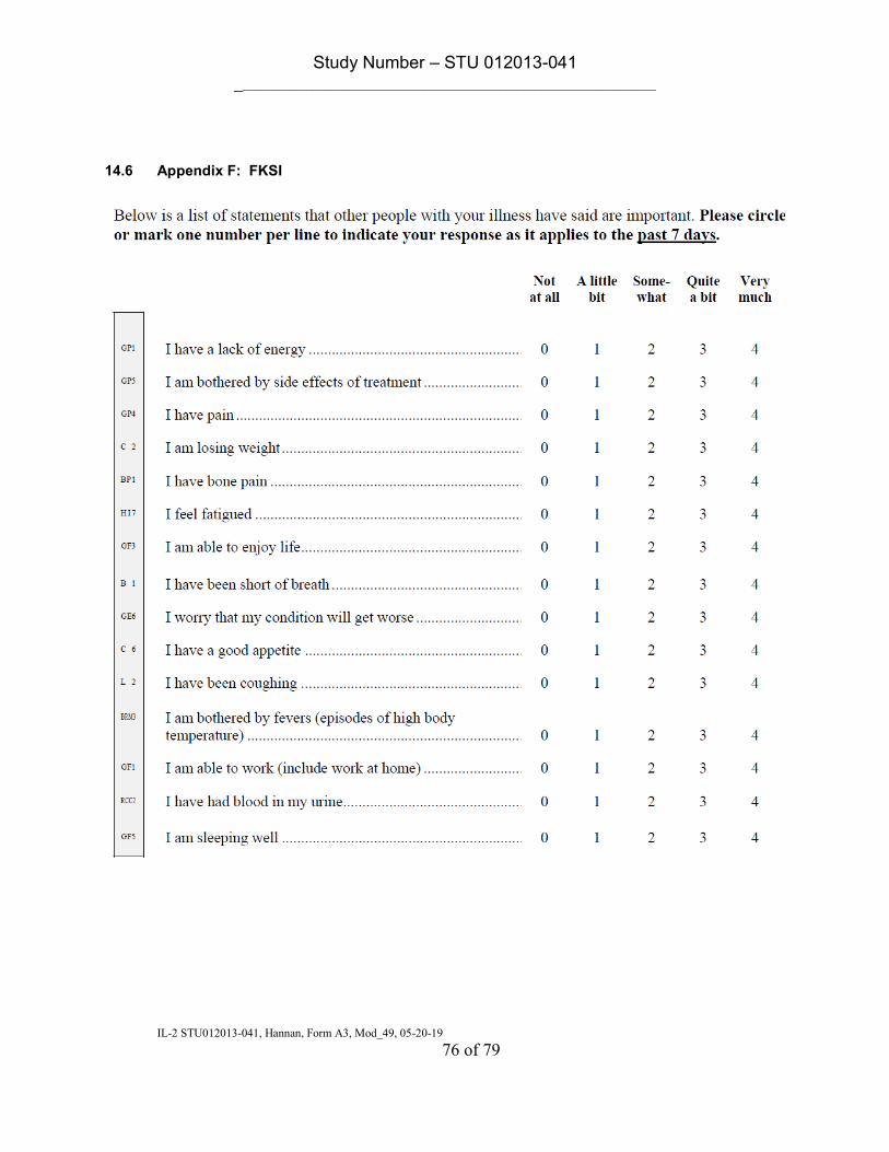
2.4.6 HRQoL: HRQoL will be measured using FACT-G, EQ-5D and FKSI questionnaire at baseline, after HD IL-2, and at four-month intervals after treatment.
2.4.7 Cost-effectiveness analysis: Health care utilization data needed to assess costs will be obtained from treatment records to include costs of hospitalization, treatment, ER visits, physician and clinic visits and medications. Markov modeling with probabilistic sensitivity analysis will be used to correlate quality-adjusted survival and cost.
3.0 Subject ELIGIBILITY
Subjects must meet all of the inclusion and exclusion criteria to be registered to the study. Study treatment may not begin until a subject is registered.
3.1 Inclusion Criteria
3.1.1 Pathologic evidence of clear cell RCC
3.1.2 Radiographic evidence of metastatic disease. CT and Bone Scan must be
performed with 21 days (+ 7 days) of registration. MRI of brain can be performed within 6 months prior to registration.
3.1.2.1 Patients with any number of metastatic site are allowed to enroll. However, only up to six sites will be selected for SBRT treatment, at the discretion of the treating radiation oncologist.
Study Number – STU 012013-041 ________________________________________________________________________
IL-2 STU012013-041, Hannan, Form A3, Mod_49, 05-20-19 14 of 79
3.1.3 Patient must have >1 lesion. Combined diameter of the lesions must be of size
>1.5cm.
3.1.4 Previous treatment with surgery, radiation, chemotherapy, immunotherapy or any targeted agents are allowed provided that:
3.1.4.1 Chemotherapy was administered > 28 days before the start of HD IL-2 3.1.4.2 Surgery, radiation, immunotherapy or any targeted agents was administered > 14 days before the start of HD IL-2
3.1.5 Age ≥ 18 years.
3.1.6 Performance status ECOG 0, 1.
3.1.7 Patient must be eligible for HD IL-2 treatment
3.1.8 Patient must be eligible for SABR to one or more extra cranial sites.
3.1.9 Adequate organ and marrow function as defined below: - leukocytes ≥ 3,000/mcL - absolute neutrophil count ≥ 1,500/mcL - platelets ≥ 50,000/mcl - total bilirubin ≤ 2mg/dL - AST(SGOT)/ALT(SPGT) ≤ 2.5 X institutional upper limit of normal
3.1.10 Women of child-bearing potential and men must agree to use adequate
contraception (hormonal or barrier method of birth control; abstinence) prior to study entry, for the duration of study participation. Should a woman become pregnant or suspect she is pregnant while participating in this study, she should inform her treating physician immediately. 3.1.10.1 A female of child-bearing potential is any woman (regardless of
sexual orientation, having undergone a tubal ligation, or remaining celibate by choice) who meets the following criteria:
Has not undergone a hysterectomy or bilateral oophorectomy; or
Has not been naturally postmenopausal for at least 12 consecutive months (i.e., has had menses at any time in the preceding 12 consecutive months).
3.1.11 Ability to understand and the willingness to sign a written informed consent
3.1.12 Adequate Renal function with Cr ≤ 1.6 mg/dL.
3.1.13 Adequate cardiac function (adequate perfusion; no ischemia) on thallium (or Tc)
stress test
3.1.14 Adequate pulmonary function on PFT (FEV1 >65%; DLCO>60%).
3.2 Exclusion Criteria
3.2.1 Subjects who have had chemotherapy within 4 weeks prior to entering the study
Study Number – STU 012013-041 ________________________________________________________________________
IL-2 STU012013-041, Hannan, Form A3, Mod_49, 05-20-19 15 of 79
3.2.2 History of HIV, Hepatitis B, Hepatitis C and HTLV
3.2.3 Subjects receiving any other investigational or standard antineoplastic agents. 3.2.4 Subjects with brain metastases are excluded from this clinical trial unless all the
metastases are adequately treated with surgery or radiation.
Follow-up imaging showing treatment adequacy is not required.
3.2.5 Subjects with life expectancy < 6 months.
3.2.6 History of allergic reactions to recombinant IL-2
3.2.7 Uncontrolled recurrent illness including, but not limited to, ongoing or active infection, symptomatic congestive heart failure, unstable angina pectoris, cardiac arrhythmia,.
3.2.8 Psychiatric illness/social situations that would limit compliance with study
requirements.
3.2.9 Subjects who are pregnant or nursing due to the potential for congenital abnormalities and the potential of this regimen to harm nursing infants.
3.2.10 Systemic or topical steroid use or other immunosuppressive therapy within the
past 14 days
3.2.11 Subjects required to take corticosteroids or other immunosuppressive therapy such as those with organ allograft
4.0 TREATMENT PLAN
4.1 SABR Dose and Techniques
4.1.1 SABR Dose
The SABR dose and fractionation scheme is generated to deliver a potent dose to ablate the targeted lesions and at the same time maximize an immune response. Since multiple studies have shown an influx of lymphocytes and monocytes within 24-48 hours after tumor irradiation [7, 12, 13, 92] and these cells play a critical role in antigen presentation and initiation of an adaptive immune response, multiple fraction irradiation which would kill these infiltrating immunocytes, is discouraged. Therefore a single fraction or a three fraction treatment regimen is allowed, and a single fraction treatment is preferred over three fractions. Due to normal organ toxicity and limits of dose constraints, sometimes a three fraction treatment must be undertaken and in those cases it is recommended that the treatment course is completed within 7-10 days. Radiation dose-(immune) response studies have shown a linear increase in immune response with increased dose per fraction of radiation without demonstration of a plateau [12, 14, 92, 93]. Two studies have compared 15Gy x 1 with 5Gy x3, and 20Gy x1 with 5Gy x4 and have showed a superior immune response generated by the single fraction radiation [12, 92]. Clinical experience with oligometastatic patients treated at 1-5 sites of disease has also showed an increase in progression free survival with the increasing radiation dose per fraction [94]. A dose of less than 7.5 Gy per fraction has demonstrated lower induction of systemic IFN-γ producing cells [93], and a previous phase II study of mRCC patients treated with HD IL-2 and singe fraction of 8Gy
Study Number – STU 012013-041 ________________________________________________________________________
IL-2 STU012013-041, Hannan, Form A3, Mod_49, 05-20-19 16 of 79
irradiation to a single lesion did now show an overall improvement in response rate [95]. Therefore 8Gy per fraction is the lowest permitted dose for this study and can be used only when administering the three fraction regimen as described in the prescription dose table below. The SABR prescription dose will be delivered to the periphery of the planning target volume (PTV, see below for definitions). Investigators will have discretion in choosing from either of the biologically equivalent dose levels using one or three fractions, although a single fraction is preferred over three fraction treatments. Treating physician will have further discretion in selecting the number and location of sites to treat if >6 sites of disease are present. Maximum number of lesions treated is deemed as feasible per the treating radiation oncologist. Physician’s will be REQUIRED to leave at least one site of disease for the purpose of measuring a radiographic response (see section 6.1) at a non-treated site, as this is part of the primary endpoint. If left untreated, this site can be treated once patient meets the definition of progressive disease (PD) (see section 6). To clarify the definition of “site”, each site is an area or organ with active extracranial disease (</=3 in the liver = one site and </=3 in the lung= one site) identified by CT scan, or PET/CT, within 4 weeks prior to initiation of SBRT (up to 2 contiguous vertebral metastasis will be considered a single site of disease). For example: a patient with 4 right axillary lymph nodes, L1-L2 bone metastasis, 3 right lung lesions, 1 left lung lesion, 2 liver lesions, and T2-T3 bone metastasis would be defined as having 6 sites of disease. Preference should be given to the largest feasible disease site, symptomatic sites and sites where palliative and preventative (i.e. to prevent a pathologic fracture in weight bearing bone, impending cord compression, impending SVC compression etc.) indications are applicable. The gross target/tumor volume (GTV) should be at least 2cm3 in size, corresponding to roughly a 1.5 cm diameter tumor. This is to ensure that adequate tumor volume and therefore adequate tumor cells (roughly 108 -109 cells/cm3 [96]) are killed for antigen presentation. Treating physicians should choose their dose based on established planning guidelines at their center including their ability to respect normal tissue tolerance listed below. It is not required that all targets be treated with the same dose fractionation. A dose from the following table should be used: Prescription Dose
Total Cumulative Dose Encompassing 95% of Planning Target Volume (Gy)
Number of Fractions
Protocol Compliant Minor Deviation * MinorDeviation
1 21-27 Gy ≥16 Gy, <21 Gy >27 Gy, <16 Gy
3 26.5-33 Gy ≥24.0 Gy, <26.5 Gy >33 Gy, <24.0 Gy
*This column is protocol compliant for tumors abutting the spinal cord (major deviation remain as listed) Dose tolerance limits should be adhered to for all treatments. Protocol compliant dose should be used in all cases, if possible. When treating tumors abutting the spinal cord, tolerance limits should not be exceeded. To facilitate this requirement, minor deviation dose ranges listed above in the table will be considered fully compliant for tumors abutting the spinal cord.
4.1.2 Planning Constraints and Concerns
The tolerance dose of SBRT to the gastrointestinal tract is not established, and patients with metastatic disease involving the esophagus, stomach, intestines, or mesenteric lymph nodes will be eligible only if no other sites of lesions are present that can be safely targeted, and the treating radiation oncologist feels that a sufficiently conservative dose constraints to these organs can be met. Patients with renal or adrenal metastases are potentially eligible if normal tissue constraints are otherwise met.
Study Number – STU 012013-041 ________________________________________________________________________
IL-2 STU012013-041, Hannan, Form A3, Mod_49, 05-20-19 17 of 79
It is well established that for palliative effect for a painful bone metastasis, a single dose of 8 Gy is usually as effective as 30 Gy [97]. However, in this protocol the goal is not just to relieve pain within an osseous metastasis but also to dramatically debulk the cancer cells present and induce in immune response, and the higher dose is more likely to accomplish this goal given a higher biological potency [98]. Long term survival after bone metastasectomy has been reported [99]. Irradiation of non-spinal skeletal sites does not generally require specialized techniques of treatment. Metastases in major lower extremity weight-bearing bones should undergo surgical stabilization if there is plain film evidence of cortical erosion.
4.1.3 SBRT Treatment Technique
SBRT will be administered the week prior to starting the first cycle of HD IL-2 with the last fraction given < 84 hours prior to the start of the first cycle of HD IL-2.
Treatment to skeletal lesions and paraspinous lesions may be accomplished with any 3D conformal radiotherapy or intensity-modulated radiotherapy (IMRT) technique suitable for this application with performance specifications adequate to provide proper tumor dose distribution and normal tissue sparing. The bone lesions can be treated with a conformal 3D or IMRT technique, which is different than SBRT/SABR technique. The difference lies primarily in dosimetric planning but otherwise all the descriptions of SBRT in terms of set-up, contouring and tissue constraints that needs to be met remains the same.
At the time of simulation for patients who will receive SBRT to the lung and/or liver, the movement of the dome of the diaphragm (superior portion of the liver) is to be observed under fluoroscopy or other acceptable means to estimate respiratory movement during treatment if no breathing control device is used. Patients will be assessed for suitability for tolerance of a respiratory control device using a breath-hold technique, respiratory gating, or abdominal compression to limit diaphragmatic excursion during respiration. Patients with severe lung disease and patients who cannot tolerate diaphragmatic or breathing control devices for other reasons will be treated without them. A larger margin to account for breathing related intra-fractional organ movement is required.
With the patient immobilized in a vacuum-type or equivalent body mold, a planning CT scan with 3-5 mm slices is performed. Intravenous contrast is recommended for lesions near mediastinal structures and lesions within the liver. The form of respiratory control to be used during treatment should also be used during the simulation. Oral GI contrast to highlight the stomach and duodenum is recommended for patients with medial liver lesions or lesions of the caudate lobe.
For treatment to the liver, the following structures are contoured: entire liver, each individual liver gross tumor volume (GTV), each kidney, and the spinal cord. The planning target volume (PTV) is constructed to account for the positional uncertainty of the GTV during treatment. The PTV for each contoured GTV should be at least 5mm larger than the GTV in the axial plane and 1.0 cm larger than the GTV in the craniocaudal plane. Larger margins may be used in cases where greater motion of the hemidiaphragm is observed in simulation despite standard maneuvers to diminish motion. For lung SBRT the same principles apply; the entire lung volumes are contoured, as are each individual GTV within the lung.
The prescription dose for each lesion is listed in the table in section 4.1, prescribed to the periphery of the PTV. There is no restriction on the dose “hotspot” except that it must be located within the PTV. A Linear Accelerator with effective photon energies of ≥ 6 MV is required. The use of a Multi-leaf collimator (MLC) or custom blocks are acceptable. A stereotactic relocalization system that relies upon stereoscopic radiographs, implanted fiducials, or near real-time CT based verification will be used.
Study Number – STU 012013-041 ________________________________________________________________________
IL-2 STU012013-041, Hannan, Form A3, Mod_49, 05-20-19 18 of 79
The PTV may be treated with any combination of coplanar or non-coplanar three-dimensional conformal fields, shaped to deliver the specified dose while restricting the dose to the normal tissues. Field arrangements will be determined by the planning system to produce the optimal conformal plan in accordance with volume definitions.
4.1.3.2 Normal Tissue Dose Constraints
In accordance with the prior Phase I studies [100], certain normal tissue dose constraints must be respected. The possibility that SBRT-induced fibrosis might cause occlusion of large central airways, thus impeding ventilation distal to the occlusion has been well considered [101]. An adjustment to the fractionation scheme may be made if, in the opinion of the treating radiation oncologist, the following conditions apply: (1) the location of a lung lesion is close enough to a large proximal bronchial airway such that occlusion might occur, and (2) compromised ventilation to the segment(s) of lung potentially affected would cause clinically significant adverse consequences. In such a case, the treating radiation oncologist should discuss any proposed dose modifications with the PI to decide whether a regimen of similar biological potency can be safely given. The same special condition applies in the setting of a patient whose primary prostate disease has been irradiated previously and is present as a site of disease; Since re-irradiation toxicity is a concern, these patients will be considered by the PI on a case-by-case basis and SBRT to a site previously irradiated with conventional fractionation within two years is not recommended. Re-irradiation to a site that has received previous SBRT is not allowed. Deviations from the intended dose regimen will be documented, with calculations of the BED of the applied regimen included in the patient’s research chart along with documentation of the discussions pertaining to the idiosyncrasies of the case. The following table lists the specific organ and dose fractionation constraints on normal tissues. For One Fraction:
Study Number – STU 012013-041 ________________________________________________________________________
IL-2 STU012013-041, Hannan, Form A3, Mod_49, 05-20-19 19 of 79
Trachea and Large
Bronchus* <4 cc 17.4 Gy 20.2 Gy stenosis/fistula
Bronchus- smaller airways <0.5 cc 12.4 Gy 13.3 Gy stenosis with
atelectasis
Rib <5 cc 28 Gy 33 Gy Pain or fracture
Skin <10 cc 25.5 Gy 27.5 Gy ulceration
Stomach <5 cc 17.4 Gy 22 Gy ulceration/fistula
Bile duct 30 Gy stenosis
Duodenum* <5 cc
<10 cc
11.2 Gy
9 Gy 17 Gy ulceration
Jejunum/Ileum* <30 cc 12.5 Gy 22 Gy enteritis/obstruction
Colon* <20 cc 18 Gy 29.2 Gy colitis/fistula
Rectum* <3.5 cc
<20 cc
39 Gy
22 Gy 44.2 Gy proctitis/fistula
Ureter 35 Gy stenosis
Bladder wall <15 cc 12 Gy 25 Gy cystitis/fistula
Penile bulb <3 cc 16 Gy impotence
Femoral Heads <10 cc 15 Gy necrosis
Renal hilum/vascular trunk 15 cc 14 Gy malignant
hypertension
Parallel Tissue Critical
Volume (cc)
Critical
Volume
Dose Max
(Gy)
Endpoint (≥Grade
3)
Lung (Right & Left) 1500 cc 7 Gy Basic Lung
Function
Lung (Right & Left) 1000 cc 7.6 Gy V-8Gy
<37% Pneumonitis
Liver 700 cc 11 Gy Basic Liver
Function
Renal cortex (Right & Left) 200 cc 9.5 Gy Basic renal function
For Three Fractions:
Serial Tissue Volume Volume Max
(Gy)
Max Point Dose
(Gy)**
Endpoint (≥Grade
3)
Optic Pathway <0.2 cc 15.3 Gy 17.4 Gy neuritis
Cochlea 14.4 Gy hearing loss
Brainstem (not
medulla) <0.5 cc 15.9 Gy 23.1 Gy cranial neuropathy
Spinal Cord and
medulla
<0.35 cc
<1.2 cc
15.9 Gy
13 Gy 22.5 Gy myelitis
Spinal Cord
Subvolume (5-6
<10% of
subvolume 18 Gy 22.5 Gy myelitis
Study Number – STU 012013-041 ________________________________________________________________________
IL-2 STU012013-041, Hannan, Form A3, Mod_49, 05-20-19 20 of 79
mm above and
below level
treated per Ryu)
Cauda Equina <5 cc 21.9 Gy 25.5 Gy neuritis
Sacral Plexus <5 cc 22.5 Gy 24 Gy neuropathy
Esophagus* <5 cc 17.7 Gy 25.2 Gy stenosis/fistula
Brachial Plexus <3 cc 22 Gy 26 Gy neuropathy
Heart/Pericardium <15 cc 24 Gy 30 Gy pericarditis
Great vessels <10 cc 39 Gy 45 Gy aneurysm
Trachea and
Large Bronchus* <5 cc 25.8 Gy 30 Gy stenosis/fistula
Bronchus- smaller
airways <0.5 cc 18.9 Gy 23.1 Gy
stenosis with
atelectasis
Rib <5 cc 40 Gy 50 Gy Pain or fracture
Skin <10 cc 31 Gy 33 Gy ulceration
Stomach <5 cc 22.5 Gy 30 Gy ulceration/fistula
Bile duct 36 Gy stenosis
Duodenum* <5 cc
<10 cc
15.6 Gy
12.9 Gy 22.2 Gy ulceration
Jejunum/Ileum* <30 cc 17.4 Gy 27 Gy enteritis/obstruction
Colon* <20 cc 24 Gy 34.5 Gy colitis/fistula
Rectum* <3.5 cc
<20 cc
45 Gy
27.5 Gy 49.5 Gy proctitis/fistula
Ureter 40 Gy stenosis
Bladder wall <15 cc 17 Gy 33 Gy cystitis/fistula
Penile bulb <3 cc 25 Gy impotence
Femoral Heads <10 cc 24 Gy necrosis
Renal
hilum/vascular
trunk
15 cc 19.5 Gy malignant
hypertension
Parallel Tissue
Critical
Volume
(cc)
Critical
Volume Dose
Max (Gy)
Endpoint (≥Grade
3)
Lung (Right &
Left) 1500 cc 10.5 Gy
Basic Lung
Function
Lung (Right &
Left) 1000 cc 11.4 Gy V-11Gy<37% Pneumonitis
Liver 700 cc 17.1 Gy Basic Liver
Function
Renal cortex
(Right & Left) 200 cc 15 Gy
Basic renal
function *Avoid circumferential irradiation. ** “point” defined as 0.035cc or less
Study Number – STU 012013-041 ________________________________________________________________________
IL-2 STU012013-041, Hannan, Form A3, Mod_49, 05-20-19 21 of 79
Exceeding these dose tolerances by more than 2.5% constitutes a minor protocol violation. Exceeding these dose tolerances by more than 5% constitutes a major protocol violation.
4.1.4 Radiation Therapy Quality Assurance
Dr. Timmerman or Dr. Hannan will perform an RT Quality Assurance Review after complete data for the first 12 cases enrolled has been received at the University of Texas Southwestern. Dr. Timmerman will perform the final review after complete data for the subsequent 12 cases enrolled has been received at the University of Texas Southwestern. These cases will be reviewed within 3 months after this study has reached the target accrual or as soon as complete data for all cases enrolled has been received, whichever occurs first.
4.2 HD IL-2
The standard and current guidelines for HD IL-2 administration developed based on Schwartzentruber [102], which has been proven to be safe at UTSW over the past years will be used for this protocol at the physician’s discretion. HD IL-2 treatment will begin within 84 hours of the last SABR fraction according to the established UTSW HD IL-2 bolus administration algorithm (Appendix B and C) and the standard procedure for admissions for HD IL-2 will be followed.
4.2.1 Treatment Dosage and Administration
HD IL-2 will be administered at a dose of 600,000 IU/kg every 8h for a total of up to 14 doses (considered as one cycle), followed by a one week break and then another cycle of the same dose over another week. Each HD IL2 course consists of two cycles of up to 14 doses each separated by a week of rest. A total of three courses may be administered for patients without progression at the discretion of the medical oncologist. Patients will be admitted to the ICU of the corresponding hospital for IL-2 treatment and close monitoring according to the HD IL-2 algorithm (appendix B). Patients will undergo placement of a central venous catheter, typically a PICC line, before each course of therapy.
4.2.1.1 Toxicities and Dosing Delays/Dose Modifications
Any subject who receives treatment on this protocol will be evaluable for toxicity. Each patient will be assessed for the development of toxicity according to the Time and Events table (5.4). Toxicity will be assessed according to the NCI Common Toxicity Criteria for Adverse Events (CTCAE), version 4.0. Dose adjustments should be made according to the system showing the greatest degree of toxicity. Treatment with HD IL-2 will be modified by withholding doses of IL-2 rather than continuing therapy at a reduced dose, according to the current guidelines followed at UTSW. Doses of IL-2 will be withheld for multiple indications as listed in the table below including hypotension refractory to fluids and pressors, anuria for more than 24 hours and unresponsive to fluid replacement and low-dose dopamine, respiratory distress requiring more than 4 L of oxygen to maintain O2 saturation greater than 95%, confusion, sustained ventricular tachycardia or any sign or symptom of myocardial ischemia or myocarditis, metabolic acidosis with HCO3 less than 18 despite attempts to correct with IV HCO3; atrial fibrillation, documented systemic infection, or any other serious toxicity that is not controlled at time of next dose. The following table published by Schwartzentruber [102] and included in the standard HD IL-2 administration guidelines of UTSW as described in Appendix B summarizes the guidelines that will be followed for discontinuation of HD IL-2:
Study Number – STU 012013-041 ________________________________________________________________________
IL-2 STU012013-041, Hannan, Form A3, Mod_49, 05-20-19 22 of 79
HD IL-2 should be delayed or discontinued if:
Schwartzentruber, J Immunotherapy, Vol. 24, No. 4, 2001. Corrective measures taken, also developed following the guidelines published by Schwartzentruber [102] in Table 5, included in the standard HD IL-2 administration guidelines of UTSW as described in Appendix C are following:
4.3 Duration of Therapy
The SABR administration followed by one course of HD IL-2 (consisting of two cycles of HD IL-2 with one week break in between) will typically be completed within four weeks. The HD IL-2 course may be repeated for a maximum of 3 times, at the discretion of the treating medical oncologist unless:
Study Number – STU 012013-041 ________________________________________________________________________
IL-2 STU012013-041, Hannan, Form A3, Mod_49, 05-20-19 23 of 79
Disease progression
Inter-current illness that prevents further administration of treatment
Unacceptable adverse event(s)
Subject decides to withdraw from the study, OR
General or specific changes in the patient’s condition render the subject unacceptable for further treatment in the judgment of the principal investigator or medical oncologist.
4.4 Duration of Follow Up
Subjects will be followed for ten years or death (although findings will be analyzed and reported at a median follow up of 2-4 years), whichever occurs first. Subjects removed from treatment for unacceptable adverse events will be followed until resolution or stabilization of the adverse event. The follow-up will be every 8 weeks (+/- 2 Weeks) from study registration with imaging studies and physical exam every 8 weeks for the first eight months, then every 12 weeks (+/- 2 Weeks) until two years, then every four months thereafter for a total of five years, and then every six months (+/- 1 month) for a total of ten years. See section 5.0 for detail. Subjects who show progressive disease will be followed for survival and will no longer strictly adhere to study calendar. QOL questionnaires will be completed every 4 months until 1 year after treatment.
4.5 Removal of Subjects from Protocol Therapy
Subjects will be removed from therapy when any of the criteria listed in Section 5.5 apply, however will continue to be followed up as per protocol described above. Notify the Principal Investigator, and document the reason for study removal and the date the subject was removed in the Case Report Form.
5.0 STUDY PROCEDURES
5.1 Screening/Baseline Procedures
Assessments performed strictly for research purposes will be done only after obtaining informed consent. Assessments performed for clinical indications (not exclusively for research purposes) may be used for baseline values even if the studies were done before informed consent was obtained. All screening procedures must be performed within 3 months prior to registration unless otherwise stated. The screening procedures include:
5.1.1 Informed Consent
5.1.2 Medical history
Complete medical and surgical history, history of infections
5.1.3 Demographics
Age, gender, race, ethnicity
Study Number – STU 012013-041 ________________________________________________________________________
IL-2 STU012013-041, Hannan, Form A3, Mod_49, 05-20-19 24 of 79
5.1.4 Review subject eligibility criteria
5.1.5 Review previous and concomitant medications
5.1.6 Physical exam including vital signs, height and weight
Performance status evaluated prior to study entry according to Appendix A (ECOG).
5.1.8 Adverse event assessment
Baseline adverse events will be assessed. See section 7 for Adverse Event monitoring and reporting.
5.1.9 Hematology
CBC with differential.
5.1.10 Blood draw for correlative studies
See Section 9.0 for details.
5.1.11 Serum chemistries
Comprehensive metabolic panel (CMP) to include: albumin, alkaline phosphatase, ALT/SGPT, AST/SGOT, BUN, creatinine, LDH, electrolytes (sodium, potassium, calcium, chloride, bicarbonate, magnesium and phosphate), glucose, uric acid, C-Reactive protein (CRP), beta-2 microglobulin and total bilirubin.
5.1.12 Pregnancy test (for females of child bearing potential)
See section 3.1.10.1 for definition.
5.1.13 Serologic Tests
HLA typing.
5.1.14 Radiographic Imaging
Bone scan, CT chest, abdomen and pelvis with IV contrast. MRI of brain with contrast, if tolerated. MRI of the abdomen should be considered on an individual basis if felt to be better by the radiation or medical oncologist. CT and Bone Scan must be performed within 21 days (+ 7 days) of registration.
5.1.15 Biopsy of metastatic lesion
5.1.15.1 Pre-treatment biopsy:
A CT-guided biopsy of a tumor lesion will be performed prior to study registration, unless previous biopsy of metastatic site within the last six months exists and a review of slides shows it to be adequate, in which case a pretreatment biopsy is optional. Soft tissue lesion will be preferred over bone biopsy for the pre-treatment biopsy. Biopsy results do not affect eligibility if previous diagnosis of clear cell histology is present. If the patient participated in protocols [such as the Urology Tissue Repository Protocol (STU 032011-187)] or procedures and the tissue confirming kidney cancer diagnosis is in storage and available at UTSW Medical
Study Number – STU 012013-041 ________________________________________________________________________
IL-2 STU012013-041, Hannan, Form A3, Mod_49, 05-20-19 25 of 79
Center or an outside institution, the study team may request a tissue sample.
5.1.15.2 Post-treatment elective biopsy:
An elective post-treatment biopsy will be performed 8 weeks after the completion of HD IL-2 and for this biopsy, any site other than a lymph node and a treated site is acceptable. In the case where all lesions are treated, any progressive or non-responding lesion will be biopsied. In case of CR no lesions may remain to be biopsied.
5.1.16 QoL Questionnaires
FACT-G, EQ-5D, FKSI, cost and convenience questionnaire. These forms will be referred to collectively as QoL Questionnaires. Cost and convenience questionnaire will only be completed at first follow-up.
5.1.17 Tumor assessment
To be performed on bone scan, CT or MRI. Please see section 6.1 for detail.
5.1.18 Pulmonary Function Tests
5.1.19 Cardiac Stress Test
5.2 Procedures During Treatment
SABR treatment requires one week for planning and one week for delivery of the 1 or 3 fractionation schemes. HD IL-2 must begin within 84 hours of the last SABR fraction. Each course of HD IL-2 consists of two cycles of approximately two weeks each (12-14 IV infusions q8h followed by about 9 days of break) requiring a total of four weeks.
5.2.1 Prior to Each Treatment Cycle
Physical exam, vital signs
Hematology
Serum chemistries
5.2.2 Prior to treatment
Registration to the study (registration must be done prior to first fraction of SABR)
CT simulation for SABR planning.
If multiple planning sessions are required, they will be completed within the first week. SABR planning completed
5.2.3 Day 1-14 (+/- 7days)
SABR treatments completed
One hour after first SABR treatment, blood collection for correlative studies
If SABR to multiple sites, an additional 5 days is allowed to complete treatment
5.2.4 Day 15-22 (+/- 7days)
Physical exam, vital signs
Hematology
Study Number – STU 012013-041 ________________________________________________________________________
IL-2 STU012013-041, Hannan, Form A3, Mod_49, 05-20-19 26 of 79
Serum chemistries
First cycle of HD IL-2 started within 84hr of the last SABR treatment
5.2.5 Day 22-29 (+/- 7days)
Break
Patient will be assessed either by clinic visit or telephone call to ensure s/he is doing well and ready for the second cycle. Telephone call can be done by any clinic staff or study personnel.
5.2.6 Day 30-37 (+/- 14 days)
Physical exam, vital signs
Hematology
Serum chemistries
Second cycle of HD IL-2
Blood collection for correlative immunologic studies (see section 9.0) on the last day of HD IL-2
5.2.7 Week 8 (+/- 2 weeks)
Physical exam, vital signs
Hematology
Serum chemistries
QoL questionnaire
Blood collection for correlative immunologic studies (see section 9.0)
Radiographic imaging o CT chest, abdomen and pelvis with IV contrast, if soft tissue metastasis o Bone scan if bone lesion was present and detected by the baseline Bone
scan o MRI, if necessary to confirm CT/bone scan findings, at the discretion of
radiologist
5.2.8 Weeks 8-45 from registration date
HD IL-2 can be repeated for a maximum of 3 courses 12 weeks apart at the discretion of the medical oncologist
5.3 Follow-up Procedures
Subject will be followed every eight-ten weeks (+/- 2 Weeks) starting from the date of registration for the first eight months, then every 12 weeks until 18 months and then every sixteen weeks for another three years and then every three – six months for the next five years. Thereafter at the discretion of the treating physician. The following procedures will be performed at each follow up:
Physical exam, vital signs
Hematology
Serum chemistries
QoL questionnaire (every other follow up)
Blood collection for correlative immunologic studies (see section 9.0) Radiographic imaging o CT chest, abdomen and pelvis with IV contrast, if soft tissue metastasis o Bone scan if bone lesion was present and detected by the baseline Bone
scan o MRI, if necessary to confirm CT/bone scan findings, at the discretion of
radiologist
Study Number – STU 012013-041 ________________________________________________________________________
IL-2 STU012013-041, Hannan, Form A3, Mod_49, 05-20-19 27 of 79
5.4 Time and Events Table
* Bone scan performed every 8 weeks only if bone lesions present in baseline bone scan. @ optional # Immunologic blood collection is needed at baseline, post SABR, post HD IL-2, at 8 week, six months and at 1 year & Comprehensive chemistry will be done once a year, after this point. % Cost and Convenience Questionnaire will only be administered at the first follow up. All other QoL surveys will take place every other follow-up. 1 Additional procedures, ICU admission, and lab requirements may apply in association with IL-2 administration, as detailed in appendix C.
Pre-study Cycle 1 , Day15 (+/- 5)
Cycle2, Day30 (+/- 14)
Cycle2, Day37 (+/- 14)
Months 1-8: q8 Weeks
Months 8-18: q12
Weeks
Months 18-120: 3-6 months
Assessment X X X X
Informed Consent
X
Vital Signs X X X X X X X
History and PE X X X X
Performance Status
X X X X
Toxicity (include DLT) Evaluations
X X X X
Bone Scan X X* X* X
CT Chest, Abd, pelvis w/ Contrast
X X X X
Biopsy of metastatic lesion
X^ X@
CBC with diff X X1 X1 X1 X X X
Basic Chemistry X X1 X1 X1 X X X
Comprehensive chemistry
X X X&
Blood collection for Immune Assays
X X X# X# X#
QOL Questionnaires
X X% X% X% X%
Cardiac Stress Test
X
PFT X
Study Number – STU 012013-041 ________________________________________________________________________
IL-2 STU012013-041, Hannan, Form A3, Mod_49, 05-20-19 28 of 79
^ if previous biopsy of metastatic site within six months with adequate review of slides is not available. If the patient participated in protocols [such as the Urology Tissue Repository Protocol (STU 032011-187)] or procedures where tissue confirming kidney cancer diagnosis was collected and the biopsy in storage is still available, the study team may request a tissue sample.
5.5 Removal of Subjects from Study
Subjects can be taken off the study treatment and/or study at any time at their own request, or they may be withdrawn at the discretion of the investigator for safety, behavioral or administrative reasons. The reason(s) for discontinuation will be documented and may include:
5.5.1 Subject voluntarily withdraws from treatment (follow-up permitted);
5.5.2 Subject withdraws consent (termination of treatment and follow-up);
5.5.3 Subject is unable to comply with protocol requirements; patients must be withdrawn from the trial and replaced if they failed to receive at least one cycle of HD IL-2
5.5.4 Subject demonstrates disease progression (unless continued treatment with HD IL-2 is deemed appropriate at the discretion of the medical oncologist);
5.5.5 Subject experiences toxicity that makes continuation in the protocol unsafe;
5.5.6 Treating physician judges continuation on the study would not be in the subject’s best interest;
5.5.7 Subject becomes pregnant (pregnancy to be reported along same timelines as a serious adverse event);
5.5.8 Development of second malignancy (except for basal cell carcinoma or squamous cell carcinoma of the skin) that requires treatment, which would interfere with this study;
5.5.9 Lost to follow-up.
6.0 Measurement of Effect
6.1 Antitumor Effect
For the primary endpoint, response and progression will be evaluated in this study using the new international criteria proposed by the Response Evaluation Criteria in Solid Tumors (RECIST) Committee [JNCI 92(3):205-216, 2000], which was also used by the two randomized trials of HD IL-2 being used as historic controls for this trial [35, 36]. The only change from RECIST v.1.1 being implemented in this study is a confirmation of new lesions or progressive disease (PD) by a second scan >6wks apart, based on the immune RECIST (irRC) criteria proposed by Wolchok. et. al that is appropriate for immune-related treatment response [91]. This is primarily because immune response often can take longer time as compared to chemotherapy before producing a radiographic response and during this time new lesions may arise which will eventually regress and if it were not for a second confirmation, these patients would be labeled to have failed therapy. In addition, the majority of mRCC responders to HD IL-2 usually produce a durable response (median response duration of 24 months), therefore, this change will not affect the outcome [35]. Additional criteria for bone lesions and clinical endpoints of pathologic fracture and cord compression are added to this study (see Section 6.1.4). Changes in only the largest diameter (unidimensional measurement) of the tumor lesions are used in the RECIST v1.1 criteria outlined in http://www.recist.com/.
Study Number – STU 012013-041 ________________________________________________________________________
IL-2 STU012013-041, Hannan, Form A3, Mod_49, 05-20-19 29 of 79
6.1.1 Definitions
Evaluable for toxicity. All subjects will be evaluable for toxicity from the time of their registration. Evaluable for objective response. Only those subjects who have undergone SABR and one course of HD IL-2, and have had their disease re-evaluated at least at two occasions (8wk and 16wk) will be considered evaluable for response. These subjects will have their response classified according to the definitions stated below.
6.1.2 Disease Parameters
Measurable disease. Measurable lesions are defined as those that can be accurately measured in at least one dimension (longest diameter to be recorded) as >10 mm with spiral (Helical) CT scan. All tumor measurements must be recorded in millimeters (or decimal fractions of centimeters). Non-measurable disease. All other lesions (or sites of disease), including small lesions (longest diameter <20 mm with conventional techniques or <10 mm using spiral CT scan), are considered non-measurable disease. Bone lesions, leptomeningeal disease (patients excluded), ascites, pleural/pericardial effusions, lymphangitis cutis/pulmonis, inflammatory breast disease, abdominal masses (not followed by CT or MRI), and cystic lesions are all non-measurable. Target lesions. All measurable lesions up to a maximum of 3 lesions per organ and 6 lesions in total, representative of all involved organs, should be identified as target lesions and recorded and measured at baseline. Target lesions should be selected on the basis of their size (lesions with the longest diameter) and their suitability for accurate repeated measurements (either by imaging techniques or clinically). A sum of the longest diameter (LD) for all target lesions will be calculated and reported as the baseline sum LD. The baseline sum LD will be used as reference by which to characterize the objective tumor response. Note: Lesions receiving SABR will be called “treated lesion” which should not be confused with “target lesions” defined here for the for the purpose of radiographic measurement. Treated lesions and target lesions are mutually exclusive lesions. Therefore, treated lesions will not be used as target lesions for evaluating response. Since patients with ANY number of metastasis is eligible for this study, in the instance where patient has only one or few site of disease and all of them are treated, appearance of new lesions (either measurable or non-measureable) will constitute PD. Non-target lesions. All other lesions (or sites of disease) including any measurable lesions over and above the 6 target lesions should be identified as non-target lesions and should also be recorded at baseline. Measurements of these lesions are not required, but the presence or absence of each should be noted throughout follow-up.
6.1.3 Methods for Evaluation of Measurable Disease
All measurements will be done digitally on the PACs system. All baseline evaluations should be performed as closely as possible to the beginning of treatment and never more than 14 days before the beginning of the treatment.
Study Number – STU 012013-041 ________________________________________________________________________
IL-2 STU012013-041, Hannan, Form A3, Mod_49, 05-20-19 30 of 79
The same method of assessment and the same technique should be used to characterize each identified and reported lesion at baseline and during follow-up using appropriate radiologic imaging. Bone scans and CT scan of Chest, abdomen and pelvis with IV contrast will be performed at baseline. Since bone scan is not reliable for RCC, it will only be used to evaluate overall response when positive in baseline scan, and any new lesions found on bone scan must be verified with a CT. Spiral CT and MRI. All CT scans will be Spiral CT and should be performed using a 5 mm contiguous reconstruction algorithm.
6.1.4 Response Criteria
6.1.4.1 Evaluation of Target Lesions
Complete Response (CR): Disappearance of all target lesions, determined by two separate observations conducted not less than 4 weeks apart. There can be no appearance of new lesions.
Partial Response (PR): At least a 30% decrease in the sum of the longest diameter (LD) of target lesions, taking as reference the baseline sum LD. There can be no appearance of new lesions. Progressive Disease (PD): At least a 20% increase in the sum of the LD of target lesions, taking as reference the smallest sum LD recorded since the treatment started, or the appearance of one or more new lesions verified by a second scan > 6 weeks apart.
Stable Disease (SD): Neither sufficient shrinkage to qualify for PR nor sufficient increase to qualify for PD, taking as reference the smallest sum LD since the treatment started.
6.1.4.2 Evaluation of Non-Target Lesions
Complete Response (CR): Disappearance of all non-target lesions and normalization of tumor marker level. Incomplete Response/Stable Disease (SD): Persistence of one or more non-target lesion(s) and/or maintenance of tumor marker level above the normal limits.
Progressive Disease (PD): Appearance of one or more new lesions and/or unequivocal progression of existing non-target lesions
6.1.4.3 Evaluation of Best Overall Response
The best overall response is the best response recorded from the start of the treatment until disease progression/recurrence (taking as reference for progressive disease the smallest measurements recorded since the treatment started). The subjects best response assignment will depend on the achievement of both measurement and confirmation criteria.
Target Lesions
Non-Target
Lesions
New Lesions Overall Response
Best Response for this Category
Study Number – STU 012013-041 ________________________________________________________________________
IL-2 STU012013-041, Hannan, Form A3, Mod_49, 05-20-19 31 of 79
Also Requires:
CR CR No CR >4 wks. confirmation
CR Non-CR/Non-
PD
No PR >4 wks.
confirmation
PR Non-PD No PR
SD Non-PD No SD documented at least once >4
wks. from baseline
PD Any Yes or No PD no prior SD, PR or CR
Any PD* Yes or No PD
Any Any Yes PD
* In exceptional circumstances, unequivocal progression in non-target lesions may be accepted as disease progression.
Note: Subjects with a global deterioration of health status requiring
discontinuation of treatment without objective evidence of disease progression at that time should be reported as “symptomatic deterioration”. Every effort should be made to document the objective progression even after discontinuation of treatment.
Note: If subjects respond to treatment and are able to have their disease resected, the patient’s response will be assessed prior to the surgery.
6.1.4.4 Evaluation of Bone Lesions:
Bone lesions will be evaluated either by CT or bone scan, whichever is deemed to be better suited by baseline studies. Since the size of the lesion is difficult to measure in a bone scan, particularly if it is not well visible in CT the following guideline will be used. Any ambiguity will require MRI for resolution:
6.1.4.4.1 Progression of bone lesions will be defined as follows:
Appearance of 1 or more new bone lesion in Bone scan, confirmed by a repeat bone scan in ≥6 weeks.
6.1.4.4.2 Response of bone lesions in bone scan will be defined by either a complete resolution (CR) at the metastatic sites or partial resolution (PR) of radiotracer uptake by a radiologist.
6.1.4.5 Evaluation of Pathologic Fracture:
Any clinical suspicion of pathologic fracture will prompt radiologic evaluation with plain film, CT or MRI as appropriate and if confirmed by a radiologist, will constitute progression, unless it is at a treated site, in which case a treatment-related toxicity will be considered.
6.1.4.6 Evaluation of spinal cord Compression or Cauda equina compression:
Any clinical suspicion of cord or cauda equine compression will prompt radiologic evaluation with MRI (CT myelography if patient is not eligible
Study Number – STU 012013-041 ________________________________________________________________________
IL-2 STU012013-041, Hannan, Form A3, Mod_49, 05-20-19 32 of 79
for MRI) as appropriate and if confirmed by a radiologist, will constitute progression.
6.1.5 Response Rate and clinical response:
Response rate (RR) is defined by combining CR and PR. Clinical response is defined by combining CR, PR, and SD.
6.1.6 Duration of Response
Duration of overall response: The duration of overall response is measured from the time measurement criteria are met for CR or PR (whichever is first recorded) until the first date that recurrent or progressive disease is objectively documented (taking as reference for progressive disease the smallest measurements recorded since the treatment started). The duration of overall CR is measured from the time measurement criteria are first met for CR until the first date that recurrent disease is objectively documented. Duration of stable disease: Stable disease is measured from the start of the study registration until the criteria for progression are met, taking as reference the smallest measurements recorded since the treatment started.
6.1.7 Progression-Free Survival
Progression-free survival (PFS) is defined as the duration of time from the date of registration to time of progression or date of death due to any cause.
6.2 Safety/tolerability
Analyses will be performed for all subjects having received at least one fraction of SABR and one cycle of HD IL-2. The study will use the CTCAE version 4.0 for reporting of non-hematologic adverse events (http://ctep.cancer.gov/reporting/ctc.html) and modified criteria for hematologic adverse events (Appendix #/letter).
6.3 Quality of Life (QoL)
6.3.1 Functional Assessment of Cancer Therapy-P (FACT-G) and Kidney Symptom Index (FKSI)
Patient-reported functional status will be assessed with prostate cancer subscales of the Functional Assessment of Cancer Therapy-General (FACT-G) (See appendix for form). The FACT-G is a 28-item questionnaire that uses 5-point Likert-type response choices (0 = not at all; 1 = a little bit; 2 =somewhat; 3 = quite a bit; 4 = very much). It will take less than 10 minutes to complete the questionnaire. The Trial Outcome Indices (TOI) also will be utilized to measure the summed functional well-being, physical well-being [103, 104]. A 5-point deterioration in the FACT-G TOI between pre-treatment and at post treatment or at year 1 will be considered clinically significant [105]. The FACT-G and FKSI will be given on Day 1 and 28 similar to several other quality of life studies [106],[82], [85] The FKSI is a 15 question validated symptom index for kidney cancer patients which has been used in several metastatic renal cell cancer studies [106]. This scale focuses on symptoms predominantly related to kidney cancer such as energy, fatigue, pain, bone
Study Number – STU 012013-041 ________________________________________________________________________
IL-2 STU012013-041, Hannan, Form A3, Mod_49, 05-20-19 33 of 79
pain, weight loss, shortness of breath, cough, fever, hematuria. This subscale of the FKSI-DRS was validated in another study as well [107] The first analysis of change in QOL from baseline to 8 weeks will only be performed on patients who are still alive at 8 weeks. Changes in QOL will be also analyzed using all available data at baseline, 8, 16, 24, and 36 weeks with semiparametric generalized estimating equations (GEE). Additionally, similarly we will also compare the percentage of patients with an effect size for the change in FKSI (FKSI) scores between pre-treatment and post treatment which will allow us to compare the percentage of patients whose functional status remains more similar to baseline levels.
6.3.2 EQ-5D
The EQ-5D is a patient self-administrated questionnaire that takes approximately 5 minutes to complete (See appendix for form). The first part consists of 5 items covering 5 dimensions including: mobility, self care, usual activities, pain/discomfort, and anxiety/depression. Each dimension can be graded on 3 levels: 1-no problems, 2-moderate problems, and 3-extreme problems. Health states are defined by the combination of the leveled responses to the 5 dimensions, generating 243 health states to which unconsciousness and death are added. The 5-item index score is transformed into a utility score between 0, “Worst health state,” and 1, “Best health state.” The index score or the cost-utility equation can be used in the quality adjusted survival analysis depending on the health state(s) of interest. Additionally the EQ 5D is utilized to establish health state utility which is used in cost-effectiveness analysis to calculate quality adjusted life years. This is the recommended health state utility form used in the United Kingdom and approved by the National Institute for Clinical Excellence (NICE) and has been used to establish health state utility and metastatic renal cell cancer patients. Unfortunately, to our knowledge there is no health state utilites established for patients receiving IL-2, much less HD-IL-2. Likewise , there is no data for metastatic renal cell carcinoma patients within the setting of SABR treatment. The EQ-5D will also be available in the Spanish language.
6.4 Cost effectiveness data collection:
Health care utilization data needed to assess costs will be obtained from treatment records. Additionally, in order to assess the treatment related indirect costs and patient out of pocket costs, a form will be administered at the first available follow up visit after completion of radiation treatment (see Appendix VII).
Hospitalizations:. For hospitalizations with physician billing records, impatient physician costs will be estimated by applying Medicare payment rates under the RBRVS-based Medicare Fee Schedule to billed procedures in the physician billing records. This is of great importance given that these patients will be admitted to the intensive care unit. Treatment Cost: Direct costs of radiation treatment including consultation, simulation, treatment planning, and treatment delivery. Patient bills related to treatment will be obtained and estimated by total billed charges adjusted by facility-specific cost-to-charge ration from Medicare cost reports as described above. Emergency Room visits: The date of ER visit and name of the facility, and whether the ER visit resulted in a hospital admission. ER costs will be estimated using
Study Number – STU 012013-041 ________________________________________________________________________
IL-2 STU012013-041, Hannan, Form A3, Mod_49, 05-20-19 34 of 79
Medicare average payment rates for facility and physician charges, using the merged MEDPAR and MBS data as described above. Physician and Clinic Visits: The date of the visit, the name of the physician or physician clinic, and the service provided (physician exam, lab test, physical therapy, etc.). Costs for physician and clinic visits will be calculated based on billing records obtained for such visits, using Medicare payment rates for procedures indicated in the clinic billing records. Medications: Prescription drugs used, including dosage strength and frequency of administration. Information about name, dose, and frequency of all prescription medications will be recorded. The medications used by the study patients will be assigned an NDC drug code. Unit costs for these drugs will be estimated as the “AWP” price published in the Red Book less 15%. Outpatient drug costs will be calculated by multiplying unit cost by the number of pills used per day times the length of time the patient received the medication. Note that costs of drugs administered through a clinic (e.g., reimbursed under Medicare Part B) are included under “clinic visit costs” and impatient drug costs are included under “inpatient facility costs.” For example: Cost estimated for the HD-IL-2 infusions will be based on Medicare allowable utilizing the appropriate J code and associated facility charges for administration of HD-IL-2.
7.0 ADVERSE EVENTS
7.1 SABR
The contraindications and adverse events for SABR are mostly related to the treatment site and its radiation dose tolerance, as discussed in detail in section 4.1
7.1.1 Contraindications: None
7.1.2 Special Warnings and Precautions for Use: N/a
7.1.3 Interaction with other medications: None
7.1.4 Adverse Reactions: Sites-specific. Please see section 4.1
7.2 HD IL-2
The standard treatment regiment for HD IL-2 which has been proven to be safe at UTSW over the past years will be used for this protocol (as detailed in section 4, Appendix B and C). For the most recent safety update, please refer to the current Study Agent Prescribing Information in the company website: http://www.proleukin.com/assets/pdf/proleukin.pdf
7.2.1 Contraindications
HD IL-2 is contraindicated in patients with a known history of hypersensitivity to IL-2 or any component of the HD IL-2 formulation. HD IL-2 is contraindicated in patients with an abnormal thallium stress test or abnormal pulmonary function tests and those with organ allografts. Retreatment with HD IL-2 is contraindicated in patients who have experienced the following drug-related toxicities while receiving an earlier course of therapy:
Study Number – STU 012013-041 ________________________________________________________________________
IL-2 STU012013-041, Hannan, Form A3, Mod_49, 05-20-19 35 of 79
• Sustained ventricular tachycardia (≥5 beats) • Cardiac arrhythmias not controlled or unresponsive to management • Chest pain with ECG changes, consistent with angina or myocardial infarction • Cardiac tamponade • Intubation for >72 hours • Renal failure requiring dialysis >72 hours • Coma or toxic psychosis lasting >48 hours • Repetitive or difficult to control seizures • Bowel ischemia/perforation • GI bleeding requiring surgery
7.2.2 Special Warnings and Precautions for Use
Because of the severe adverse events which generally accompany HD IL-2 therapy at the recommended dosages, thorough clinical evaluation should be performed to identify patients with significant cardiac, pulmonary, renal, hepatic, or CNS impairment in whom HD IL-2 is contraindicated. Patients with normal cardiovascular, pulmonary, hepatic, and CNS function may experience serious, life threatening or fatal adverse events. Adverse events are frequent, often serious, and sometimes fatal. Should adverse events, which require dose modification occur, dosage should be withheld rather than reduced (See “DOSAGE AND ADMINISTRATION” section, “Dose Modifications” subsection of the manufacturer’s prescribing information). HD IL-2 has been associated with exacerbation of pre-existing or initial presentation of autoimmune disease and inflammatory disorders. Exacerbation of Crohn’s disease, scleroderma, thyroiditis, inflammatory arthritis, diabetes mellitus, oculo-bulbar myasthenia gravis, crescentic IgA glomerulonephritis, cholecystitis, cerebral vasculitis, Stevens-Johnson syndrome and bullous pemphigoid, has been reported following treatment with IL-2. All patients should have thorough evaluation and treatment of CNS metastases and have a negative scan prior to receiving HD IL-2 therapy. New neurologic signs, symptoms, and anatomic lesions following HD IL-2 therapy have been reported in patients without evidence of CNS metastases. Clinical manifestations included changes in mental status, speech difficulties, cortical blindness, limb or gait ataxia, hallucinations, agitation, obtundation, and coma. Radiological findings included multiple and, less commonly, single cortical lesions on MRI and evidence of demyelination. Neurologic signs and symptoms associated with HD IL-2 therapy usually improve after discontinuation of HD IL-2 therapy; however, there are reports of permanent neurologic defects. One case of possible cerebral vasculitis, responsive to dexamethasone, has been reported. In patients with known seizure disorders, extreme caution should be exercised as HD IL-2 may cause seizures. Patients should have normal cardiac, pulmonary, hepatic, and CNS function at the start of therapy. (See “PRECAUTIONS” section, “Laboratory Tests” subsection of manufacturer’s prescribing information). Capillary leak syndrome (CLS) begins immediately after HD IL-2 treatment starts and is marked by increased capillary permeability to protein and fluids and reduced vascular tone. In most patients, this results in a concomitant drop in mean arterial blood pressure within 2 to 12 hours after the start of treatment. With continued therapy, clinically significant hypotension will occur. In addition, extravasation of protein and fluids into the extravascular space will lead to the formation of edema and creation of new effusions. Medical management of CLS begins with careful monitoring of the patient’s fluid and organ perfusion status, as described in Section 4, Appendices B and C, in accordance with the established and standard practice guidelines followed at UTSW. This is achieved by frequent determination of blood pressure and pulse, and by monitoring organ function, which includes assessment of mental status and urine output. Hypovolemia is assessed by catheterization and central pressure monitoring.
Study Number – STU 012013-041 ________________________________________________________________________
IL-2 STU012013-041, Hannan, Form A3, Mod_49, 05-20-19 36 of 79
Flexibility in fluid and pressor management is essential for maintaining organ perfusion and blood pressure. Correction of hypovolemia may require large volumes of IV fluids but caution is required because unrestrained fluid administration may exacerbate problems associated with edema, in particular leading to pulmonary edema. With extravascular fluid accumulation, edema is common and ascites, pleural or pericardial effusions may develop. Management of these events depends on a careful balancing of the effects of fluid shifts so that neither the consequences of hypovolemia (e.g., impaired organ perfusion) nor the consequences of fluid accumulations (e.g., pulmonary edema) exceed the patient’s tolerance. Clinical experience has shown that early administration of dopamine (1 to 5 μg/kg/min) to patients manifesting capillary leak syndrome, before the onset of hypotension, can help to maintain organ perfusion particularly to the kidney and thus preserve urine output. Weight and urine output should be carefully monitored. If organ perfusion and blood pressure are not sustained by dopamine therapy, the dose of dopamine can be titrated as described in section 4 and in Appendix C, to 5mcg/kg/min to maintain SBP equal to or greater than 90 mm Hg, or add phenylephrine hydrochloride 20 mg/500 ml NS
at 1 mcg/kg/min and titrate to 1.5 mcg/kg/min. (See “ADVERSE REACTIONS” section below). Prolonged
use of pressors, either in combination or as individual agents, at relatively high doses, may be associated with cardiac rhythm disturbances. If there has been excessive weight gain or edema formation, particularly if associated with shortness of breath from pulmonary congestion, use of diuretics, once blood pressure has normalized, has been shown to hasten recovery. NOTE: Prior to the use of any product mentioned, the physician should refer to the package insert for the respective product. HD IL-2 treatment should be withheld for failure to maintain organ perfusion as demonstrated by altered mental status, reduced urine output, a fall in the systolic blood pressure below 90 mm Hg or onset of cardiac arrhythmias (See “DOSAGE AND ADMINISTRATION” section 4.2 and “Dose Modifications” subsection 4.3). Recovery from CLS begins soon after cessation of HD IL-2 therapy. Usually, within a few hours, the blood pressure rises, organ perfusion is restored and reabsorption of extravasated fluid and protein begins. Kidney and liver function are impaired during HD IL-2 treatment. Use of concomitant nephrotoxic or hepatotoxic medications may further increase toxicity to the kidney or liver. Mental status changes including irritability, confusion, or depression which occur while receiving HD IL-2 may be indicators of bacteremia or early bacterial sepsis, hypoperfusion, occult CNS malignancy, or direct HD IL-2-induced CNS toxicity. Alterations in mental status due solely to HD IL-2 therapy may progress for several days before recovery begins. Rarely, patients have sustained permanent neurologic deficits (See “Drug Interactions” section below). Exacerbation of pre-existing autoimmune disease or initial presentation of autoimmune and inflammatory disorders has been reported following HD IL-2 alone or in combination with interferon. Hypothyroidism, sometimes preceded by hyperthyroidism, has been reported following HD IL-2 treatment. Some of these patients required thyroid replacement therapy. Changes in thyroid function may be a manifestation of autoimmunity. Onset of symptomatic hyperglycemia and/or diabetes mellitus has been reported during HD IL-2 therapy. HD IL-2 enhancement of cellular immune function may increase the risk of allograft rejection in transplant patients.
7.2.1 Drug Interactions
HD IL-2 may affect central nervous function. Therefore, interactions could occur following concomitant administration of psychotropic drugs (e.g., narcotics, analgesics, antiemetics, sedatives, tranquilizers). Concurrent administration of drugs possessing nephrotoxic (e.g., aminoglycosides, indomethacin), myelotoxic (e.g., cytotoxic chemotherapy), cardiotoxic (e.g., doxorubicin) or hepatotoxic (e.g., methotrexate, asparaginase) effects with HD IL-2 may increase toxicity in these organ systems. The
Study Number – STU 012013-041 ________________________________________________________________________
IL-2 STU012013-041, Hannan, Form A3, Mod_49, 05-20-19 37 of 79
safety and efficacy of HD IL-2 in combination with any antineoplastic agents have not been established. In addition, radiographic contrast material should be avoided one week pre and post HD IL-2 therapy, unless absolutely necessary, to avoid inducing an allergic reaction. In addition, reduced kidney and liver function secondary to HD IL-2 treatment may delay elimination of concomitant medications and increase the risk of adverse events from those drugs. Hypersensitivity reactions have been reported in patients receiving combination regimens containing sequential high dose HD IL-2 and antineoplastic agents, specifically, dacarbazine, cis-platinum, tamoxifen and interferon-alfa. These reactions consisted of erythema, pruritus, and hypotension and occurred within hours of administration of chemotherapy. These events required medical intervention in some patients. Myocardial injury, including myocardial infarction, myocarditis, ventricular hypokinesia, and severe rhabdomyolysis appear to be increased in patients receiving HD IL-2 and interferon-alfa concurrently. Exacerbation or the initial presentation of a number of autoimmune and inflammatory disorders has been observed following concurrent use of interferon-alfa and HD IL-2, including crescentic IgA glomerulonephritis, oculo-bulbar myasthenia gravis, inflammatory arthritis, thyroiditis, bullous pemphigoid, and Stevens-Johnson syndrome. Although glucocorticoids have been shown to reduce HD IL-2-induced side effects including fever, renal insufficiency, hyperbilirubinemia, confusion, and dyspnea, concomitant administration of these agents with HD IL-2 may reduce the antitumor effectiveness of HD IL-2 and thus should be avoided Beta-blockers and other antihypertensives may potentiate the hypotension seen with HD IL-2.
Adverse event data collection and reporting, which are required as part of every clinical trial, are done to ensure the safety of subjects enrolled in the studies as well as those who will enroll in future studies using similar agents. Adverse events are reported in a routine manner at scheduled times during a trial. Additionally, certain adverse events must be reported in an expedited manner to allow for optimal monitoring of subject safety and care.
All subjects experiencing an adverse event, regardless of its relationship to study drug, will be monitored until:
the adverse event resolves or the symptoms or signs that constitute the adverse event return to baseline;
any abnormal laboratory values have returned to baseline; there is a satisfactory explanation other than the study drug for the changes
observed
Acute Adverse Events
An adverse event is defined as any untoward or unfavorable medical occurrence in a human research study participant, including any abnormal sign (for example, abnormal physical exam or laboratory finding), symptom, clinical event, or disease, temporarily associated with the subject’s participation in the research, whether or not it is considered related to the subject’s participation in the research. Adverse events occurring through the time period of the start of treatment through the first follow up occurring 12 weeks post treatment will be considered acute adverse events. All acute adverse events will be assessed and reported as per below. Late Adverse Events
Study Number – STU 012013-041 ________________________________________________________________________
IL-2 STU012013-041, Hannan, Form A3, Mod_49, 05-20-19 39 of 79
Adverse effects occurring in the time period from the end of acute monitoring, to 3 years post treatment for progression or death (whichever comes first), will be defined as late adverse events. These events will include all adverse events reported directly to a member of the study team and will be captured, assessed, graded and reported as appropriate.
In addition, the study team will review encounters in a select specialty category relevant to study endpoints. These select specialties include hospitalizations, medical oncology, and radiation oncology records and will be limited in scope based on categorization of events (GU/GI) and also the type of records that will be queried (hospitalizations, medical oncology, and radiation oncology).
7.4 Definitions
7.4.1 Definition of Adverse Event
An adverse event (AE) is any untoward medical occurrence in a subject receiving study treatment and which does not necessarily have a causal relationship with this treatment. An AE can therefore be any unfavorable and unintended sign (including an abnormal laboratory finding), symptom, or disease temporally associated with the use of an experimental intervention, whether or not related to the intervention.
7.4.2 Severity of Adverse Events
All non-hematologic adverse events will be graded according to the NCI Common Terminology Criteria for Adverse Events (CTCAE) version 4.0. The CTCAE v4 is available at http://ctep.cancer.gov/reporting/ctc.html If no CTCAE grading is available, the severity of an AE is graded as follows:
Mild (grade 1): the event causes discomfort without disruption of normal daily
activities.
Moderate (grade 2): the event causes discomfort that affects normal daily
activities.
Severe (grade 3): the event makes the patient unable to perform normal daily
activities or significantly affects his/her clinical status.
Life-threatening (grade 4): the subject was at risk of death at the time of the
event.
Fatal (grade 5): the event caused death.
7.4.3 Serious Adverse Events
A “serious” adverse event is defined in regulatory terminology as any untoward
medical occurrence that:
7.4.3.1 Results in death.
If death results from (progression of) the disease, the disease should
Study Number – STU 012013-041 ________________________________________________________________________
IL-2 STU012013-041, Hannan, Form A3, Mod_49, 05-20-19 40 of 79
7.4.3.2 Is life-threatening.
(the subject was at risk of death at the time of the event; it does not
refer to an event that hypothetically might have caused death if it were
more severe).
7.4.3.3 Requires in-patient hospitalization or prolongation of existing hospitalization for ≥ 24 hours.
7.4.3.4 Results in persistent or significant disability or incapacity.
7.4.3.5 Is a congenital anomaly/birth defect
7.4.3.6 Is an important medical event
Any event that does not meet the above criteria, but that in the judgment of the investigator jeopardizes the subject, may be considered for reporting as a serious adverse event. The event may require medical or surgical intervention to prevent one of the outcomes listed in the definition of “Serious Adverse Event“. For example: allergic bronchospasm requiring intensive treatment in an emergency room or at home; convulsions that may not result in hospitalization; development of drug abuse or drug dependency.
Note: A “Serious adverse event” is by definition an event that meets any of the above criteria. Serious adverse events may or may not be related to the research project. A serious adverse event determination does not require the event to be related to the research. That is, both events completely unrelated to the condition under study and events that are expected in the context of the condition under study may be serious adverse events, independent of relatedness to the study itself. As examples, a car accident requiring >24 hour inpatient admission to the hospital would be a serious adverse event for any research participant; likewise, in a study investigating end-stage cancer care, any hospitalization or death which occurs during the protocol-specified period of monitoring for adverse and serious adverse events would be a serious adverse event, even if the event observed is a primary clinical endpoint of the study.
1Pre-planned hospitalizations or elective surgeries are not considered SAEs. Note: If events occur during a pre-planned hospitalization or surgery, that prolong the existing hospitalization, those events should be evaluated and/or reported as SAEs.
2 NCI defines hospitalization for expedited AE reporting purposes as an inpatient hospital stay equal to or greater than 24 hours. Hospitalization is used as an indicator of the seriousness of the adverse event and should only be used for situations where the AE truly fits this definition and NOT for hospitalizations associated with less serious events. For example: a hospital visit where a patient is admitted for observation or minor treatment (e.g. hydration) and released in less than 24 hours. Furthermore, hospitalization for pharmacokinetic sampling is not an AE and therefore is not to be reported either as a routine AE or in an expedited report.
7.5 Unanticipated Problems Involving Risks to Subjects or Others (UPIRSOs):
The phrase “unanticipated problems involving risks to subjects or others” is found, but not defined in the HHS regulations at 45 CFR 46, and the FDA regulations at 21 CFR 56.108(b)(1) and 21 CFR 312.66. For device studies, part 812 uses the term
Study Number – STU 012013-041 ________________________________________________________________________
IL-2 STU012013-041, Hannan, Form A3, Mod_49, 05-20-19 41 of 79
unanticipated adverse device effect, which is defined in 21 CFR 812.3(s). Guidance from the regulatory agencies considers unanticipated problems to include any incident, experience, or outcome that meets ALL three (3) of the following criteria:
• Unexpected in terms of nature, severity or frequency given (a) the research procedures that are described in the protocol-related documents, such as the IRB-approved research protocol and informed consent document; and (b) the characteristics of the subject population being studied; AND
• Related or possibly related to participation in the research (possibly related means there is a reasonable possibility that the incident, experience, or outcome may have been caused by the procedures involved in the research);
AND • Suggests that the research places subjects or others at greater risk of harm (including
physical, psychological, economic, or social harm) than was previously known or recognized. Note: According to OHRP, if the adverse event is serious, it would always suggest a greater risk of harm.
Follow-up
All adverse events will be followed up according to good medical practices.
7.5.1 Reporting
The UTSW IRB requires reporting of all UPIRSOs according to the guidance below. For participating centers other than UTSW, local IRB guidance should be followed for local reporting of serious adverse events. All SAEs occurring during the protocol-specified monitoring period should be submitted to the UTSW study team within 2 business days of the center learning of the event.
7.5.2 UPIRSOs occurring on the study require expedited reporting, and are submitted to the UTSW IRB through the UTSW eIRB by the UTSW study team and to the SCCC DSMC Coordinator. Hardcopies or electronic versions of the eIRB report; FDA Form #3500A forms, or other sponsor forms, if applicable; and/or any other supporting documentation available should be submitted to the UTSW study team and will be forwarded to the DSMC Coordinator. The DSMC Coordinator forwards the information onto the DSMC Chairman who determines if immediate action is required. Follow-up eIRB reports, and all subsequent SAE documentation that is available are also submitted to the DSMC Chair who determines if further action is required. (See Appendix IV of the SCCC DSMC Plan for a template Serious Adverse Event Form which may be utilized when a sponsor form is unavailable and SAE submission to the eIRB is not required).
All serious adverse events which occur on research subjects on protocols for which the SCCC is the DSMC of record require reporting to the DSMC regardless of whether IRB reporting is required. Hardcopies or electronic versions of the FDA Form #3500A forms, or other sponsor forms, if applicable; and/or any other supporting documentation available should be forwarded to the DSMC Coordinator. If the event occurs on a multi-institutional clinical trial coordinated by the UTSW Simmons Cancer Center, the DOT Manager or lead coordinator ensures that all participating sites are notified of the event and resulting action, according to FDA guidance for expedited reporting. DSMC Chairperson reviews all serious adverse events upon receipt from the DSMC Coordinator. The DSMC Chairperson determines whether action is required and either takes action immediately, convenes a special DSMC session (physical or electronic), or defers the action until a regularly scheduled DSMC meeting.
Study Number – STU 012013-041 ________________________________________________________________________
IL-2 STU012013-041, Hannan, Form A3, Mod_49, 05-20-19 42 of 79
The following instructions section may be modified as needed to ensure clear guidance for institutions participating in the trial who will not report directly to the UTSW Institutional Review Board. If needed, this reporting may be facilitated by the UTSW study team for example.
Telephone reports to: (Investigator/study team: Insert names and phone numbers for required notifications)
Written reports to: (Investigator/study team: Insert names, fax numbers, an addresses for required notifications)
UTSW SCCC Data Safety Monitoring Committee Coordinator Email: [email protected] Fax: 214-648-5949 or deliver to BLB.306
UTSW Institutional Review Board (IRB) Submit via eIRB with a copy of the final sponsor report as attached supporting documentation
1. SAEs Serious adverse events (SAEs) for studies where the SCCC DSMC is the DSMC of record require reporting to the DSMC coordinator within 5 working days of PI awareness, or as described in the protocol.
2. Unanticipated Problems Involving Risks to Subjects or Others (UPIRSOs)
Local Serious Adverse Event UPIRSOs require reporting to the UTSW IRB within 48 hours of PI awareness of the event (life threatening or fatal events experienced by subjects enrolled by the investigator(s) under UTSW IRB jurisdiction).
Local UPIRSOs (non-serious events experienced by subjects enrolled by the investigator(s) under UTSW IRB jurisdiction) require reporting to the UTSW IRB within 5 business days of PI awareness of the event.
External UPIRSOs including those that occur as non-local events require reporting to the UTSW IRB within 10 working days of PI awareness of the event.
For further guidance for Investigators regarding safety reporting requirements for INDs and BA/BE studies, refer to FDA Draft Guidance document: http://www.fda.gov/downloads/Drugs/GuidanceComplianceRegulatoryInformation/Guidances/UCM227351.pdf
Study Number – STU 012013-041 ________________________________________________________________________
IL-2 STU012013-041, Hannan, Form A3, Mod_49, 05-20-19 43 of 79
7.6 Steps to Determine If an Adverse Event Requires Expedited Reporting to the SCCC DSMC and/or HRPP.
Step 1: Identify the type of adverse event using the NCI Common Terminology Criteria for Adverse Events (CTCAE v4).
Step 2:Grade the adverse event using the NCI CTCAE v4. Step 3:Determine whether the adverse event is related to the protocol therapy Attribution categories are as follows: - Definite – The AE is clearly related to the study treatment. - Probable – The AE is likely related to the study treatment. - Possible – The AE may be related to the study treatment. - Unrelated – The AE is clearly NOT related to the study treatment.
Note:This includes all events that occur within 30 days of the last dose of protocol treatment. Any event that occurs more than 30 days after the last dose of treatment and is attributed (possibly, probably, or definitely) to the agent(s) must also be reported accordingly.
Step 4: Determine the prior experience of the adverse event. Expected events are those that have been previously identified as resulting from administration of the agent. An adverse event is considered unexpected, for expedited reporting purposes only, when either the type of event or the severity of the event is not listed in:
the current known adverse events listed in the Agent Information Section of this protocol;
the drug package insert;
the current Investigator’s Brochure
7.6.1 Reporting SAEs and UPIRSOs to the Simmons Comprehensive Cancer Center (SCCC) Data Safety Monitoring Committee (DSMC)
All SAE/UPIRSOs at all sites, which occur in research subjects on protocols for which the SCCC is the DSMC of record require reporting to the DSMC regardless of whether IRB reporting is required. All SAEs/UPIRSOs occurring during the protocol-specified monitoring period should be submitted to the SCCC DSMC within 5 business days of the PI or delegated study team members awareness of the event(s). In addition, for participating centers other than UTSW, local IRB guidance should be followed for local reporting of serious adverse events.
The UTSW study team is responsible for submitting SAEs/UPIRSOs to the SCCC DSMC Coordinator. Hardcopies or electronic versions of the eIRB Reportable Event report; FDA Form #3500A forms, or other sponsor forms, if applicable; and/or any other supporting documentation available should be submitted to the DSMC Coordinator. The DSMC Coordinator forwards the information onto the DSMC Chairman who determines if immediate action is required. Follow-up eIRB reports, and all subsequent SAE/UPIRSO documentation that is available are also submitted to the DSMC Chair who determines if further action is required. (See Appendix III of the SCCC DSMC Plan for a template Serious Adverse Event Form which may be utilized when a sponsor form is unavailable and SAE submission to the eIRB is not required).
If the event occurs on a multi-institutional clinical trial coordinated by the UTSW Simmons Comprehensive Cancer Center, the DOT Manager or lead coordinator ensures that all
Study Number – STU 012013-041 ________________________________________________________________________
IL-2 STU012013-041, Hannan, Form A3, Mod_49, 05-20-19 44 of 79
participating sites are notified of the event and resulting action, according to FDA guidance for expedited reporting. DSMC Chairperson reviews all SAEs/UPIRSOs upon receipt from the DSMC Coordinator. The DSMC Chairperson determines whether action is required and either takes action immediately, convenes a special DSMC session (physical or electronic), or defers the action until a regularly scheduled DSMC meeting.
Telephone reports to: (Investigator/study team: Insert names and phone numbers for required notifications) Sarmistha Sen 214-645-1477
Written reports to: (Investigator/study team: Insert names, fax numbers, an addresses for required notifications)
UTSW SCCC Data Safety Monitoring Committee Coordinator Email: [email protected] Fax: 214-648-5949 or deliver to BLB.306
UTSW Institutional Review Board (IRB) Submit a Reportable Event via eIRB with a copy of the final sponsor report as attached supporting documentation
Reporting Unanticipated Problems Involving Risks to Subjects or Others (UPIRSOs) to the UTSW HRPP/IRB
UTSW reportable event guidance applies to all research conducted by or on behalf of UT Southwestern, its affiliates, and investigators, sites, or institutions relying on the UT Southwestern IRB. Additional reporting requirements apply for research relying on a non-UT Southwestern IRB. According to UTSW HRPP/IRB policy, UPIRSOs are incidents, experiences, outcomes, etc. that meet ALL three (3) of the following criteria: 1. Unexpected in nature, frequency, or severity (i.e., generally not expected in a
subject’s underlying condition or not expected as a risk of the study; therefore, not included in the investigator’s brochure, protocol, or informed consent document),AND
2. Probably or definitely related to participation in the research, AND 3. Suggests that the research places subjects or others at a greater risk of harm
(including physical, psychological, economic, or social harm) than was previously known or recognized. Note: According to OHRP, if the adverse event is serious, it would always suggest a greater risk of harm.
For purposes of this policy, UPIRSOs include unanticipated adverse device effects (UADEs) and death or serious injury related to a humanitarian use device (HUD). UPIRSOs must be promptly reported to the UTSW IRB within 5 working days of PI awareness. For research relying on a non-UT Southwestern IRB (external, central, or single IRB): Investigators relying on an external IRB who are conducting research on behalf of UT Southwestern or its affiliates are responsible for submitting LOCAL UPIRSOs to the UT
Study Number – STU 012013-041 ________________________________________________________________________
IL-2 STU012013-041, Hannan, Form A3, Mod_49, 05-20-19 45 of 79
Southwestern IRB within 5 working days of PI awareness. Investigators must report to their relying IRB according to the relying IRB’s policy. In addition, the external IRB’s responses or determinations on these local events must be submitted to the UT Southwestern IRB within 10 working days of receipt. Events NOT meeting UPIRSO criteria: Events that do NOT meet UPIRSO criteria should be tracked, evaluated, summarized, and submitted to the UTSW HRPP/IRB at continuing review. For more information on UTSW HRPP/IRB reportable event policy, see https://www.utsouthwestern.edu/research/research-administration/irb/assets/policies-combined.pdf.
7.7 Reporting Requirements for Adverse Events
7.4.4 Expedited Reporting
The Principal Investigator must be notified within 24 hours of learning of any serious adverse events, regardless of attribution, occurring during the study or within 30 days of the last administration of the study drug.
Suspected adverse reactions will also be reported to Prometheus Laboratories. A copy of any SAE report submitted to any IRB must be sent to Prometheus Laboratories. If the “IL-2” is a suspect or co-suspect drug reported on the FDA Form 3500A MedWatch report, Prometheus Laboratories also requests a courtesy copy of the FDA Form 3500A MedWatch report that was submitted to the US Food and Drug Administration, via email or fax, to Drug Safety and Pharmacovigilance at Prometheus Laboratories, Inc. provided below. Please also include your contact information.
Suspected adverse reactions will also be reported to FDA at 1-800-FDA-1088 or www.fda.gov/medwatch.
The IRB must be notified within 10 business days of “any unanticipated problems involving risk to subjects or others” (UPR/UPIRSO).
The following events meet the definition of UPR:
1. Any serious event (injuries, side effects, deaths or other problems), which in the opinion of the Principal Investigator was unanticipated, involved risk to subjects or others, and was possibly related to the research procedures.
2. Any serious accidental or unintentional change to the IRB-approved protocol that alters the level of risk.
3. Any deviation from the protocol taken without prior IRB review to eliminate apparent immediate hazard to a research subject.
4. Any new information (e.g., publication, safety monitoring report, updated sponsor safety report), interim result or other finding that indicates an unexpected change to the risk/benefit ratio for the research.
Study Number – STU 012013-041 ________________________________________________________________________
IL-2 STU012013-041, Hannan, Form A3, Mod_49, 05-20-19 46 of 79
5. Any breach in confidentiality that may involve risk to the subject or others.
6. Any complaint of a subject that indicates an unanticipated risk or that cannot be resolved by the Principal Investigator.
The FDA should be notified within 7 business days of any unexpected fatal or life-threatening adverse event with possible relationship to study drug, and 15 business days of any event that is considered: 1) serious, 2) unexpected, and 3) at least possibly related to study participation.
7.5 Stopping Rules
The study will be stopped if the combination treatment of SABR and HD IL-2 in the interim annual analysis is determined to confer significantly increased Grade 3-5 toxicity as reported in the literature from the treatments performed alone.
8.0 DRUG INFORMATION
8.1 HD IL-2
Other names for the drug(s): Proleukin, Aldesleukin
Classification - type of agent: Cytokine, Antineoplastic Agent, Miscellaneous; Biological Response Modulator
Mode of action: Immune-stimulator; Binds to IL-2-receptor and activates proliferation of lymphocytes. Aldesleukin is a human recombinant interleukin-2 product which promotes proliferation, differentiation, and recruitment of T and B cells, natural killer (NK) cells, and thymocytes; causes cytolytic activity in a subset of lymphocytes and subsequent interactions between the immune system and malignant cells; can stimulate lymphokine-activated killer (LAK) cells and tumor-infiltrating lymphocytes (TIL) cells.
Storage and stability: Refrigerator at 2° to 8°C (36° to 46°F). Avoid exposure to heat and light. Stability after reconstitution—24 hours.
Protocol dose: 600,000 U/kg q8h up to 14 doses. Please see Appendix B.
Preparation: PROLEUKIN is supplied as a sterile, white to off-white, lyophilized cake in single-use vials intended for intravenous (IV) administration. When reconstituted with 1.2 mL Sterile Water for Injection, USP, each mL contains 18 million IU (1.1 mg) PROLEUKIN, 50 mg mannitol, and 0.18 mg sodium dodecyl sulfate, buffered with approximately 0.17 mg monobasic and 0.89 mg dibasic sodium phosphate to a pH of 7.5 (range 7.2 to 7.8).
Route of administration for this study: IV infusion over 15 minutes
Incompatibility: Reconstitution and dilution procedures other than those recommended may result in incomplete delivery of bioactivity and/or formation of biologically inactive protein. Use of Bacteriostatic Water for Injection or Sodium Chloride Injection 0.9% should be avoided because of increased aggregation. Proleukin must not be mixed with other medicinal products except those mentioned in section 6.6. It is recommended that devices or administration sets containing in-line
Study Number – STU 012013-041 ________________________________________________________________________
IL-2 STU012013-041, Hannan, Form A3, Mod_49, 05-20-19 47 of 79
filters are not used for delivery of Proleukin. Bioassays have shown significant loss of aldesleukin when filters are used.
Availability: Commercially available from Novartis.
Nursing implications: Close monitoring in ICU setting.
9.0 CORRELATIVES/SPECIAL STUDIES
The goal of the planned laboratory correlative studies is to measure the induced immune response to patient’s pre-treatment tumor tissue antigens (see section 1.5 for detail). In addition, the correlative studies will evaluate the immune response generated by the regiment. The submission of collected whole blood before, during and post treatment as indicated in Section 5 is mandatory and will be performed at baseline, during RT (one hour after first SABR fraction), after HD IL-2 cycle 2, at 8 weeks, 6 months and 1 year. Biopsy specimen of metastatic sites prior to initiation of treatment is required if previous biopsy of metastatic site within six months with adequate review of slides is not performed or available, in which case it is optional. If the patient participated in protocols [such as the Urology Tissue Repository Protocol (STU 032011-187)] or procedures and the tissue confirming kidney cancer diagnosis is in storage and available at UTSW Medical Center or an outside institution, the study team may request a tissue sample. The 8wks post-treatment biopsy is optional.
9.1 Sample Collection Guidelines
Samples will be labeled with the subject’s de-identified study number and collection date and delivered for analysis during regular business hours to: NC7: 208; Attn Dr. Raquibul Hannan
9.1.1 Whole blood sample: Patient’s whole blood will be collected in EDTA (Lavender top) tubes for ~ 100 ml at baseline, during RT (one hour after first SABR fraction), post HD IL-2, at 8 week and at 24 week (+/- 2 weeks) and at 1 year starting from the first day of study registration for immunologic assays. In addition, 10 ml will be collected in anti-coagulant-free tubes (Red top) for the collection of sera. The
Study Number – STU 012013-041 ________________________________________________________________________
IL-2 STU012013-041, Hannan, Form A3, Mod_49, 05-20-19 48 of 79
only exception is the blood collection one hour post first SABR fraction which will be for 20ml in EDTA and 20 ml in anti-coagulant-free tubes The blood will immediately be processed (within 2 hours) by centrifugation (1000g, 15min, 4 oC), collecting the supernatant and freezing at -80 oC in 5 aliquots for future experiments. The pellet will be re-suspended in PBS and PBMC will be isolated using standard protocol. Briefly, the cell suspension will be carefully placed on 10ml polystyrene tube containing 1ml ficoll and centrifuged (400g, 30min, RT). Collect the PBMC region from the ficoll and washed 3x with PBS. Count and freeze cells in 5 aliquots with 10%DMSO 90%FBS in -80oC.
9.1.2 Tumor Biopsy Sample: A CT-guided biopsy of tumor lesion consisting of 4-5 18G needle cores is recommended at the time of registration of the patients to the study. If the patient participated in protocols [such as the Urology Tissue Repository Protocol (STU 032011-187)] or procedures and the tissue confirming kidney cancer diagnosis is in storage and available at UTSW Medical Center or an outside institution, the study team may request a tissue sample. A second biopsy at 8 weeks (+/- 2 week) after the last HD IL-2 treatment is elective.
9.1.2.1 Initial required biopsy: 3-4 core biopsies will be processed as routine diagnostic specimens by Pathology for the purpose of diagnosis and Immumo-histochemistry (IHC). After on site adequacy check using touch imprint slides, the cores will be fixed in 10% buffered formalin for up to 8 hours and processed routinely to obtain formalin fixed paraffin embedded blocks. Eight, 3-micron thick sections will be cut. The first and last sections will be stained with hematoxylin and eosin (H&E) stain to evaluate the presence, extent, and grade of renal carcinoma. The remaining sections will be used to perform immunohistochemical staining if needed. (see section 9.2 for IHC detail). Two additional cores will be placed in normal saline on wet ice and brought to NC9.208 (Dr. Raquibul Hannan) for generation of tumor lysates to be used as a source of antigen in the immunoassays. The biopsy cores will be chopped into minute pieces. Small volume of normal saline is added and the mixture is passed through a 19G needle, attached to a 5 ml syringe, several times, until the passage of the mixture occurred without difficulty. The process is repeated with 21G, 23G, and if possible 25G needle. The entire mixture is placed in liquid nitrogen until frozen, and then thawed in a water bath at 42°C. The freezing and thawing is repeated for a total of five times. The sample is passed through another 23G or 25G needle to disperse any clumps. The sample is then centrifuged at high speed, the supernatant collected, protein concentration measured using NanoDrop2000 and frozen at -80 oC in 5 aliquots.
9.1.2.2 8 week Elective Biopsy: One core from the biopsy will be processed by pathology to generate slides for histology and IHC. The second and third core will be placed in normal saline on wet ice and brought to NC9.208 (Dr. Raquibul Hannan) for processing for flow cytometry of cells. The tumor tissue is first cut into small pieces and incubated in PBS containing DNAse I 1mg/ml (Roche Diagnostics, Indianapolis, IN) and Collagenase 2mg/ml (Fisher Scientific, Pittsburgh, PA) for 1h at 37oC. The lysate is then passed through cell strainer in PBS and washed 2x in 10ml of PBS
Study Number – STU 012013-041 ________________________________________________________________________
IL-2 STU012013-041, Hannan, Form A3, Mod_49, 05-20-19 49 of 79
followed by RBC lysis buffer. The cells are then frozen in 4 aliquots with 10%DMSO 90%FBS in -80oC for future flow cytometry analysis.
9.2 Assay Methodology
9.2.1 Elispot: IFN- ELISpot assays will be performed according to manufacturer’s protocol using a commercial ELISpot kit (MabTECH). Briefly, 96 well plates are coated overnight with 0.015 mg/ml of an anti-human IFN-g monoclonal antibody. PBMC from patients will be incubated in triplicates wells and stimulated in the presence of either PA2024 (10µg/ml), protein lysate from patient biopsy (50µg/ml) or 5 ng/ml PMA and 0.5 ng/ml ionomycin as positive control and albumin as negative control. For ELISPOT assays, plates are incubated for 48 hours, washed, probed with biotinylated anti-IFNg, further washed, and then incubated with streptavidin alkaline phosphatase. Spot development is achieved with 5-bromo-4-chloro-3-indolyl phosphate/nitroblue tetrazolium (BCIP/NBT; Invitrogen) and spots are enumerated by an automatic ELISpot reader.
9.2.2 ELISA: In this procedure, patient tumor tissue lysate is first adsorbed to an EIA 96 well microplate (Fischer). Patient plasma is then added to each well as a source of primary antibody and serially diluted. After extensive washes, detection enzyme (HRP)-linked anti-human mAb is then added to each well and allowed to bind. Appropriate substrate is then added to each well and color development occurs within 5-60 min. UV Microplate Reader will be used to read the plates
9.2.3 3H-thymidine Proliferation Assay: PBMC from patients will be incubated in a similar manner as above for five days at 37 oC then overnight with 0.5 mCi tritiated 3H-thymidine, harvested onto a glass-fiber filter using a 96-well FilterMate cell harvester. The radioactivity of the 3H-thymidine is detected by a direct betaplate counter. The degree of antigen-specific clonal T cell expansion will be expressed as a stimulation index (SI) of the ratio of 3H-thymidine incorporation by cells incubated with patient tumor lysate compared with media controls. An alternate method utilizing FACS analysis with carboxy fluorescein diacetate succinimidyl ester (CFSE) is also available [108].
9.2.4 Chromium Release Cytotoxicity Assays: For cell-mediated cytotoxicity analysis, A 50 µl sample of 51Cr-labeled target cells (Caki-2 and ACHIN human renal cancer cells) is mixed with 100µl of effector cells (patient PBMC) at various target to effector ratio (E:T ratios). After centrifugation at 100 X G, the cells are incubated for 2 hr at 37°C. The radioactivity of culture supernatant is measured using a gamma counter and percentage of cytotoxicity is calculated. For antibody-dependent cytotoxicity analysis this procedure will be performed with patient’s plasma instead of PBMC and the percentage of cytotoxicity is calculated in similar manner. An alternate and non-radioactive labeling method utilizes GAPDH enzyme release from lysed cells called Bioluminescence Non Radioactive Cytotoxicity Assay (aCella-TOX, T Cell Technology, INC) [109, 110].
9.2.5 Flow cytometric analysis (FACS): For FACS analysis of cell-surface molecules, the cell samples are stained with fluorescent dye – conjugated monoclonal antibodies against the selected markers on ice followed by fixation with 4% paraformaldahyde. Data are acquired on a LSRII (BD Biosciences) and analyzed using FACSDiva software (BD Biosciences). The PBMC of each patient before and after treatment will be analyzed to identify the relative sub-population of CTLs,
Study Number – STU 012013-041 ________________________________________________________________________
IL-2 STU012013-041, Hannan, Form A3, Mod_49, 05-20-19 50 of 79
regulatory T-Cells, effector memory T cells, MDSCs, neutrophils and NK cells utilizing appropriate cell surface markers (see section 1.5).
9.2.6 Immunohistochemical staining (IHC): Standard immunohistochemistry staining procedure for will be performed using the Benchmark XT automated stainer (Ventana) for both antibodies. Briefly, formalin-fixed, paraffin-embedded tissue sections will be cut at 3-4 micron and air-dried overnight. The sections will be deparaffinized, rehydrated, and subjected to heat-induced epitope retrieval. Sections will then be incubated with appropriate primary antibody. For signal detection, ultraView universal detection system (Ventana) will be used. The slides will be developed using 3-3’-diaminobenzidine chromogen and counterstained with hematoxylin. Appropriate positive and negative controls will be utilized for each run of immunostains. The evaluation of the immunostaining will be carried out by a genitourinary pathologist without knowledge of any clinicopathologic data. Only nuclear reactivity will be considered positive. An H score will be assigned as the product of average intensity of staining (0 for negative, 1 for weakly positive, 2 for moderately positive, and 3 for strongly positive) and extent of immunoexpression (0-100% percentage of cells staining). In addition, Dual-antibody ISH will be performed to identify and analyze TILs, CTL (CD3+,CD8+), Tregs (CD4+FoxP3), DC (CD11c) , NK/T (CD3+, CD1d), neutrophils (CD11b, Ly6G} and MDSC (CD14+,CD11b) in the tumor tissue before and after treatment, when available (see section 1.5).
9.2.7 Serum Cytokine Analysis: Multiplex cytokine analysis in patient’s plasma will be performed in precoated 96 well plates (Human TH1/TH2 10 plex ultrasensitive assay, Meso Scale Discovery – MSD, Maryland, USA) according to manufacturer’s instructions. 25 µL of diluent 2 is dispersed into each well. The plate is sealed and incubated by vigorous horizontal shaking for 30 minutes at RT. 25 µL of the patient plasma is added per well and all samples measured in triplicates. Plates are sealed and incubated by vigorous horizontal shaking for two hours at RT. Plates are washed three times with 0.05% Tween 20 in PBS . 25 µL of 1× detection antibody solution is placed per well and sealed plates are incubated by vigorous horizontal shaking for two hours at RT. Plates are washed three times with 0.05% Tween 20 in PBS. 150 µL of 2× Read Buffer T is added to each well. Plates are analysed using the MSD SECTOR Imager 2400 and Discovery Workbench 3.0 software (both from Meso Scale Discovery, USA). The mean value of two wells is taken as the recorded reading, provided that the coefficient of variation (CV) was less than 10%. Concentrations recorded lower than the standard curve are kept as absolute values. For purposes of logarithmic analysis, readings of 0 are adjusted to 0.01 pg/ml. The following cytokines will be measured before and after treatment for each patients: Th1/Th2/Th17 cytokines, IFN-γ, IL-1β, IL-2, IL-4, IL-5, IL-8, IL-10, IL-12p70, IL-13, IL-17 TNF-α; pro-inflammatory cytokines: GM-CSF, IFN-γ, IL-1β, IL-2, IL-6, IL-8, IL-10, IL-12p70, TNF-α; Chemokines: Eotaxin, MIP-1β, TARC, IP-10, IL-8, MCP-1, MCP-4 and others including IL-6, TGF-β and HMGB1 (see section 1.5).
9.2.8 Western-blot/Immuno-blot: Caki-2 and ACHIN human renal cancer cells lysate will be used to perform immune-blott using plasma collected from patients before and after treatment. The Caki-2 and ACHIN cell 106 cells/mL will be lysed in immunoprecipitation assay buffer on ice for 30 min. Standard western blott methodology will be utilized. Briefly, 400 µg of protein will be separated using pre-made gradient 4% to 12% Bis-Tris gels (Invitrogen, Burlington, ON, Canada) and transferred to nitrocellulose. Patient sera/plasma will be diluted 1/500 in Blotto (5% dry milk powder; 0.1% Tween 20; 50 mmol/L Tris; 150 mmol/L NaCl) and
Study Number – STU 012013-041 ________________________________________________________________________
IL-2 STU012013-041, Hannan, Form A3, Mod_49, 05-20-19 51 of 79
incubated with nitrocellulose membranes for 1 h at room temperature using a multichannel immuoblotting device (Mini Protean II Multiscreen, Bio-Rad, Missassauga, ON, Canada). The membrane will then incubated for 1 h at room temperature with horseradish peroxidase–conjugated goat anti-human IgG (H+L; Jackson ImmunoResearch, West Grove, PA) diluted 1/10,000 in Blotto and visualized by enhanced chemiluminescence.
9.3 Specimen Banking
Subject samples collected for this study will be retained at the department of pathology and at the lab of Dr. Hannan (NC7. 208). Specimen may be shipped to companies or outside institutions to perform specialized assays, a non-extensive list of which is provided above. Specimen will be shipped de-identified. Specimens will be stored indefinitely or until they are used up. If future use is denied or withdrawn by the subject, best efforts will be made to stop any additional studies and to destroy the specimens. Raquibul Hannan will be responsible for reviewing and approving requests for clinical specimen from potential research collaborators outside of UTSW. Collaborators will be required to complete an agreement (a Material Transfer Agreement or recharge agreement) that states specimens will only be released for use in disclosed research. Any data obtained from the use of clinical specimen will be the property of UTSW for publication and any licensing agreement will be strictly adhered to. The specimens, DNA, and their derivatives may have significant therapeutic or commercial value. The Informed Consent form contains this information and informs the subject that there is the potential for financial gain by UTSW, the investigator or a collaborating researcher or entity. The following information obtained from the subject's medical record may be provided, among other, to research collaborators when specimens are made available:
Diagnosis
Collection time in relation to study treatment
Clinical outcome – if available
Demographic data
10.0 QUALITY OF LIFE AND COST-EFFECTIVENESS
10.1 Health-Related Quality of Life (HRQOL) Analysis
The study design is to prospectively analyze the HRQOL among patients with mRCC treated with SABR and HD-IL-2. While hypofractionation is hypothesized to yield greater tumor cell kill, it may also increase the normal tissue toxicity, in which case there may be a decrease in HRQOL. The primary normal tissue toxicities in patients receiving radiation depends on the location of the treatment. Prior studies have demonstrated that the most sensitive and clinically meaningful method for accurately capturing the normal tissue toxicities is via patients reported outcomes (PROs), such as HRQOL. In this non-randomized trial, we plan to assess the FACT-G at specific time points to minimize patient burden: baseline (pretreatment), end of HD-IL-2 cycle 2 treatment, and at subsequent follow ups (See Appendix). In order to analyze the QOL, we plan to use a brief, validated instrument that is user friendly and has clinical relevance [111]. FACT-G is a measure that sums the functional well being (FWB), physical well being (PWB), the social/family well-being (S/FWB), and emotional well being(EMB). FKSI adds to the FACT–G (27 items) by including 15 items specific to prostate cancer patients. The FACT-G has been validated as well and
Study Number – STU 012013-041 ________________________________________________________________________
IL-2 STU012013-041, Hannan, Form A3, Mod_49, 05-20-19 52 of 79
used in other studies evaluating treatment options for patients with mRCC [106]. It takes about 5-10 minutes to complete and has been written at the 6th grade level. FACT has been translated into 26 languages and is available free of charge to institutions with the completion of an agreement to share data, accessible at http://www.facit.org/translation/licensure.aspx. In a HRQOL study focused on patient with mRCC a symptoms subscale questionnaire was developed and will also be administed in this study. It is 15 questions and should take less than 5 minutes to complete. This form focuses on symptoms frequently experienced by renal cell carcinoma patients and has been used in several recent mRCC studies [112] In addition, the EQ-5D. EQ-5D is a standardized instrument for use as a measure of health outcome. Applicable to a wide range of health conditions and treatments, it provides a simple descriptive profile and a single index value for health status. The US version of the EQ-5D will be used, to enable mapping of general HR-QoL scores from EQ-5D scores into health state utility scores (ranging from 0 to 1) for the US population. These utility scores are needed for cost-utility analysis (estimates of costs per “quality adjusted” life-year gained) [113, 114]. HRQoL of patients with mRCC is unfortunately not well described in the literature for either treatment modalities in this study or other standard of care treatments as well. A review of patient reported outcomes and health-related quality of life studies in the modern era of metastatic renal cell carcinoma is available highlighting the great need to assess patient reported outcomes in this patient population [84].
There is little reported on the effect on HRQOL for patients treated with HD-IL-2, No specific data has been published other than in abstract form and this data showed stable QoL between several IL-2 dosing patterns [81] However, there are several detailed reports evaluating quality of life for sunitinib, sorafanib. temsirolimus, everolimus, pazopanib [115]. Thus, we propose utilizing the FACT-G, FKSI, and EQ-5D in the patients enrolled on this study for descriptive purposes given the lack of HR-QoL data for either IL-2, HD-IL-2, or SABR in this patient population [116]. Additionally, in order to calculate the indirect costs associated with hypofractionated radiation treatment, a single administration of a short economic questionnaire will take place at the end of radiation treatment or first available follow up or whichever occurs first. This questionnaire which has been adapted for administration in the United States has been used in economic assessments in rural Canadian cancer health service research [117].
10.2 Cost-Effectiveness Analysis (CEA)
For the primary CEA analysis, we will estimate cost accumulated within 1 years after enrollment. A larger limit is possible if we have a reasonable number of people surviving at that time.
Since patients are enrolled into the study over time and some patients are still alive at the end of the study, their survival time and costs are censored. Due to the presence censoring, we cannot use a simple average of the patients’ total costs, a simple average of the patients’ costs for those with complete cost information, or a Kaplan-Meier estimator on censored costs, since these all produce biased estimators of the mean costs[113]. Instead, we will use the inverse-probability weighting method to calculate average costs.[114, 118] The assumption used in this method is that censoring is independent of the survival time, or cost collection process, which is often satisfied in well-conducted clinical trials. If the new treatment can both extend patients’ survival time (or quality-adjusted survival time), and save costs at the same time, the new treatment will be preferred to the current standard treatment under any willingness to pay threshold.
Study Number – STU 012013-041 ________________________________________________________________________
IL-2 STU012013-041, Hannan, Form A3, Mod_49, 05-20-19 53 of 79
However, if the new treatment extends survival time but costs more, cost-effectiveness analysis provides an estimate of the incremental cost of greater incremental effectiveness. For traditional cost-effectiveness analysis, treatment effectiveness is measured simply as survival time. The incremental cost-effectiveness ratio indicates the additional cost required to attain one additional year of survival. For cost-utility analysis, treatment effectiveness is measured as quality-adjusted survival time (which accounts for the impact of treatment on both mortality and morbidity, including any differences in adverse affects of treatment affecting HR-QoL). For cost-utility analysis, the incremental cost-effectiveness ratio indicates the additional cost required to attain one additional year of quality adjusted survival.
10.2.1 Projection Model and Sensitivity Analysis
If the new treatment is implemented in usual practice, some of its potential benefits to patients may extend beyond the time horizon of the clinical trial. We will explore the potential to use results from the clinical trial based cost-effectiveness analysis, augmented with information from secondary sources, to develop a model to project costs and effectiveness beyond the time horizon included in the clinical trial. Any such model projections would be subjected to probabilistic sensitivity analysis, to assess the impact of parameter uncertainty on estimated cost effectiveness results. This is typically done via Markov Modeling with probabilistic sensitivity analysis.
10.3 Quality adjusted survival time:
The quality-adjusted survival time estimates need to account for the presence of censoring. Due to the induced informative censoring problem, the ordinary survival method (e.g., Kaplan-Meier estimator) cannot be applied in this case [114, 118, 119]. Accordingly, we will use the inverse-probability weighted method of Zhao and Tsiatis to carry out the survival time analysis [114, 118]. To estimate quality adjusted survival time, data from EQ-5D will first be translated into utility measures. These measures are obtained at discrete time points, so they will be interpolated into the time intervals between the visits. The quality-adjusted survival time is just an integration of the utility measures over a patient’s survival time, or until the time limit similar as the cost calculation, whichever occurs earlier.
11.0 STATISTICAL CONSIDERATIONS
11.1 Study Design/Study Endpoints
This is an open label phase II non-randomized single arm prospective clinical trial. The primary end point is RR, while the secondary end points include improvement in OS, PFS, immunologic response, toxicity and quality of life (Please see endpoint details in section 2 and 6).
11.2 Sample Size and Accrual
The previous studies show that the maximum RR for mRCC patients treated with HD IL-2 is 23% [35]. We expect a >60% improvement in this rate leading to a 36.8% RR for mRCC patients when treated with SABR and IL2. The calculation of the sample size is based on the RR and Simon’s optimal two-stage design will be used with (α,β)=(0.20, 0.20) [120]. The RR of 23% is set as the lowest desirable rate and 36.8% is the rate we are targeting. We chose type I error of 0.20 and type II error of 0.20. That is, if the true response rate is less than 23%, the probability of wrongly accepting the treatment for further study is 20%, and if the true response rate exceeds 36.8%, the probability of wrongly rejecting it for further study is 20%. According to the optimal two-stage design, a maximum of 33 patients will be needed. In the first stage, 17 patients will be evaluated. If 3 or fewer responses are observed, then the trial will be terminated for futility. Otherwise an additional 16 patients (second stage) will enter the trial. Finally, if more than 9 responses are observed among 33 patients, then the regimen
Study Number – STU 012013-041 ________________________________________________________________________
IL-2 STU012013-041, Hannan, Form A3, Mod_49, 05-20-19 54 of 79
(SABR+IL-2) may be considered as the experimental arm of a Phase III trial. The average sample size for this phase II trial is 26.2 patients, and the probability of early termination is 0.43 for a drug with response probability of 23%.
11.3 Data Analyses Plans
This is a single-arm Phase II trial of SABR and HD IL-2 for mRCC patients. PFS and OS, will be estimated using the Kaplan-Meier approach along with the 95% confidence interval. Exact binomial method will be used to calculate the response rate, toxicity and the corresponding 95% confidence interval. One-sample log-rank test [121] will be used to test if the survival endpoints such as OS or PFS are significantly different from those in the historical control reported in McDermott et. al [35]. Generalized estimating equation (GEE) analysis will be conducted to test if the median number of spots in Elispot of the PBMC collected from patients are significantly different over time (before starting of treatment and after SBRT, at 8 weeks and six months). T-Cell proliferation SI will also be evaluated in the same manner. HRQol, CEA and quality adjusted survival time analysis is described in Section 10.
12.0 STUDY MANAGEMENT
12.1 Conflict of Interest
Any investigator who has a conflict of interest with this study (patent ownership, royalties, or financial gain greater than the minimum allowable by their institution, etc.) must have the conflict reviewed by the principal investigator. All investigators will follow the University conflict of interest policy.
11.2 Institutional Review Board (IRB) Approval and Consent
The IRB must approve the consent form and protocol. In obtaining and documenting informed consent, the investigator should comply with the applicable regulatory requirement(s), and should adhere to Good Clinical Practice (GCP) and to ethical principles that have their origin in the Declaration of Helsinki. Before recruitment and enrollment onto this study, the subject will be given a full explanation of the study and will be given the opportunity to review the consent form. Each consent form must include all the relevant elements currently required by the FDA Regulations and local or state regulations. Once this essential information has been provided to the subject and the investigator is assured that the subject understands the implications of participating in the study, the subject will be asked to give consent to participate in the study by signing an IRB-approved consent form. Prior to a patient’s participation in the trial, the written informed consent form should be signed and personally dated by the subject and by the person who conducted the informed consent discussion.
Before the study can be initiated at any site, the following documentation must be provided to the Clinical Research Office, Department of Radiation Oncology, UTSW.
A copy of the official IRB approval letter for the protocol and informed consent
IRB membership list or Federalwide Assurance letter
Study Number – STU 012013-041 ________________________________________________________________________
IL-2 STU012013-041, Hannan, Form A3, Mod_49, 05-20-19 55 of 79
CVs and medical licensure for the principal investigator and any associate investigators who will be involved in the study
Form FDA 1572 appropriately filled out and signed with appropriate documentation (NOTE: this is required if (institution) holds the IND. Otherwise, the affiliate Investigator’s signature on the protocol is sufficient to ensure compliance)
A copy of the IRB-approved consent form
CAP and CLIA Laboratory certification numbers and institution lab normal values
Executed clinical research contract
12.4 Registration Procedures
All subjects must be registered with the Clinical Research Office, Department of Radiation Oncology, UTSW, before enrollment to study. Prior to registration, eligibility criteria must be confirmed with the Clinical research office Study Coordinator. To register a subject, call 214-633-1753 Monday through Friday, 9:00AM-5:00PM.
12.5 Data Management and Monitoring/Auditing
REDCap is the UTSW SCCC institutional choice for the electronic data capture of case report forms for this and all SCCC Investigator Initiated Trials. REDCap will be used for electronic case report forms in accordance with Simmons Comprehensive Cancer Center requirements. Trial monitoring will be conducted no less than annually and refers to a regular interval review of trial related activity and documentation performed by the DOT and/or the CRO Multi-Center IIT Monitor. This review includes but is not limited to accuracy of case report forms, protocol compliance, timeless and accuracy of Velos entries and AE/SAE management and reporting. Documentation of trial monitoring will be maintained along with other protocol related documents and will be reviewed during internal audit.
Audits will be performed according to the DSMC plan. These reviews will be documented by (insert method for documenting reviews and distribution of reports to the study team and SCCC-DSMC, if needed)
The UTSW Simmons Comprehensive Cancer Center (SCCC) Data Safety Monitoring Committee (DSMC) is responsible for monitoring data quality and patient safety for all UTSW SCCC clinical trials. As part of that responsibility, the DSMC reviews all local serious adverse events and UPIRSOs in real time as they are reported and reviews adverse events on a quarterly basis. The quality assurance activity for the Clinical Research Office provides for periodic auditing of clinical research documents to ensure data integrity and regulatory compliance. A copy of the DSMC plan is available upon request.
The SCCC DSMC meets quarterly and conducts annual comprehensive reviews of ongoing clinical trials, for which it serves as the DSMC of record. The QAC works as part of the DSMC to conduct regular audits based on the level of risk. Audit findings are reviewed at the next available DSMC meeting. In this way, frequency of DSMC monitoring is dependent upon the level of risk. Risk level is determined by the DSMC Chairman and a number of factors such as the phase of the study; the type of investigational agent, device or intervention being studied; and monitoring required to ensure the safety of study subjects based on the associated risks of the study. Protocol-specific DSMC plans must be consistent with these principles.
Study Number – STU 012013-041 ________________________________________________________________________
IL-2 STU012013-041, Hannan, Form A3, Mod_49, 05-20-19 56 of 79
12.6 Adherence to the Protocol
Except for an emergency situation in which proper care for the protection, safety, and well-being of the study subject requires alternative treatment, the study shall be conducted exactly as described in the approved protocol.
12.6.1 Exceptions (also called single-subject exceptions or single-subject waivers): include any departure from IRB-approved research that is not due to an emergency and is:
intentional on part of the investigator; or
in the investigator’s control; or
not intended as a systemic change (e.g., single-subject exceptions to eligibility
[inclusion/exclusion] criteria)
Reporting requirement: Exceptions are non-emergency deviations that require prospective
IRB approval before being implemented. Call the IRB if your request is
urgent. If IRB approval is not obtained beforehand, this constitutes a major
deviation.
12.6.2 Emergency Deviations: include any departure from IRB-approved research that is necessary to:
avoid immediate apparent harm, or
protect the life or physical well-being of subjects or others
Reporting requirement: Emergency deviations must be promptly reported to the IRB within 5 working days of occurrence.
12.6.3 Major Deviations (also called violations): include any departure from IRB-approved research that:
Harmed or placed subject(s) or others at risk of harm (i.e., did or has the potential to negatively affect the safety, rights, or welfare of subjects or others), or
Affect data quality (e.g., the completeness, accuracy, reliability, or validity of the data) or the science of the research (e.g., the primary outcome/endpoint of the study)
Reporting requirement: Major deviations must be promptly reported to
the IRB within 5 working days of PI awareness.
12.6.4 Minor Deviations: include any departure from IRB-approved research that:
Did not harm or place subject(s) or others at risk of harm (i.e., did not or did not have the potential to negatively affect the safety, rights, or welfare of subjects or others), or
Did not affect data quality (e.g., the completeness, accuracy, reliability, or validity of the data) or the science of the research (e.g., the primary outcome/endpoint of the study)
Reporting requirement: Minor deviations should be tracked and summarized in the progress report at the next IRB continuing review
12.7 Amendments to the Protocol
Should amendments to the protocol be required, the amendments will be originated and documented by the Principal Investigator. When an amendment to the protocol
Study Number – STU 012013-041 ________________________________________________________________________
IL-2 STU012013-041, Hannan, Form A3, Mod_49, 05-20-19 57 of 79
substantially alters the study design or the potential risk to the patient, a revised consent form might be required. The written amendment, and if required the amended consent form, must be sent to the IRB for approval prior to implementation.
12.8 Record Retention
Study documentation includes all Case Report Forms, data correction forms or queries, source documents, Sponsor-Investigator correspondence, monitoring logs/letters, and regulatory documents (e.g., protocol and amendments, IRB correspondence and approval, signed patient consent forms). Source documents include all recordings of observations or notations of clinical activities and all reports and records necessary for the evaluation and reconstruction of the clinical research study. Government agency regulations and directives require that the study investigator retain all study documentation pertaining to the conduct of a clinical trial. In the case of a study with a drug seeking regulatory approval and marketing, these documents shall be retained for at least two years after the last approval of marketing application in an International Conference on Harmonization (ICH) region. In all other cases, study documents should be kept on file until three years after the completion and final study report of this investigational study.
12.9 Obligations of Investigators
The Principal Investigator is responsible for the conduct of the clinical trial at the site in accordance with Title 21 of the Code of Federal Regulations and/or the Declaration of Helsinki. The Principal Investigator is responsible for personally overseeing the treatment of all study patients. The Principal Investigator must assure that all study site personnel, including sub-investigators and other study staff members, adhere to the study protocol and all FDA/GCP/NCI regulations and guidelines regarding clinical trials both during and after study completion. The Principal Investigator at each institution or site will be responsible for assuring that all the required data will be collected and entered onto the Case Report Forms. Periodically, monitoring visits may be conducted and the Principal Investigator will provide access to his/her original records to permit verification of proper entry of data. At the completion of the study, all case report forms will be reviewed by the Principal Investigator and will require his/her final signature to verify the accuracy of the data.
13.0 REFERENCES
1. Potters, L., et al., American Society for Therapeutic Radiology and Oncology (ASTRO)
and American College of Radiology (ACR) practice guideline for the performance of
stereotactic body radiation therapy. Int J Radiat Oncol Biol Phys, 2010. 76(2): p. 326-32.
2. Kavanagh, B.D., R.C. McGarry, and R.D. Timmerman, Extracranial radiosurgery
(stereotactic body radiation therapy) for oligometastases. Semin Radiat Oncol, 2006.
16(2): p. 77-84.
3. Milano, M.T., et al., Oligometastases treated with stereotactic body radiotherapy: long-
term follow-up of prospective study. Int J Radiat Oncol Biol Phys, 2012. 83(3): p. 878-86.
Study Number – STU 012013-041 ________________________________________________________________________
IL-2 STU012013-041, Hannan, Form A3, Mod_49, 05-20-19 58 of 79
4. Salama, J.K., et al., Stereotactic body radiotherapy for multisite extracranial
oligometastases: Final report of a dose escalation trial in patients with 1 to 5 sites of
metastatic disease. Cancer, 2012. 118(11): p. 2962-70.
5. Finkelstein, S.E., et al., The confluence of stereotactic ablative radiotherapy and tumor
immunology. Clin Dev Immunol, 2011. 2011: p. 439752.
6. Corso, C.D., A.N. Ali, and R. Diaz, Radiation-induced tumor neoantigens: imaging and
therapeutic implications. Am J Cancer Res, 2011. 1(3): p. 390-412.
7. Rubner, Y., et al., How does ionizing irradiation contribute to the induction of anti-tumor
immunity? Front Oncol, 2012. 2: p. 75.
8. Ma, Y., et al., Chemotherapy and radiotherapy: cryptic anticancer vaccines. Semin
Immunol, 2010. 22(3): p. 113-24.
9. Apetoh, L., et al., Toll-like receptor 4-dependent contribution of the immune system to
anticancer chemotherapy and radiotherapy. Nat Med, 2007. 13(9): p. 1050-9.
10. Schmid, T.E. and G. Multhoff, Radiation-induced stress proteins - the role of heat shock
proteins (HSP) in anti- tumor responses. Curr Med Chem, 2012. 19(12): p. 1765-70.
11. Obeid, M., et al., Calreticulin exposure dictates the immunogenicity of cancer cell death.
Nat Med, 2007. 13(1): p. 54-61.
12. Lugade, A.A., et al., Local radiation therapy of B16 melanoma tumors increases the
generation of tumor antigen-specific effector cells that traffic to the tumor. J Immunol,
2005. 174(12): p. 7516-23.
13. Takeshima, T., et al., Local radiation therapy inhibits tumor growth through the
generation of tumor-specific CTL: its potentiation by combination with Th1 cell therapy.
Cancer Res, 2010. 70(7): p. 2697-706.
14. Reits, E.A., et al., Radiation modulates the peptide repertoire, enhances MHC class I
expression, and induces successful antitumor immunotherapy. J Exp Med, 2006.
203(16636135): p. 1259-1271.
15. Ogawa, Y., et al., Expression of fas (CD95/APO-1) antigen induced by radiation therapy
for diffuse B-cell lymphoma: immunohistochemical study. Clin Cancer Res, 1997. 3(12 Pt
1): p. 2211-6.
16. Sheard, M.A., et al., Up-regulation of Fas (CD95) in human p53wild-type cancer cells
treated with ionizing radiation. Int J Cancer, 1997. 73(5): p. 757-62.
17. Chakravarty, P.K., et al., Flt3L therapy following localized tumor irradiation generates
long-term protective immune response in metastatic lung cancer: its implication in
designing a vaccination strategy. Oncology, 2006. 70(4): p. 245-54.
18. Hannan, R., et al., Combined immunotherapy with Listeria monocytogenes-based PSA
vaccine and radiation therapy leads to a therapeutic response in a murine model of
prostate cancer. Cancer Immunol Immunother, 2012.
19. Hodge, J.W., et al., Synergizing radiation therapy and immunotherapy for curing
incurable cancers. Opportunities and challenges. Oncology (Williston Park), 2008.
22(9): p. 1064-70; discussion 1075, 1080-1, 1084.
20. Ruocco, M.G., et al., Suppressing T cell motility induced by anti-CTLA-4 monotherapy
improves antitumor effects. J Clin Invest, 2012. 122(10): p. 3718-30.
21. Konoeda, K., [Therapeutic efficacy of pre-operative radiotherapy on breast carcinoma:
in special reference to its abscopal effect on metastatic lymph-nodes]. Nihon Gan Chiryo
Gakkai Shi, 1990. 25(6): p. 1204-14.
Study Number – STU 012013-041 ________________________________________________________________________
IL-2 STU012013-041, Hannan, Form A3, Mod_49, 05-20-19 59 of 79
22. Ohba, K., et al., Abscopal regression of hepatocellular carcinoma after radiotherapy for
bone metastasis. Gut, 1998. 43(4): p. 575-7.
23. Postow, M.A., et al., Immunologic correlates of the abscopal effect in a patient with
melanoma. N Engl J Med, 2012. 366(10): p. 925-31.
24. Wersall, P.J., et al., Regression of non-irradiated metastases after extracranial
stereotactic radiotherapy in metastatic renal cell carcinoma. Acta Oncol, 2006. 45(4): p.
493-7.
25. Demaria, S., et al., Ionizing radiation inhibition of distant untreated tumors (abscopal
effect) is immune mediated. Int J Radiat Oncol Biol Phys, 2004. 58(3): p. 862-70.
26. Malek, T.R. and A.L. Bayer, Tolerance, not immunity, crucially depends on IL-2. Nat
Rev Immunol, 2004. 4(9): p. 665-74.
27. Rosenberg, S.A., et al., Regression of established pulmonary metastases and
subcutaneous tumor mediated by the systemic administration of high-dose recombinant
interleukin 2. J Exp Med, 1985. 161(5): p. 1169-88.
28. Murphy, K., P. Travers, and M. Walport, T Cell-Mediated Immunity, in Janeway's
Immunobiology, K. Murphy, P. Travers, and M. Walport, Editors. 2008, Garland Science:
New York. p. 323-379.
29. Cousens, L.P., J.S. Orange, and C.A. Biron, Endogenous IL-2 contributes to T cell
expansion and IFN-gamma production during lymphocytic choriomeningitis virus
infection. J Immunol, 1995. 155(12): p. 5690-9.
30. Su, H.C., et al., CD4+ and CD8+ T cell interactions in IFN-gamma and IL-4 responses
to viral infections: requirements for IL-2. J Immunol, 1998. 160(10): p. 5007-17.
31. Blattman, J.N., et al., Therapeutic use of IL-2 to enhance antiviral T-cell responses in
vivo. Nat Med, 2003. 9(5): p. 540-7.
32. D'Souza, W.N., et al., Essential role for IL-2 in the regulation of antiviral extralymphoid
CD8 T cell responses. J Immunol, 2002. 168(11): p. 5566-72.
33. Suzuki, H., et al., Abnormal development of intestinal intraepithelial lymphocytes and
peripheral natural killer cells in mice lacking the IL-2 receptor beta chain. J Exp Med,
1997. 185(3): p. 499-505.
34. Klapper, J.A., et al., High-dose interleukin-2 for the treatment of metastatic renal cell
carcinoma : a retrospective analysis of response and survival in patients treated in the
surgery branch at the National Cancer Institute between 1986 and 2006. Cancer, 2008.
113(2): p. 293-301.
35. McDermott, D.F., et al., Randomized phase III trial of high-dose interleukin-2 versus
subcutaneous interleukin-2 and interferon in patients with metastatic renal cell
carcinoma. J Clin Oncol, 2005. 23(1): p. 133-41.
36. Yang, J.C., et al., Randomized study of high-dose and low-dose interleukin-2 in patients
with metastatic renal cancer. J Clin Oncol, 2003. 21(16): p. 3127-32.
37. Fyfe, G., et al., Results of treatment of 255 patients with metastatic renal cell carcinoma
who received high-dose recombinant interleukin-2 therapy. J Clin Oncol, 1995. 13(3): p.
688-96.
38. Belldegrun, A.S., et al., Cancer-specific survival outcomes among patients treated during
the cytokine era of kidney cancer (1989-2005): a benchmark for emerging targeted
cancer therapies. Cancer, 2008. 113(9): p. 2457-63.
Study Number – STU 012013-041 ________________________________________________________________________
IL-2 STU012013-041, Hannan, Form A3, Mod_49, 05-20-19 60 of 79
39. Negrier, S., et al., Recombinant human interleukin-2, recombinant human interferon alfa-
2a, or both in metastatic renal-cell carcinoma. Groupe Francais d'Immunotherapie. N
Engl J Med, 1998. 338(18): p. 1272-8.
40. Lee, J., et al., Interleukin 2 expression by tumor cells alters both the immune response
and the tumor microenvironment. Cancer Res, 1998. 58(7): p. 1478-85.
41. Lee, J., et al., Alteration of tumour response to radiation by interleukin-2 gene transfer.
Br J Cancer, 2000. 82(4): p. 937-44.
42. Shiraishi, K., et al., Enhancement of antitumor radiation efficacy and consistent induction
of the abscopal effect in mice by ECI301, an active variant of macrophage inflammatory
protein-1alpha. Clin Cancer Res, 2008. 14(4): p. 1159-66.
43. Everse, L.A., et al., Local low-dose interleukin-2 induces systemic immunity when
combined with radiotherapy of cancer. A pre-clinical study. Int J Cancer, 1997. 72(6): p.
1003-7.
44. Jurgenliemk-Schulz, I.M., et al., Anti-tumor effects of local irradiation in combination
with peritumoral administration of low doses of recombinant interleukin-2 (rIL-2). Radiat
Oncol Investig, 1997. 5(2): p. 54-61.
45. Vivier, E., et al., Innate or adaptive immunity? The example of natural killer cells.
Science, 2011. 331(6013): p. 44-9.
46. Manjunath, S.R., et al., Autologous immune enhancement therapy in recurrent ovarian
cancer with metastases: a case report. Case Rep Oncol, 2012. 5(1): p. 114-8.
47. Tseng, H.C., et al., Increased lysis of stem cells but not their differentiated cells by
natural killer cells; de-differentiation or reprogramming activates NK cells. PLoS One,
2010. 5(7): p. e11590.
48. Bauer, S., et al., Activation of NK cells and T cells by NKG2D, a receptor for stress-
inducible MICA. Science, 1999. 285(5428): p. 727-9.
49. Montie, J.E., et al., The role of adjunctive nephrectomy in patients with metastatic renal
cell carcinoma. J Urol, 1977. 117(3): p. 272-5.
50. Flanigan, R.C., et al., Nephrectomy followed by interferon alfa-2b compared with
interferon alfa-2b alone for metastatic renal-cell cancer. N Engl J Med, 2001. 345(23):
p. 1655-9.
51. Mickisch, G.H., et al., Radical nephrectomy plus interferon-alfa-based immunotherapy
compared with interferon alfa alone in metastatic renal-cell carcinoma: a randomised
trial. Lancet, 2001. 358(9286): p. 966-70.
52. Chioda, M., et al., Myeloid cell diversification and complexity: an old concept with new
turns in oncology. Cancer Metastasis Rev, 2011. 30(1): p. 27-43.
53. Gabrilovich, D.I. and S. Nagaraj, Myeloid-derived suppressor cells as regulators of the
immune system. Nat Rev Immunol, 2009. 9(3): p. 162-74.
54. Dunn, G.P., L.J. Old, and R.D. Schreiber, The immunobiology of cancer
immunosurveillance and immunoediting. Immunity, 2004. 21(2): p. 137-48.
55. Staehler, M., The role of metastasectomy in metastatic renal cell carcinoma. Nat Rev
Urol, 2011. 8(4): p. 180-1.
56. van der Poel, H.G., et al., Metastasectomy in renal cell carcinoma: A multicenter
retrospective analysis. Eur Urol, 1999. 35(3): p. 197-203.
57. Alt, A.L., et al., Survival after complete surgical resection of multiple metastases from
renal cell carcinoma. Cancer, 2011. 117(13): p. 2873-82.
Study Number – STU 012013-041 ________________________________________________________________________
IL-2 STU012013-041, Hannan, Form A3, Mod_49, 05-20-19 61 of 79
58. Lo, S.S., et al., Stereotactic body radiation therapy: a novel treatment modality. Nat Rev
Clin Oncol, 2010. 7(1): p. 44-54.
59. Timmerman, R.D., et al., Stereotactic body radiation therapy in multiple organ sites. J
Clin Oncol, 2007. 25(8): p. 947-52.
60. Teh, B.S., et al., Stereotactic body radiation therapy (SBRT) for genitourinary
malignancies. Discov Med, 2010. 10(52): p. 255-62.
61. Seung, S.K., et al., Phase 1 study of stereotactic body radiotherapy and interleukin-2--
tumor and immunological responses. Sci Transl Med, 2012. 4(137): p. 137ra74.
62. Nemunaitis, J., Vaccines in cancer: GVAX, a GM-CSF gene vaccine. Expert Rev
Vaccines, 2005. 4(3): p. 259-74.
63. Le, D.T., D.M. Pardoll, and E.M. Jaffee, Cellular vaccine approaches. Cancer J, 2010.
16(4): p. 304-10.
64. Simons, J.W., et al., Induction of immunity to prostate cancer antigens: results of a
clinical trial of vaccination with irradiated autologous prostate tumor cells engineered to
secrete granulocyte-macrophage colony-stimulating factor using ex vivo gene transfer.
Cancer Res, 1999. 59(20): p. 5160-8.
65. Correale, P., et al., In vitro generation of human cytotoxic T lymphocytes specific for
peptides derived from prostate-specific antigen. J Natl Cancer Inst, 1997. 89(4): p. 293-
300.
66. Horoszewicz, J.S., et al., LNCaP model of human prostatic carcinoma. Cancer Res, 1983.
43(4): p. 1809-18.
67. Kaighn, M.E., et al., Establishment and characterization of a human prostatic carcinoma
cell line (PC-3). Invest Urol, 1979. 17(1): p. 16-23.
68. Su, Z.Z., et al., Surface-epitope masking and expression cloning identifies the human
prostate carcinoma tumor antigen gene PCTA-1 a member of the galectin gene family.
Proc Natl Acad Sci U S A, 1996. 93(14): p. 7252-7.
69. Berger, A., Th1 and Th2 responses: what are they? BMJ, 2000. 321(7258): p. 424.
70. Kidd, P., Th1/Th2 balance: the hypothesis, its limitations, and implications for health and
disease. Altern Med Rev, 2003. 8(3): p. 223-46.
71. Lohr, J., et al., Role of IL-17 and regulatory T lymphocytes in a systemic autoimmune
disease. J Exp Med, 2006. 203(13): p. 2785-91.
72. Finkelstein, S.E., et al., Combination of External Beam Radiotherapy (EBRT) With
Intratumoral Injection of Dendritic Cells as Neo-Adjuvant Treatment of High-Risk Soft
Tissue Sarcoma Patients. Int J Radiat Oncol Biol Phys, 2012. 82(2): p. 924-32.
73. Sheikh, N.A., et al., Sipuleucel-T immune parameters correlate with survival: an analysis
of the randomized phase 3 clinical trials in men with castration-resistant prostate cancer.
Cancer Immunol Immunother, 2012.
74. Rubio, V., et al., Ex vivo identification, isolation and analysis of tumor-cytolytic T cells.
Nat Med, 2003. 9(11): p. 1377-82.
75. Sheikh, N.A. and L.A. Jones, CD54 is a surrogate marker of antigen presenting cell
activation. Cancer Immunol Immunother, 2008. 57(9): p. 1381-90.
76. Nguyen, X.D., et al., Flow cytometric analysis of T cell proliferation in a mixed
lymphocyte reaction with dendritic cells. J Immunol Methods, 2003. 275(1-2): p. 57-68.
77. Dong, C., et al., ICOS co-stimulatory receptor is essential for T-cell activation and
function. Nature, 2001. 409(6816): p. 97-101.
Study Number – STU 012013-041 ________________________________________________________________________
IL-2 STU012013-041, Hannan, Form A3, Mod_49, 05-20-19 62 of 79
78. Centers for Medicare and Medicaid Services, “NHE Fact Sheet” 2009 September;