Mini-review Oleanolic acid and its synthetic derivatives for the prevention and therapy of cancer: Preclinical and clinical evidence Muthu K. Shanmugam a , Xiaoyun Dai a , Alan Prem Kumar a,b,c,d , Benny K.H. Tan a , Gautam Sethi a,b,⇑ , Anupam Bishayee e,⇑ a Department of Pharmacology, Yong Loo Lin School of Medicine, National University of Singapore, Singapore b Cancer Science Institute of Singapore, National University of Singapore, Centre for Translational Medicine, Singapore c School of Biomedical Sciences, Faculty of Health Sciences, Curtin University, Western Australia, Australia d Department of Biological Sciences, University of North Texas, Denton, TX, USA e Department of Pharmaceutical Sciences, School of Pharmacy, American University of Health Sciences, Signal Hill, CA, USA article info Article history: Received 30 November 2013 Received in revised form 6 January 2014 Accepted 20 January 2014 Keywords: Oleanolic acid Synthetic triterpenoids CDDO Pentacyclic triterpenoids Inflammation cancer abstract Oleanolic acid (OA, 3b-hydroxyolean-12-en-28-oic acid) is a ubiquitous pentacyclic multifunctional tri- terpenoid, widely found in several dietary and medicinal plants. Natural and synthetic OA derivatives can modulate multiple signaling pathways including nuclear factor-jB, AKT, signal transducer and acti- vator of transcription 3, mammalian target of rapamycin, caspases, intercellular adhesion molecule 1, vascular endothelial growth factor, and poly (ADP-ribose) polymerase in a variety of tumor cells. Impor- tantly, synthetic derivative of OA, 2-cyano-3,12-dioxoolean-1,9-dien-28-oic acid (CDDO), and its C-28 methyl ester (CDDO-Me) and C28 imidazole (CDDO-Im) have demonstrated potent antiangiogenic and antitumor activities in rodent cancer models. These agents are presently under evaluation in phase I stud- ies in cancer patients. This review summarizes the diverse molecular targets of OA and its derivatives and also provides clear evidence on their promising potential in preclinical and clinical situations. Ó 2014 Elsevier Ireland Ltd. All rights reserved. 1. Introduction Triterpenes have existed in nature from ancient times and have been identified in prehistoric geological sediments [1]. Triterpenes are widespread in nature and are highly abundant in medicinal plants especially in the leaves, bark, fruits and seeds of the herbs [2,3]. Based on the number of isoprene units, triterpenes can be acyclic, mono-, bi-, tri-, tetra- and pentacyclic. Pentacyclic triter- penes have six isoprene units with a basic formula of C 30 H 48 . They are synthesized in plants by cyclization of squalene. Latest esti- mate indicates the existence of approximately 20,000 different tri- terpene saponins from various sources [1,3,4]. The most studied triterpenes are the tetracyclic triterpenes, such as cycloartanes, dammaranes, euphanes and protostanes, and pentacyclic triterpenes, such as gammaceranes, hopanes, lupanes, oleananes and ursanes. In the past decade, numerous publications have indi- cated the various bioactivities of pentacyclic triterpenoids. Penta- cyclic triterpenes in general possess unique biological properties. These bioactivities include antitumor, anti-inflammatory, antiviral, antidiabetic, antimicrobial, antiparasitic, cardioprotective, hepato- protective, gastroprotective and wound healing effects [5]. The antitumor and anti-inflammatory effects of pentacyclic triterpe- noids have received the most attention and a couple of synthetic oleanolic acid derivatives are now in clinical trials [3,4,6–9]. 2. Oleanolic acid Oleanolic acid (OA, 3b-hydroxyolean-12-en-28-oic acid) (Fig. 1A) is a bioactive pentacyclic triterpenoid belonging to the family Oleaceae and has been isolated from more than 1600 plant species, the majority of them are edible plants and medicinal herbs [5,10,11]. OA is abundant in ginseng root [12] and in olive plant (Olea europaea) from which the compound derives its name [13]. The olive plant is the primary commercial source for the compound but other sources include Arctostaphyllos uva-ursi (Bearberry), Cal- luna vulgaris (Heather), Crataeva nurvala (Three leaved caper) http://dx.doi.org/10.1016/j.canlet.2014.01.016 0304-3835/Ó 2014 Elsevier Ireland Ltd. All rights reserved. ⇑ Corresponding authors. Address: Department of Pharmacology, Yong Loo Lin School of Medicine, National University of Singapore, Singapore. Tel.: +65 65163267; fax: +65 68737690 (G. Sethi). Address: Department of Pharmaceutical Sciences, School of Pharmacy, American University of Health Sciences, 1600 East Hill Street, Signal Hill, CA 90755, USA. Tel.: +1 (562) 988 2278x2038; fax: +1 (562) 988 1791 (A. Bishayee). E-mail addresses: [email protected](G. Sethi), [email protected](A. Bishay- ee). Cancer Letters 346 (2014) 206–216 Contents lists available at ScienceDirect Cancer Letters journal homepage: www.elsevier.com/locate/canlet
Oleanolic acid and its synthetic derivatives for the preventionand therapy of cancer: Preclinical and clinical evidence
http://dx.doi.org/10.1016/j.canlet.2014.01.0160304-3835/� 2014 Elsevier Ireland Ltd. All rights reserved.
⇑ Corresponding authors. Address: Department of Pharmacology, Yong Loo LinSchool of Medicine, National University of Singapore, Singapore. Tel.: +6565163267; fax: +65 68737690 (G. Sethi). Address: Department of PharmaceuticalSciences, School of Pharmacy, American University of Health Sciences, 1600 EastHill Street, Signal Hill, CA 90755, USA. Tel.: +1 (562) 988 2278x2038; fax: +1 (562)988 1791 (A. Bishayee).
Muthu K. Shanmugam a, Xiaoyun Dai a, Alan Prem Kumar a,b,c,d, Benny K.H. Tan a, Gautam Sethi a,b,⇑,Anupam Bishayee e,⇑a Department of Pharmacology, Yong Loo Lin School of Medicine, National University of Singapore, Singaporeb Cancer Science Institute of Singapore, National University of Singapore, Centre for Translational Medicine, Singaporec School of Biomedical Sciences, Faculty of Health Sciences, Curtin University, Western Australia, Australiad Department of Biological Sciences, University of North Texas, Denton, TX, USAe Department of Pharmaceutical Sciences, School of Pharmacy, American University of Health Sciences, Signal Hill, CA, USA
a r t i c l e i n f o
Article history:Received 30 November 2013Received in revised form 6 January 2014Accepted 20 January 2014
Keywords:Oleanolic acidSynthetic triterpenoidsCDDOPentacyclic triterpenoidsInflammation cancer
a b s t r a c t
Oleanolic acid (OA, 3b-hydroxyolean-12-en-28-oic acid) is a ubiquitous pentacyclic multifunctional tri-terpenoid, widely found in several dietary and medicinal plants. Natural and synthetic OA derivativescan modulate multiple signaling pathways including nuclear factor-jB, AKT, signal transducer and acti-vator of transcription 3, mammalian target of rapamycin, caspases, intercellular adhesion molecule 1,vascular endothelial growth factor, and poly (ADP-ribose) polymerase in a variety of tumor cells. Impor-tantly, synthetic derivative of OA, 2-cyano-3,12-dioxoolean-1,9-dien-28-oic acid (CDDO), and its C-28methyl ester (CDDO-Me) and C28 imidazole (CDDO-Im) have demonstrated potent antiangiogenic andantitumor activities in rodent cancer models. These agents are presently under evaluation in phase I stud-ies in cancer patients. This review summarizes the diverse molecular targets of OA and its derivatives andalso provides clear evidence on their promising potential in preclinical and clinical situations.
� 2014 Elsevier Ireland Ltd. All rights reserved.
1. Introduction
Triterpenes have existed in nature from ancient times and havebeen identified in prehistoric geological sediments [1]. Triterpenesare widespread in nature and are highly abundant in medicinalplants especially in the leaves, bark, fruits and seeds of the herbs[2,3]. Based on the number of isoprene units, triterpenes can beacyclic, mono-, bi-, tri-, tetra- and pentacyclic. Pentacyclic triter-penes have six isoprene units with a basic formula of C30H48. Theyare synthesized in plants by cyclization of squalene. Latest esti-mate indicates the existence of approximately 20,000 different tri-terpene saponins from various sources [1,3,4]. The most studiedtriterpenes are the tetracyclic triterpenes, such as cycloartanes,dammaranes, euphanes and protostanes, and pentacyclic
triterpenes, such as gammaceranes, hopanes, lupanes, oleananesand ursanes. In the past decade, numerous publications have indi-cated the various bioactivities of pentacyclic triterpenoids. Penta-cyclic triterpenes in general possess unique biological properties.These bioactivities include antitumor, anti-inflammatory, antiviral,antidiabetic, antimicrobial, antiparasitic, cardioprotective, hepato-protective, gastroprotective and wound healing effects [5]. Theantitumor and anti-inflammatory effects of pentacyclic triterpe-noids have received the most attention and a couple of syntheticoleanolic acid derivatives are now in clinical trials [3,4,6–9].
2. Oleanolic acid
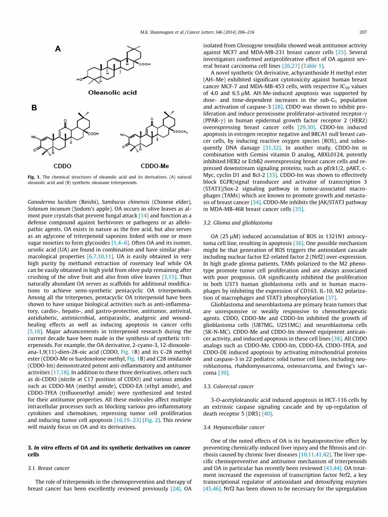
Oleanolic acid (OA, 3b-hydroxyolean-12-en-28-oic acid)(Fig. 1A) is a bioactive pentacyclic triterpenoid belonging to thefamily Oleaceae and has been isolated from more than 1600 plantspecies, the majority of them are edible plants and medicinal herbs[5,10,11]. OA is abundant in ginseng root [12] and in olive plant(Olea europaea) from which the compound derives its name [13].The olive plant is the primary commercial source for the compoundbut other sources include Arctostaphyllos uva-ursi (Bearberry), Cal-luna vulgaris (Heather), Crataeva nurvala (Three leaved caper)
Fig. 1. The chemical structures of oleanolic acid and its derivatives. (A) naturaloleanolic acid and (B) synthetic oleanane triterpenoids.
M.K. Shanmugam et al. / Cancer Letters 346 (2014) 206–216 207
Ganoderma lucidum (Reishi), Sambucus chinensis (Chinese elder),Solanum incanum (Sodom’s apple). OA occurs in olive leaves as al-most pure crystals that prevent fungal attack [14] and function as adefense compound against herbivores or pathogens or as allelo-pathic agents. OA exists in nature as the free acid, but also servesas an aglycone of triterpenoid saponins linked with one or moresugar moieties to form glycosides [1,4–6]. Often OA and its isomer,ursolic acid (UA) are found in combination and have similar phar-macological properties [6,7,10,11]. UA is easily obtained in veryhigh purity by methanol extraction of rosemary leaf while OAcan be easily obtained in high yield from olive pulp remaining aftercrushing of the olive fruit and also from olive leaves [3,15]. Thusnaturally abundant OA serves as scaffolds for additional modifica-tions to achieve semi-synthetic pentacyclic OA triterpenoids.Among all the triterpenes, pentacyclic OA triterpenoid have beenshown to have unique biological activities such as anti-inflamma-tory, cardio-, hepato-, and gastro-protective, antitumor, antiviral,antidiabetic, antimicrobial, antiparasitic, analgesic and wound-healing effects as well as inducing apoptosis in cancer cells[5,16]. Major advancements in triterpenoid research during thecurrent decade have been made in the synthesis of synthetic trit-erpenoids. For example, the OA derivative, 2-cyano-3, 12-dioxoole-ana-1,9(11)-dien-28-oic acid (CDDO, Fig. 1B) and its C-28 methylester (CDDO-Me or bardoxolone methyl, Fig. 1B) and C28 imidazole(CDDO-Im) demonstrated potent anti-inflammatory and antitumoractivities [17,18]. In addition to these three derivatives, others suchas di-CDDO (nitrile at C17 position of CDDO) and various amidessuch as CDDO-MA (methyl amide), CDDO-EA (ethyl amide), andCDDO-TFEA (trifluoroethyl amide) were synthesized and testedfor their antitumor properties. All these molecules affect multipleintracellular processes such as blocking various pro-inflammatorycytokines and chemokines, repressing tumor cell proliferationand inducing tumor cell apoptosis [16,19–23] (Fig. 2). This reviewwill mainly focus on OA and its derivatives.
3. In vitro effects of OA and its synthetic derivatives on cancercells
3.1. Breast cancer
The role of triterpenoids in the chemoprevention and therapy ofbreast cancer has been excellently reviewed previously [24]. OA
isolated from Glossogyne tenuifolia showed weak antitumor activityagainst MCF7 and MDA-MB-231 breast cancer cells [25]. Severalinvestigators confirmed antiproliferative effect of OA against sev-eral breast carcinoma cell lines [26,27] (Table 1).
A novel synthetic OA derivative, achyranthoside H methyl ester(AH–Me) exhibited significant cytotoxicity against human breastcancer MCF-7 and MDA-MB-453 cells, with respective IC50 valuesof 4.0 and 6.5 lM. AH-Me-induced apoptosis was supported bydose- and time-dependent increases in the sub-G1 populationand activation of caspase-3 [28]. CDDO was shown to inhibit pro-liferation and induce peroxisome proliferator-activated receptor-c(PPAR-c) in human epidermal growth factor receptor 2 (HER2)overexpressing breast cancer cells [29,30]. CDDO-Im inducedapoptosis in estrogen receptor negative and BRCA1 null breast can-cer cells, by inducing reactive oxygen species (ROS), and subse-quently DNA damage [31,32]. In another study, CDDO-Im incombination with Gemini vitamin D analog, ABXL0124, potentlyinhibited HER2 or ErbB2 overexpressing breast cancer cells and re-pressed downstream signaling proteins, such as pErk1/2, pAKT, c-Myc, cyclin D1 and Bcl-2 [33]. CDDO-Im was shown to effectivelyblock EGFR/signal transducer and activator of transcription 3(STAT3)/Sox-2 signaling pathway in tumor-associated macro-phages (TAMs) which are known to promote growth and metasta-sis of breast cancer [34]. CDDO-Me inhibits the JAK/STAT3 pathwayin MDA-MB-468 breast cancer cells [35].
3.2. Glioma and glioblastoma
OA (25 lM) induced accumulation of ROS in 1321N1 astrocy-toma cell line, resulting in apoptosis [36]. One possible mechanismmight be that generation of ROS triggers the antioxidant cascadeincluding nuclear factor E2-related factor 2 (Nrf2) over-expression.In high grade glioma patients, TAMs polarized to the M2 pheno-type promote tumor cell proliferation and are always associatedwith poor prognosis. OA significantly inhibited the proliferationin both U373 human glioblastoma cells and in human macro-phages by inhibiting the expression of CD163, IL-10, M2 polariza-tion of macrophages and STAT3 phosphorylation [37].
Glioblastoma and neuroblastoma are primary brain tumors thatare unresponsive or weakly responsive to chemotherapeuticagents. CDDO, CDDO-Me and CDDO-Im inhibited the growth ofglioblastoma cells (U87MG, U251MG) and neuroblastoma cells(SK-N-MC). CDDO-Me and CDDO-Im showed equipotent antican-cer activity, and induced apoptosis in these cell lines [38]. All CDDOanalogs such as CDDO-Me, CDDO-Im, CDDO-EA, CDDO-TFEA, andCDDO-DE induced apoptosis by activating mitochondrial proteinsand caspase-3 in 22 pediatric solid tumor cell lines, including neu-roblastoma, rhabdomyosarcoma, osteosarcoma, and Ewing’s sar-coma [39].
3.3. Colorectal cancer
3-O-acetyloleanolic acid induced apoptosis in HCT-116 cells byan extrinsic caspase signaling cascade and by up-regulation ofdeath receptor 5 (DR5) [40].
3.4. Hepatocellular cancer
One of the noted effects of OA is its hepatoprotective effect bypreventing chemically-induced liver injury and the fibrosis and cir-rhosis caused by chronic liver diseases [10,11,41,42]. The liver spe-cific chemopreventive and antitumor mechanism of triterpenoidsand OA in particular has recently been reviewed [43,44]. OA treat-ment increased the expression of transcription factor Nrf2, a keytranscriptional regulator of antioxidant and detoxifying enzymes[45,46]. Nrf2 has been shown to be necessary for the upregulation
P P
P
P P
P
P
P
P
P
P
P P
P
P
STAT3
TNFIL6
STAT3STAT3
STAT3
TRAIL
Ser 32Ser 36
RelA p50
IκB RelA p50
IκB
RelA p50 RelA p50
Apaf-1
Caspase-9 Caspase-3
Cytochrome c
Caspase-8 (Caspase-10)
PI3K /PI3K
Akt
mTOR
NF-κB
p
p
STAT3STAT3
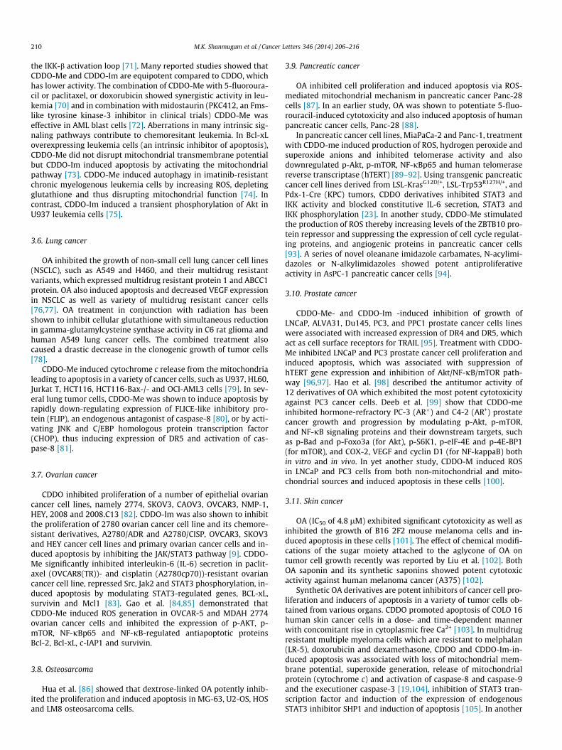
Fig. 2. Major inflammation-associated signaling pathways inhibited by synthetic derivatives of oleanolic acid. These pathways include NF-jB, STAT3, TRAIL signaltransduction pathways and the Keap1/Nrf2/ARE activation cascade that have been shown to be modulated both in vitro and in vivo.
208 M.K. Shanmugam et al. / Cancer Letters 346 (2014) 206–216
of genes involved in oxidative stress, such as glutathione S-trans-ferase or superoxide dismutase-containing antioxidant responseelement (ARE) [47]. In a recent study, it was shown that OA bindsto the ligand-binding domain of the farnesoid X receptor (FXR), aligand-regulated transcription factor that regulates the biosynthe-sis of bile acid and its excretion from liver cells [48] and modulatesthe expression of FXR target genes, such as CYP7A1 [49]. Hence apart of Nrf2-mediated hepatoprotective effect of OA may be partlymediated through FXR and by inhibiting NF-jB activation path-way. OA is also reported to have anti-inflammatory and anticancereffects [8,9]. OA was found to induce cell cycle arrest by modulat-ing ERK-p53 mediated cell cycle arrest and induced apoptosis inHCC cells via the mitochondrial pathway [50]. OA induced apopto-sis by modulating the mitochondrial pathway and down regulatingXIAP in HuH7 hepatocellular carcinoma cells [51].
A series of furoxan and glycosyl-based nitric oxide releasingderivatives of OA have been reported to have potent anticanceractivity against HCC cell lines [52–54]. In another series of O(2)-glycosylated diazeniumdiolate-based derivatives of OA were syn-thesized and evaluated for their anti-HCC activity. In this series,one particular compound 6, (O2-b-d-Galactopyranosyl 1-4-[(12-en-28-b-d-glucopyranosyloleanolate-3-yl-oxy)-succinyl-oxy]piperidin-1-yldiazen-1-ium-1,2-diolate) induced HCC cell apopto-sis, characterized by a decrease in mitochondrial membrane poten-tials and Bcl-2 expression, with greater cytochrome c release, Bax,caspase-3 and -9 expression in HCC cells [55,56]. Mallavadhaniet al. [57] synthesized a series of 17 OA C-17 ester chains consistingof olefinic, acetoacetyl, and bromoalkyl compounds and tested forits antiproliferative activity against SiHa and HeLa (cervix), A-549(lung), and IMR-32 (neuroblastoma) cancer cell lines. However,
all these compounds showed similar activity as their parent com-pound [57]. OA with azaheterocyclic groups at the 2, 3 positionof the A-ring was shown to have cytotoxic activity against HCCline, BEL-7404 cells and induced apoptosis through down-regula-tion of Bcl-2 and mitochondrial membrane potential, releasingcytochrome c, and upregulation of Bax and caspase-3 [58]. A novelPABA/NO derivative of OA was shown to have significant and selec-tive activity against HepG2 cells and induced apoptosis by modu-lating ROS/MAPK-mediated mitochondrial pathway [59]. OAderivative, with 1-en-2-cyano-3-oxo in ring A and a nitro groupat C-17, was shown to be important for its cytotoxicity againstHepG2 and Col-02 cells [60]. In the liver, most of the syntheticCDDO analogs also protect against toxic insults such as acetamino-phen, aflatoxin, concanavalin A, or cisplatin and against injury fromischemia by up-regulating the Nrf2/ARE pathway [16,20].
3.5. Hematological malignancies
OA (80 lM) induced apoptosis in HL60 cells via activation ofcaspase-9 and caspase-3 and induced cleavage of poly(ADP-ribose)polymerase [61].
Synthetic OA derivatives inhibited proliferation and inducedapoptosis in vitro in a wide variety of human tumor cells includingleukemia cells. Olean-12-Eno[2,3-c] [1,2,5] oxadiazol-28-oic acid(OEOA), synthetic derivative of OA, induced G1 cell cycle arrest aswell as differentiation in human leukemia cell lines, K562, HELand JURKAT [62]. Three new active oleanolic vinyl bornates inhib-ited the growth of leukemia cells (Jurkat and K562) and Burkitt’slymphoma cells (Jijoye) without concomitant inhibition of non-tu-moral human fibroblasts [63]. CDDO primarily activated the
Table 1In vitro anticancer effects of natural and synthetic oleanolic acid.
Molecules Biological effects References
Breast cancerOA Inhibits proliferation of human breast cancer cell lines MCF-7 and MDA-MB-231 [26]OA Induces apoptosis in MCF7, T47D, SKBR3 cells and in tomoxifen resistant MCF7 cells [27]AH-Me Induces apoptosis in MCF7 and MDA-MB 435 cells [28]CDDO Inhibits proliferation and induce PPARc in HER2 overexpressing cells [29,30]CDDO-Me Inhibits JAK/STAT pathway in MDA-MB-468 cells [35]CDDO-Im Induces apoptosis in estrogen receptor negative and in BRCA1 null cells; Inhibits EGFR/STAT3/Sox-2
pathway[31–34]
Glioma and glioblastomaOA Induces apoptosis of astrocytoma cells [36]
Inhibits proliferation of U373 glioblastoma cells and suppresses STAT3 phosphorylation [37]CDDO; CDDO-Me; CDDO-Im Inhibits proliferation of glioblastoma cells U87MG and U251MG and neuroblastoma cells (SK-N-MC) [38]All CDDO analogs Induces apoptosis in 22 pediatric solid tumor cells [39]
Hepatocellular cancerOA Activates Keap1-Nrf1-ARE pathway; Exhibits hepatoprotective and chemopreventive effects [16,20,41–44]Furoxan- and glycosyl- based NO releasing
OAInduces apoptosis in HCC cells [50–52]
O(2)-glycosylated diazeniumdiolate-OA Decreases mitochondrial membrane potential and induces apoptosis in HCC cells [55,56]Azaheterocyclic-OA Exerts cytotoxic activity in BEL-7404 cell line [58]PABA/NO-OA Modulates ROS/MAPK pathway [59]
Leukemia and lymphomaOEOA Induces G1 cell cycle arrest and differentiation in K562 HEL and JURKAT cells [62]OA vinyl boronates Inhibits proliferation of JURKAT, K562 and Jijoye cells [63]CDDO; CDDO-Me; CDDO-Im Induces apoptosis in myeloid leukemia cells, pediatric acute lymphoblastic leukemia, MOLT-4, chronic
lymphocytic leukemia, U937, HL-60, in diffuse large B-cell lymphoma cells;[64–68,74]
Inhibits NF-jB activation in U-937 cells and induces autophagy in imatinib resistant chronicmyelogenous leukemia cells
Lung cancerOA Induces apoptosis of normal and multidrug resistant non-small cell lung cancer cell line [77]CDDO-Me Induces apoptosis by modulation of mitochondrial membrane potential in lung cancer cells [79–81]
Ovarian cancerCDDO Inhibits proliferation of 2774, SKOV3, CAOV3, OVCAR3, NMP-1, HEY, 2008 and 2008.C13 ovarian cancer
cells[82]
CDDO-Im Induces apoptosis and inhibits JAK/STAT activation in normal 2780, SKOV3, OVCAR3, HEY and multidrugresistant variants
[9]
CDDO-Me Inhibits IL-6 secretion, STAT3-regulated gene products in paclitaxel and cisplatin resistant cells and inOVCAR-5 and MDAH2774 cells
[83–85]
Osteosarcoma cancerDextrose-OA Induces apoptosis in MG-63, U2-OS, HOS and LM8 cells [86]Pancreatic cancerOA Combination with 5-fluorouracil exhibits synergistic cytotoxic effect in Panc-28 cells [88]CDDO-Me Inhibits NF-jB in MiaPaCa-2 and Panc1 cells; Inhibits IL6 secretion and STAT3 phosphorylation in
acylimidazoles; N-alkylimidazolesInhibits proliferation in AsPC-1 cells [94]
Prostate cancerCDDO-Me; CDDO-Im Increases DR4 and DR5 expression; Inhibits the growth of LNCaP, ALVA31, PC3 and PPC1 cells [95–97,99]
Skin cancerOA Induces apoptosis of B16F2 melanoma cells [101]CDDO Increases cytoplasmic fee calcium and induces apoptosis of COLO16 cells [103]CDDO-Im Induces apoptosis of melphalan, dexamethasone and doxorubicin resistant cells; Inhibits STAT3
phosphorylation[104,105]
M.K. Shanmugam et al. / Cancer Letters 346 (2014) 206–216 209
extrinsic apoptotic pathway in myeloid leukemia cells [64]. In an-other study, CDDO, CDDO-Me and CDDO-Im suppressed thegrowth of pediatric acute lymphoblastic leukemia. The observedcytotoxicity was independent of induced ceramide synthesis inMOLT-4 cells [65]. CDDO and CDDO-Im also displayed antitumoractivity against chronic lymphocytic leukemia (CLL) derived frompatients and in a mouse model of CLL and small B cell lymphoma(SBL). In in vitro studies, these triterpenoids induced apoptosis ofCLL cells [66]. When CDDO was compared to several PPAR-c li-gands, including BRL49653 (rosiglitazone) and 15-deoxy-Delta12,14-prostaglandin J(2), in leukemia (U937 and HL-60) and lym-phoid cells (Su-DHL, Sup-M2, Ramos, Raji, Hodgkin’s cells, and pri-mary CLL), CDDO-induced differentiation and apoptosis was of
greater potency when compared to PPAR-c ligand-induced apopto-sis, and it was characterized by loss of mitochondrial membranepotential and caspase activation [67]. Similar results were reportedin human diffuse large B-cell lymphoma (DLBCL) [68]. In anotherstudy, Lon protease inhibition was shown to mediate CDDO-in-duced B-lymphoid cell apoptosis, a novel anticancer drug target[69]. Shishodia et al. [70] reported that CDDO-Me inhibited humanleukemia cell proliferation, inhibited constitutive and inducibleNF-jB activation, and NF-jB-regulated gene products, such as vas-cular endothelial growth factor (VEGF), cyclooxygenase-2 (COX-2),and matrix metalloproteinase-9. In human U-937 myeloid leuke-mia cells, CDDO and CDDO-Me directly blocked IKK-b activityand thereby the NF-jB pathway by interacting with Cys-179 in
210 M.K. Shanmugam et al. / Cancer Letters 346 (2014) 206–216
the IKK-b activation loop [71]. Many reported studies showed thatCDDO-Me and CDDO-Im are equipotent compared to CDDO, whichhas lower activity. The combination of CDDO-Me with 5-fluoroura-cil or paclitaxel, or doxorubicin showed synergistic activity in leu-kemia [70] and in combination with midostaurin (PKC412, an Fms-like tyrosine kinase-3 inhibitor in clinical trials) CDDO-Me waseffective in AML blast cells [72]. Aberrations in many intrinsic sig-naling pathways contribute to chemoresitant leukemia. In Bcl-xLoverexpressing leukemia cells (an intrinsic inhibitor of apoptosis),CDDO-Me did not disrupt mitochondrial transmembrane potentialbut CDDO-Im induced apoptosis by activating the mitochondrialpathway [73]. CDDO-Me induced autophagy in imatinib-resistantchronic myelogenous leukemia cells by increasing ROS, depletingglutathione and thus disrupting mitochondrial function [74]. Incontrast, CDDO-Im induced a transient phosphorylation of Akt inU937 leukemia cells [75].
3.6. Lung cancer
OA inhibited the growth of non-small cell lung cancer cell lines(NSCLC), such as A549 and H460, and their multidrug resistantvariants, which expressed multidrug resistant protein 1 and ABCC1protein. OA also induced apoptosis and decreased VEGF expressionin NSCLC as well as variety of multidrug resistant cancer cells[76,77]. OA treatment in conjunction with radiation has beenshown to inhibit cellular glutathione with simultaneous reductionin gamma-glutamylcysteine synthase activity in C6 rat glioma andhuman A549 lung cancer cells. The combined treatment alsocaused a drastic decrease in the clonogenic growth of tumor cells[78].
CDDO-Me induced cytochrome c release from the mitochondrialeading to apoptosis in a variety of cancer cells, such as U937, HL60,Jurkat T, HCT116, HCT116-Bax-/- and OCI-AML3 cells [79]. In sev-eral lung tumor cells, CDDO-Me was shown to induce apoptosis byrapidly down-regulating expression of FLICE-like inhibitory pro-tein (FLIP), an endogenous antagonist of caspase-8 [80], or by acti-vating JNK and C/EBP homologous protein transcription factor(CHOP), thus inducing expression of DR5 and activation of cas-pase-8 [81].
3.7. Ovarian cancer
CDDO inhibited proliferation of a number of epithelial ovariancancer cell lines, namely 2774, SKOV3, CAOV3, OVCAR3, NMP-1,HEY, 2008 and 2008.C13 [82]. CDDO-Im was also shown to inhibitthe proliferation of 2780 ovarian cancer cell line and its chemore-sistant derivatives, A2780/ADR and A2780/CISP, OVCAR3, SKOV3and HEY cancer cell lines and primary ovarian cancer cells and in-duced apoptosis by inhibiting the JAK/STAT3 pathway [9]. CDDO-Me significantly inhibited interleukin-6 (IL-6) secretion in paclit-axel (OVCAR8(TR))- and cisplatin (A2780cp70))-resistant ovariancancer cell line, repressed Src, Jak2 and STAT3 phosphorylation, in-duced apoptosis by modulating STAT3-regulated genes, BCL-xL,survivin and Mcl1 [83]. Gao et al. [84,85] demonstrated thatCDDO-Me induced ROS generation in OVCAR-5 and MDAH 2774ovarian cancer cells and inhibited the expression of p-AKT, p-mTOR, NF-jBp65 and NF-jB-regulated antiapoptotic proteinsBcl-2, Bcl-xL, c-IAP1 and survivin.
3.8. Osteosarcoma
Hua et al. [86] showed that dextrose-linked OA potently inhib-ited the proliferation and induced apoptosis in MG-63, U2-OS, HOSand LM8 osteosarcoma cells.
3.9. Pancreatic cancer
OA inhibited cell proliferation and induced apoptosis via ROS-mediated mitochondrial mechanism in pancreatic cancer Panc-28cells [87]. In an earlier study, OA was shown to potentiate 5-fluo-rouracil-induced cytotoxicity and also induced apoptosis of humanpancreatic cancer cells, Panc-28 [88].
In pancreatic cancer cell lines, MiaPaCa-2 and Panc-1, treatmentwith CDDO-me induced production of ROS, hydrogen peroxide andsuperoxide anions and inhibited telomerase activity and alsodownregulated p-Akt, p-mTOR, NF-jBp65 and human telomerasereverse transcriptase (hTERT) [89–92]. Using transgenic pancreaticcancer cell lines derived from LSL-KrasG12D/+, LSL-Trp53R127H/+, andPdx-1-Cre (KPC) tumors, CDDO derivatives inhibited STAT3 andIKK activity and blocked constitutive IL-6 secretion, STAT3 andIKK phosphorylation [23]. In another study, CDDO-Me stimulatedthe production of ROS thereby increasing levels of the ZBTB10 pro-tein repressor and suppressing the expression of cell cycle regulat-ing proteins, and angiogenic proteins in pancreatic cancer cells[93]. A series of novel oleanane imidazole carbamates, N-acylimi-dazoles or N-alkylimidazoles showed potent antiproliferativeactivity in AsPC-1 pancreatic cancer cells [94].
3.10. Prostate cancer
CDDO-Me- and CDDO-Im -induced inhibition of growth ofLNCaP, ALVA31, Du145, PC3, and PPC1 prostate cancer cells lineswere associated with increased expression of DR4 and DR5, whichact as cell surface receptors for TRAIL [95]. Treatment with CDDO-Me inhibited LNCaP and PC3 prostate cancer cell proliferation andinduced apoptosis, which was associated with suppression ofhTERT gene expression and inhibition of Akt/NF-jB/mTOR path-way [96,97]. Hao et al. [98] described the antitumor activity of12 derivatives of OA which exhibited the most potent cytotoxicityagainst PC3 cancer cells. Deeb et al. [99] show that CDDO-meinhibited hormone-refractory PC-3 (AR�) and C4-2 (AR+) prostatecancer growth and progression by modulating p-Akt, p-mTOR,and NF-jB signaling proteins and their downstream targets, suchas p-Bad and p-Foxo3a (for Akt), p-S6K1, p-eIF-4E and p-4E-BP1(for mTOR), and COX-2, VEGF and cyclin D1 (for NF-kappaB) bothin vitro and in vivo. In yet another study, CDDO-M induced ROSin LNCaP and PC3 cells from both non-mitochondrial and mito-chondrial sources and induced apoptosis in these cells [100].
3.11. Skin cancer
OA (IC50 of 4.8 lM) exhibited significant cytotoxicity as well asinhibited the growth of B16 2F2 mouse melanoma cells and in-duced apoptosis in these cells [101]. The effect of chemical modifi-cations of the sugar moiety attached to the aglycone of OA ontumor cell growth recently was reported by Liu et al. [102]. BothOA saponin and its synthetic saponins showed potent cytotoxicactivity against human melanoma cancer (A375) [102].
Synthetic OA derivatives are potent inhibitors of cancer cell pro-liferation and inducers of apoptosis in a variety of tumor cells ob-tained from various organs. CDDO promoted apoptosis of COLO 16human skin cancer cells in a dose- and time-dependent mannerwith concomitant rise in cytoplasmic free Ca2+ [103]. In multidrugresistant multiple myeloma cells which are resistant to melphalan(LR-5), doxorubicin and dexamethasone, CDDO and CDDO-Im-in-duced apoptosis was associated with loss of mitochondrial mem-brane potential, superoxide generation, release of mitochondrialprotein (cytochrome c) and activation of caspase-8 and caspase-9and the executioner caspase-3 [19,104], inhibition of STAT3 tran-scription factor and induction of the expression of endogenousSTAT3 inhibitor SHP1 and induction of apoptosis [105]. In another
M.K. Shanmugam et al. / Cancer Letters 346 (2014) 206–216 211
recent study, it was demonstrated that CDDO-Im and CDDO-Mecan protect human keratinocytes against toxicity from the sulfurmustard analog, 2-chloroethyl ethyl sulfide, by inducing the syn-thesis of glutathione, which is depleted by sulfur mustard [106].
4. In vivo antitumor activity of OA and its synthetic derivatives
In several reports, OA displayed potent in vitro inhibitory activ-ity against tumor cell proliferation and also powerful induction ofapoptosis. However, to determine the in vivo bioactivity, OA wastested in several rodent models of organ-specific cancer (Table 2).
4.1. Hepatocellular carcinoma
In a liver cancer model, OA inhibited HCC tumors in Balb/C mice[50]. In this study, mice were randomly divided into three groups:control, low dose of OA and high dose of OA. In OA-treated groups,mice were administered with 75 or 150 mg/kg/day OA intraperito-neally respectively for 3 weeks. OA significantly inhibited thegrowth of HCC tumors [50]. Intraperitoneal administration of OAshowed a LD50 of 1500 mg/ml in mice and a single subcutaneousdose of 1000 mg/ml caused no toxic effects in rats [107].
Synthetic OA derivatives have been used to treat established tu-mors in experimental animal models. Furoxan- and glycosyl-basednitric oxide releasing OA derivatives displayed low acute toxicity inmice while significantly inhibiting the growth of HCC tumorsin vivo [53,54]. A series of O(2)-glycosylated diazeniumdiolate-based OA derivatives were tested for their anti-HCC activity. Com-pound 6 in this series exhibited low acute toxicity (LD50 = 173.3 -mg/kg) and potently inhibited HCC tumor growth in mice (3 mg/kg, iv) [55]. A novel PABA/NO OA derivative showed potent antitu-mor activity and significantly reduced tumor volume and tumorweight in a H22 solid tumor model [59]. CDDO-Im when adminis-tered for 8 weeks was shown to reduce metastasized tumor burdenin the liver after intravenous inoculation of tumor cells [108]. Inaddition, short-and long-term clinical trials using OA for acuteand chronic hepatitis, respectively, demonstrated the safety of thiscompound [10].
4.2. Breast carcinoma
A novel synthetic oleanane triterpenoid (methyl-25-hydroxy-3-oxoolean-12-en-28-oate, AMR-Me) when administered orally atdoses of 0.8, 1.2 or 1.6 mg/kg, three times a week for eighteenweeks inhibited the growth of 7,12-dimethylbenz(a)antracene(DMBA)-initiated mammary carcinoma in rats [109]. AMR-Medownregulated the expression of estrogen receptor-a (ER-a), ER-b and cyclin D1 and diminished Wnt/b-catenin signaling duringDMBA mammary tumorigenesis in rats [110]. Very recently, ithas been shown that AMR-Me downregulated the expression ofCOX-2 and heat shock protein 90 (HSP90), suppressed the degrada-tion of inhibitory jB-a (IjB-a) and reduced the translocation ofNF-jB from cytosol to nucleus in DMBA-induced mammary tu-mors in rats [111].
In female BALB/c or FVB/NJ mice orthotopically implanted withbreast tumor cells (4TO7 or MMTVB-neu), CDDO-Im formulated asnanoparticles when combined with HER-2 DNA vaccine producedsignificant antitumor activity and was associated with parallelreduction in the production of pro-inflammatory cytokines suchas transforming growth factor-b, IL-6 and IL-10 and enhanced tu-mor-specific cytotoxic T-lymphocyte response [112]. When fedwith CDDO-Me mixed with diet beginning at 10 weeks of age, sig-nificantly delayed mammary tumor growth by over 3 months in amouse transgenic model with overexpressing MMTV-neu (ErbB2/HER2) receptor tyrosine kinase [113]. In another breast cancer
mouse model with deletion of BRCA1 gene and a single allelemutation in p53 tumor suppressor, CDDO-Me diet increased life-span of mice by 5 weeks compared to control mice [114] and in-duced tumor growth arrest in MDA-MB-435 ER, MDA-MB-468 ERand MCF7 ER xenograft breast cancer mouse models[29,112,115]. Intraperitoneal injection of CDDO-Me nanoparticlesshown to inhibit invasion and metastasis to lungs in a spontane-ously developing mammary tumor derived from chemoresistant4T1 breast cancer cells subcutaneously implanted in Balb/c mice[30]. In a recent report, potent chemopreventive activity was ob-served when CDDO-Im was administered orally in combinationwith BXL0124 (Gemini vitamin D analog) in MMTV-ErbB2/neumice [33]. In another model of ER-negative breast cancer inMMTV- polyoma middle T (PyMT) mice fed with CDDO-Me(50 mg/kg diet) at starting 4 weeks of age, CDDO-Me significantlyincreased the overall survival by 5.2 weeks [116].
4.3. Colon carcinoma
Chemopreventive activity of OA was observed in rats subjectedto chemical carcinogenesis. Oral treatment of rats with OA (25 mg/kg body weight) prevented 1,2-dimethylhydrazine-induced coloncarcinoma [117,118].
4.4. Prostate carcinoma
CDDO and CDDO-Me prevented the progression of prostate can-cer in the TRAMP mice model [119]. In addition, inhibition of pro-gression of pre-neoplastic lesions (i.e., low and high-grade prostateintraepithelial neoplasms) to adenocarcinoma of the prostate byCDDO-Me in TRAMP mice was associated with significant decreasein TERT and its regulatory proteins in the prostate gland. Thesedata provide evidence that telomerase is a potential target ofCDDO-Me for the prevention and treatment of prostate cancer[96,97].
4.5. Leukemia and lymphoma
Leukemia cells seem to be especially sensitive to triterpenoids.Liposome-formulated CDDO or CDDO-Im triterpenoids reducedleukemia and lymphoma growth in vivo in a TRAF2DN/Bcl-2 trans-genic mouse model of chronic lymphocytic leukemia and small B-cell lymphoma. CDDO-Im was more potent than CDDO and in-duced apoptosis of circulating B cells by 60 to 90% [66]. In anotherstudy, CDDO-Im was shown to be more potent than CDDO in bothB16 mice melanoma tumors in BDF1 mice and L1210 murine leu-kemia. CDDO-Im was injected intraperitonially twice a day for7 days, at doses of 50, 100, and 200 mg/kg. CDDO-Im induced sig-nificant dose-dependent decrease in tumor burden [120]. Combi-nation of all-trans retinoic acid and CDDO-Me significantlyimproved survival in a syngeneic mouse model of acute promyelo-cytic leukemia [121].
4.6. Melanoma
OA was shown to inhibit mouse skin tumor promotion by 12-O-tetradecanoylphorbol-13-acetate (TPA) [122]. Furthermore, OA de-creased the development of melanoma-induced lung metastasis[76]. OA when administered at doses of 5 or 10 mg/kg/day de-creased pulmonary metastasis on day 18 in groups of mice injectedintravenously with B16F10 melanoma cells [76].
In SKH1 hairless mice, Di-CDDO (10 nM, twice/week for17 weeks) applied topically to the skin of mice significantly de-creased the incidence of skin tumors induced by chronic low-levelUBV radiation [123].
Table 2In vivo antitumor activities of natural and synthetic oleanolic acid.
Molecules Tumor model Dose References
OA Inhibits TPA-induced skin tumors in mice 2.5, 5 or 10 lmol/0.2 ml/mouse;30 minpretreatment
[122]
Inhibits HCC tumors in Balb/c mice 75 or 150 mg/day, i.p.; 3 weeks [50]Inhibits 1,2-dimethyhydrazine induced colon carcinoma in rats 25 mg/kg, oral; 4 weeks [117]Inhibits melanoma-induced lung metastasis 5 mg/kg/day or 10 mg/kg/day, i.v.; 18 days [76]
CDDO Inhibits leukemia and lymphoma growth in TRAF2DN/Bcl2 transgenicmodel of CLL and SBL
5, 10 or 20 mg/kg, nine administrations; 21-25 days
[66]
CDDO-Me Inhibits breast cancer growth in a MMTV-neu (ErbB2/HER2) transgenicmouse model
60 or 100 mg/kg, diet; 45 weeks [113]
Inhibits breast cancer growth in BRCA1 null mice model 50 mg/kg, diet; 18–24 weeks [114]Inhibits MDA-MB-435 ER, MDA-MB-468 ER and MCF7 ER xenografts inmice
20 mg/kg/mouse/day, i.v., thrice weekly;3 weeks
[29,112,115]
Inhibits ER-negative breast cancer in PyMT mice 50 mg/kg, diet; various time points [116]Inhibits the progression of prostate cancer in transgenic TRAMP micemodel
7.5 mg/kg, oral, 5 days/week; 20 weeks [119]
Significantly improves survival in a syngenic acute promyelocyticleukemia mice model
5 mg/kg; 2 day intervals for 23 days [121]
Inhibits pancreatic tumor growth in a xenograft mouse model 7.5 mg/kg/day, oral; 4 weeks [93]Inhibits pancreatic tumor growth in a transgenic mouse model ofpancreatic cancer
60 mg/kg, diet; variable time points [23]
Inhibits lung carcinogenesis in A/J mouse model CDDO-Me 60 mg/kg, diet; CDDO-EA 400 mg/kg, diet; 15 weeks
[21,124]
CDDO-Me nanoparticles Inhibits subcutaneously implanted 4T1 breast cancer growth in Balb/cmice
2 days interval, 5 injections [30]
CDDO-Im Inhibits hepatocellular carcinoma and reduces metastatic tumorburden
800 mg/kg, diet; 5 days/week for 56 days [33,108]
Exerts chemopreventive effect when administered in combinationwith BXL0124 to MMTV-ErbB2/neu mice
BXL0124 (0.3 lg/kg), oral, for 56 weeks;(3 lmol/kg); 6 times/week for 3 weeks
[33]
Inhibits melanoma tumors in BDF1 mice and L1201 murine leukemia 50, 100 or 200 mg/kg, i.p.; twice a day for7 days
[120]
CDDO-Im nanoparticles Inhibits orthotopically implanted breast tumor cells (4TO7 or MMTVB-neu) in female Balb/c nude mice or FVB/NJ mice
200 ml PBS (1.36 � 1013 particles), i.v.; 8 Timesin 46 days
[112]
Inhibits leukemia and lymphoma growth in TRAF2DN/Bcl2 transgenicmodel of CLL and SBL
5, 10 or 20 mg/kg, i.p.; 21–25 days [66]
Dicyano-CDDO Inhibits UV-irradiated skin tumor in SKH1 hairless mice 10 nM, topically; Twice/week for 17 weeks [123]
Furoxan- and glycosyl-based OA
Inhibits SMMC-7721 HCC tumors in mice 12.5 or 25 mg/kg, i.p.; 3 times/week for 21 days [53,54]
O(2)-glyco-sylated diaz-eniumdiolate-based OA
Inhibits SMMC-7721 HCC tumor in mice 3 mg/kg, i.v.; 3 times/week for 3 weeks [55]
PABA/NO-based OA Inhibits H22 solid tumors in mice 10, 20 or 40 mg/kg/day, i.p; 14 days [59]
AMR-Me Inhibits DMBA-induced breast tumors in rats and interferes with Wnt/b-catenin and NF-jB signaling
0.8, 1.2 or 1.6 mg/kg, oral; 3 times/week for18 weeks
[109–111]
Dextrose OA Suppresses LM8 osteosarcoma growth and lung metastasis in mice 25, 50 or 100 mg/kg i.p.; 4 weeks [86]
212 M.K. Shanmugam et al. / Cancer Letters 346 (2014) 206–216
4.7. Pancreatic carcinoma
In xenograft models of pancreatic cancer, oral administration ofCDDO-Me (7.5 mg/kg) daily for 4 weeks significantly decreased tu-mor volume and the expression of VEGF, cyclin D1 and survivin[93]. In addition to their efficacy in various xenograft models, OAderivatives also significantly delayed tumor development in trans-genic models. In a transgenic mouse model of pancreatic cancerwith mutations (LSL-KrasG12D/+, LSL-Trp53R127H/+, Pdx-1-Cre)[KPC] synthetic OA derivatives increased survival of KPC mice by3–4 weeks. In this particular experiment, mice were fed powderedcontrol diet or a diet containing the triterpenoids, CDDO-Me(60 mg/kg diet) or CDDO-EA (400 mg/kg diet) or their respectivecombinations [23].
4.8. Lung carcinoma
OA derivatives are also potent inhibitors of lung carcinogenesis.When mixed in diet and fed to A/J mice one week after initiationwith vinyl carbamate, CDDO-Me, CDDO-EA and CDDO-MA signifi-
cantly decreased lung adenocarcinoma tumor burden by 86–96%,compared to controls [124].
4.9. Osteosarcoma
Synthetic OA derivative, dextrose-OA, dose-dependently inhib-ited LM8 osteosarcoma growth in vivo [86]. Dextrose-OA (25, 50and 100 mg/kg body weight) was intraperitoneally administeredfor 4 weeks. At the end of the 4-week treatment, dextrose-OA sig-nificantly inhibited the growth of tumor compared to vehicle con-trol and inhibited metastasis of LM8 osteosarcoma tumor cells tolungs [86].
5. Clinical trials of synthetic OA derivatives
CDDO, a multifunctional molecule with apoptosis-inducingactivity in cancer cells, was evaluated in a phase 1 clinical trial con-ducted by Speranza et al. [125]. In this clinical study, seven pa-tients were enrolled for phase I dose-escalation study to
M.K. Shanmugam et al. / Cancer Letters 346 (2014) 206–216 213
determine toxicity, maximum tolerated dose (MTD), and pharma-cokinetic profiles of CDDO. Following administration of CDDO asa 5-day continuous infusion every 28 days in patients with ad-vanced cancers, this particular compound showed rapid increasein plasma concentration and achieved steady-state plasma levelwithin 48 h. Bardoxolone methyl, a novel synthetic OA triterpe-noid, exhibits potent anti-inflammatory activity and anticanceractivity. Hong et al. [126] evaluated the first-in-human phase Iclinical trial of bardoxolone methyl in patients with advanced solidtumor and lymphoma to delineate the dose-limiting toxicities,MTD, and to characterize its pharmacokinetic and pharmacody-namics parameters. Bardoxolone methyl was administered orallyonce a day for 21 days and showed a MTD of 900 mg/d associatedwith the anti-tumor activity [126]. In an earlier dose escalationstudy with bardoxolone methyl in 34 or 47 patients with advancedrefractory lymphoid solid tumors, bardoxolone methyl was admin-istered orally for 21 days at doses ranging from 5 mg/day or 1.3 g/day and modulated NF-jB, STAT3 and Nrf2 targets in these tumors.Bardoxolone methyl was well tolerated in 91% of patients andshowed minimal toxicity when administered for up to 1 year in aphase 3 trial [16]. In all clinical trials, bardoxolone methyl was rel-atively safe [127].
6. Preclinical and clinical pharmacokinetic studies of OA and itssynthetic derivatives
A highly sensitive HPLC–ES–MS–MS method was developed bySong et al. [128] to determine the bioavailability of OA in healthyChinese male volunteers. Following administration of oral OA cap-sules (40 mg/volunteer, single dose) to 18 male volunteers, themean values of Cmax, Tmax, AUC0–48, AUC0-infinity, t1/2, CL/F, and V/Fwere found to be 12.12 ± 6.84 ng/ml, 5.2 ± 2.9 h,114.34 ± 74.87 ng h/ml, 124.29 ± 106.77 ng h/ml, 8.73 ± 6.11 h,555.3 ± 347.7 l/h, and 3371.1 ± 1990.1 l, respectively [128]. In an-other study, OA (0.5%) mixed in diet was fed to C57BL/6 mice for8 weeks and evaluated for its bioavailability, tissue distribution,and its antioxidant activity. Results from this study showed thatOA was easily detected by HPLC–MS system and its bioavailabilitywas 0.55 lg/ml in mice plasma, 1.7 lg/g in brain tissue, 4.2 lg/g inheart tissue, 10.3 lg/g in liver tissue, 5.5 lg/g in kidney tissue,6.0 lg/g in colon tissue and 3.7 lg/g in bladder tissue [129]. Thedose-independent pharmacokinetic behavior of OA was investi-gated after intravenous and oral administration in rats with thedoses ranging from 0.5–2 and 25–50 mg/kg, respectively [130].Following oral administration, the systemic absorption was extre-mely low (F1=4 was 0.7%). The low oral bioavailability of OA mightbe due to poor gastrointestinal absorption and subsequent hepaticfirst-pass metabolism [15,130]. Different formulations of OA, suchas freeze-dried polyvinylpyrrolidone and sodium caprate OA, in-creased dissolution rate and intestinal permeability when testedin vitro in Caco-2 cells and in vivo in Sprague-Dawley rats, respec-tively [130,131]. Pharmacokinetic parameters of OA and other pen-tacyclic triterpene saponins have been reported in detail includingthe metabolism of OA [132]. Cao et al. [133] synthetized numerouswater-soluble amino acid analogues of OA and tested for their bio-availability. Interestingly, aqueous solubility of OA increased from0.012 lg/ml to 2.5–3.1 lg/ml and absolute oral bioavailability in-creased 2-fold [15,133]. In a recent study, Cao et al. [134] showedthat propylene glycol-linked amino acid/dipeptide diester pro-drugs of OA showed better stability, permeability, affinity, and bio-availability. In order to increase OA bioavailability, sucrose-esterstabilized nanosuspension of OA was synthesized and tested inin vitro cancer cell cultures and in vivo in mice [135]. The investi-gators found that OA nanosuspension bioavailability in A549 hu-man non-small-cell lung cancer cell line was concentration-,
temperature- and time-dependent and the formulation showedexcellent in vivo oral and intravenous bioavailability in rats [135]and in self-nanoemulsified drug delivery systems [136].
7. Conclusions and perspectives
Pentacyclic triterpenoids obtained from natural plant materialshave been shown to inhibit tumor cell proliferation, induce apop-tosis, increase the life span of tumor-bearing mice compared tocontrol group, as well as prevent angiogenesis, invasion and metas-tasis of tumor cells to distant organ sites in preclinical models ofcancer. They also exhibit multi-functionality by targeting multipletumor cell promoting extracellular and intracellular protein targetsand are thus named multifunctional compounds. In this review, wehave highlighted the significance of both natural and synthetic OAderivatives in various organ-based tumor models and discussedthe potential of these compounds in chemoprevention and ther-apy. We have also summarized the reported chemopreventiveand therapeutic efficacy of pentacyclic triterpene OA in transgenic,orthotopic and xenograft tumor models. Indications from bothin vitro and in vivo studies suggest that OA can indeed suppressmultiple molecular targets that play a fundamental role in bothdevelopment and progression of chronic inflammation and cancer.In this decade alone, several synthetic OA derivatives were synthe-sized that exhibited potent antitumor activity both in in vitro andin vivo studies with phase-1 and phase-2 clinical trials reportedfor bardoxolone methyl. Bardoxolone methyl seems promisingwith a good safety profile in human clinical trials. The evidencealso supports the similarity of inhibiting common molecular tar-gets in addition to the novel target proteins that play a pivotal rolein tumor progression. Using several sensitive instruments, OAabsorption, distribution, metabolism and excretion profiles havebeen reported. OA is bioavailable following oral administration inmice and human pharmacokinetic and pharmacodynamics profilesof OA and its synthetic derivatives are also discussed. All thesestudies uphold the traditional use of OA as well as its usefulnessin modern day traditional Chinese medicine clinics. Additionalclinical trials are warranted to bring these exciting molecules toclinical use for the benefit of mankind.
Conflict of Interest
None declared.
Acknowledgments
This research work was supported by grants from the SingaporeMinistry of Health’s National Medical Research Council to GS underits Individual Research Grants Funding scheme. APK was supportedby grants from Singapore Ministry of Education Tier 2 [MOE2012-T2-2-139], Academic Research Fund Tier 1 [R-184-000-228-112]and Cancer Science Institute of Singapore, Experimental Therapeu-tics I Program [Grant R-713-001-011-271]. The research on AMR-Me and breast cancer prevention as presented here was supportedby the award R03CA136014 from the National Cancer Institute/Na-tional Institutes of Health to AB. The content of this article is solelythe responsibility of the authors and does not necessarily representthe official views of the National Cancer Institute or the NationalInstitutes of Health.
References
[1] R.A. Hill, J.D. Connolly, Triterpenoids, Nat. Prod. Rep. 30 (2013) 1028–1065.[2] D.R. Phillips, J.M. Rasbery, B. Bartel, S.P. Matsuda, Biosynthetic diversity in
214 M.K. Shanmugam et al. / Cancer Letters 346 (2014) 206–216
[3] M.N. Laszczyk, Pentacyclic triterpenes of the lupane, oleanane and ursanegroup as tools in cancer therapy, Planta Med. 75 (2009) 1549–1560.
[4] M.K. Shanmugam, A.H. Nguyen, A.P. Kumar, B.K. Tan, G. Sethi, Targetedinhibition of tumor proliferation, survival, and metastasis by pentacyclictriterpenoids: potential role in prevention and therapy of cancer, Cancer Lett.320 (2012) 158–170.
[5] H. Sheng, H. Sun, Synthesis, biology and clinical significance of pentacyclictriterpenes: a multi-target approach to prevention and treatment ofmetabolic and vascular diseases, Nat. Prod. Rep. 28 (2011) 543–593.
[6] M.K. Shanmugam, X. Dai, A.P. Kumar, B.K. Tan, G. Sethi, A. Bishayee, Ursolicacid in cancer prevention and treatment: molecular targets,pharmacokinetics and clinical studies, Biochem. Pharmacol. 85 (2013)1579–1587.
[7] V.R. Yadav, S. Prasad, B. Sung, R. Kannappan, B.B. Aggarwal, Targetinginflammatory pathways by triterpenoids for prevention and treatment ofcancer, Toxins (Basel) 2 (2010) 2428–2466.
[8] P. Dzubak, M. Hajduch, D. Vydra, A. Hustova, M. Kvasnica, D. Biedermann, L.Markova, M. Urban, J. Sarek, Pharmacological activities of naturaltriterpenoids and their therapeutic implications, Nat. Prod. Rep. 23 (2006)394–411.
[9] A. Petronelli, G. Pannitteri, U. Testa, Triterpenoids as new promisinganticancer drugs, Anticancer Drugs 20 (2009) 880–892.
[10] J. Liu, Pharmacology of oleanolic acid and ursolic acid, J. Ethnopharmacol. 49(1995) 57–68.
[11] J. Liu, Oleanolic acid and ursolic acid: research perspectives, J.Ethnopharmacol. 100 (2005) 92–94.
[12] S. Shibata, New natural products and plant drugs with pharmacological andtherapeutical activity, in: H. Wagner, P. Wolff, (Eds.), Springer-Verlag, 1977,pp. 177–196.
[13] J.L. Simonsen, W.C.J. Ross, Hydroxy acids, hydroxy lactones, hydroxyaldehydoacids, hydroxyketo acids and the stereochemistry of the triterpenes, in: TheTerpenes: The Triterpenes and Their Derivatives, Cambridge University Press,Cambridge, 1957.
[14] M.A. Kubo I, Secreted oleanolic acid on the cuticle Olea europaea (Oleaceae); achemical barrier to fungal attack, Cell. Mol. Life Sci. 40 (1984) 937–938.
[15] S. Jager, K. Winkler, U. Pfuller, A. Scheffler, Solubility studies of oleanolic acidand betulinic acid in aqueous solutions and plant extracts of Viscum album L,Planta Med. 73 (2007) 157–162.
[16] K.T. Liby, M.B. Sporn, Synthetic oleanane triterpenoids: multifunctional drugswith a broad range of applications for prevention and treatment of chronicdisease, Pharmacol. Rev. 64 (2012) 972–1003.
[17] N. Suh, Y. Wang, T. Honda, G.W. Gribble, E. Dmitrovsky, W.F. Hickey, R.A.Maue, A.E. Place, D.M. Porter, M.J. Spinella, C.R. Williams, G. Wu, A.J.Dannenberg, K.C. Flanders, J.J. Letterio, D.J. Mangelsdorf, C.F. Nathan, L.Nguyen, W.W. Porter, R.F. Ren, A.B. Roberts, N.S. Roche, K. Subbaramaiah, M.B.Sporn, A novel synthetic oleanane triterpenoid, 2-cyano-3,12-dioxoolean-1,9-dien-28-oic acid, with potent differentiating, antiproliferative, and anti-inflammatory activity, Cancer Res. 59 (1999) 336–341.
[18] T. Honda, B.V. Rounds, L. Bore, H.J. Finlay, F.G. Favaloro Jr., N. Suh, Y. Wang,M.B. Sporn, G.W. Gribble, Synthetic oleanane and ursane triterpenoids withmodified rings A and C: a series of highly active inhibitors of nitric oxideproduction in mouse macrophages, J. Med. Chem. 43 (2000) 4233–4246.
[19] D. Chauhan, G. Li, K. Podar, T. Hideshima, R. Shringarpure, L. Catley, C.Mitsiades, N. Munshi, Y.T. Tai, N. Suh, G.W. Gribble, T. Honda, R. Schlossman,P. Richardson, M.B. Sporn, K.C. Anderson, The bortezomib/proteasomeinhibitor PS-341 and triterpenoid CDDO-Im induce synergistic anti-multiplemyeloma (MM) activity and overcome bortezomib resistance, Blood 103(2004) 3158–3166.
[20] R.K. Thimmulappa, C. Scollick, K. Traore, M. Yates, M.A. Trush, K.T. Liby, M.B.Sporn, M. Yamamoto, T.W. Kensler, S. Biswal, Nrf2-dependent protectionfrom LPS induced inflammatory response and mortality by CDDO-Imidazolide, Biochem. Biophys. Res. Commun. 351 (2006) 883–889.
[21] K. Liby, R. Risingsong, D.B. Royce, C.R. Williams, T. Ma, M.M. Yore, M.B. Sporn,Triterpenoids CDDO-methyl ester or CDDO-ethyl amide and rexinoidsLG100268 or NRX194204 for prevention and treatment of lung cancer inmice, Cancer Prev. Res. (Phila.) 2 (2009) 1050–1058.
[22] J.J. Auletta, J.L. Alabran, B.G. Kim, C.J. Meyer, J.J. Letterio, The synthetictriterpenoid, CDDO-Me, modulates the proinflammatory response to in vivolipopolysaccharide challenge, J. Interferon Cytokine Res. 30 (2010) 497–508.
[23] K.T. Liby, D.B. Royce, R. Risingsong, C.R. Williams, A. Maitra, R.H. Hruban, M.B.Sporn, Synthetic triterpenoids prolong survival in a transgenic mouse modelof pancreatic cancer, Cancer Prev. Res. (Phila.) 3 (2010) 1427–1434.
[24] A. Bishayee, S. Ahmed, N. Brankov, M. Perloff, Triterpenoids as potentialagents for the chemoprevention and therapy of breast cancer, Front. Biosci.(Landmark Ed.) 16 (2011) 980–996.
[25] H.F. Hsu, J.Y. Houng, C.L. Chang, C.C. Wu, F.R. Chang, Y.C. Wu, Antioxidantactivity, cytotoxicity, and DNA information of Glossogyne tenuifolia, J. Agric.Food Chem. 53 (2005) 6117–6125.
[26] Y. Allouche, F. Warleta, M. Campos, C. Sanchez-Quesada, M. Uceda, G. Beltran,J.J. Gaforio, Antioxidant, antiproliferative, and pro-apoptotic capacities ofpentacyclic triterpenes found in the skin of olives on MCF-7 human breastcancer cells and their effects on DNA damage, J. Agric. Food Chem. 59 (2011)121–130.
[27] G. Gu, I. Barone, L. Gelsomino, C. Giordano, D. Bonofiglio, G. Statti, F.Menichini, S. Catalano, S. Ando, Oldenlandia diffusa extracts exertantiproliferative and apoptotic effects on human breast cancer cells
through ERalpha/Sp1-mediated p53 activation, J. Cell. Physiol. 227 (2012)3363–3372.
[28] M. Fukumura, H. Ando, Y. Hirai, K. Toriizuka, Y. Ida, Y. Kuchino,Achyranthoside H methyl ester, a novel oleanolic acid saponin derivativefrom Achyranthes fauriei roots, induces apoptosis in human breast cancerMCF-7 and MDA-MB-453 cells via a caspase activation pathway, J. Nat. Med.63 (2009) 181–188.
[29] M. Konopleva, W. Zhang, Y.X. Shi, T. McQueen, T. Tsao, M. Abdelrahim, M.F.Munsell, M. Johansen, D. Yu, T. Madden, S.H. Safe, M.C. Hung, M. Andreeff,Synthetic triterpenoid 2-cyano-3,12-dioxooleana-1,9-dien-28-oic acidinduces growth arrest in HER2-overexpressing breast cancer cells, Mol.Cancer Ther. 5 (2006) 317–328.
[30] X. Ling, M. Konopleva, Z. Zeng, V. Ruvolo, L.C. Stephens, W. Schober, T.McQueen, M. Dietrich, T.L. Madden, M. Andreeff, The novel triterpenoid C-28methyl ester of 2-cyano-3, 12-dioxoolen-1, 9-dien-28-oic acid inhibitsmetastatic murine breast tumor growth through inactivation of STAT3signaling, Cancer Res. 67 (2007) 4210–4218.
[31] M.L. Hyer, R. Croxton, M. Krajewska, S. Krajewski, C.L. Kress, M. Lu, N. Suh,M.B. Sporn, V.L. Cryns, J.M. Zapata, J.C. Reed, Synthetic triterpenoidscooperate with tumor necrosis factor-related apoptosis-inducing ligand toinduce apoptosis of breast cancer cells, Cancer Res. 65 (2005) 4799–4808.
[32] E.H. Kim, C.X. Deng, M.B. Sporn, K.T. Liby, CDDO-imidazolide induces DNAdamage, G2/M arrest and apoptosis in BRCA1-mutated breast cancer cells,Cancer Prev. Res. (Phila.) 4 (2011) 425–434.
[33] J.Y. So, J.E. Wahler, T. Yoon, A.K. Smolarek, Y. Lin, W.J. Shih, H. Maehr, M.Uskokovic, K.T. Liby, M.B. Sporn, N. Suh, Oral administration of a Geminivitamin D analog, a synthetic triterpenoid and the combination preventsmammary tumorigenesis driven by ErbB2 overexpression, Cancer Prev. Res.(Phila.) 6 (2013) 959–970.
[34] J. Yang, D. Liao, C. Chen, Y. Liu, T.H. Chuang, R. Xiang, D. Markowitz, R.A.Reisfeld, Y. Luo, Tumor-associated macrophages regulate murine breastcancer stem cells through a novel paracrine EGFR/Stat3/Sox-2 signalingpathway, Stem cells 31 (2013) 248–258.
[35] R. Ahmad, D. Raina, C. Meyer, D. Kufe, Triterpenoid CDDO-methyl esterinhibits the Janus-activated kinase-1 (JAK1)signal transducer and activator oftranscription-3 (STAT3) pathway by direct inhibition of JAK1 and STAT3,Cancer Res. 68 (2008) 2920–2926.
[36] R. Martin, J. Carvalho-Tavares, E. Ibeas, M. Hernandez, V. Ruiz-Gutierrez, M.L.Nieto, Acidic triterpenes compromise growth and survival of astrocytoma celllines by regulating reactive oxygen species accumulation, Cancer Res. 67(2007) 3741–3751.
[37] Y. Fujiwara, Y. Komohara, R. Kudo, K. Tsurushima, K. Ohnishi, T. Ikeda, M.Takeya, Oleanolic acid inhibits macrophage differentiation into the M2phenotype and glioblastoma cell proliferation by suppressing the activationof STAT3, Oncol. Rep. 26 (2011) 1533–1537.
[38] X. Gao, D. Deeb, H. Jiang, Y. Liu, S.A. Dulchavsky, S.C. Gautam, Synthetictriterpenoids inhibit growth and induce apoptosis in human glioblastomaand neuroblastoma cells through inhibition of prosurvival Akt, NF-kappaBand Notch1 signaling, J. Neurooncol. 84 (2007) 147–157.
[39] J.L. Alabran, A. Cheuk, K. Liby, M. Sporn, J. Khan, J. Letterio, K.S. Leskov, Humanneuroblastoma cells rapidly enter cell cycle arrest and apoptosis followingexposure to C-28 derivatives of the synthetic triterpenoid CDDO, Cancer Biol.Ther. 7 (2008) 709–717.
[40] K.H. Yoo, J.H. Park, E.J. Cui, K.I. Kim, J.Y. Kim, J. Kim, S.G. Hong, N.I. Baek, I.S.Chung, 3-O-acetyloleanolic acid induces apoptosis in human colon carcinomaHCT-116 cells, Phytother. Res. 26 (2012) 1541–1546.
[41] S.A. Reisman, L.M. Aleksunes, C.D. Klaassen, Oleanolic acid activates Nrf2 andprotects from acetaminophen hepatotoxicity via Nrf2-dependent and Nrf2-independent processes, Biochem. Pharmacol. 77 (2009) 1273–1282.
[42] X. Wang, X.L. Ye, R. Liu, H.L. Chen, H. Bai, X. Liang, X.D. Zhang, Z. Wang, W.L. Li,C.X. Hai, Antioxidant activities of oleanolic acid in vitro: possible role of Nrf2and MAP kinases, Chem. Biol. Interact. 184 (2010) 328–337.
[43] J.D. Hayes, M. McMahon, S. Chowdhry, A.T. Dinkova-Kostova, Cancerchemoprevention mechanisms mediated through the Keap1-Nrf2 pathway,Antioxid. Redox Signal. 13 (2010) 1713–1748.
[44] R.J. Thoppil, A. Bishayee, Terpenoids as potential chemopreventive andtherapeutic agents in liver cancer, World J. Hepatol. 3 (2011) 228–249.
[45] C.D. Klaassen, S.A. Reisman, Nrf2 the rescue: effects of the antioxidative/electrophilic response on the liver, Toxicol. Appl. Pharmacol. 244 (2010) 57–65.
[46] J. Liu, Q. Wu, Y.F. Lu, J. Pi, New insights into generalized hepatoprotectiveeffects of oleanolic acid: key roles of metallothionein and Nrf2 induction,Biochem. Pharmacol. 76 (2008) 922–928.
[47] A.L. Eggler, K.A. Gay, A.D. Mesecar, Molecular mechanisms of natural productsin chemoprevention: induction of cytoprotective enzymes by Nrf2, Mol. Nutr.Food Res. 52 (Suppl. 1) (2008) S84–S94.
[48] M. Ananthanarayanan, N. Balasubramanian, M. Makishima, D.J. Mangelsdorf,F.J. Suchy, Human bile salt export pump promoter is transactivated by thefarnesoid X receptor/bile acid receptor, J. Biol. Chem. 276 (2001) 28857–28865.
[49] W. Liu, C. Wong, Oleanolic acid is a selective farnesoid X receptor modulator,Phytother. Res. 24 (2010) 369–373.
[50] X. Wang, H. Bai, X. Zhang, J. Liu, P. Cao, N. Liao, W. Zhang, Z. Wang, C. Hai,Inhibitory effect of oleanolic acid on hepatocellular carcinoma via ERK-p53-mediated cell cycle arrest and mitochondrial-dependent apoptosis,Carcinogenesis 34 (2013) 1323–1330.
M.K. Shanmugam et al. / Cancer Letters 346 (2014) 206–216 215
[51] M.H. Shyu, T.C. Kao, G.C. Yen, Oleanolic acid and ursolic acid induce apoptosisin HuH7 human hepatocellular carcinoma cells through a mitochondrial-dependent pathway and downregulation of XIAP, J. Agric. Food Chem. 58(2010) 6110–6118.
[52] L. Chen, Y. Zhang, X. Kong, E. Lan, Z. Huang, S. Peng, D.L. Kaufman, J. Tian,Design, synthesis, and antihepatocellular carcinoma activity of nitric oxidereleasing derivatives of oleanolic acid, J. Med. Chem. 51 (2008) 4834–4838.
[53] Z. Huang, Y. Zhang, L. Zhao, Y. Jing, Y. Lai, L. Zhang, Q. Guo, S. Yuan, J. Zhang, L.Chen, S. Peng, J. Tian, Synthesis and anti-human hepatocellular carcinomaactivity of new nitric oxide-releasing glycosyl derivatives of oleanolic acid,Org. Biomol. Chem. 8 (2010) 632–639.
[54] J. Zhang, Y. Gao, F. Su, Z. Gong, Y. Zhang, Interaction characteristics withbovine serum albumin and retarded nitric oxide release of ZCVI(4)-2, a newnitric oxide-releasing derivative of oleanolic acid, Chem. Pharm. Bull. (Tokyo)59 (2011) 734–741.
[55] Z. Huang, J. Fu, L. Liu, Y. Sun, Y. Lai, H. Ji, E.E. Knaus, J. Tian, Y. Zhang,Glycosylated diazeniumdiolate-based oleanolic acid derivatives: synthesis,in vitro and in vivo biological evaluation as anti-human hepatocellularcarcinoma agents, Org. Biomol. Chem. 10 (2012) 3882–3891.
[56] J. Fu, L. Liu, Z. Huang, Y. Lai, H. Ji, S. Peng, J. Tian, Y. Zhang, Hybrid moleculefrom O2-(2,4-dinitrophenyl)diazeniumdiolate and oleanolic acid: aglutathione S-transferase pi-activated nitric oxide prodrug with selectiveanti-human hepatocellular carcinoma activity and improved stability, J. Med.Chem. 56 (2013) 4641–4655.
[57] U.V. Mallavadhani, A. Mahapatra, B. Pattnaik, N. Vanga, N. Suri, A.K. Saxena,Synthesis and anti-cancer activity of some novel C-17 analogs of ursolic andoleanolic acids, Med. Chem. Res. 22 (2013) 1263–1269.
[58] X. Kang, J. Hu, Z.B. Gao, Y. Ju, C.L. Xu, Synthesis, anti-proliferative andproapoptotic activity of novel oleanolic acid azaheterocyclic derivatives, Med.Chem. Comm. 3 (2012) 1245–1249.
[59] L. Liu, J. Fu, T. Li, R. Cui, J. Ling, X. Yu, H. Ji, Y. Zhang, NG, a novel PABA/NO-based oleanolic acid derivative, induces human hepatoma cell apoptosis via aROS/MAPK-dependent mitochondrial pathway, Eur. J. Pharmacol. 691 (2012)61–68.
[60] L. Chen, J.B. Wu, F. Lei, S. Qian, L. Hai, Y. Wu, Synthesis and biologicalevaluation of oleanolic acid derivatives as antitumor agents, J. Asian Nat.Prod. Res. 14 (2012) 355–363.
[61] P. Zhang, H. Li, D. Chen, J. Ni, Y. Kang, S. Wang, Oleanolic acid inducesapoptosis in human leukemia cells through caspase activation and poly(ADP-ribose) polymerase cleavage, Acta Biochim. Biophys. Sin. (Shanghai) 39(2007) 803–809.
[62] Y.P. Ng, Y. Chen, Y. Hu, F.C. Ip, N.Y. Ip, Olean-12-eno[2,3-c] [1,2,5]oxadiazol-28-oic acid (OEOA) induces G1 cell cycle arrest and differentiation in humanleukemia cell lines, PLoS ONE 8 (2013) e63580.
[63] V.M. Moreira, J.A. Salvador, S. Simoes, F. Destro, R. Gavioli, Novel oleanolicvinyl boronates: synthesis and antitumor activity, Eur. J. Med. Chem. 63(2013) 46–56.
[64] Y. Ito, P. Pandey, A. Place, M.B. Sporn, G.W. Gribble, T. Honda, S. Kharbanda, D.Kufe, The novel triterpenoid 2-cyano-3,12-dioxoolean-1,9-dien-28-oic acidinduces apoptosis of human myeloid leukemia cells by a caspase-8-dependent mechanism, Cell Growth Differ. 11 (2000) 261–267.
[65] G. Kong, D. Wang, H. Wang, J. Wu, J. Bielawski, M. Konopleva, M. Andreeff, P.P.Ruvolo, B.J. Maurer, Synthetic triterpenoids have cytotoxicity in pediatricacute lymphoblastic leukemia cell lines but cytotoxicity is independent ofinduced ceramide increase in MOLT-4 cells, Leukemia 22 (2008) 1258–1262.
[66] C.L. Kress, M. Konopleva, V. Martinez-Garcia, M. Krajewska, S. Lefebvre, M.L.Hyer, T. McQueen, M. Andreeff, J.C. Reed, J.M. Zapata, Triterpenoids displaysingle agent anti-tumor activity in a transgenic mouse model of chroniclymphocytic leukemia and small B cell lymphoma, PLoS ONE 2 (2007) e559.
[67] P.S. Brookes, K. Morse, D. Ray, A. Tompkins, S.M. Young, S. Hilchey, S. Salim,M. Konopleva, M. Andreeff, R. Phipps, S.H. Bernstein, The triterpenoid 2-cyano-3,12-dioxooleana-1,9-dien-28-oic acid and its derivatives elicit humanlymphoid cell apoptosis through a novel pathway involving the unregulatedmitochondrial permeability transition pore, Cancer Res. 67 (2007) 1793–1802.
[68] D.M. Ray, K.M. Morse, S.P. Hilchey, T.M. Garcia, R.E. Felgar, S.B. Maggirwar,R.P. Phipps, S.H. Bernstein, The novel triterpenoid 2-cyano-3,12-dioxooleana-1,9-dien-28-oic acid (CDDO) induces apoptosis of human diffuse large B-celllymphoma cells through a peroxisome proliferator-activated receptorgamma-independent pathway, Exp. Hematol. 34 (2006) 1202–1211.
[69] S.H. Bernstein, S. Venkatesh, M. Li, J. Lee, B. Lu, S.P. Hilchey, K.M. Morse, H.M.Metcalfe, J. Skalska, M. Andreeff, P.S. Brookes, C.K. Suzuki, The mitochondrialATP-dependent Lon protease: a novel target in lymphoma death mediated bythe synthetic triterpenoid CDDO and its derivatives, Blood 119 (2012) 3321–3329.
[70] S. Shishodia, G. Sethi, M. Konopleva, M. Andreeff, B.B. Aggarwal, A synthetictriterpenoid, CDDO-Me, inhibits IkappaBalpha kinase and enhances apoptosisinduced by TNF and chemotherapeutic agents through down-regulation ofexpression of nuclear factor kappaB-regulated gene products in humanleukemic cells, Clin. Cancer Res. 12 (2006) 1828–1838.
[71] R. Ahmad, D. Raina, C. Meyer, S. Kharbanda, D. Kufe, Triterpenoid CDDO-Meblocks the NF-kappaB pathway by direct inhibition of IKKbeta on Cys-179, J.Biol. Chem. 281 (2006) 35764–35769.
[72] R. Ahmad, S. Liu, E. Weisberg, E. Nelson, I. Galinsky, C. Meyer, D. Kufe, S.Kharbanda, R. Stone, Combining the FLT3 inhibitor PKC412 and thetriterpenoid CDDO-Me synergistically induces apoptosis in acute myeloid
leukemia with the internal tandem duplication mutation, Mol. Cancer Res. 8(2010) 986–993.
[73] T. Ikeda, M. Sporn, T. Honda, G.W. Gribble, D. Kufe, The novel triterpenoidCDDO and its derivatives induce apoptosis by disruption of intracellularredox balance, Cancer Res. 63 (2003) 5551–5558.
[74] I. Samudio, S. Kurinna, P. Ruvolo, B. Korchin, H. Kantarjian, M. Beran, K.Dunner Jr., S. Kondo, M. Andreeff, M. Konopleva, Inhibition of mitochondrialmetabolism by methyl-2-cyano-3,12-dioxooleana-1,9-diene-28-oate inducesapoptotic or autophagic cell death in chronic myeloid leukemia cells, Mol.Cancer Ther. 7 (2008) 1130–1139.
[75] K. Liby, T. Hock, M.M. Yore, N. Suh, A.E. Place, R. Risingsong, C.R. Williams, D.B.Royce, T. Honda, Y. Honda, G.W. Gribble, N. Hill-Kapturczak, A. Agarwal, M.B.Sporn, The synthetic triterpenoids, CDDO and CDDO-imidazolide, are potentinducers of heme oxygenase-1 and Nrf2/ARE signaling, Cancer Res. 65 (2005)4789–4798.
[76] K.A. Lucio, G. Rocha Gda, L.C. Moncao-Ribeiro, J. Fernandes, C.M. Takiya, C.R.Gattass, Oleanolic acid initiates apoptosis in non-small cell lung cancer celllines and reduces metastasis of a B16F10 melanoma model in vivo, PLoS One6 (2011) e28596.
[77] J.Z. Shan, Y.Y. Xuan, S.Q. Ruan, M. Sun, Proliferation-inhibiting and apoptosis-inducing effects of ursolic acid and oleanolic acid on multi-drug resistancecancer cells in vitro, Chin. J. Integrat. Med. 17 (2011) 607–611.
[78] J. Wang, M. Yu, L. Xiao, S. Xu, Q. Yi, W. Jin, Radiosensitizing effect of oleanolicacid on tumor cells through the inhibition of GSH synthesis in vitro, Oncol.Rep. 30 (2013) 917–924.
[79] I. Samudio, M. Konopleva, H. Pelicano, P. Huang, O. Frolova, W. Bornmann, Y.Ying, R. Evans, R. Contractor, M. Andreeff, A novel mechanism of action ofmethyl-2-cyano-3,12 dioxoolean-1,9 diene-28-oate: direct permeabilizationof the inner mitochondrial membrane to inhibit electron transport andinduce apoptosis, Mol. Pharmacol. 69 (2006) 1182–1193.
[80] W. Zou, S. Chen, X. Liu, P. Yue, M.B. Sporn, F.R. Khuri, S.Y. Sun, C-FLIPdownregulation contributes to apoptosis induction by the novel synthetictriterpenoid methyl-2-cyano-3, 12-dioxooleana-1, 9-dien-28-oate (CDDO-Me) in human lung cancer cells, Cancer Biol. Ther. 6 (2007) 1614–1620.
[81] W. Zou, X. Liu, P. Yue, Z. Zhou, M.B. Sporn, R. Lotan, F.R. Khuri, S.Y. Sun, C-JunNH2-terminal kinase-mediated up-regulation of death receptor 5 contributesto induction of apoptosis by the novel synthetic triterpenoid methyl-2-cyano-3,12-dioxooleana-1, 9-dien-28-oate in human lung cancer cells,Cancer Res. 64 (2004) 7570–7578.
[82] B. Melichar, M. Konopleva, W. Hu, K. Melicharova, M. Andreeff, R.S. Freedman,Growth-inhibitory effect of a novel synthetic triterpenoid, 2-cyano-3,12-dioxoolean-1,9-dien-28-oic acid, on ovarian carcinoma cell lines notdependent on peroxisome proliferator-activated receptor-gammaexpression, Gynecol. Oncol. 93 (2004) 149–154.
[83] Z. Duan, R.Y. Ames, M. Ryan, F.J. Hornicek, H. Mankin, M.V. Seiden, CDDO-Me,a synthetic triterpenoid, inhibits expression of IL-6 and Stat3 phosphorylationin multi-drug resistant ovarian cancer cells, Cancer Chemother. Pharmacol.63 (2009) 681–689.
[84] X. Gao, Y. Liu, D. Deeb, A.S. Arbab, A.M. Guo, S.A. Dulchavsky, S.C. Gautam,Synthetic oleanane triterpenoid, CDDO-Me, induces apoptosis in ovariancancer cells by inhibiting prosurvival AKT/NF-kappaB/mTOR signaling,Anticancer Res. 31 (2011) 3673–3681.
[85] X. Gao, Y. Liu, D. Deeb, P. Liu, A. Liu, A.S. Arbab, S.C. Gautam, ROS mediateproapoptotic and antisurvival activity of oleanane triterpenoid CDDO-Me inovarian cancer cells, Anticancer Res. 33 (2013) 215–221.
[86] Y. Hua, Z. Zhang, J. Li, Q. Li, S. Hu, M. Sun, Z. Cai, Oleanolic acid derivative Dex-OA has potent anti-tumor and anti-metastatic activity on osteosarcoma cellsin vitro and in vivo, Invest. New Drugs 29 (2011) 258–265.
[87] J. Wei, M. Liu, H. Liu, H. Wang, F. Wang, Y. Zhang, L. Han, X. Lin, Oleanolic acidarrests cell cycle and induces apoptosis via ROS-mediated mitochondrialdepolarization and lysosomal membrane permeabilization in humanpancreatic cancer cells, J. Appl. Toxicol. 33 (2013) 756–765.
[88] J. Wei, H. Liu, M. Liu, N. Wu, J. Zhao, L. Xiao, L. Han, E. Chu, X. Lin, Oleanolicacid potentiates the antitumor activity of 5-fluorouracil in pancreatic cancercells, Oncol. Rep. 28 (2012) 1339–1345.
[89] D. Deeb, X. Gao, A.S. Arbab, K. Barton, S.A. Dulchavsky, S.C. Gautam, CDDO-Me: a novel synthetic triterpenoid for the treatment of pancreatic cancer,Cancers (Basel) 2 (2010) 1779–1793.
[90] D. Deeb, X. Gao, Y. Liu, S.H. Kim, K.R. Pindolia, A.S. Arbab, S.C. Gautam,Inhibition of cell proliferation and induction of apoptosis by oleananetriterpenoid (CDDO-Me) in pancreatic cancer cells is associated with thesuppression of hTERT gene expression and its telomerase activity, Biochem.Biophys. Res. Commun. 422 (2012) 561–567.
[91] D. Deeb, X. Gao, Y. Liu, N.R. Varma, A.S. Arbab, S.C. Gautam, Inhibition oftelomerase activity by oleanane triterpenoid CDDO-Me in pancreatic cancercells is ROS-dependent, Molecules 18 (2013) 3250–3265.
[92] I. Samudio, M. Konopleva, N. Hail Jr., Y.X. Shi, T. McQueen, T. Hsu, R. Evans, T.Honda, G.W. Gribble, M. Sporn, H.F. Gilbert, S. Safe, M. Andreeff, 2-Cyano-3,12-dioxooleana-1,9-dien-28-imidazolide (CDDO-Im) directly targetsmitochondrial glutathione to induce apoptosis in pancreatic cancer, J. Biol.Chem. 280 (2005) 36273–36282.
[93] I. Jutooru, G. Chadalapaka, M. Abdelrahim, M.R. Basha, I. Samudio, M.Konopleva, M. Andreeff, S. Safe, Methyl 2-cyano-3,12-dioxooleana-1,9-dien-28-oate decreases specificity protein transcription factors and inhibitspancreatic tumor growth: role of microRNA-27a, Mol. Pharmacol. 78 (2010)226–236.
216 M.K. Shanmugam et al. / Cancer Letters 346 (2014) 206–216
[94] A.S. Leal, R. Wang, J.A. Salvador, Y. Jing, Synthesis of novel heterocyclicoleanolic acid derivatives with improved antiproliferative activity in solidtumor cells, Org. Biomol. Chem. 11 (2013) 1726–1738.
[95] M.L. Hyer, R. Shi, M. Krajewska, C. Meyer, I.V. Lebedeva, P.B. Fisher, J.C. Reed,Apoptotic activity and mechanism of 2-cyano-3,12-dioxoolean-1,9-dien-28-oic-acid and related synthetic triterpenoids in prostate cancer, Cancer Res. 68(2008) 2927–2933.
[96] Y. Liu, X. Gao, D. Deeb, S.C. Gautam, Oleanane triterpenoid CDDO-Me inhibitsAkt activity without affecting PDK1 kinase or PP2A phosphatase activity incancer cells, Biochem. Biophys. Res. Commun. 417 (2012) 570–575.
[97] Y. Liu, X. Gao, D. Deeb, A.S. Arbab, S.C. Gautam, Telomerase reversetranscriptase (TERT) is a therapeutic target of oleanane triterpenoid CDDO-Me in prostate cancer, Molecules 17 (2012) 14795–14809.
[98] J. Hao, J. Liu, X. Wen, H. Sun, Synthesis and cytotoxicity evaluation of oleanolicacid derivatives, Bioorg. Med. Chem. Lett. 23 (2013) 2074–2077.
[99] D. Deeb, X. Gao, H. Jiang, S.A. Dulchavsky, S.C. Gautam, Oleanane triterpenoidCDDO-Me inhibits growth and induces apoptosis in prostate cancer cells byindependently targeting pro-survival Akt and mTOR, Prostate 69 (2009) 851–860.
[100] D. Deeb, X. Gao, H. Jiang, B. Janic, A.S. Arbab, Y. Rojanasakul, S.A. Dulchavsky,S.C. Gautam, Oleanane triterpenoid CDDO-Me inhibits growth and inducesapoptosis in prostate cancer cells through a ROS-dependent mechanism,Biochem. Pharmacol. 79 (2010) 350–360.
[101] K. Hata, K. Hori, S. Takahashi, Differentiation- and apoptosis-inducingactivities by pentacyclic triterpenes on a mouse melanoma cell line, J. Nat.Prod. 65 (2002) 645–648.
[102] Q. Liu, H. Liu, L. Zhang, T. Guo, P. Wang, M. Geng, Y. Li, Synthesis andantitumor activities of naturally occurring oleanolic acid triterpenoidsaponins and their derivatives, Eur. J. Med. Chem. 64 (2013) 1–15.
[103] N. Hail Jr., M. Konopleva, M. Sporn, R. Lotan, M. Andreeff, Evidence supportinga role for calcium in apoptosis induction by the synthetic triterpenoid 2-cyano-3,12-dioxooleana-1,9-dien-28-oic acid (CDDO), J. Biol. Chem. 279(2004) 11179–11187.
[104] T. Ikeda, Y. Nakata, F. Kimura, K. Sato, K. Anderson, K. Motoyoshi, M. Sporn, D.Kufe, Induction of redox imbalance and apoptosis in multiple myeloma cellsby the novel triterpenoid 2-cyano-3,12-dioxoolean-1,9-dien-28-oic acid, Mol.Cancer Ther. 3 (2004) 39–45.
[105] K. Liby, N. Voong, C.R. Williams, R. Risingsong, D.B. Royce, T. Honda, G.W.Gribble, M.B. Sporn, J.J. Letterio, The synthetic triterpenoid CDDO-Imidazolidesuppresses STAT phosphorylation and induces apoptosis in myeloma andlung cancer cells, Clin. Cancer Res. 12 (2006) 4288–4293.
[106] E.L. Abel, J.D. Bubel, M.S. Simper, L. Powell, S.A. McClellan, M. Andreeff, M.C.MacLeod, J. DiGiovanni, Protection against 2-chloroethyl ethyl sulfide (CEES)-induced cytotoxicity in human keratinocytes by an inducer of the glutathionedetoxification pathway, Toxicol. Appl. Pharmacol. 255 (2011) 176–183.
[107] G.B. Singh, S. Singh, S. Bani, B.D. Gupta, S.K. Banerjee, Anti-inflammatoryactivity of oleanolic acid in rats and mice, J. Pharm. Pharmacol. 44 (1992)456–458.
[109] A. Bishayee, A. Mandal, R.J. Thoppil, A.S. Darvesh, D. Bhatia, Chemopreventiveeffect of a novel oleanane triterpenoid in a chemically induced rodent modelof breast cancer, Int. J. Cancer 133 (2013) 1054–1063.
[110] A. Mandal, D. Bhatia, A. Bishayee, Simultaneous disruption of estrogenreceptor and Wnt/beta-catenin signaling is involved in methyl amooranin-mediated chemoprevention of mammary gland carcinogenesis in rats, Mol.Cell. Biochem. 384 (2013) 239–250.
[111] A. Mandal, D. Bhatia, A. Bishayee, Suppression of inflammatory cascade isimplicated in methyl amooranin-mediated inhibition of experimentalmammary carcinogenesis, Mol. Carcinog. (2013).
[112] D. Liao, Z. Liu, W.J. Wrasidlo, Y. Luo, G. Nguyen, T. Chen, R. Xiang, R.A. Reisfeld,Targeted therapeutic remodeling of the tumor microenvironment improvesan HER-2 DNA vaccine and prevents recurrence in a murine breast cancermodel, Cancer Res. 71 (2011) 5688–5696.
[113] K. Liby, R. Risingsong, D.B. Royce, C.R. Williams, M.M. Yore, T. Honda, G.W.Gribble, W.W. Lamph, N. Vannini, I. Sogno, A. Albini, M.B. Sporn, Preventionand treatment of experimental estrogen receptor-negative mammarycarcinogenesis by the synthetic triterpenoid CDDO-methyl ester and therexinoid LG100268, Clin. Cancer Res. 14 (2008) 4556–4563.
[114] E.H. Kim, C. Deng, M.B. Sporn, D.B. Royce, R. Risingsong, C.R. Williams, K.T.Liby, CDDO-methyl ester delays breast cancer development in BRCA1-mutated mice, Cancer Prev. Res. (Phila.) 5 (2012) 89–97.
[115] H. Lapillonne, M. Konopleva, T. Tsao, D. Gold, T. McQueen, R.L. Sutherland, T.Madden, M. Andreeff, Activation of peroxisome proliferator-activatedreceptor gamma by a novel synthetic triterpenoid 2-cyano-3,12-dioxooleana-1,9-dien-28-oic acid induces growth arrest and apoptosis inbreast cancer cells, Cancer Res. 63 (2003) 5926–5939.
[116] K. Tran, R. Risingsong, D. Royce, C.R. Williams, M.B. Sporn, K. Liby, Thesynthetic triterpenoid CDDO-methyl ester delays estrogen receptor-negative
mammary carcinogenesis in polyoma middle T mice, Cancer Prev. Res.(Phila.) 5 (2012) 726–734.
[117] R.A. Furtado, E.P. Rodrigues, F.R. Araujo, W.L. Oliveira, M.A. Furtado, M.B.Castro, W.R. Cunha, D.C. Tavares, Ursolic acid and oleanolic acid suppresspreneoplastic lesions induced by 1,2-dimethylhydrazine in rat colon, Toxicol.Pathol. 36 (2008) 576–580.
[118] N.B. Janakiram, C. Indranie, S.V. Malisetty, P. Jagan, V.E. Steele, C.V. Rao,Chemoprevention of colon carcinogenesis by oleanolic acid and its analog inmale F344 rats and modulation of COX-2 and apoptosis in human colon HT-29 cancer cells, Pharm. Res. 25 (2008) 2151–2157.
[119] X. Gao, D. Deeb, Y. Liu, A.S. Arbab, G.W. Divine, S.A. Dulchavsky, S.C. Gautam,Prevention of prostate cancer with oleanane synthetic triterpenoid CDDO-Mein the TRAMP mouse model of prostate cancer, Cancers (Basel) 3 (2011)3353–3369.
[120] A.E. Place, N. Suh, C.R. Williams, R. Risingsong, T. Honda, Y. Honda, G.W.Gribble, L.M. Leesnitzer, J.B. Stimmel, T.M. Willson, E. Rosen, M.B. Sporn, Thenovel synthetic triterpenoid, CDDO-imidazolide, inhibits inflammatoryresponse and tumor growth in vivo, Clin. Cancer Res. 9 (2003) 2798–2806.
[121] Y. Tabe, M. Konopleva, Y. Kondo, R. Contractor, T. Tsao, S. Konoplev, Y. Shi, X.Ling, J.C. Watt, Y. Tsutsumi-Ishii, A. Ohsaka, I. Nagaoka, J.P. Issa, S.C. Kogan, M.Andreeff, PPARgamma-active triterpenoid CDDO enhances ATRA-induceddifferentiation in APL, Cancer Biol. Ther. 6 (2007) 1967–1977.
[122] T. Oguro, J. Liu, C.D. Klaassen, T. Yoshida, Inhibitory effect of oleanolic acid on12-O-tetradecanoylphorbol-13-acetate-induced gene expression in mouseskin, Toxicol. Sci. 45 (1998) 88–93.
[123] A.T. Dinkova-Kostova, S.N. Jenkins, S.L. Wehage, D.L. Huso, A.L. Benedict, K.K.Stephenson, J.W. Fahey, H. Liu, K.T. Liby, T. Honda, G.W. Gribble, M.B. Sporn,P. Talalay, A dicyanotriterpenoid induces cytoprotective enzymes andreduces multiplicity of skin tumors in UV-irradiated mice, Biochem.Biophys. Res. Commun. 367 (2008) 859–865.
[124] K. Liby, D.B. Royce, C.R. Williams, R. Risingsong, M.M. Yore, T. Honda, G.W.Gribble, E. Dmitrovsky, T.A. Sporn, M.B. Sporn, The synthetic triterpenoidsCDDO-methyl ester and CDDO-ethyl amide prevent lung cancer induced byvinyl carbamate in A/J mice, Cancer Res. 67 (2007) 2414–2419.
[125] G. Speranza, M.E. Gutierrez, S. Kummar, J.M. Strong, R.J. Parker, J. Collins, Y.Yu, L. Cao, A.J. Murgo, J.H. Doroshow, A. Chen, Phase I study of the synthetictriterpenoid, 2-cyano-3, 12-dioxoolean-1, 9-dien-28-oic acid (CDDO), inadvanced solid tumors, Cancer Chemother. Pharmacol. 69 (2012) 431–438.
[126] D.S. Hong, R. Kurzrock, J.G. Supko, X. He, A. Naing, J. Wheler, D. Lawrence, J.P.Eder, C.J. Meyer, D.A. Ferguson, J. Mier, M. Konopleva, S. Konoplev, M.Andreeff, D. Kufe, H. Lazarus, G.I. Shapiro, B.J. Dezube, A phase I first-in-human trial of bardoxolone methyl in patients with advanced solid tumorsand lymphomas, Clin. Cancer Res. 18 (2012) 3396–3406.
[127] P.E. Pergola, P. Raskin, R.D. Toto, C.J. Meyer, J.W. Huff, E.B. Grossman, M.Krauth, S. Ruiz, P. Audhya, H. Christ-Schmidt, J. Wittes, D.G. Warnock,Bardoxolone methyl and kidney function in CKD with type 2 diabetes, N.Engl. J. Med. 365 (2011) 327–336.
[128] M. Song, T.J. Hang, Y. Wang, L. Jiang, X.L. Wu, Z. Zhang, J. Shen, Y. Zhang,Determination of oleanolic acid in human plasma and study of itspharmacokinetics in Chinese healthy male volunteers by HPLC tandemmass spectrometry, J. Pharm. Biomed. Anal. 40 (2006) 190–196.
[129] M.C. Yin, M.C. Lin, M.C. Mong, C.Y. Lin, Bioavailability, distribution, andantioxidative effects of selected triterpenes in mice, J. Agric. Food Chem. 60(2012) 7697–7701.
[130] D.W. Jeong, Y.H. Kim, H.H. Kim, H.Y. Ji, S.D. Yoo, W.R. Choi, S.M. Lee, C.K. Han,H.S. Lee, Dose-linear pharmacokinetics of oleanolic acid after intravenous andoral administration in rats, Biopharm. Drug Dispos. 28 (2007) 51–57.
[131] H.H. Tong, Z. Du, G.N. Wang, H.M. Chan, Q. Chang, L.C. Lai, A.H. Chow, Y.Zheng, Spray freeze drying with polyvinylpyrrolidone and sodium caprate forimproved dissolution and oral bioavailability of oleanolic acid, a BCS Class IVcompound, Int. J. Pharm. 404 (2011) 148–158.
[132] S. Gao, S. Basu, Z. Yang, A. Deb, M. Hu, Bioavailability challenges associatedwith development of saponins as therapeutic and chemopreventive agents,Curr. Drug Targets 13 (2012) 1885–1899.
[133] F. Cao, J. Jia, Z. Yin, Y. Gao, L. Sha, Y. Lai, Q. Ping, Y. Zhang, Ethylene glycol-linked amino acid diester prodrugs of oleanolic acid for pepT1-mediatedtransport: synthesis, intestinal permeability and pharmacokinetics, Mol.Pharm. (2012).
[134] F. Cao, Y. Gao, M. Wang, L. Fang, Q. Ping, Propylene glycol-linked amino acid/dipeptide diester prodrugs of oleanolic acid for PepT1-mediated transport:synthesis, intestinal permeability, and pharmacokinetics, Mol. Pharm. 10(2013) 1378–1387.
[135] W. Li, S. Das, K.Y. Ng, P.W. Heng, Formulation, biological and pharmacokineticstudies of sucrose ester-stabilized nanosuspensions of oleanolic acid, Pharm.Res. 28 (2011) 2020–2033.
[136] J. Xi, Q. Chang, C.K. Chan, Z.Y. Meng, G.N. Wang, J.B. Sun, Y.T. Wang, H.H. Tong,Y. Zheng, Formulation development and bioavailability evaluation of a self-nanoemulsified drug delivery system of oleanolic acid, AAPS Pharm. Sci. Tech.10 (2009) 172–182.