THE JOURNAL OF THE LINNEAN socwry, On Cerebral Homologies in Vertebrates and Invertsbrates. By Professor OWEN, C.B., F.R.S., F.L.S., &c. [Read November 16, 1882.1 EN a study of the homologies of the Divisions of the Vertebrate brain with Nerve-centres in Invertebrates, the subjects of com- parison should be the best-developed anterior and special-sense masses in the latter and the least-developed ones in the former subkingdom. In many Fishes-Lepidosteus, Angzcilla, e. g.*, the neural masses in direct relation to nerves of special sense are as large as, or larger than, those not so related beariug the names of “cere- brum” and ‘I cerebellum,” these being the homologues of those centres which receive, in higher Vertebrates, such vast accessions of grey and white neuritie as to represent, or seemingly compose, the whole organ known as the “brain” in man and most mammals. The chief accumulation rises and expands from the parial nerve-tracts or I‘ crura” between those portions of the tracts which, in front of the cerebral hemispheres, develop the masses or ganglia related to the sense of ‘I smell ” and those behind the hemispheres, related to the sense of “ sight.” Next in retral succession are enlargements related to the sense of ‘I taste ” and * ‘ Anatomy of Vertebrates,’ TO, vol. i. (1866) p. 275, figs. 174, 175. LINN. J0URN.-ZOOLOGT, VOL. XTII. 1
Transcript
THE J O U R N A L
OF
THE L I N N E A N s o c w r y ,
On Cerebral Homologies in Vertebrates and Invertsbrates. By Professor OWEN, C.B., F.R.S., F.L.S., &c.
[Read November 16, 1882.1
EN a study of the homologies of the Divisions of the Vertebrate brain with Nerve-centres in Invertebrates, the subjects of com- parison should be the best-developed anterior and special-sense masses in the latter and the least-developed ones in the former subkingdom. In many Fishes-Lepidosteus, Angzcilla, e. g.*, the neural
masses in direct relation to nerves of special sense are as large as, or larger than, those not so related beariug the names of “cere- brum” and ‘ I cerebellum,” these being the homologues of those centres which receive, in higher Vertebrates, such vast accessions of grey and white neuritie as to represent, or seemingly compose, the whole organ known as the “brain” in man and most mammals.
The chief accumulation rises and expands from the parial nerve-tracts or I‘ crura” between those portions of the tracts which, in front of the cerebral hemispheres, develop the masses or ganglia related to the sense of ‘ I smell ” and those behind the hemispheres, related to the sense of “ sight.” Next in retral succession are enlargements related to the sense of ‘ I taste ” and
* ‘ Anatomy of Vertebrates,’ TO, vol. i. (1866) p. 275, figs. 174, 175. LINN. J0URN.-ZOOLOGT, VOL. XTII. 1
2 PROF. OWEN ON CrREBRAL HOMOLOOIEY
to movements of parts of the mouth ; behind the “ trigeminal ’’ centres are these subserving the sense of “ hearing ; ” above these centres rises the “ cerebellum.”
Thus the central masses of the neural axis iu relation to the ‘‘ special senses ” run in longitudinal sequence from before back- ward, and might be called the “ ganglions ’’ of smell, sight, taste, and hearing.
These several sense-centres are not in contact with one ano- ther in all Vertebrates. The olfactory ganglia are connected by long cords with the optic ganglia in many fishes (Cyprinoids e. 9.”). The tracts intercommunicating with the trigeminal lobes recall the corresponding ones known as “ oesophageal cords ” in Mollusks and Insects. Short and thick in all Vertebrates are the tracts of the macromyelon, or “ medulla oblonga,” con- necting the gustatory with the auditory nerve-centres ; but ail such centres, with superadded masses, are reckoned parts of the “ brain .”
The condition which affects the length and tenuity of the tracts connecting the optic (diagram, p. 13, a f) with the oral (ib. a) nerve-centres in Invertebrates is the course of the alimentary canal (ib. c I) neuradt, along the interspace between the fore- most and the next neural centres.
The elongated homologues of the vertebrate ‘‘ crura cerebri ” are termed by Lyonnet, with sound homological views, “ conduits de Is moelle Qpinii?re ” 0 ; by later anatomists, rejecting his views, “ cesophageal cords ” or “ commissures.”
In illustration of the present suggestions of the homologies in question, I propose to take, from the group of Arthropods, the nervous system of the Locusts.
The first, commonly foremost, neural mass (diagram, a), which, by the course of the oesophagus, c, in Mollusks and Articulates, is turned to the hsmal aspect of the alimentary canal, is that
* Tom. czt. p. 275, figs. 177, 178. t Ib. p. 276, fig. 179 (Chimera). See also fig. 3, “ O n the Romology of the
Conario-hypophysisl Tract,” Journal of the Linnean Society, Zoology, vol. XI i. p. 135.
$ ‘Trait6 anatomique de la Chenille qui ronge la bois d u Saule,’ 4 0 , 1762.
J As represented in Caloptenus femzwrubrzlm, C. sprehis , and C. biuzttatzks, by the exemplary difisections and microscopic sections by MM. Burgess and Mason, described and figured by Prof. Packsrd in the ‘ Second Report of the U.S. Entomological Commission,’ 1880, pp. 223-242. pls. ix.-xv.
IN VEBTEBRATES AND INVESI’EDRATES. 3
which is usually designated the “ supracesophageal ganglion,” or, after Lyonnet and Cuvier *, “ the brain.”
This consists of a pair of neural masses, or “ hemispheres,” confluent mesirrlly for one half of their longitudinal extent, before and behind which confluent tract they are free. Each moiety presents three lobes or enlargements, the smallest of which re- ceives the antenna1 nerve, e, a second, the largest, the optic nerve,f: the third the ocellar nerve, gt. From the cesopha- geal surface of each moiety proceeds the tract or “ commissure,” d, which, traversing its own side of the gullet, converges to and, with its fellow, expands into the neural mass termed the ‘‘ sub- oeeophageal ganglion,” b.
With this neural mass me connected by origin or insertion the nerves to the “trophi,” i. e. the labrum, the mandibles, the maxillse, the labium with its tongue-like extension, and the sense-organs called “ maxillary ” end ‘‘ labial palpi,” together with the complex muscles of these several parts.
The properties of the vertebrate mouth, viz. taste and motions, may be reasonably assigned to the foregoing invertebrate oral organs : accordingly the nerves connected therewith, endowing the mouth with the same characteristic powers and properties for testing, seizing, and comminuting alimentary substances, I deem, with their neural centres, to be homologous with those of like endowments in the vertebrate animals.
The part of the vertebrate brain to which, therefore, the so-called “ subcesophageal ganglion ” in Invertebrates is analogous and, I conceive also, homologous, is the basis of the epencephalon known as the “medulla oblongata” (macromyelon), or SO much of that rnyelencephalous tract as may be in connevion with the trige- mind and hypoglossal nerves-the neural machinery, to wit, for the sensations and motions of the parts forming or being lodged within, or furnishing secretions to, the vertebrate mouth.
Through the different course of the gullet, in relation to cer- tain nerve-centres in Vertebrates and Invertebratest, a greater degree of juxtaposition and concentration of those centres con- nected with the special senses, and the neural mechanism relating
* 1‘ Le cerveau proprement dit,”Let;ons d’Anat. comparke, ed. 1845, tom. iii.
t 1 omit the filaments connecting the foremost minute mesial ganglion of the
; Linnean Society’s Journal, vol. xvi. p. 135, figs. 2 and 3.
pp. 305, 335.
sympathetic" or stomato-gestrtc” system with the above cerebral mass.
1*
4 PROF. OWEN ON CEREBRAL HOMOLOOIES
to the reception of their impressions, is possible in the group in which the “brain,” or sum of such centres, is not traversed by the alimentary canal.
We are thus prepared for the conception that, as the oral nerve-centres in Invertebrates me so far removed from the narial nerve-centres, so the ear-organs and their centres may be corre- spondingly remote from the oral ones.
Johannes Miiller recognized a structure in the fore leg of the Gryllzcs Aieroglyphzcs, which von Siebold detected in other Ortho- ptera ; and this structure was by both regarded its the true seat of the auditory sense. The vesicle, in connexion with a pzcast tympanic membrane closing an orifice in the fore leg, receives two unusually large nerves f Porn the foremost “ thoracic gan- glion,” o ; these nerves accompany the tracheal branch of the vesicle j the lesser nerve attaches itself t o the vesicular dilatation, and there expands into a flattened tract, displaying a structure akin to that of the acoustic-nerve lining of the semicircular canals in Vertebrates. This interpretation is accepted by the experienced anatomist of the Arthropoda, Prof. Packard, who writes :-“ I n the green Grasshoppers, such as the Katydes and their allies, whose ears are situated in their fore legs, the first thoracic ganglion ’ is a complex one ’’ *, such “ auditory nerves ” communicating therewith.
Although, physiologically, the remoter neural mass may be compared with the part of the epencephalon in connexion with the auditory organ, it may be too much to look for consent to a corresponding homology. And, if such be denied, yet the retral transfer of a sense-character beyond the gustatory one to the foremost or even a remoter thoracic nerve-mass may not, con- sequentially, affect the grounds for homologizing both the 80-
called “ supra-” and ‘’ subcesophageal ” ganglia, which are con- stant in regard to their special sense-nerves, with the parts of the vertebrate brain similarly distinguished by relations to nerves of special sense.
Conclusions counter t o these homologies either limit the term ‘‘ brain ” to what is called the “ supraesophageal ganglion ” in Invertebrates, or, more consistently, involve a negation of the homology of any part of the central neural system in Inverte- brates with any part of that systeni in Vertebrates.
* ‘ Second Report ’ &c. p. 223.
IN VERTEBRATES AND INVERTEBRATE 8. 5
The latest neurotornist of the Arthropoda, for example, con- cludes, emphatically, as follows :-" It should be remembered that the word " brain " is applied to the compound (supracesophageal) ganglion simply by courtesy and as a matter of convenience, as it does not correspond to the brain of a vertebrate animal, the brain of the horse or man being composed of several distinct pairs of ganglia. Moreover, the brain and nervous cord of the fish or man is fundamentally different, or not homologoiis with that of the lower or invertebrate animals." " The nervous cord of the insect consists of a chain of ganglia connected by nerves or com- rhissures " #.
The " nervous cord I' here signifies the central tracts-gan- glidnic or otherwise-occupying in Invertebrates what is held to be, and ie described LLB, the "ventral region '' of the body-
The structukl phenomena cited in support of the foregoing negation are :-" The entire brain of an insect is white, as are all the ganglia "t; while '' the spinal cord of the fish or man consists of two kinds of substances or tissues, called " grey " and " white substance "2.
But the associated microscopical investigators and mani- I)uIators, Burgess and Mason, found in the "entire brain" (my " fore brain " or " haemcesophageal centres," a) :-" I. An outer, slightly darker, usually pale greyish-white portion, made up of ' cortical cells ' " +" ; and " 11. The medullary or inner part of the brain consists of matter which remains white or unstained after the preparation has remained thoroughly exposed to the action of carmine. It consists of minute granules and interlacing fibres, The latter often forms a fine irregular network enclosing masses of finely granulated nerve-matter " 9.
Remembering the transposition of the grey and white neurine in different parts of the vertebrate neural axis, I cannot give the value to a similar transposition in parts of the invertebrate neural axis which Professor Packard assigns thereto.
The eyes of the Cuttlefish are the homologues of those of the Lump-fish, as are the optic nerve8 and the cerebral mass super- added, in both, to the centre receiving the impressions of those nerves. Such homology legitinintely extends from Cephaloyods
cavity.
* Packard, ' Second Report ' &C., p 224. 4 Ib. p 226.
1 Ill. p 224. $ Ill p. 227.
6 PROF. OWEN ON CEREBRAL HOMOLOGIES
to the Invertebrates in which a homologue of the vertebrate hemispheres may not be so largely developed or superadded.
Accordingly I conclude that the collective neural centres and their intercommunicating tracts in Invertebrates are the homo- logues of those centres and tracts called “ braiii and spinal cord ” in Vertebrates, and that such “neural axis” marks, in both grades of the animal series, the same position in the body, and the same local relations to the vascular centre, m, and the alimen- tary canal, 2. As a corollary, the neural axis, o r“ ganglionic cord ” in Arthropods ( b o a ) denotes the neural position, and supports the inference that its foremost portion, a, is simply displaced by the course of the gullet through the brain in order to open by a mouth upon the neural aspect of the body. The suppression of such transcerebral tract in Vertebrates allows the continuation of the alimentary canal forwai ds t o an oral opening on the hzmai aspect of the body. Here the esophagus offers no obstacle to the approximation of the main cerebral centres to each other-the fore brain to the hind brain. Henee that juxtaposed allocatiou of the primary encephalic divisions, associated with the progressive accu- mulations of grey and white neurine, which the cerebrum and cerebellum preseut, in relation to the ceutres subservient to the ingoing conductors of sensations and the outgoing ones of motions, as we pass in their contemplation from the fish to the ape and the man.
The so-called “ brain ” in the Locusts answers to a part only of the brain of a fish ; moreover it is n o t a “ supraesophageal ganglion,” but a (‘ sub ” or “ hzemcesophageal ” one.
The next neural mass in the brain of the Locust (b) answers to the epencephalon of the fish ; i t is not a ‘ I subesophageal gan- glion,” but a “ supra-” or “ neurcesophageal ” one, and the fore- most of that series of the neural centres or “ ganglions.”
The homologue of the vertebrate myelon in Invertebrates is not protected by a special bony case or “ vertebral column.” The ‘‘ ganglionic cord ” is nevertheless the most precious, as it is the most delicate and crushable of an insect’s organs. Hence it has been, so to epeak, ordained that the part of the body’s surface to which the neural axis is nearest should not be, as in the beast, along the part most exposed and liable to blows. By a modified flexure of the limb-segments the trunk of a beetle or lobster is turned so as to hold the same relative position to the ground as does the part of the beast’s body least exposed to injuries.
7N VRRTEURATEB AND INVERTEBRATES. 7
The aspects of the trunk in loconiotioii are no primary or essen- tial characters of a natural group. Some insects, indeed, swim with their neural surface upwards, as does the fish.
Active Birnana, in the as1)ects of the trunk, differ from both beasts and beetles : when a man stands, his body is a t right angles to the ground, and the limbs are in the same line with the trunk. But the heart in man indicates the “ hsmal,” aspect, the myelon the “ neural ” aspect, as in the animals of lower grade, whether vertebrate or invertebrate.
The restriction by Cuvier of cerebral homologies to the so-called ‘ I supraaesophageal ganglion ’’ in the latter zoological division leads me to add a few remarks on what may be derived from the molluscous subkingdom in iliustration of my present subject. I n this groiip, indeed, the great anatomist admitted an exception in favour of the highest Cephalopoda *.
In fact, the encephalon in the Dibranchiate order resembles that of Vertebrates in the mutual proximity of the “ fore ” and
hind brains ;” so approximated, they are both also protected partially by a cartilaginous case which, with some histological modification, is analogous to, if not homologous with, the verte- brate cranium.
Bu t the cephalopodic brain retains the invertebrate condition of giving passage to the gullet along the tract or part auswering to the third ventricle ; only the lateral boundaries or crural tracts are much shorter and thicker than in inferior Mollusks or in Articulates.
Still it is plain that the nervous mass on one side of the gullet answers to the ‘‘ supraoesophageal ganglion,” and that on the opposite side to the “ subcesophageal ganglion ” of lower Inver- tebrates.
The latter, in Cephalopods, sends off the acoustic nerves, and is continued into the cords which endow the muscles and skin of the trunk with the motory and sensory powers. A closer resemblance than is usually seen in Invertebrates to the Vertebrate myelon is moreover manifested by the conspicuous ganglions developed on the sensory tracts or cords of the trunk?, and the non-ganglionic continuation of the motory division of the body-cords continued from the Cephalopod’s brain.
* Op. cit. tom. iii. p. 297. t ‘ Anatomy of the Pearly Nautilus,’ 4to, 1852, p. 38, pl. 7. fig. 3.
8 PROF. OWEN ON CEREBRAL HOMOLOQIES
From the beginning of the short and thick side tracts which indicate, if they do not represent, the parts of the vertebrate brain intervening between the ‘‘ pros-” and LL epencephalon ” t h e large optic nerves are given off. 1 need not repeat their well- known characters and developments in relation to the large and complex eyes of the Dibranchiates.
Beyond the origin of the optic nerves each side tract termi- nates iu a “ suboesophageal ” mass, divided into two portions and supplying the parts corresponding with those in Vertebrates which send and receive their nervous influences through t h e “ medulla oblongata ” (macromyelon) and the “ spinal cord ” (myelon).
The dibranchiate homologue of the supraesophageal ganglion iiioreover supports a part of the vertebrate cerebrum, less mani- festly, if at all, shown in other Invertebrates ; it is a superposed mass of a whiter colour than the rest of the encephalic centres, with an indication of a division into a lateral paip of lobes, and, in Eepia, presentiug a subtriangular form with the apex anterior. From the deeper-seated part of the “ supramophageal ” mass are sent off, besides smaller filaments, a pair of nerves, or “ crura,” which converge and are lost in a more anterior ganglionic mass -the “ ganglion Bus-buccal,)’ or the superornl ganglion, of Cuvie? -which distributes nerves to the delicate membranous folds and processes developed from the interqaces of the cephalic arms, and to the plicated and papillose lips which surround and project anterior to the beak, and which soft and lubricous parts we may reasonably suppose to receive from their supercesophageal, o r cerebral, centres the faculty of judging of the odorous qualities of the substances to be seized by the beak.
From the anterior portion of the larger “ subesophageal ” mass are sent off nerves to the rasping and gustatory organs within the mouth, and the larger nerves which supply the eight cephalic acetabuliferous arms and tentacles. From the POS-
terior division of the subesophageal mass are sent off the moto- sensory nerves of the trunk already noticed, and also visceral nerves *.
I n the Tetrabranchiate Cephalopods the foregoing primary di- visions and functions of the brain are simplified, and so are more clearly manifested. The cartilaginous defensive case protects only the homologue of the “ sub-” or, rather, L‘ neuresophageal ”
* Memoir on the Pearly Nautilus,’ 4t0, 1832, p. 37, pl. 7. fig. 3.
I N VERTEBRATES AND INVERTEBRATES. 9
ganglion, which is more distinctly divided into a fore and hind mass. The first of these supplies the anterior or cephalic mus- cular and tegumentary parts, the second the posterior or corporal ones; and from this division or cerebral centre are derived the nerves of the acoustic organs developed or imbedded in the cor- responding supporting cartilage *.
The super- (ham-)Esophageal body develops no peripheral lobe, is in the form of a thick cord which sends forward nerves to oral parts suggestive of an olfactory function, and, laterally, the large short cords, swelling into ganglions, subserving the retinal supply of the pedunculate eyes.
The brain-space traversed by the gullet is wider than in the Dibranchiates, the annectant tracts between the “ supra-” and “ suboesophageal ” masses are longer ; but their resemblance to the cesophageal cordR in the Articulates is still closer in the rno- difications of the cephalopodal type of the nervous system, espe- cially of its encephalic centres, which are seen in Aplysia and all lower Mollusca.
And here I need only to refer to the rich series of monographs on this branch of comparative neurology, for which we are in- debted to our fellow Member and labourer Mr. Robert Garner, of Stoke-upon-Trent -f, still in enjoyment of health and intellectual vigour ; also to another, whose loss we lament, the late Dr. Blbany Hancock, F.R.S.:
In his admirable researches on the Nervous System of Insects, Newport 3 discovered that “ the nervous cords between the ganglia included two columns,” and that “the inferior column alone goes to the formation of the ganglia, whilst the superior lies upon them ~1 ithout any perceptible edargement.” Upon this he founded his distinction of the “motor ” and “ sensitive ” columns in Insects as in Vertebrates. This, of itself, must m eigh in the question of the homology of the ganglionic cords of Arti- culates with the niyeloo of Vertebrates ; and acceptors of such homology gain by a determination of the corresponding surfaces
* Macdonald, Anat. of the ATnutiZus umbilicatzcs, Phil. Trans. 1855, p. 279. t See his beautifully illustrated memoirs in the Transactions of the Linnean
Society, rol. xvii. (1837), and in the Transactions of the Zoological Society, POI. ii. (1835).
$ By monographs in the publications by the Ray Society, in the ’Annals of Natural Histoi-y,’ and in the ‘ Philosophical Transactions,’ with his associate workers Embleton and Alder.
5 Philosophical Transictions, 1843, p. 243.
10
of the entire frame in the two groups. If the ganglionic cord be the homologue of the myelon, the surface of the body next to which those nerve-centres respectively extend must be the s m e . I f such surface be turned downward in the ordinary station and progression of an Insect, the columns on which the sensory gan- glions are formed will be “ inferior ;” while in Vertebrates, accord- ing to the position in which the body may be carried, the gangli- onic or sensory columns will be “ superior ” in the beast and “ pos- terior ” in the man. Terms, therefore, defining aspect and position independent of the accident of limb-direction, should be accept- able : ‘‘ neural ” and “ haemal ” are as applicable to parts as to wholes.
A heart, whether compact or elongate, has a surface looking toward the ‘‘ neural aspect,” and a surface with an opposite aspect. One may predicate of the h s m a l side of a “ heart ” or “ dorsal vessel ” whether i t be at the fore side of the body (in a man), or a t the under side (in a beast), or along t h e upper side (in an insect). So likewise with regard to the nervous axis : Newport’a sensory ganglions in that of the Insect are developed in and from the cords on the “neural” side of such axis, a s they are in the “ neural ” colunins of the Vertebrate myelon, as distinguished from the “ hsemal ’) columns.
Developmental researches may gaiu by such appreciation. The admirable Investig&or whose recent loss morphologists deplore, thus writes:-“The embryo of Peripatus shows what was oiiee part of a continuous slit running nearly its whole length ; ” . . . “ it a t first leads into the alimentary canal, like the neureiiteric canal of the vertebrate embryo ; but this communication is closed prior to the appearamnee of the first rudiments of the ventral nerve- cords”*.
The primitive streak, or slit, ’prior to its closure as the medul- lary canal, occupies the same position or aspect of the body in the vertebrate embryo as does the so-termed ventral position in Per& patus-that, namely, which in Vertebrates is called “ dorsal ” as arbitrarily as in Invertebrates it is called “ ventral.” It is the homologous aspect or position of the body in both.
But, to resume, my contention here is, that the hoinolognes of t h e primary divisions of the brain in Mollusks are the parts known in Articulates as the “ snpra-” and “ subcesophageal gnugliniis ” \\ ith their comtnissural or annectant cords or “ crurib,” that
* Balfour, ‘ Cornparatire Eiubrgology,’ 8v0, 1881, vol. ii p. 312.
PROF. OWEN ox CERFIIR LL HOMO LOO IF^
IN VERTEBRAFES AND INFER’rEBR 1TES. 11
the topical relations of these parts to the gullet are the same in both great divisions of Invertebrates, and that the homologies of the aforesaid parts Kith the primary divisions of the Vertebrate brain are affected solely by the altered relation thereto of the gullet and mouth.
The homologies of the Dibranchiate brain, notwithstanding the esophageal and oral differences and a non-appreciation of their essential natnre and cause, were recognized and affirmed by the Father of the anatomy of the Mollusca. They are clearly ex- pressed in the first of his immortal ‘ Mkmoires ’* on that subject ; and are briefly summarized in the ‘ LeGons d’Anatomie comparke.’ After describing the “ sub-” and “ supraesophageal ” centres, Cuvier affirms :-‘‘ On pourrait comparer le premier au cervelet, l’autre au cervwu des VertBbrBs.” I f for ‘ I cerebellum ” one writes epencephalon,” this defined correspondence of the brain of the highest Mollusks with that of the lowest Vertebrates would square with nip own convictions.
But now I am driven to ask, Why did Cuvier refuse to extend his views, whether homological or analogical, of the answerable parts of the brain in Vertebrates and Invertebrates beyond the “ supraesophageal” mass or ganglion in Mollusks aud Articu- lates ? Because lie declined to extend those views in relation to the Vertebrate and Invertebrate encephalic centres beyond or below the higher order of Cephalopoda; and he logically pro- nounced, a t the conclusion of his admirable anatomical mono- graph of the “Poulpe” (Octopus vulgnris), that the class of which it was the type-my Cephalopoda Dibranchiata-formed not the passage to any other group, and that they have not re- sulted from the de>elopment of other animals, and that their own development has produced nothing superior to them?. It must be remembered, horn ever, that the transitional modifications of the Tetrabranchiate Cephalopods had not a t that date been made known.
If, however, the cerebral homoIogies may be traced, with the guidance of the Pearly Nautilus, through the still lower, more simplified Mollusea, notwithstanding their retaining more of the lower and primitive circumoral type, my next contention is that
* ‘ MBnoires pour servir B 1’Histoire et l’ilnatornie des Mollusques,’ 4to,
t ‘ Mdmoire sur le Poalpe,’ op. cit. p. 43. 1816, MBm. le’, “ Sur le Poulpe (Ocfoprcs vulgari8).”
12 CEREB ItAL HOMOT,OGIES IN VERTEBRATES AND INVERTFBRATFR.
those homologies may be predicated of the modifications of the brain in the Articulata.
So plain, so obvious, indeed, seem to me the grounds for such homologies, tbat I should have shrunk from urging them before my fellow-labourers of this Society were not views very analo- gous to the restricted ones of Cuvier maintained and asserted by the accomplished aud experienced comparative anatomist, espe- cially of Invertebrate animals, in the United States, to whose valuable Monograph * I have already referred.
I gladly, however, welcome the alliance of my Master in predi- cating corresponding parts of t h e nervous centres in the whole series of brain-possessing animals, so far as he felt himself justi- fied to go. And I avail myself of this concordance to define, agreeably with our cnmmon views, the aspects of the body in the adult Cephalopod, but in the terms which have been suggested by conclusions as to the essential conditions and wide extent of a possible predication of neural homologies.
The side of the body of a Cuttlefish or Squid denoted by the “ neurcesophageal ” (“ suboesophageal ” so called) brain-part, with the chief nervous extensions therefrom along the trunk, is the “ neural aspc ct,” its superficies the “ neural surface.” The side of the body to which the “ haemesophageal ” (so-called ‘‘ supraesophageal ”) brain-part has been turned by the course of the gullet is the “ haemal aspect ; ” its superficies is the ‘‘ haemal surface.” The “ narrow space enclosed by the arms, which cou- tains the mouth,” together with the entire acetabular surface of those cephalic arms, is the anterior or “ oral surface,” answering to that so termed in all other Iuvertebrates, as is the homologous part in all Vertebrates. The opposite end of the body, with its appended fins, i s the posterior or caudal end; what is usually called the upper surface in adult Cephalopods, as in all lower Mollusks and in Articulates, is the “ haemal one ; ” the opposite surface is the “ iieural ” one. As here defined, and as illustrated and named in a former contribution to the Bocietyt, there can be a t least no doubt as to the answerable aspects and surfaces in auy Invertebrate possessing comparable centres and cords of the nervous system, with comparable centres, or hearts, of the vascular system. So the heart in man indicates the “ Iiaenial ”
* Antk, p. 2. t Journal of the Linnean Society (Zoolopy), January 1882, p. 131, figs. 2,3,
7. 8.
ON VARIATIONS I N FORM IN SbLXO FONTINALIS. 13
aspect, the myelon the “ neural ” aspect of his body, as in the animals below hiin whether vertebrate or invertebrate.
, o
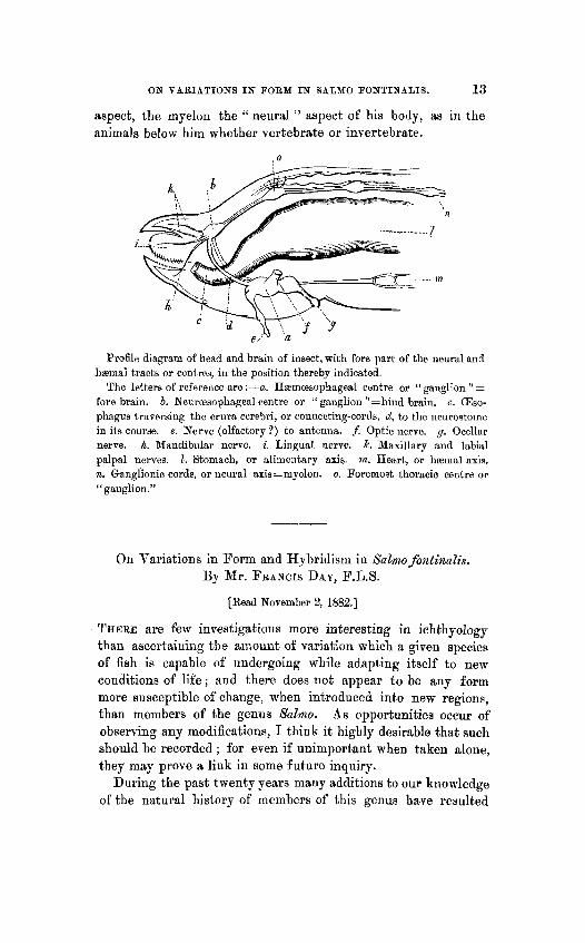
Profile diagram of head and brain of insect, with fore part of the neural and haemal tracts or centres, iu the position thereby indicated.
The letters of reference are :-a. Hamcesophageal centre or “ganglion ” = fore brain. b. Neurcesophsgeal centre or “ ganglion ”=hind brain. c. (Eso- phagus traversing the crura cerebri, or connecting-cords, d, to the neurostoine in its course. e. Nerve (olfactory?) to antenna. f. Optic nerve. g. Ocellar nerve. h. Mandibular nerve. i. Lingual nerve. k. Maxillary and labial palpal nerves. 1. Stomach, or alimentary axis. m. Heart, or haeinal axis. n. Ganglionic cords, or neural axis=myelon. 0. Foremost thoracic centre or “ganglion.”
On Variatioiis in Form and Hybridism iu Salmo fontinatis. By Mr. FRANCIS DAY, F . L . S .
[Read November 2, 1882.1
THERE are few investigations more interesting in ichthyology than ascertaining the amount of variation which a given species of fish is capable of undergoing while adapting itself to new conditions of life; and there does not appear to be any form more Rusceptible of change, when introduced into new regions, than members of the genus Salmo. As opportunities occur of observing any modifications, I think it highly desirable that such should be recorded ; for even if unimportant when taken alone, they may prove a link in some future inquiry.
During the past twenty years many additions t o our knowledge of the natural history of members of this genus have resulted
ON VARIATIONS I N FORM IN SbLXO FONTINALIS. 13
aspect, the myelon the “ neural ” aspect of his body, as in the animals below hiin whether vertebrate or invertebrate.
, o
Profile diagram of head and brain of insect, with fore part of the neural and haemal tracts or centres, iu the position thereby indicated.
The letters of reference are :-a. Hamcesophageal centre or “ganglion ” = fore brain. b. Neurcesophsgeal centre or “ ganglion ”=hind brain. c. (Eso- phagus traversing the crura cerebri, or connecting-cords, d, to the neurostoine in its course. e. Nerve (olfactory?) to antenna. f. Optic nerve. g. Ocellar nerve. h. Mandibular nerve. i. Lingual nerve. k. Maxillary and labial palpal nerves. 1. Stomach, or alimentary axis. m. Heart, or haeinal axis. n. Ganglionic cords, or neural axis=myelon. 0. Foremost thoracic centre or “ganglion.”
On Variatioiis in Form and Hybridism iu Salmo fontinatis. By Mr. FRANCIS DAY, F . L . S .
[Read November 2, 1882.1
THERE are few investigations more interesting in ichthyology than ascertaining the amount of variation which a given species of fish is capable of undergoing while adapting itself to new conditions of life; and there does not appear to be any form more Rusceptible of change, when introduced into new regions, than members of the genus Salmo. As opportunities occur of observing any modifications, I think it highly desirable that such should be recorded ; for even if unimportant when taken alone, they may prove a link in some future inquiry.
During the past twenty years many additions t o our knowledge of the natural history of members of this genus have resulted