viable bacteria, with a limit of detection of 7.6 × 10³ and 1.1 × 10³ CFU/mL for S. typhi and E. coli O157, respec-tively, while eliminating amplification from non-viable cells. Furthermore, we studied the effects of greater flow rates to expedite the labeling process and identified a maxi-mum flow rate of 0.7 μL/min for complete labeling with the current design.
The World Health Organization (WHO) established food-borne diseases as one of the eleven priorities and challenges for the millennium (Torgerson et al. 2014). WHO estimates there are 2.2 million deaths every year related to foodborne diseases, having infants contributing more than 80 % of fatalities with an estimate of 1.9 million cases. Moreover, in the USA alone, 128.000 food poisoning hospitalizations and around 3.000 related deaths result in an estimated cost of $77.7 billion dollars annually (Scharff 2012). One of the reasons for the persistence and prevalence of foodborne ill-nesses is the poor performance of current microorganism detection techniques that undermines effective prevention (Roda et al. 2012). Traditionally, foodborne pathogenic bacteria are monitored using a series of detection assays that include growth in selective culture media, biochemi-cal nucleic acid confirmations, and immunological capture (Elizaquível et al. 2014). The full screening process is time-consuming, expensive and labor-intensive, and requires specialized equipment and trained personnel. Novel sys-tems and devices have been reported to address these chal-lenges inherent to the detection of foodborne pathogenic
Abstract Propidium monoazide (PMA) is a membrane impermeable molecule that covalently bonds to double stranded DNA when exposed to light and inhibits the poly-merase activity, thus enabling DNA amplification detection protocols that discriminate between viable and non-viable entities. Here, we present a microfluidic device for inexpen-sive, fast, and simple PMA labeling for viable qPCR and qLAMP assays. The three labeling stages of mixing, incu-bation, and cross-linking are completed within a microflu-idic device that is designed with Tesla structures for passive microfluidic mixing, bubble trappers to improve flow uni-formity, and a blue LED to cross-link the molecules. Our results show that the on-chip PMA labeling is equivalent to the standard manual protocols and prevents the replica-tion of DNA from non-viable cells in amplification assays. However, the on-chip process is faster and simpler (30 min of hands-off work), has a reduced likelihood of false nega-tives, and it is less expensive because it only uses 1/20th of the reagents normally consumed in standard bench pro-tocols. We used our microfluidic device to perform viable qPCR and qLAMP for the detection of S. typhi and E. coli O157. With this device, we are able to specifically detect
1 Micro and Nanotechnology Lab, University of Illinois at Urbana-Champaign, 208 N. Wright St., Urbana, IL 61801, USA
2 Department of Electrical and Computer Engineering, University of Illinois at Urbana-Champaign, 306 N. Wright St., Urbana, IL 61801, USA
3 1270 Digital Computer Laboratory, Department of Bioengineering, University of Illinois at Urbana-Champaign, 1304 W. Springfield Ave., Urbana, IL 61801, USA
bacteria. Particularly, strategies that target to expedite the process (Li et al. 2013), minimize cost (Cho et al. 2015), or automate analysis (Kim et al. 2014) have been pursued by multiple researchers. Unfortunately, these strategies are still under development and fail to provide complete solu-tions that enhanced the monitoring ability of control agen-cies. Therefore, there is still a pressing need to create novel and more effective foodborne pathogen screening methods that improve prevention and control strategies.
Powerful screening assays rely on biomolecular tech-niques that have already been incorporated in standard detection protocols in recent years (Nugen and Baeum-ner 2008). These molecular techniques are based on DNA amplification processes, such as the real-time quantitative polymerase chain reaction (qPCR), and have demonstrated outstanding sensitivity, versatility, and specificity (Guan et al. 2013). The DNA-based tests also enable the detection of subdominant bacteria population even without the pres-ence of selective culture media (Postollec et al. 2011) and allow the miniaturization of detection devices (Zhao et al. 2014). Also, alternative methods for DNA amplification, like the loop-mediated isothermal amplification (LAMP), have been incorporated in pathogenic detection (Mori et al. 2013). These new methods not only have major advan-tages, such as a higher specificity and reliability, but also minimize the complexity of the detection protocol enabling novel detection systems and devices (Duarte et al. 2013). However, DNA amplification methods still present a major drawback due to their inability to discriminate between live and dead bacteria present in food samples (Elizaquível et al. 2012). The lack of discrimination between DNA tem-plates coming from viable versus non-viable pathogens is especially relevant for food safety applications where food processing steps are designed to sterilize samples. DNA amplification techniques can lead to the overestimation of the objective microorganisms and false positives, because DNA from dead bacteria is conserved during long periods and can interfere in the amplification process (Wagner et al. 2015).
Fortunately, DNA amplification detection techniques can gain the ability to discriminate viable from non-viable pathogens using viability dyes in the template molecules. DNA-based viability assays are achieved by adding inter-calating dyes, such as propidium monoazide (PMA) or ethidium monoazide (EMA), to the sample prior to the amplification reaction (Elizaquível et al. 2012; Dinu and Bach 2013). These viability dyes have two important char-acteristics: They are cell-membrane impermeable and they inhibit DNA replication. Molecules like PMA can only penetrate the membranes of non-viable cells, intercalate with the genomic DNA, and then form covalent bonds with
the DNA in a light-triggered cross-linking process. Later, during the nucleic acid amplification stage, the modified DNA strand, now strongly bounded to the viability dye, is not replicated by the polymerase. The process is described in the schematic of Fig. 1d, which shows how the incor-poration of viability dyes yields DNA amplification assays that only report viable pathogens. Nevertheless, the use of viability dyes adds complications to the sample preparation stage. The current PMA labeling protocol consists of three steps: mixing, incubation, and cross-linking. Although the protocol is not complex and requires only standard labo-ratory equipment, each step is time-consuming and labor-intensive. This often leads to experimental errors and pro-cess-to-process variations which undermine reliability and repeatability (Desneux et al. 2015).
In this paper, we report the fabrication and characteriza-tion of a microfluidic device for PMA labeling designed to facilitate the incorporation of viability dyes into the qPCR and qLAMP detection assays. The objective of our device is to simplify and automate the PMA labeling process to make it faster and less expensive. The microfluidic device that is used to perform the labeling process is presented in Fig. 1a. It is designed with a series of ‘Tesla structures’ for simple single-level passive fluidic mixing (Yang et al. 2015; Hong et al. 2004), and bubble trapping elements to improve reliability (Lochovsky et al. 2012). The Tesla structure, presented in Fig. 1b, consists of bifurcating channels that use the Coanda effect to produce a transverse dispersion of fluids. This transverse dispersion increases the speed of the mixing process by promoting a turbulent flow (Hos-sain et al. 2010). On the other hand, the in-plane bubble traps, shown in Fig. 1c, take advantage of the PDMS gas permeability to remove undesired bubbles that will prevent appropriate mixing. The trapping structure consists in an array of closely paced pillars, a reservoir, and input/output channels. The pillars act as a sieve for bubbles, confining them in the reservoir and preventing their flow into the flu-idic channel. Then, the fluid can surround the trapped bub-bles minimizing undesired bubble blocks and resulting in a more consistent flow (Lochovsky et al. 2012). With these two microfluidic elements in the chip we achieve adequate and reliable mixing, and the incorporation of a blue LED in close proximity to the channels allows the full labeling process of mixing, incubation, and cross-linking. The per-formance of the microfluidic device for PMA labeling was tested with Salmonella typhimurium (S. typhi) and Escheri-chia coli (E. coli) O157 samples. After on-chip labeling, the bacterial samples were used as template in qPCR and qLAMP assays. The following sections present chip fab-rication, experimental methods, and viable qPCR/qLAMP results with microfluidic preparation.
Microfluid Nanofluid (2016) 20:114
1 3
Page 3 of 9 114
2 Materials and methods
2.1 Microfluidic chip fabrication
The microfluidic devices are fabricated with standard soft lithography using a mold made with a single layer of 45 μm thick SU8 (Microchem, Westborough, MA) pattern on a regular silicon wafer (University Wafer, Boston, MA). To prevent PDMS residues depositing inside the small gaps in the ‘Tesla structures’, it is important to silianize the wafer for 30 min with Perfluorooctyl silane (Sigma-Aldrich, St Louis, MO) using vacuum-assisted evapora-tion. Sylgar PDMS (Sigma-Aldrich) in the standard ratio of 10:1 is poured on the mold, de-gassed, and cured over-night at 60 °C to create a fluidic layer made of PDMS. The diced PDMS is then plasma-bonded to a glass slide in Pico Diener Oxygen Plasma System (Diener electronics, DE), and a standard T-1¾ blue LED is bonded to the back of the glass with a thin layer of transparent epoxy (Thorlabs, Newton, NJ).
2.2 Foodborne pathogen testing samples
Samples of E. coli O157 and S. typhi are cultivated in Bovine heart infusion (BHI) and Lysogenic Broth (LB) media, respectively. Eight milliliters of media was spiked with 5 μL of cells and incubated for 18 h to reach a car-rying capacity of 1.1 × 10⁹ CFU/mL for E. coli and of 7.6 × 10⁸ CFU/mL for S. typhi. Genomic DNA from the bacterial cultures is used as template for the viable qPCR and qLAMP assays. The extraction consisted in a heat lysis at 95 °C during 20 min and a series of centrifugations at 8600 rcf to remove debris.
2.3 Microfluidic device operation
The two inputs of the microfluidic device are connected to 10-mL syringes (Becton–Dickinson, Franklin Lanes, NJ) via standard Tygon tubing (Component supply, Fort Meade, FL). One of the syringes is loaded with the culture media (with growing or lysed cells) and 1× PMA enhancer
Fig. 1 a Image of the microfluidic device for PMA labeling. b Detail of the Tesla structures used in the device for passive mixing. c Detail of the bubble trappers used in the device to improve reliability. d Schematic of the working principle of viability dyes in DNA amplification assays
Microfluid Nanofluid (2016) 20:114
1 3
114 Page 4 of 9
(Biotium, Hayward, CA). The second syringe is loaded with 0.2 mM of PMA in DI with 1 % (v/v) DMSO. Both syringes are set in a standard PHD Ultra syringe pump (Harvard Apparatus, Holliston, MA) and unloaded at a constant rate between 0.2 and 2 μL/min, while the LED is excited with a conventional DC source at a potential of 30 V dissipating 360 mW. The pump is programmed to col-lect 15 μL of labeled sample that then is treated as template following the same DNA extraction protocol described in the previous subsection. Overall, the on-chip labeling pro-tocol is faster and consumes less reagents than the stand-ard process. The microfluidic device requires around 30 min without user intervention, while the bench protocol requires at least an hour of hands-on work. In addition, the lower reagent volumes required in microfluidics reduce the PMA consumption by a factor of 20, thereby significantly reducing the cost per assay.
2.4 qPCR or qLAMP
Eiken’s Loopamp® kits and Biotium’s Real-time PCR kits were purchased for both E. coli O157 and S. typhi
amplification assays. The reactions are prepared accord-ing to the manufacturer’s protocol but using the template DNA collected from the microfluidic device. The qPCR and qLAMP reactions are performed using an Eppendorf Mastercycler® ep RealPlex Real-Time PCR system. The PCR protocol consists of a pre-incubation at 95 °C for 10 min, followed by 40 amplification cycles of denatura-tion at 94 °C for 5 s, annealing/elongation at 52 °C for 30 s, and fluorescence imaging in every annealing step. On the other hand, the qLAMP reaction is performed at a constant temperature of 65 °C during 90 min taking fluorescence images every minute.
3 Results and discussion
3.1 Off‑chip versus on‑chip sample labeling
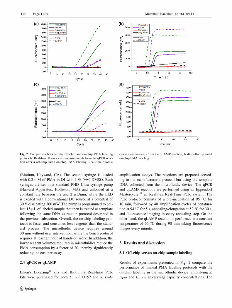
Results of experiments presented in Fig. 2 compare the performance of manual PMA labeling protocols with the on-chip labeling in the microfluidic device, amplifying S. typhi and E. coli at carrying capacity concentrations. The
Fig. 2 Comparison between the off-chip and on-chip PMA labeling protocols. Real-time fluorescence measurements from the qPCR reac-tion after a off-chip and c on-chip PMA labeling. Real-time fluores-
cence measurements from the qLAMP reaction, b after off-chip and d on-chip PMA labeling
Microfluid Nanofluid (2016) 20:114
1 3
Page 5 of 9 114
manual protocol follows the PMA manufacturer recom-mendation: 10 min of mixing, 10 min of incubation, and 20 min of cross-linking with a halogen lamp with the sam-ple immersed in an ice bath (Fittipaldi et al. 2012). On the other hand, the on-chip protocol simply involves flow-ing the dye and sample through the microfluidic chip as described in the previous section. The comparisons of the labeling methods show that on-chip treatment is as effec-tive as the manual process, inhibiting the amplification of non-viable cells. The color codes in Fig. 2 present four different experimental scenarios. A positive control (Pos-Control) indicates no labeling (no PMA dye) to assure complete amplification, while a negative control (NegCon-trol) indicates no bacteria flown through the chip (replaced with DI water). The viable assay (Viable) flows PMA and media with viable cells through the chip, and the non-viable assay (Non-Viable) flows heat-lysed bacteria with PMA. No amplification was expected from the non-viable assay, while amplification was anticipated for the viable sample. All the four assays presented in Fig. 2 panels had the expected results, with amplification from positive and viable samples but no amplification from non-viable or negative samples. Therefore, the comparison of the labeling methods shows that on-chip labeling is as effective as the manual labeling. In fact, we observe significant increase in threshold cycles/times for the manual samples both for PCR and LAMP assays. As others have reported (Nocker et al. 2007b; Bae and Wuertz 2009), the standard PMA pro-tocol can stress the cells, compromise their membrane, and trigger the inhibition caused by the viability dye. Longer threshold cycles and times in the viable but PMA-treated samples can be related to partial DNA amplification inhibi-tion due to stress in the cells during the labeling process. Comparative experiments of Fig. 2 lead to the conclu-sion that the microfluidic device is capable of effectively performing the three stages of the PMA labeling process and likely reduce the stress on the target analyte reducing
the likelihood of preparation-related false negatives (Eli-zaquível et al. 2012; Nocker et al. 2007a, b). These results align with previously reported viability assays on PDMS devices that have demonstrated low unintended lysis of cells by PDMS materials (Halldorsson et al. 2015). Prior reports indicate that PDMS has beneficial properties for cell viability, such as transparency and gas permeability, which result in the lower lysis that we observe in our plat-form. Furthermore, the use of only blue light as the cross-linking catalyzer has been previously identified as more reliable method for PMA labeling (Oh et al. 2015), further explaining the lower probability of preparation-related false negatives that we observe on-chip.
3.2 Repeatability of on‑chip labeling
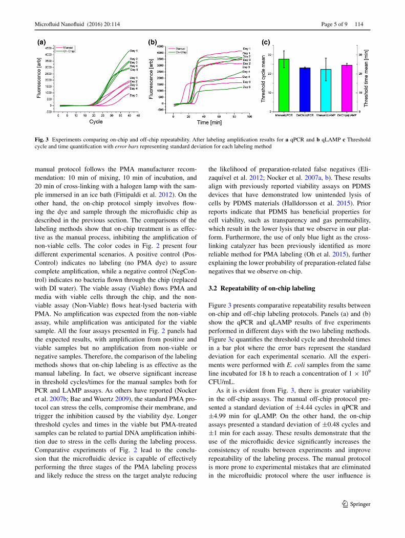
Figure 3 presents comparative repeatability results between on-chip and off-chip labeling protocols. Panels (a) and (b) show the qPCR and qLAMP results of five experiments performed in different days with the two labeling methods. Figure 3c quantifies the threshold cycle and threshold times in a bar plot where the error bars represent the standard deviation for each experimental scenario. All the experi-ments were performed with E. coli samples from the same line incubated for 18 h to reach a concentration of 1 × 10⁹ CFU/mL.
As it is evident from Fig. 3, there is greater variability in the off-chip assays. The manual off-chip protocol pre-sented a standard deviation of ±4.44 cycles in qPCR and ±4.99 min for qLAMP. On the other hand, the on-chip assays presented a standard deviation of ±0.48 cycles and ±1 min for each assay. These results demonstrate that the use of the microfluidic device significantly increases the consistency of results between experiments and improve repeatability of the labeling process. The manual protocol is more prone to experimental mistakes that are eliminated in the microfluidic protocol where the user influence is
Fig. 3 Experiments comparing on-chip and off-chip repeatability. After labeling amplification results for a qPCR and b qLAMP c Threshold cycle and time quantification with error bars representing standard deviation for each labeling method
Microfluid Nanofluid (2016) 20:114
1 3
114 Page 6 of 9
diminished. Therefore, by minimizing hands-on steps dur-ing the labeling process it has been possible to reduce vari-ations between assays.
3.3 Limit of detection for E. coli and S. typhi
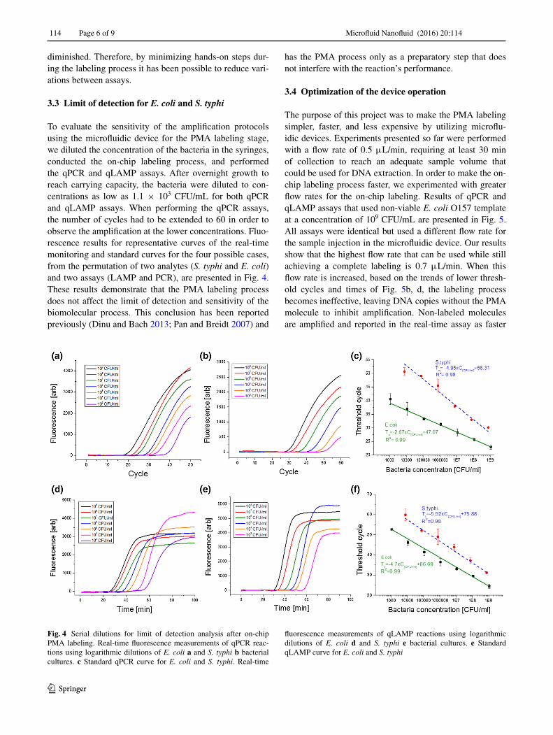
To evaluate the sensitivity of the amplification protocols using the microfluidic device for the PMA labeling stage, we diluted the concentration of the bacteria in the syringes, conducted the on-chip labeling process, and performed the qPCR and qLAMP assays. After overnight growth to reach carrying capacity, the bacteria were diluted to con-centrations as low as 1.1 × 10³ CFU/mL for both qPCR and qLAMP assays. When performing the qPCR assays, the number of cycles had to be extended to 60 in order to observe the amplification at the lower concentrations. Fluo-rescence results for representative curves of the real-time monitoring and standard curves for the four possible cases, from the permutation of two analytes (S. typhi and E. coli) and two assays (LAMP and PCR), are presented in Fig. 4. These results demonstrate that the PMA labeling process does not affect the limit of detection and sensitivity of the biomolecular process. This conclusion has been reported previously (Dinu and Bach 2013; Pan and Breidt 2007) and
has the PMA process only as a preparatory step that does not interfere with the reaction’s performance.
3.4 Optimization of the device operation
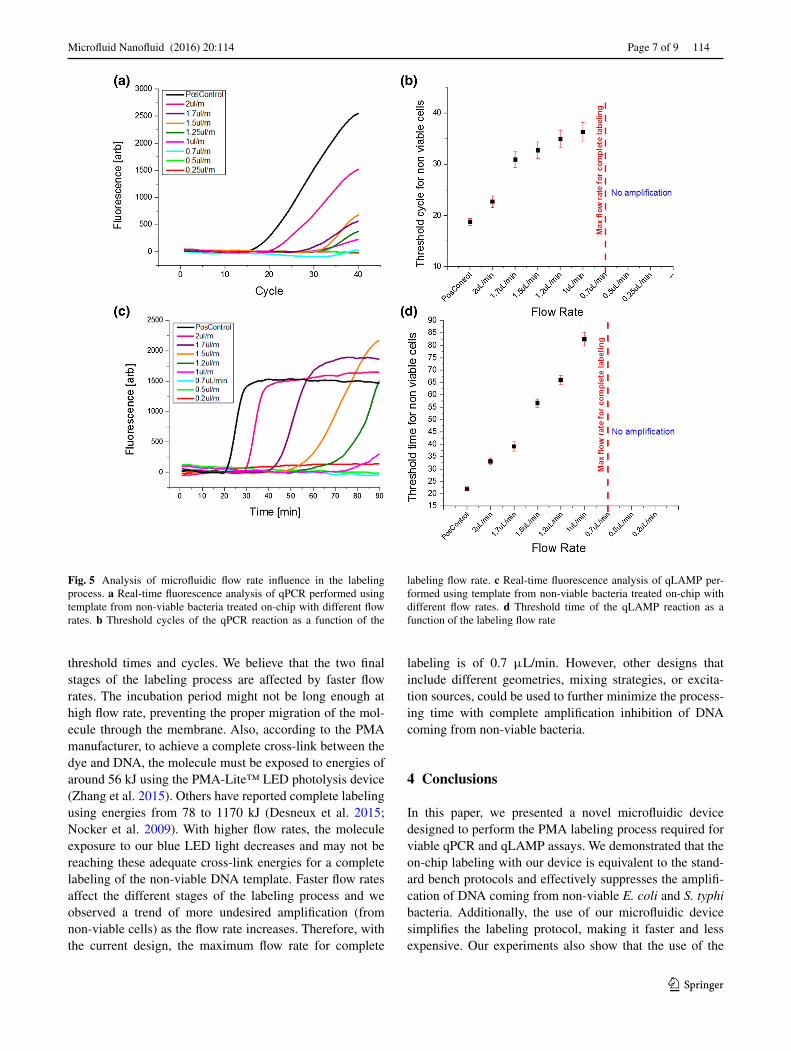
The purpose of this project was to make the PMA labeling simpler, faster, and less expensive by utilizing microflu-idic devices. Experiments presented so far were performed with a flow rate of 0.5 μL/min, requiring at least 30 min of collection to reach an adequate sample volume that could be used for DNA extraction. In order to make the on-chip labeling process faster, we experimented with greater flow rates for the on-chip labeling. Results of qPCR and qLAMP assays that used non-viable E. coli O157 template at a concentration of 109 CFU/mL are presented in Fig. 5. All assays were identical but used a different flow rate for the sample injection in the microfluidic device. Our results show that the highest flow rate that can be used while still achieving a complete labeling is 0.7 μL/min. When this flow rate is increased, based on the trends of lower thresh-old cycles and times of Fig. 5b, d, the labeling process becomes ineffective, leaving DNA copies without the PMA molecule to inhibit amplification. Non-labeled molecules are amplified and reported in the real-time assay as faster
Fig. 4 Serial dilutions for limit of detection analysis after on-chip PMA labeling. Real-time fluorescence measurements of qPCR reac-tions using logarithmic dilutions of E. coli a and S. typhi b bacterial cultures. c Standard qPCR curve for E. coli and S. typhi. Real-time
fluorescence measurements of qLAMP reactions using logarithmic dilutions of E. coli d and S. typhi e bacterial cultures. e Standard qLAMP curve for E. coli and S. typhi
Microfluid Nanofluid (2016) 20:114
1 3
Page 7 of 9 114
threshold times and cycles. We believe that the two final stages of the labeling process are affected by faster flow rates. The incubation period might not be long enough at high flow rate, preventing the proper migration of the mol-ecule through the membrane. Also, according to the PMA manufacturer, to achieve a complete cross-link between the dye and DNA, the molecule must be exposed to energies of around 56 kJ using the PMA-Lite™ LED photolysis device (Zhang et al. 2015). Others have reported complete labeling using energies from 78 to 1170 kJ (Desneux et al. 2015; Nocker et al. 2009). With higher flow rates, the molecule exposure to our blue LED light decreases and may not be reaching these adequate cross-link energies for a complete labeling of the non-viable DNA template. Faster flow rates affect the different stages of the labeling process and we observed a trend of more undesired amplification (from non-viable cells) as the flow rate increases. Therefore, with the current design, the maximum flow rate for complete
labeling is of 0.7 μL/min. However, other designs that include different geometries, mixing strategies, or excita-tion sources, could be used to further minimize the process-ing time with complete amplification inhibition of DNA coming from non-viable bacteria.
4 Conclusions
In this paper, we presented a novel microfluidic device designed to perform the PMA labeling process required for viable qPCR and qLAMP assays. We demonstrated that the on-chip labeling with our device is equivalent to the stand-ard bench protocols and effectively suppresses the amplifi-cation of DNA coming from non-viable E. coli and S. typhi bacteria. Additionally, the use of our microfluidic device simplifies the labeling protocol, making it faster and less expensive. Our experiments also show that the use of the
Fig. 5 Analysis of microfluidic flow rate influence in the labeling process. a Real-time fluorescence analysis of qPCR performed using template from non-viable bacteria treated on-chip with different flow rates. b Threshold cycles of the qPCR reaction as a function of the
labeling flow rate. c Real-time fluorescence analysis of qLAMP per-formed using template from non-viable bacteria treated on-chip with different flow rates. d Threshold time of the qLAMP reaction as a function of the labeling flow rate
Microfluid Nanofluid (2016) 20:114
1 3
114 Page 8 of 9
microfluidic device and the viability dye does not affect the sensitivity of the DNA amplification methods. The limit of detection we found was of 1.1 × 10³ CFU/mL of bacte-ria or ten copies per reaction, a similar metric to the one obtained in other methods that do not use the PMA stage. We also studied the effect of fluidic rates in the labeling process, identifying a threshold of 0.7 μL/min.
Detection methods based on DNA amplification have demonstrated outstanding sensitivity and specificity. They are currently used in many applications that include food safety, disease diagnostics, or patient monitoring (Dhama et al. 2014). The incorporation of viability dyes like PMA expands these abilities and enables other uses such as the quantification of viable beneficial microbiota (Elizaquível et al. 2013). By miniaturizing the PMA labeling process with microfluidics, it is possible to facilitate its incorpora-tion in novel miniaturized DNA amplification systems. For example, the PMA labeling device could receive sample from concentration and filtering systems that use dielectro-phoretic (Yang et al. 2006) or mechanical filtering (Li et al. 2013) to reduce volumes. Additionally, detection devices that use smartphones (Damhorst et al. 2015), semiconduc-tor devices (Duarte-Guevara et al. 2014), or compact-disk technology (Furutani et al. 2010) for miniaturized DNA-based assays can be used to analyze sample that has been labeled with the microfluidic device. These combinations of modules and devices are poised to create highly effi-cient pathogen screening systems. Moreover, the recent adaptation of industrial manufacturing techniques (e.g., polycarbonate micro molding) for microfluidic devices will minimize the cost of equipment and cartridges further facilitating the development of cost-effective tools (Volpatti and Yetisen 2014). Therefore, our demonstration of on-chip PMA labeling opens the opportunity to improve portable DNA amplification and expand its possible applications. Future studies will focus on the integration of sample prep-aration and detection stages, improve labeling speed, and further minimize cost.
Acknowledgments The authors would like to thank the center for innovative instrumentation technology (http://cabpn.illinois.edu/) for funding support and technical guidance from the industry partners.
References
Bae S, Wuertz S (2009) Discrimination of viable and dead fecal Bac-teroidales bacteria by quantitative PCR with propidium mono-azide. Appl Environ Microbiol 75(9):2940–2944
Cho IH, Bhunia A, Irudayaraj J (2015) Rapid pathogen detection by lateral-flow immunochromatographic assay with gold nanopar-ticle-assisted enzyme signal amplification. Int J Food Microbiol 206:60–66
loop-mediated isothermal amplification (RT-LAMP) on a chip from whole blood. Engineering (Beijing, China) 1(3):324
Desneux J, Chemaly M, Pourcher AM (2015) Experimental design for the optimization of propidium monoazide treatment to quan-tify viable and non-viable bacteria in piggery effluents. BMC Microbiol 15(1):1
Dhama K, Karthik K, Chakraborty S, Tiwari R, Kapoor S, Kumar A, Thomas P (2014) Loop-mediated isothermal amplification of DNA (LAMP): A new diagnostic tool lights the world of diagnosis of animal and human pathogens: A review. Pak J Biol Sci 17(2):151
Dinu LD, Bach S (2013) Detection of viable but non-culturable Escheri-chia coli O157: H7 from vegetable samples using quantitative PCR with propidium monoazide and immunological assays. Food Con-trol 31(2):268–273
Duarte C, Salm E, Dorvel B, Reddy B Jr, Bashir R (2013) On-chip parallel detection of foodborne pathogens using loop-mediated isothermal amplification. Biomed Microdevices 15(5):821–830
Duarte-Guevara C, Lai FL, Cheng CW, Reddy B Jr, Salm E, Swami-nathan V, Bashir R (2014) Enhanced biosensing resolution with foundry fabricated individually addressable dual-gated ISFETs. Anal Chem 86(16):8359–8367
Elizaquível P, Sánchez G, Aznar R (2012) Quantitative detection of viable foodborne E. coli O157: H7, Listeria monocytogenes and Salmonella in fresh-cut vegetables combining propidium mono-azide and real-time PCR. Food Control 25(2):704–708
Elizaquível P, Aznar R, Sánchez G (2014) Recent developments in the use of viability dyes and quantitative PCR in the food microbiol-ogy field. J Appl Microbiol 116(1):1–13
Fittipaldi M, Nocker A, Codony F (2012) Progress in understanding preferential detection of live cells using viability dyes in combina-tion with DNA amplification. J Microbiol Methods 91(2):276–289
Furutani S, Nagai H, Takamura Y, Kubo I (2010) Compact disk (CD)-shaped device for single cell isolation and PCR of a specific gene in the isolated cell. Anal Bioanal Chem 398(7–8):2997–3004
Guan ZP, Jiang Y, Gao F, Zhang L, Zhou GH, Guan ZJ (2013) Rapid and simultaneous analysis of five foodborne pathogenic bacteria using multiplex PCR. Eur Food Res Technol 237(4):627–637
Halldorsson S, Lucumi E, Gómez-Sjöberg R, Fleming RM (2015) Advantages and challenges of microfluidic cell culture in polydi-methylsiloxane devices. Biosens Bioelectron 63:218–231
Hong C, Choo J, Ahn C (2004) A novel in-plane microfluidic mixer with modified Tesla structures. Lab Chip 4(2):109–113
Hossain S, Ansari MA, Husain A, Kim KY (2010) Analysis and opti-mization of a micromixer with a modified Tesla structure. Chem Eng J 158(2):305–314
Kim TH, Park J, Kim CJ, Cho YK (2014) Fully integrated lab-on-a-disc for nucleic acid analysis of food-borne pathogens. Anal Chem 86(8):3841–3848
Li X, Ximenes E, Amalaradjou MAR, Vibbert HB, Foster K, Jones J, Liu X, Bhunia A, Ladisch MR (2013) Rapid sample processing for detection of food-borne pathogens via cross-flow microfiltra-tion. Appl Environ Microbiol 79(22):7048–7054
Lochovsky C, Yasotharan S, Günther A (2012) Bubbles no more: in-plane trapping and removal of bubbles in microfluidic devices. Lab Chip 12(3):595–601
Mori Y, Kanda H, Notomi T (2013) Loop-mediated isothermal ampli-fication (LAMP): recent progress in research and development. J Infect Chemother 19(3):404–411
Nocker A, Sossa-Fernandez P, Burr MD, Camper AK (2007a) Use of propidium monoazide for live/dead distinction in microbial ecol-ogy. Appl Environ Microbiol 73(16):5111–5117
Nocker A, Sossa KE, Camper AK (2007b) Molecular monitoring of disinfection efficacy using propidium monoazide in combination with quantitative PCR. J Microbiol Methods 70(2):252–260
Nocker A, Mazza A, Masson L, Camper AK, Brousseau R (2009) Selective detection of live bacteria combining propidium
monoazide sample treatment with microarray technology. J Microbiol Methods 76(3):253–261
Nugen SR, Baeumner AJ (2008) Trends and opportunities in food pathogen detection. Anal Bioanal Chem 391(2):451–454. doi:10.1007/s00216-008-1886-2
Oh E, McMullen L, Jeon B (2015) Impact of oxidative stress defense on bacterial survival and morphological change in Campylobac-ter jejuni under aerobic conditions. Front Microbiol 6:295
Pan Y, Breidt F (2007) Enumeration of viable Listeria monocytogenes cells by real-time PCR with propidium monoazide and ethidium monoazide in the presence of dead cells. Appl Environ Microbiol 73(24):8028–8031
Postollec F, Falentin H, Pavan S, Combrisson J, Sohier D (2011) Recent advances in quantitative PCR (qPCR) applications in food microbiology. Food Microbiol 28(5):848–861
Roda A, Mirasoli M, Roda B, Bonvicini F, Colliva C, Reschiglian P (2012) Recent developments in rapid multiplexed bioanalytical methods for foodborne pathogenic bacteria detection. Microchim Acta 178(1–2):7–28
Scharff RL (2012) Economic burden form health losses due to food-borne illness in the United States. J Food Prot 175:123–131
Torgerson PR, de Silva NR, Fevre EM, Kasuga F, Rokni MB, Zhou XN, Sripa B, Gargouri N, Willingham AN, Stein C (2014) The global burden of foodborne parasitic diseases: an update. Trends Parasitol 30(1):20–26
Volpatti LR, Yetisen AK (2014) Commercialization of microfluidic devices. Trends Biotechnol 32(7):347–350
Wagner AO, Praeg N, Reitschuler C, Illmer P (2015) Effect of DNA extraction procedure, repeated extraction and ethidium mono-azide (EMA)/propidium monoazide (PMA) treatment on over-all DNA yield and impact on microbial fingerprints for bacteria, fungi and archaea in a reference soil. Appl Soil Ecol 93:56–64
Yang L, Banada PP, Chatni MR, Lim KS, Bhunia AK, Ladisch M, Bashir R (2006) A multifunctional micro-fluidic system for dielectrophoretic concentration coupled with immuno-capture of low numbers of Listeria monocytogenes. Lab on a Chip 6(7):896–905
Yang AS, Chuang FC, Chen CK, Lee MH, Chen SW, Su TL, Yang YC (2015) A high-performance micromixer using three-dimensional Tesla structures for bio-applications. Chem Eng J 263:444–451
Zhang Z, Liu H, Lou Y, Xiao L, Liao C, Malakar PK, Pan Y, Zhao Y (2015) Quantifying viable Vibrio parahaemolyticus and Listeria monocytogenes simultaneously in raw shrimp. Appl Microbiol Biotechnol 99(15):6451–6462
Zhao X, Lin CW, Wang J, Oh DH (2014) Advances in rapid detec-tion methods for foodborne pathogens. J Microbiol Biotechnol 24(3):297–312