1113 loins. He had no difficulty on micturition nor did his urine seem to him to be abnormal in quality or quantity. lIe was daily at his business and felt well excepting this aching over the region described and which did not at this time amount to an actual pain. On exami- nation I found his temperature and pulse normal. The urine to the naked eye did not suggest haematuria or pyuria but on testing it was found to contain a small quantity of albumin. My diagnosis was sub- acute nephritis and I prescribed rest and the usual routine treatment for that complaint. He made a good recovery although a trace of albumin was present in his urine when he returned to business again. A year later, in April, 1903, he again consulted me and this time he complained of pain across the lumbar region and especially in the right loin. There was not the shadow of a doubt of the pain at this time being on the right side and not on the left side, for the patient, who was a shrewd business man and a most trustworthy observer, afterwards reminded me that among other local applications I prescribed to relieve the dull pain on the right side was a fly blister I about three inches square. The pain at this time had not the character I or the direction of that in renal colic. Whilst he had no diffi- culty on micturition his urine was of a smoky tint and on testing it not only albumin but blood also was present. No microscopical exa- mination was made. The question was now raised of a renal calculus on the right side, and an operation was even hinted at, but as the patient seemed to improve rapidly with absolute rest in bed, a milk dietary, and diuretics and was most anxious to return to business nothing further was done. Two months later, on June 2nd, he sent for me hurriedly one morning and said that several times during the previous night he had had acute attacks of pain on the right side over his right kidney and shooting down towards his right testicle. According to his description of these attacks they suggested to my mind renal colic, although not of such an agonising character as is often seen. His urine again contained blood. I then told him that there was no doubt that he had a calculus or calculi in the right kidney and insisted on his seeing Dr. Newman in regard to an operation. He consulted him a few days later and an operation on the right side was recommended. The patient had fortunately for himself as the sequel showed, a strong objection to operative interference, so he sought the waters of Harrogate in the hope of a cure. Whilst there he consulted a physician in Harrogate who also recommended an operation on the right side. On the advice of a friend he went to London and consulted a surgeon who wrote to me that my patient had most probably right pyelitis due to calculi and he recommended operation on the right side. It is only fair to say here that the patient told the consultant that he had been cystoscopically examined and skia- graphed with no definite result. The remaining history of the case will be related by Dr. Newman. I have only to add that the case was a most instructive one to me in many ways and among other facts deeply impressed on my mind the extreme value of the x rays in renal diagnosis. I am firmly convinced that if my patien had gone to any medical man with the symptoms I have mentioned his advice would have been to operate on the right side. There was only one other point. Was the right kidney normal ? To that question my reply would be that the x rays were faithful in bringing out calculi on the left side and none on the right side. Moreover, the operation was eminently success’nl and the patient is now free from all pain or dis- comfort and to be absolutely certain that he will remain so I shall report the case later to Dr. Newman. When I was asked to see the patient on May 4th, 1903, I came to the conclusion that he was suffering from a calculus in the right kidney but to confirm the diagnosis I had an x-ray photograph taken and on the llth I examined the bladder with the cystoscope. From neither of those observa- tions was any benefit derived. The bladder was strictly healthy and there was nothing remarkable about the appear- ance of the orifice of either ureter and the photograph failed to show any distinct shadow of a calculus. On August 7th three photographs were taken and in one of them there was a faint indication of a shadow correspond- ing to the position of the pelvis of the left kidney, but as this was indistinct no reliance was placed upon it. Again on Oct. 12th other three photographs were taken and in two of them the same shadow was found (Fig. 2). This pointed clearly to the presence of a calculus and notwithstanding the predominance of pain upon the right side I concluded that the stone was in the left kidney. At this time there was some pus in the urine and on making a cystoscopic examination I found that it proceeded from the left kidney. This corrobo- rated the view of the position of the calculus. On Oct. 17th I explored the left kidney and found two stones, each weigh- ing about two drachms. The patient made a good recovery, as indicated in Dr. Colvin’s report. CASE 3. Pyonephrosis limited to the right 7zidney ; pus and albumin in the urine; pain most marked in left lumbar -region ; nephrotomy lJure.-A man, aged 26 years, con- sulted me first in July, 1898. He then complained of pain across the back and extending downwards to the crest of the ilium. The history of the case showed that the first departure from health was noticed two and a half years previously. At first the pain was generally distributed over the lumbar regions on both sides. As the disease advanced the pain was most marked in the region of the left kidney and frequently limited to that part. The patient was admitted to the Central Home where on examination I found that on palpation there was a distinct enlargement of the right kidney while the left kidney could hardly be felt. On admission the patient complained of frequent micturition ; the urine was copious, pale in colour, contained a siderable deposit of pus, the reaction was acid, and the specific gravity was 1010, but no blood had ever been observed nor had he passed gravel or stones. A cysto- scopic examination showed the pus to be escaping from the right ureter only and this was confirmed by two subsequent FIG. 2. Reduced-x-ray photograph from Case 2. The arrow points to the shadow of the calculi. examinations. The patient was advised to have an opera- tion performed but he preferred to wait and to test the effect of complete rest and medicinal treatment for three months. In November I saw the patient again and he then told me that little benefit had been derived from the treatment pre- scribed and he was now anxious to have an operation per- formed. During the last three months the pain was almost always referred to the left renal region but as the patient was convinced by the swelling on the right side that it was the right kidney that was diseased he consented to have an ex- ploratory incision made. On Nov. 15th I incised the kidney and evacuated ten ounces of pus but no stone was found. The patient made a good recovery and when I heard from him last from Cape Town, South Africa, he reported that there was no recurrence of the pain and that the pus had dis- appeared from the urine entirely (October, 1902). Glasgow. ON SOME FORMS OF PSEUDO-APPENDI- CITIS (ENTERO-TYPHLO-COLITIS.— INTESTINAL LITHIASIS). BY DR. BOTTENTUIT, CONSULTING PHYSICIAN AT PLOMBIÈRES. THE frequent occurrence of appendicitis and the con- sternation it causes both to physician and patient has led Mr. A. H. Tubby to remark that as soon as a subject is seized with pain in the right abdominal region ’’ it might be safely said that their brain is no longer in their cranial cavity but in the right iliac fossa, for so fixed is their attention on that region that every ache and pain there, any delayed action of the bowels, prompts them to expect an attack of appendicitis and to make their lives a burden to themselves and others. " This prejudice has seized upon the medical faculty. For a long time appendicitis has been overlooked. How often has it been mistaken for hepatic or nephritic colic, salpingitis, &c. ? It is necessary to dispel this fascination for McBurney’s spot. To me it appears of interest to direct my readers’
Transcript
1113
loins. He had no difficulty on micturition nor did his urine seem tohim to be abnormal in quality or quantity. lIe was daily at hisbusiness and felt well excepting this aching over the region describedand which did not at this time amount to an actual pain. On exami-nation I found his temperature and pulse normal. The urine to thenaked eye did not suggest haematuria or pyuria but on testing it wasfound to contain a small quantity of albumin. My diagnosis was sub-acute nephritis and I prescribed rest and the usual routine treatmentfor that complaint. He made a good recovery although a trace ofalbumin was present in his urine when he returned to business
again. A year later, in April, 1903, he again consulted me andthis time he complained of pain across the lumbar region andespecially in the right loin. There was not the shadow of a doubt ofthe pain at this time being on the right side and not on the left side,for the patient, who was a shrewd business man and a most trustworthyobserver, afterwards reminded me that among other local applicationsI prescribed to relieve the dull pain on the right side was a fly blister
Iabout three inches square. The pain at this time had not the character Ior the direction of that in renal colic. Whilst he had no diffi-culty on micturition his urine was of a smoky tint and on testing itnot only albumin but blood also was present. No microscopical exa-mination was made. The question was now raised of a renal calculuson the right side, and an operation was even hinted at, but as thepatient seemed to improve rapidly with absolute rest in bed, a milkdietary, and diuretics and was most anxious to return to businessnothing further was done. Two months later, on June 2nd, hesent for me hurriedly one morning and said that several timesduring the previous night he had had acute attacks of pain on theright side over his right kidney and shooting down towards hisright testicle. According to his description of these attacks
they suggested to my mind renal colic, although not of such anagonising character as is often seen. His urine again contained blood.I then told him that there was no doubt that he had a calculus orcalculi in the right kidney and insisted on his seeing Dr. Newman inregard to an operation. He consulted him a few days later and anoperation on the right side was recommended. The patient hadfortunately for himself as the sequel showed, a strong objection tooperative interference, so he sought the waters of Harrogate in thehope of a cure. Whilst there he consulted a physician in Harrogatewho also recommended an operation on the right side. On theadvice of a friend he went to London and consulted a surgeonwho wrote to me that my patient had most probably rightpyelitis due to calculi and he recommended operation on theright side. It is only fair to say here that the patient told theconsultant that he had been cystoscopically examined and skia-graphed with no definite result. The remaining history of the casewill be related by Dr. Newman. I have only to add that the case wasa most instructive one to me in many ways and among other factsdeeply impressed on my mind the extreme value of the x rays inrenal diagnosis. I am firmly convinced that if my patien had goneto any medical man with the symptoms I have mentioned his advicewould have been to operate on the right side. There was only oneother point. Was the right kidney normal ? To that question myreply would be that the x rays were faithful in bringing out calculi onthe left side and none on the right side. Moreover, the operation waseminently success’nl and the patient is now free from all pain or dis-comfort and to be absolutely certain that he will remain so I shallreport the case later to Dr. Newman.
When I was asked to see the patient on May 4th, 1903, Icame to the conclusion that he was suffering from a calculusin the right kidney but to confirm the diagnosis I had anx-ray photograph taken and on the llth I examined thebladder with the cystoscope. From neither of those observa-tions was any benefit derived. The bladder was strictlyhealthy and there was nothing remarkable about the appear-ance of the orifice of either ureter and the photographfailed to show any distinct shadow of a calculus. On
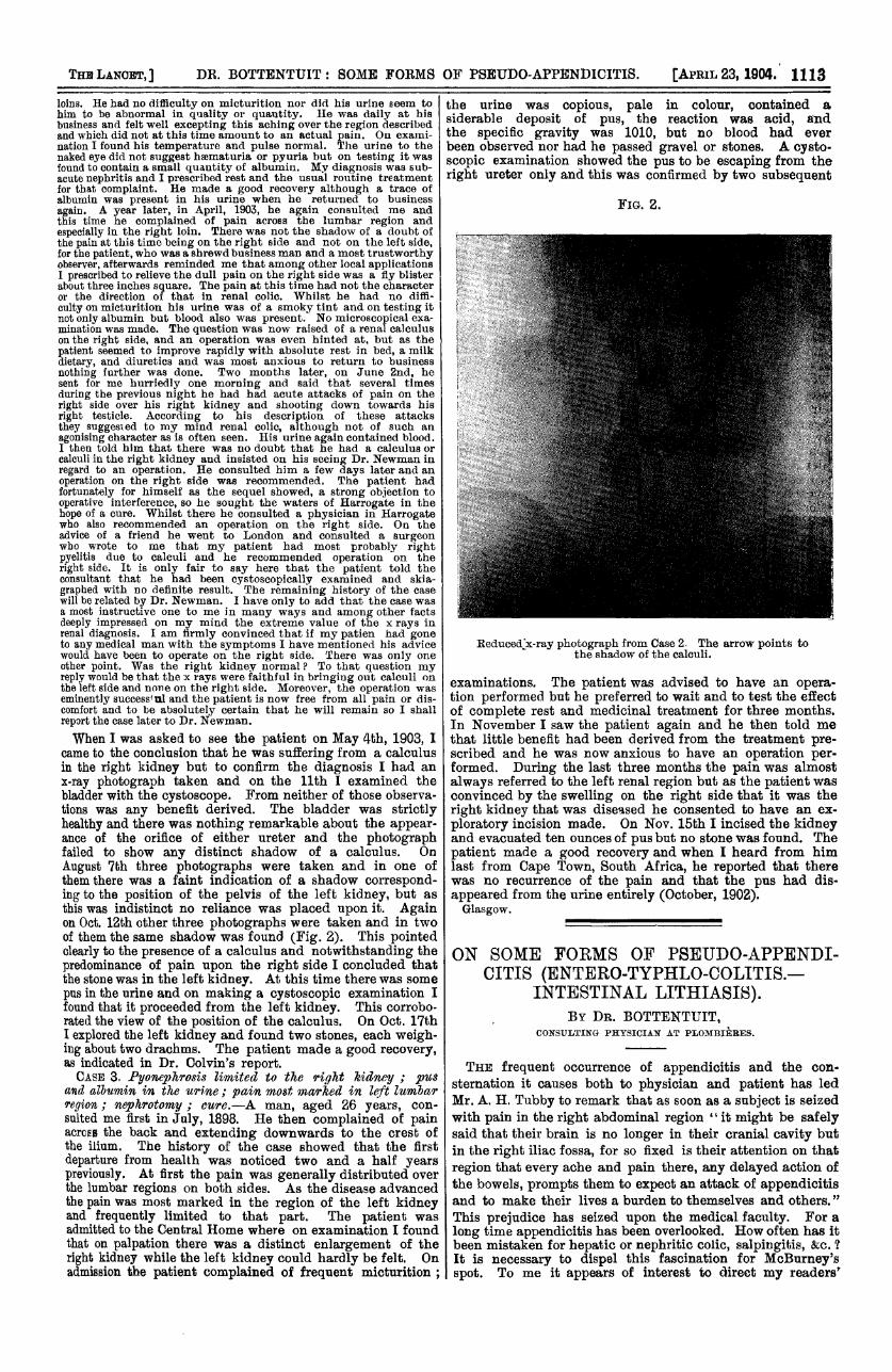
August 7th three photographs were taken and in one ofthem there was a faint indication of a shadow correspond-ing to the position of the pelvis of the left kidney, but asthis was indistinct no reliance was placed upon it. Againon Oct. 12th other three photographs were taken and in twoof them the same shadow was found (Fig. 2). This pointedclearly to the presence of a calculus and notwithstanding thepredominance of pain upon the right side I concluded thatthe stone was in the left kidney. At this time there was somepus in the urine and on making a cystoscopic examination Ifound that it proceeded from the left kidney. This corrobo-rated the view of the position of the calculus. On Oct. 17th
I explored the left kidney and found two stones, each weigh-ing about two drachms. The patient made a good recovery,as indicated in Dr. Colvin’s report.CASE 3. Pyonephrosis limited to the right 7zidney ; pus
and albumin in the urine; pain most marked in left lumbar-region ; nephrotomy lJure.-A man, aged 26 years, con-
sulted me first in July, 1898. He then complained of painacross the back and extending downwards to the crest ofthe ilium. The history of the case showed that the firstdeparture from health was noticed two and a half yearspreviously. At first the pain was generally distributed overthe lumbar regions on both sides. As the disease advancedthe pain was most marked in the region of the left kidneyand frequently limited to that part. The patient wasadmitted to the Central Home where on examination I foundthat on palpation there was a distinct enlargement of theright kidney while the left kidney could hardly be felt. Onadmission the patient complained of frequent micturition ;
the urine was copious, pale in colour, contained a
siderable deposit of pus, the reaction was acid, andthe specific gravity was 1010, but no blood had ever
been observed nor had he passed gravel or stones. A cysto-scopic examination showed the pus to be escaping from theright ureter only and this was confirmed by two subsequent
FIG. 2.
Reduced-x-ray photograph from Case 2. The arrow points tothe shadow of the calculi.
examinations. The patient was advised to have an opera-tion performed but he preferred to wait and to test the effectof complete rest and medicinal treatment for three months.In November I saw the patient again and he then told methat little benefit had been derived from the treatment pre-scribed and he was now anxious to have an operation per-formed. During the last three months the pain was almostalways referred to the left renal region but as the patient wasconvinced by the swelling on the right side that it was theright kidney that was diseased he consented to have an ex-ploratory incision made. On Nov. 15th I incised the kidneyand evacuated ten ounces of pus but no stone was found. The
patient made a good recovery and when I heard from himlast from Cape Town, South Africa, he reported that therewas no recurrence of the pain and that the pus had dis-appeared from the urine entirely (October, 1902).Glasgow.
ON SOME FORMS OF PSEUDO-APPENDI-CITIS (ENTERO-TYPHLO-COLITIS.—
INTESTINAL LITHIASIS).BY DR. BOTTENTUIT,
CONSULTING PHYSICIAN AT PLOMBIÈRES.
THE frequent occurrence of appendicitis and the con-
sternation it causes both to physician and patient has ledMr. A. H. Tubby to remark that as soon as a subject is seizedwith pain in the right abdominal region ’’ it might be safelysaid that their brain is no longer in their cranial cavity butin the right iliac fossa, for so fixed is their attention on that
region that every ache and pain there, any delayed action ofthe bowels, prompts them to expect an attack of appendicitisand to make their lives a burden to themselves and others. "
This prejudice has seized upon the medical faculty. For a
long time appendicitis has been overlooked. How often has itbeen mistaken for hepatic or nephritic colic, salpingitis, &c. ?It is necessary to dispel this fascination for McBurney’sspot. To me it appears of interest to direct my readers’
1114
attention to certain cases in which pain in the right iliac Ifossa, even increased at McBurney’s spot, or intestinalcdses, or abdominal symptoms, can deceive us or lead usto expect an appendicitis without a real appendicitisexisting in the true sense of the word. If it has sometimes
happened that appendicitis has been overlooked and surgicalintervention rejected at a time when it was necessary, onthe other hand operations have been undertaken for appen-dicitis which never existed. Works on appendicitis havemultiplied, the importance of lesions of the appendix hasbeen demonstrated, and nearly all symptoms heretofore attri-buted to typhlitis have been recognised to have their seat orlocal origin in the vermiform appendix and not in the cseoum.Treves (1885) showed that the cseoum is always covered byperitoneum but is not attached by areolar tissue to the iliacfascia. According to Treves the mesocascum formerlydescribed has no existence at all. All surgeons who have
performed the operation for appendicitis for urgent sym-ptoms formerly attributed to typhlitis maintained that thesehad their origin not in the cseoum but in the appendix.
Typhlitis was accordingly denied ; this word was even
proscribed by certain writers who devoted no space to it intheir works on intestinal maladies. It is evidently anexaggeration to deny the existence of typhlitis. The caecum
may be the seat of the same inflammatory processes as thecolon and the small intestine. Intestinal affections know7runder the name of follicular, membranous, or calculusenterocolitis are plentiful ; the mucous membrane of thecaecum is injured but these lesions do not induce gangrene,perforation of the caecum, or peritonitis. In this way theyare differentiated from appendicular lesions or the sequelof appendicitis (Dieulafoy). This author, also, whileattributing to the casoum its due importance, formerlyexaggerated and to-day underrated by certain writers,proposes to call inflammation of the intestine entero-typhlo-colitis. The term ’ entero-colitis " had the disadvantage ofignoring the- caecum which, however, has a share with thesmall intestine and colon in the intestinal lesions. I adopt,therefore, the term "entero-typhlo-colitis" in place of theterms I entero-colitis or "catarrhal enteritis." "
The study of muco-membranous entero-typhlo-colitis pre-sents the greatest interest in regard to - its relations withappendicitis and from the point of view of differentialdiagnosis. It is about 12 years since the first publicationson this malady appeared. In 1892 I published an articleentitled "Catarrhal Enteritis,"’ describing the symptomsand pathogeny of this affection. Since that time numerousessays on this subject have been published. The relation ofentero-typhlo-colitis to appendicitis has given rise to numerousdiscussions. The authors who first described muco-mem-branous colitis failed to note its coincidence with appendicitis.In 1892, out of 460 of my patients suffering from muco-mem-branous colitis not one to my knowledge had an attack oftrue appendicitis (infectious), though I was able to follow upthese cases for several years in succession. Since that time Ihave observed more than 2000 similar cases and my statisticsshow an almost similar result. A small number of patientshave had, after suffering from muco-membranous colitis,attacks of appendicitis, but the average is so small thatthese cases must be considered exceptional.As Dieulafoy has remarked, we have here to deal for
the most part with crises which simulate appendicitis, Icrises which he proposes calling pseudo-appendicitis indistinction to infectious appendicitis. Some writers, z,however, have affirmed that muco-membranous entero-typhlo-colitis led to appendicitis. This position mightbe maintained if we followed them up in six or seven
cases per cent., but it is only right to add that these writersacknowledge that their patients did not show the symptomsof classic appendicitis, with its dramatic train of maladiesand its terrible consequences, but only very light andtrivial forms signalised by some fleeting symptomswhich were cured concomitantly with the colitis whichcaused them. This might be called, perhaps, appendicismand I am of opinion that such cases might be called pseudo-appendicitis. I have frequently observed similar cases atPlombieres but have had no fatal ones to deplore, but thesewere not cases of infectious appendicitis. I do not mean tosay that gastro-intestinal troubles have no predisposinginfluence towards appendicitis but we must not exaggeratetheir importance. If we compare the number of cases of true
appendicitis with the number of people who diet themselves
1 Brit. Med. Jour., April, 1892.
improperly and have gastro-intestinal troubles it will beseen that the figures are reassuring to dyspeptics. It is thesame with subjects who have muco-membranous entero.
typhlo-colitis. Appendicitis only supervenes in quite an ex.ceptional way in the course of entero-colitis.
Intestinal lithiasis has been specially studied since 1896.E. C Jones, Edward Shattock, R. S. Thompson, Delepine, andDieulafoy called attention to this condition. Sir Dyce Duck-worth and Dr. A. E. Garrod have written interesting paperson this subject.2 Intestinal lithiasis may be latent for manyyears. Patients are sometiuies made aware of it by thespecial sensation arising from the evacuation of earthysubstances. The presence of the sand in the stools, a morefrequent symptom than is generally realised, arouses thepatient’s attention, but in most cases it is attributed indefault of chemical analysis to the existence of biliary calculi.Patients who are observant maintain that they have painfulattacks periodically recurring which after lasting severaldays finish in collapse. We almost always find in the vessel,along with sand, slimy and mucous substances and falsemembrane. Intestinal lithiasis nearly always coincideswith entero-typhlo-colitis, although some cases have beennoted in which lithiasis existed alone. These facts havebeen mentioned by Hayem and A. Robin. I have observeda great many cases and they coincided with muco-mem-’branous colitis. Intestinal lithiasis exists much more
’frequently than is supposed in enteritis and if it were notso difficult to get patients to keep a look-out for it-a taskwhich is more than disagreeable, repugnant in fact-I amsure we should find it in many subjects. To find thesand with certainty the fasces must be placed on a finesieve and water must be poured on them, sufficientin quantity to break them up. The stream of watermust be continued till the water, received in a basin,has ceased to be coloured. The water must be decantedand the sand is then found, after the water has stood for alittle, in the bottom of the basin. These salts are composedof phosphates and carbonate of lime and also of ammonio-magnesian phosphates. They also contain some organicmatter. I have said that the presence of sand often sets uppainful attacks which might lead to the suspicion of, or betaken for, attacks of appendicitis. Yet I have seen patients-who had intestinal lithiasis and did not experience anyattacks of pain. Dieulafoy considers that’ this intestinallithiasis is of the same kind as the others and is dependenton a gouty or arthritic diathesis. All the patients I haveseen afflicted with muco-membranous entero-typhlo-colitis(several thousands) showed characteristics of the gouty orarthritic diathesis.
Diagnosis.-The two most frequent forms of enteritiswhich may be confounded with appendicitis are muco-
membranous entero-typhlo-colitis and intestinal lithiasis.Diagnosis is not always easy and its possible confusionwith inflammation of the vermiform appendix explains myusage of the term
’’ pseudo-appendicitis." " Let me recall ina few words what are the principal symptoms of appendicitisin its most frequent form. Most often the attack ofappendicitis comes on suddenly; sometimes, however, it is
preceded by intestinal troubles, alternation of diarrhoea andconstipation, flatulence, expulsion of gas or extremely foetidmatter. Sometimes there are nausea and vomiting. Thefever is generally insignificant ; abdominal pains are quicklylocalised in the right abdominal fossa. These intestinaltroubles may even at this stage be caused by the absorptionof toxins secreted by the bacillus coli whose virulencebegins to increase. At the end of some days, or even hours,the crisis breaks out and it is usually not till this momentthat the medical man is called in.
I need not here describe the symptoms of an attack ofappendicitis ; I need only recall the three principal signs:1. The pain, either spontaneous or provoked by pressure atMcBurney’s spot, more or less acute, which spreads some-times to the epigastric region or other parts of the abdomen,but which can always be easily localised by a careful exa-mination. 2. The defensive state of the abdominal muscles.At first there is a general rigidity of all the abdominalmuscles but later the rigidity becomes localised in the rightiliac fossa. 3. The hyperassthesia of the skin correspondingto the appendicular zone. If the skin be lightly rubbed inthis region either with a pencil or with the end of a glove ahyperaesthesia is revealed which does not exist elsewhere.Sometimes, indeed, by exciting this hyperassthesia we give
2 Transactions of the Royal Medical and Chirurgical Society, 1902.
1115
rise to painful reflex cramps in the underlying muscle. 4.Nausea and vomiting set in sometimes with the premonitoryperiod. These are generally bilious.
Stercoral typhlitis is met with fairly often but is frequentlypassed over because the physician’s attention is absorbedwith the idea of appendicitis. Renvers cites the statisticsof the German army to show the frequency and also thebenignity of this malady. Stercoral typhlitis is cured bymedical treatment. In certain cases the necropsy revealstumefaction in the walls of the caecum, following on pro-longed constipation, and accumulation of faeces in thecsecum, but the appendix is healthy. To sum up, ster-coral typhlitis is nearly always a benignant affection.Hence we can understand that Sir Dyce Duckworth hasnot found mention of a single case of typhlitis in the
post-mortem registers of St. Bartholomew’s Hospital duringa period of eight years. I have seen several cases dia-
gnosed as appendicitis. There were puffiness in the rightiliac fossa and pain at McBurney’s spot, but the sensa-
tion given by palpation of the tumour, its clammy con-sistence, its sausage-shape, and the more or less habitualconstipation, accentuated during the last few days, causedme to reserve my diagnosis. The use of an enema wasfollowed by expulsion of fmces. Then it was seen to be acase of stercoral typhlitis, not appendicitis. As soon as theintestine was freed the symptoms improved and tumefactionceased. Mr. Tubby cites an analogous case seen withDr. W. H. Allchin. I have seen habitually constipatedpatients who have often experienced the same trouble. Brocamentions the following case. He was urgently summoned tothe Trousseau Hospital to a little girl who was convalescentfrom scarlet fever and seemed to be entering on an attack ofappendicitis with fever. There were, in fact, pain on pressureat McBurney’s spot, vomiting, and constipation, but theevenness of the pulse caused him to delay measures. Thenext day all the symptoms passed away after the expulsionof a thread-worm.
I have already described the premonitory period whichmay last a long time, years even (Albers de Bonn). It ischaracterised by troubles which establish themselves slowlyand in so imperceptible a manner that the patient scarcelyperceives it. Digestive troubles are especially noticeableowing to paucity of stools which are passed every three orfour days. They are very scanty. The amount of defeca-tion is not proportionate to the amount of food taken. Thefseees are viscous and brownish, sometimes hard and as ifmoulded. The appetite is preserved but disturbed from timeto time. The abdomen is often inflated, especially inthe right iliac fossa. Work becomes difficult and sleepirregular. Gradually stercoral dilatation of the caecum
will be set up, the most rapid and natural consequenceof prolonged retention of matter in the intestine. Incertain cases typhlitis may set in suddenly and assume
an active form. Following on slight symptoms of gastricdisturbance the medical man administers a purgativeand is surprised at the extraordinary quantity of matter,often with a foetid odour, which is expelled. By question-ing the patient he will get at the history and find out ifhe was generally constipated, or if he had alternations of
constipation and diarrhoea, or, lastly, if he had already hadsimilar attacks. The puffiness of the iliac fossa, the shape ofthe stercoral tumour, the localisation of the pain, and theabsence of cutaneous hyperassthesia, and generally of fever,will cause us to delay diagnosis and to temporise. Thepatient should be closely watched. The employment ofinjections and purgatives will suffice to bring on abundantstools and to clear up the diagnosis. In this case thesymptoms mend rapidly and the attack ceases until itappears later, for this disease is subject to reappearance.Cases of entero-typhlo-colitis are usually easy to diagnose ;
the expulsion of slimy or mucous matter or false membrane,constipation or alternating constipation and diarrhoea, or
diarrhoea alone, or pains in the abdomen, enable us to
recognise the disease. Diagnosis is difficult only when thereare paroxysmal crises. In such cases the intensity of thepain, the feverish condition, and the vomiting may makethe medical man mistake them for appendicitis. Theseenteralgic or paroxysmal crises are frequent in muco-
membranous entero-typhlo-colitis.I have said that entero-typhlo-colitis in its benignant form
sometimes escapes detection. Patients attach little im-portance to it and if an attack of paroxysms comes on andthe medical man is not informed of it confusion will bepossible if he be unacquainted with the habitual condition of
the patient. These crises may arise suddenly: errors of dietor a strong purgative, or fatigue, or any kind of excess, oreven psychical causes may produce them. The pains whenvery sharp are generally localised along the course of thetransverse colon. This pain is usually more severe on theright side over the hepatic flexure and on the left side overthe splenic flexure. Next in frequency to these are painfulpoints in the iliac fossa on the right or left side, near thecaecum or the sigmoid flexure. According to its position thepain may simulate the painful paroxysms of hepatic or
nephritic colic, or yet again those of appendicitis. It isaccompanied by reactional phenomena, such as feebleness ofpulse, cold sweats, contracted facies, and the like. In manycases there is cutaneous hypermsthesia at the level of the
sigmoid flexure, sometimes on the left and sometimes on theright side. We can understand that when the maximum ofpain is seated on the right illac fossa on a level withMeBurney’s spot a mistake may be easily made. I haveremarked that in entero-typhlo-colitis cutaneous hyper-aesthesia is observed, now on the right side, now on the left.If this bypersesthesia is specially marked in the csecal region,with muscular resistance, we are very much tempted at theoutset to diagnose appendicitis. The physician will have tomake a careful study of symptoms and antecedents.The subject who is attacked by appendicitis is nearly
always seized unawares in the midst of robust health. Inattacks of entero typhlo-colitis there has nearly always beena period of digestive troubles which the patient willremember even if he has never examined his stools and ifhe is unaware that he is suffering from entero-typhlo-colitis.He will recollect, when interrogated, that he has already hadattacks of pain in the left side or in the transverse region ofthe abdomen. He will tell you that he is subject to intes-tinal troubles, constipation, diarrhoea, especially under theinfluence of cold or departure from regular diet. In appen-dicitis the maximum of pain is in the caecum, very near theappendix, but in entero-typhlo-colitis the pain is not definitelylocalised ; it radiates over the path of the colon, principallyon a level with the hepatic flexure and splenic flexure. Thereis another point often very painful between the umbilicusand the ensiform appendix. In this case it is the transversecolon which is the seat of the pain.
Diagnosis, then, will embrace examination of the stoolsto see if they contain slimy substance or even false mem-brane or sand. When the diagnosis is once determined andall idea of appendicitis is rejected, treatment will be givenfor a paroxysmal attack of entero-typhlo-colitis. But it willbe well to watch carefully McBurney’s region and always tobear in mind the possible inflammation of the vermiformappendix with its protean forms which are so deceitful andits progress, often irregular, which will induce fatal conse-quences from an error of diagnosis.
Paris. ______________
A NEW FORM OF REMOVEABLE DEEPSUTURE FOR THE PREVENTION
OF STITCH SINUSES.BY LAWRIE H. McGAVIN, F.R.C.S. ENG.,
SURGEON TO THE SEAMEN’S HOSPITAL SOCIETY.
THAT sutures which were at the time of their intro-duction into the tissues perfectly aseptic will from timeto time reappear with the formation of a sinus in thecicatrix of a wound healed by perfect primary union is anundoubted fact, and the period which may elapse betweenthe operation and their appearance varies from a matterof a few weeks to several years. It is difficult to say upto what time from the date of operation the blame forsuch an occurrence should be attributed to primary in-fection of the sutures, but probably from six weeks totwo months is a fairly accurate estimate, for although theprocess is necessarily an extremely slow one, the organismsconcerned being of very low vitality in most cases, fewwounds are of sufficient depth to prevent the pus resultingfrom the circumscribed area of necrosis from reaching thesurface of the body well within those limits. There islittle doubt that the occurrence of stitch sinuses followingthe use of perfectly aseptic sutures may be favoured, first,by the tension of the suture ; secondly, by the method of itsapplication; and thirdly, by the varying condition of the