58
Oncologic Emergencies Mauricio Burotto MD National Cancer Institute National Institutes of Health

Oncologic EmergenciesMauricio Burotto MD
National Cancer Institute
National Institutes of Health

Oncologic EmergenciesConcepts
• Can be at presentation or during the evolution of the malignancy
• Usually caused by the tumor itself (primary or metastasis) or secondary to humoral factors
• Also an oncologic emergency can be related to the cancer treatment

Oncologic EmergenciesConcepts
–Primary : SVC syndrome related to a NSCLC–Metastasis : Cord compression –Humoral : Hypercalcemia, Hyponatremia
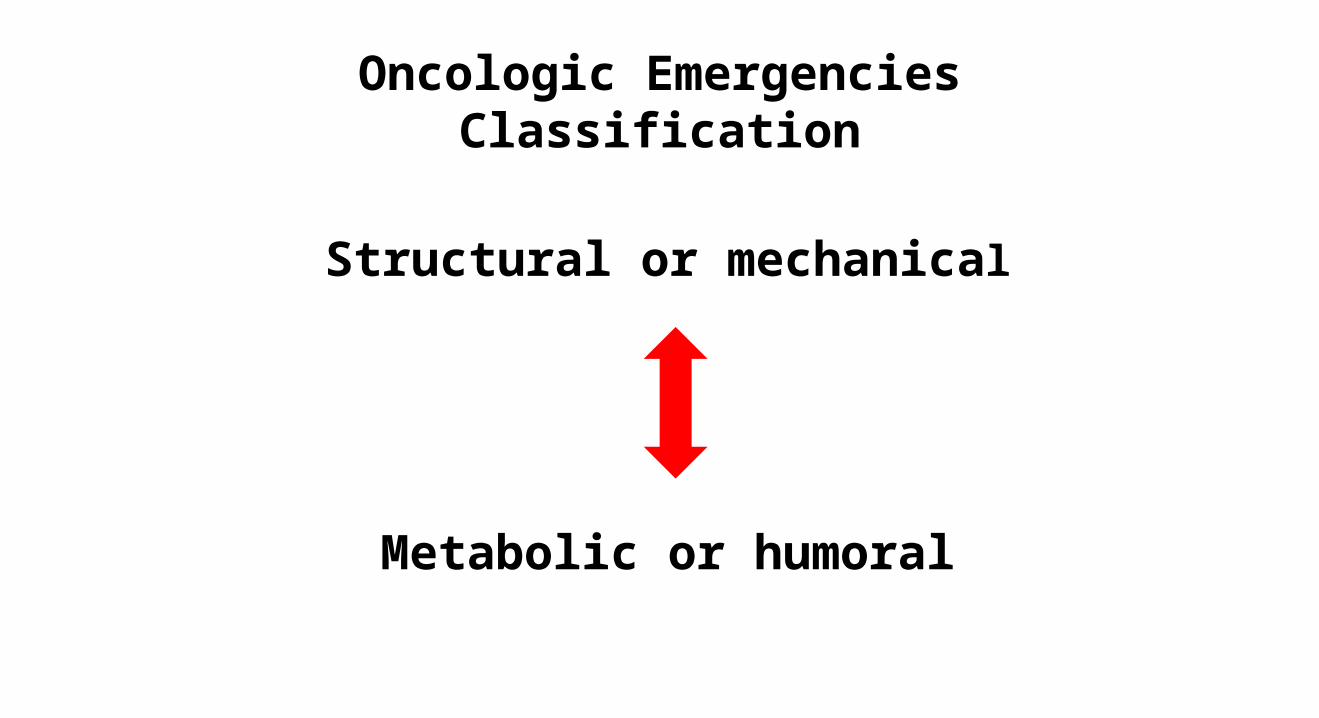
Oncologic EmergenciesClassification
Structural or mechanical
Metabolic or humoral

Oncologic EmergenciesConcepts
• Emergency related to the cancer treatment
– Chemotherapy• TLS
– Targeted Agents• Hemorrhage• Bowel perforation • “Allergic reaction” → Infusion reaction
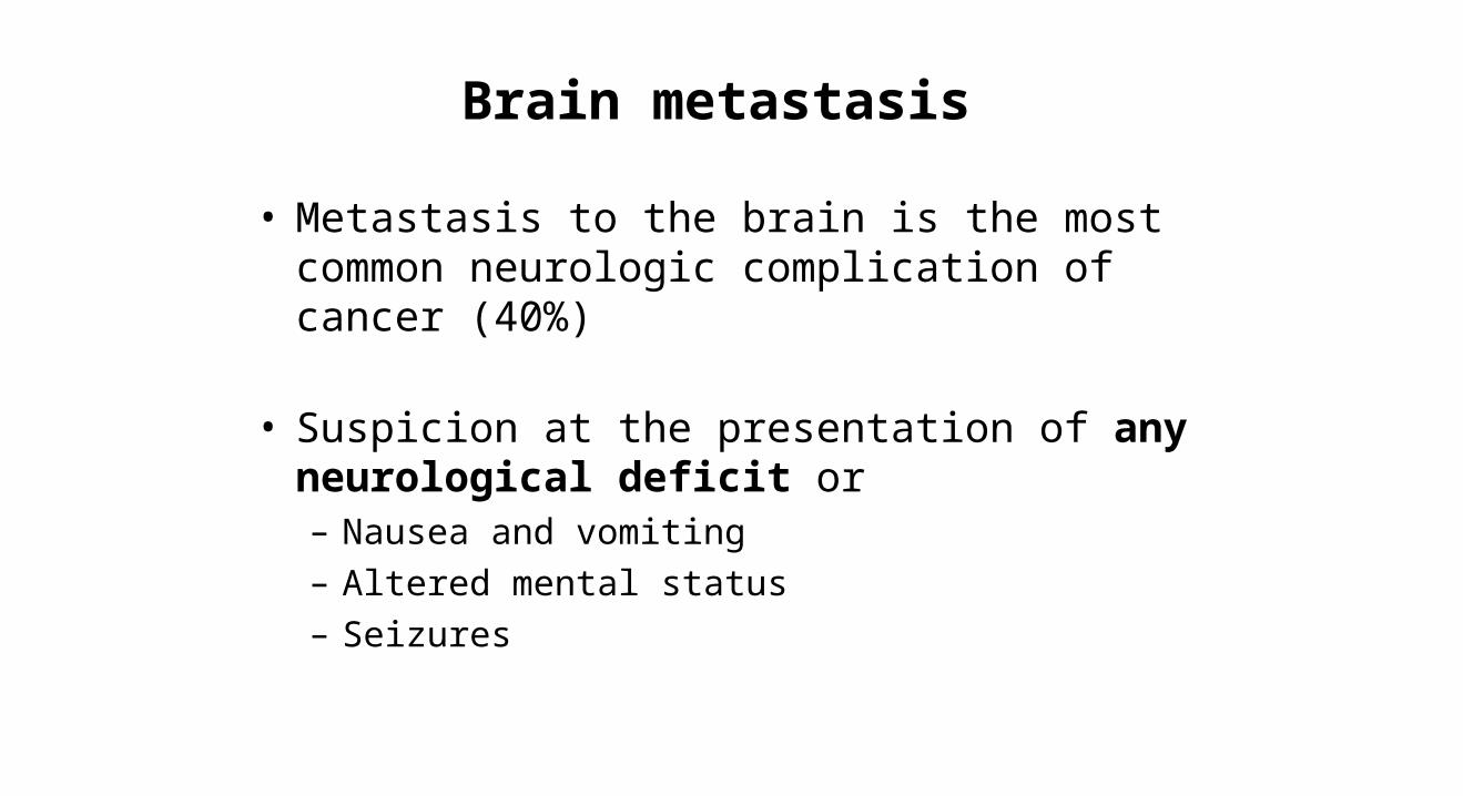
Brain metastasis
• Metastasis to the brain is the most common neurologic complication of cancer (40%)
• Suspicion at the presentation of any neurological deficit or– Nausea and vomiting– Altered mental status– Seizures

Brain metastasisDiagnosis
• Contrast-enhanced CT– Without contrast in patient with known lesions when
hemorrhage is suspected
• MRI with gadolinium phase– More sensitive and better for posterior fossa– Expensive – Longer
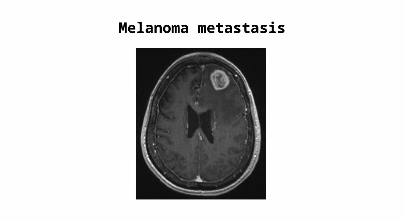
Melanoma metastasis

Brain metastasisEmergencies
• Intracranial hypertension (ICP)– Herniation
• Hemorrhagic stroke
• Seizures

Increased ICPManagement
• Corticosteroids are routinely used to treat increased ICP caused by brain metastasis by reducing vasogenic cerebral edema. Dexamethasone
• Osmotic diuresis: Mannitol and hypertonic saline
• Intensive Care: ICP monitoring and Hyperventilation


Malignant cord compression
• Called epidural spine compression (ESCC)
• Defined as : Compression of the dural sac and its contents by an extradural tumor mass. The minimal radiological evidence for SCC is indentation of the theca at the level of the clinical symptoms
• Anatomical Causes: Epidural compression by tumor mass or pathological Fx of the vertebrae and retropulsion of bony fragments into the canal


Malignant cord compression

Malignant cord compression
• Probability in patient with cancer 2-5%– LUNG, BREAST, MYELOMA, PROSTATE
• Consequences: irreversible loss of neurologic function
• Clinical presentation: pain, (seven weeks before neurologic symptoms), weakness, bladder and bowel dysfunction (later)

Malignant cord compressionDistribution
• Thoracic spine 50-70%
• Cervical 10-30%
• Lumbosacral 20-30%

Malignant cord compressionDiagnosis
• History and physical examination
• Imaging– Spine X ray– CT scan– MRI

Malignant cord compressionDiagnosis
• History and physical examination– Basic neurologic examination ALWAYS!!!
• Imaging– Spine X ray– CT scan
– MRI of the entire spine

Malignant cord compression Treatment
• Symptomatic treatment: – Pain control, anticoagulation*,avoid constipation
• Glucocorticoids – Not clarity between initial high vs low dose (range 10
to 100 Dexamethasone ) then 16 mg in divided doses

Malignant cord compression Treatment
• Definitive treatment: spinal stability, type of tumor and grade of cord compression
• Surgery: resection + stabilization → Rdt
• Radiation :– Radiosensitive : Lymphoma, SCLC, MM, Seminoma– Radioresistance : Melanoma, RCC, CRC, Sarcoma– Dose and schedule : Range (8Gy x1 to 40Gy x20 )
• Prognostic factors associated with shortened survival after RT include relatively radioresistant histology, the presence of visceral metastases or other bone metastases, non-ambulatory status at treatment, an interval from the original diagnosis to ESCC ≤15 months, and an interval <14 days from the onset of motor symptoms to the initiation of RT

Malignant cord compression Treatment
• Stereotactic body radiotherapy (STBR)– SBRT with a single 16-24 Gy fraction gives excellent tumor control, even in patients who
have relatively radioresistant tumors.
• Chemotherapy
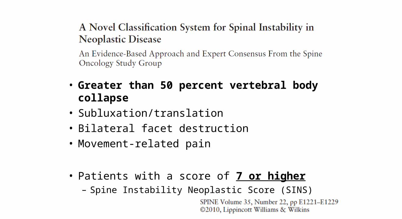
• Greater than 50 percent vertebral body collapse• Subluxation/translation• Bilateral facet destruction• Movement-related pain
• Patients with a score of 7 or higher– Spine Instability Neoplastic Score (SINS)

Superior vena cava syndrome

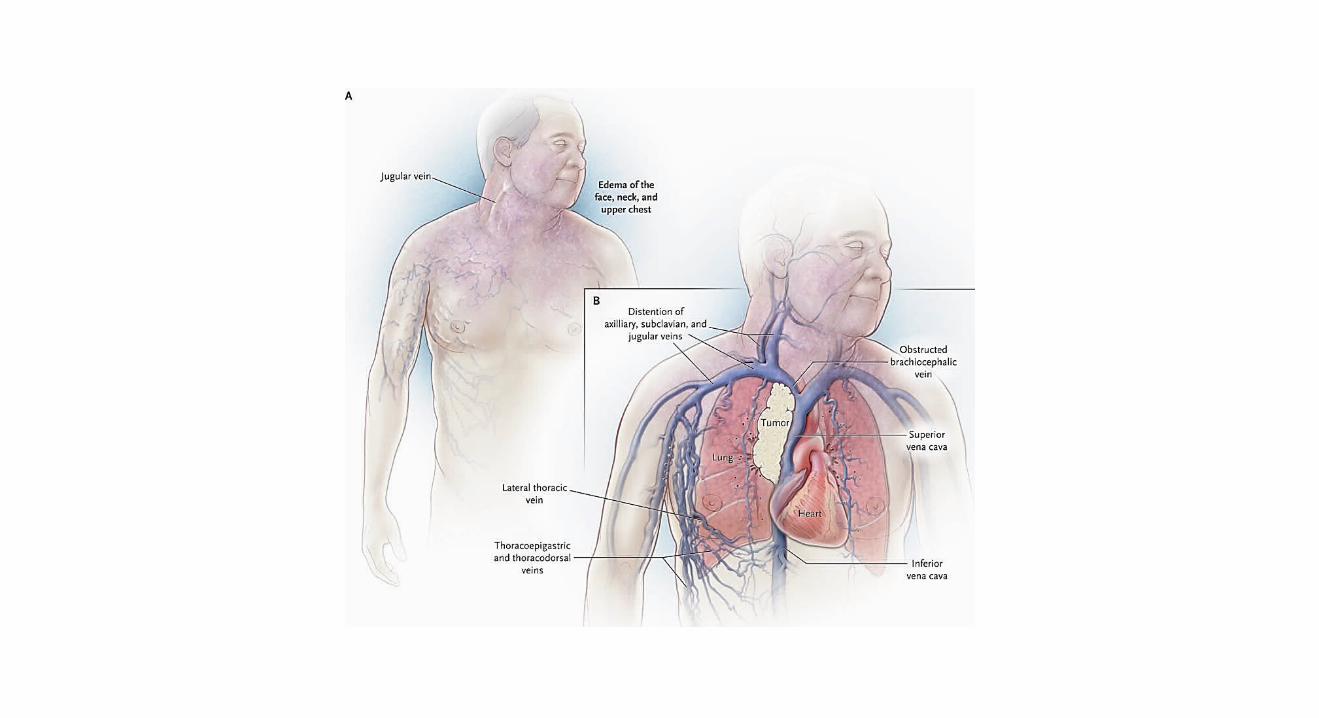

Any condition that leads to obstruction of blood flow
through the SVC

Superior vena cava syndrome
• Malignancy accounts for 60% to 85% of cases– 20% to 40% intravascular devices causing thrombosis. – Lung cancer and non-Hodgkin lymphoma are responsible for most cases
• Symptom severity depends on the extent of the obstruction and its rapidity of onset– Dyspnea, facial swelling, and distended neck veins – Collaterals develop over time and slow the progression symptoms

Superior vena cava syndromeDiagnosis
• Chest x ray– Abnormal in 80% of the cases : Widening and pleural effusion
• US– First test in patients with vascular devices who present with extremity
swelling.
• CT Chest– Test of choice : identification of the cause and collaterals

Superior vena cava syndromeManagement
• Emergent treatment is indicated in patients with airway obstruction or laryngeal edema. However, SVC syndrome most commonly develops gradually, and treatment can be delayed until the primary diagnosis is established.
• Radiation (NSCLC)
• Steroids (NHL and Thymoma)
• Chemotherapy (SCLC, testicular, NHL)

Superior vena cava syndromeManagement
• Angioplasty and stenting
• Surgery
• Thrombolytic therapy
• The American College of Chest Physicians recommends establishment of a histologic diagnosis before instituting treatment in stable patients

Superior vena cava syndromeManagement
Always try to obtain and histologic diagnosis
Exceptions
Central airway obstruction
Coma because of cerebral edema

Pericardial tamponade

Pericardial effusion
•Thoracic radiation– During radiation of early after finished– Recall reaction
•Infectious•Paraneoplastic autoimmune
– Immunotherapies for cancer
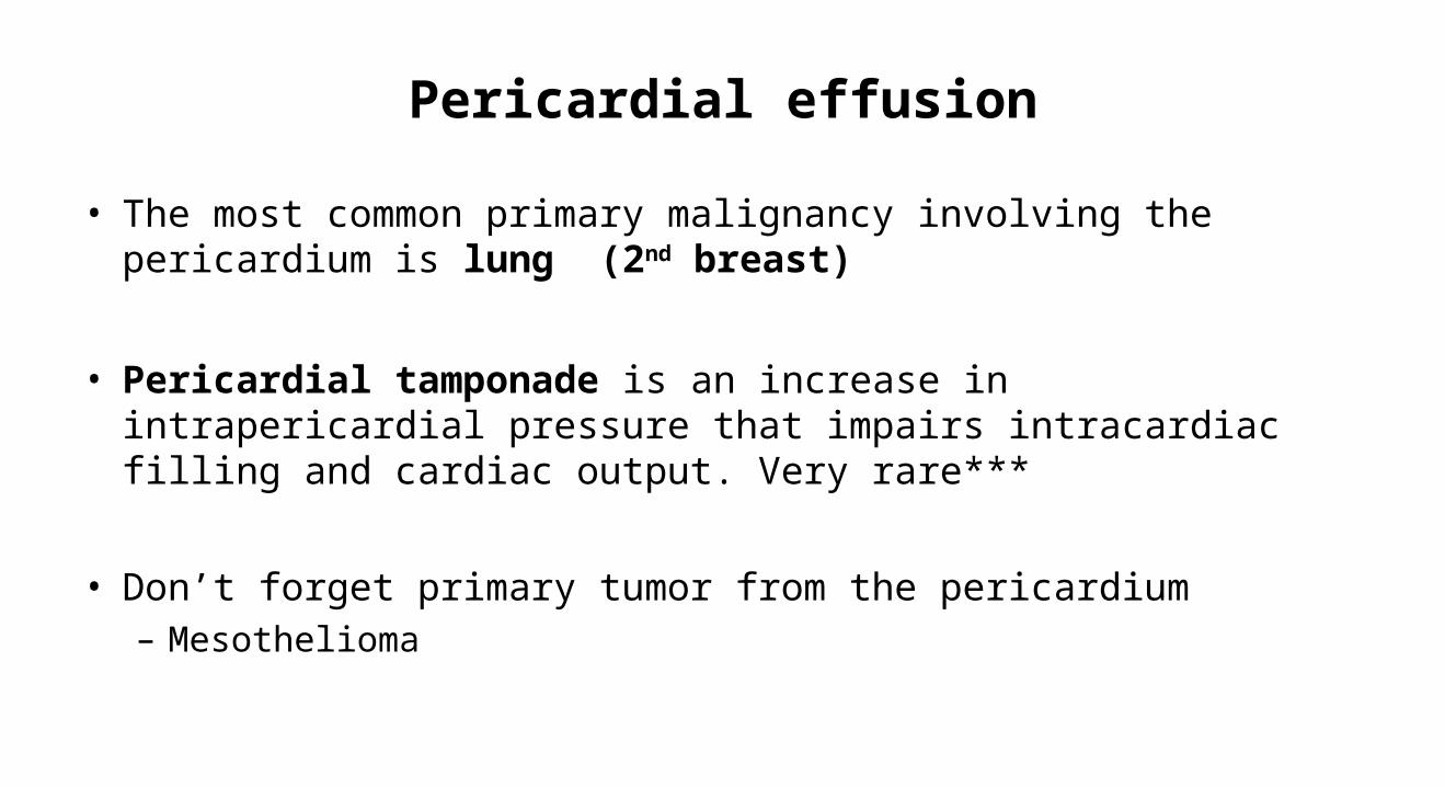
Pericardial effusion
• The most common primary malignancy involving the pericardium is lung (2nd breast)
• Pericardial tamponade is an increase in intrapericardial pressure that impairs intracardiac filling and cardiac output. Very rare***
• Don’t forget primary tumor from the pericardium– Mesothelioma

Pericardial tamponadeDiagnosis and Treatment
• Echocardiography – Right atrial collapse is a more sensitive marker of
pericardial tamponade, whereas right ventricular collapse is more specific.
• Emergent pericardiocentesis– Indwelling catheter– Window

Intestinal emergencies
• Acute bowel obstruction– CRC 10-30%– Ovarian Cancer 20-50%*– Treatment options: surgery, endoscopic intervention, and pharmacologic palliation; self-
expanding metallic stents
• Perforation – CRC and lymphomas– Targeted agents: Anti-angiogenic agents Bevacizumab
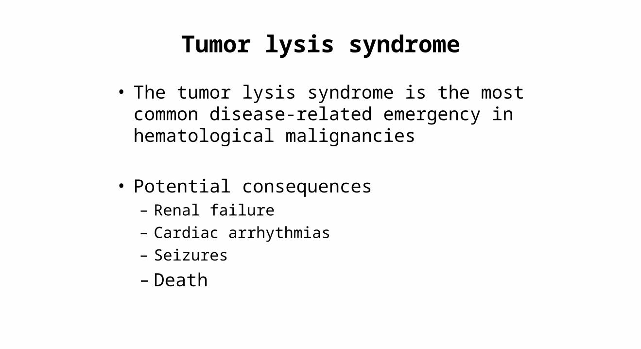
Tumor lysis syndrome
• The tumor lysis syndrome is the most common disease-related emergency in hematological malignancies
• Potential consequences – Renal failure– Cardiac arrhythmias– Seizures
– Death

Tumor lysis syndrome
• Usually after treatment (most effective most probable)
– Can be spontaneous
• Most common hematological malignancies compared with solid tumors but…– LMA and NHL– Combination chemotherapy – Targeted agents : started to be reported

Tumor lysis syndrome
• Syndrome that may include :
• hyperkalemia• hyperphosphatemia• hypocalcemia• hyperuricemia
massive tumor cell death
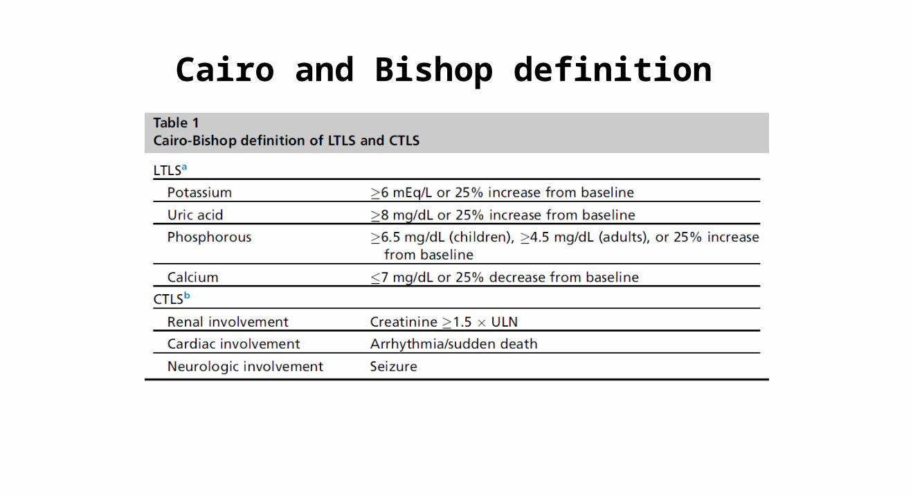
Cairo and Bishop definition


Management
• Hydration
• Allopurinol*
• Rasburicase: most widely accepted dosing is based on the 2008 International Expert Panel on TLS, which suggests a dose of 0.1 mg/kg daily for TLS prevention and 0.2 mg/kg daily for TLS treatment
• Alkalinization: not longer recommended


Hypercalcemia
• Hypercalcemia is the most common oncologic metabolic emergency (10% to 30% at some point during disease course)
• It is defined as a total serum calcium concentration greater than 10 mg/dL or an ionized calcium concentration greater than 5.6 mg/dL.
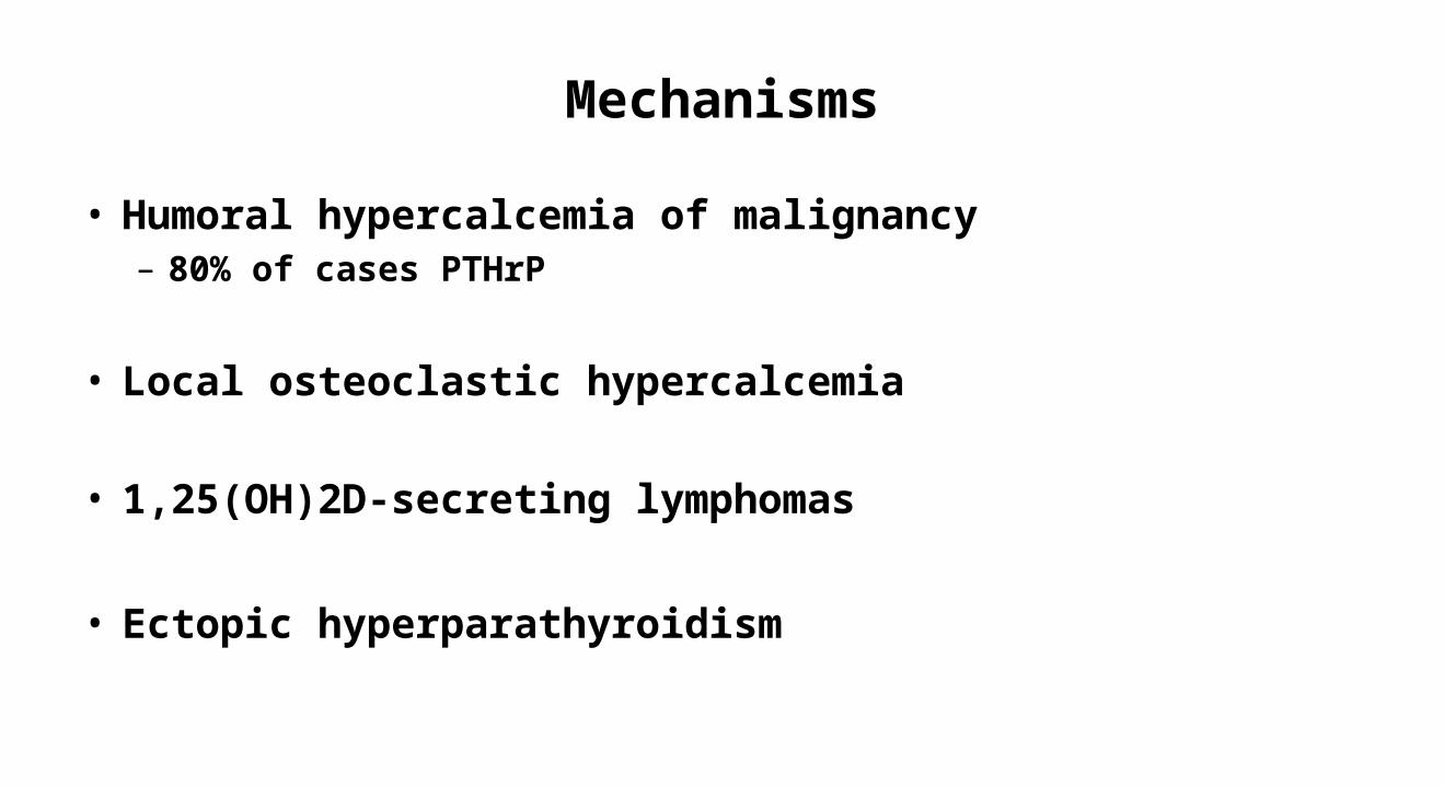
Mechanisms
• Humoral hypercalcemia of malignancy– 80% of cases PTHrP
• Local osteoclastic hypercalcemia
• 1,25(OH)2D-secreting lymphomas
• Ectopic hyperparathyroidism

HypercalcemiaPresentation
• Neurologic symptoms – Lethargy, confusion (coma)
• Gastrointestinal– Constipation, Nausea, Anorexia – Pancreatitis
• Renal– Nephrogenic diabetes insipidus (Poliuria, polidipsea)

Diagnosis
• Measurement – Always check with albumin and correct– Ideally check ionized calcium
• Levels– Mild <12 mg/dL [3 mmol/L]
– Moderate 2 to 14 mg/dL [3 to 3.5 mmol/L]
– High >14 mg/dL [3.5 mmol/L]

HypercalcemiaTreatment
• Hydration (volume expansion)– NaCL 0,9% 150-200 ml urine output 80-100 ml
– Loop Diuretics ???
• Bisphosphonates– Always in high Ca+ almost always in moderate– Check renal function– Maximum effect in two to four days– Zolendronic Acid (4mg over 15-20 min)– Pamidronate ( 60-90-mg over 2 hours)

HypercalcemiaTreatment
• RANKL inhibitors– Denusumab **recommended patients with zoledronic acid
(ZA)-refractory hypercalcemia 6-120 sc
– Can be used in the setting of renal dysfunction– Can cause hypocalcemia (Check vit D levels before )
• Steroids: Lymphomas (granulomatous component)
• Calcitonin (only in symptomatic patients with high Ca+)
• Hemodialysis
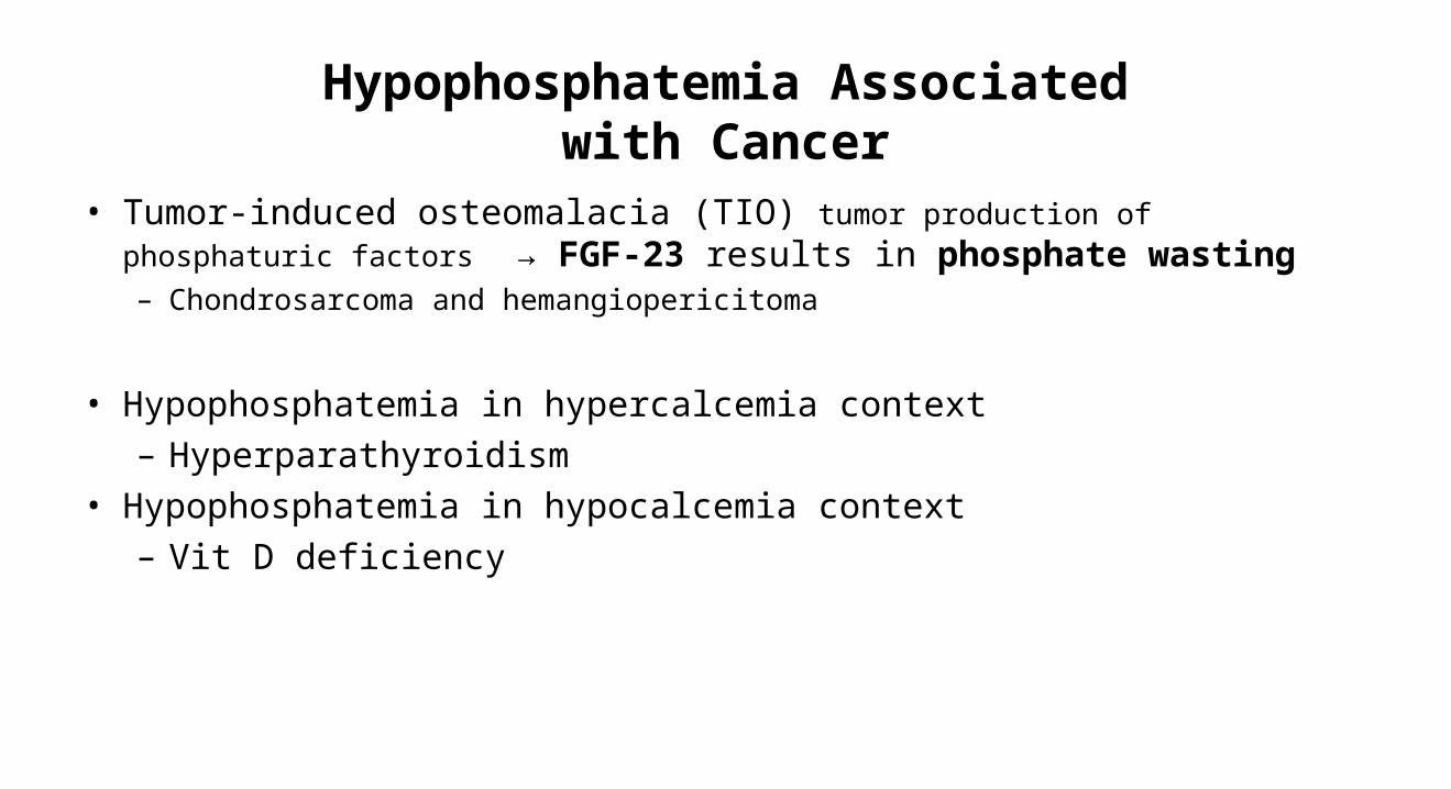
Hypophosphatemia Associated with Cancer
• Tumor-induced osteomalacia (TIO) tumor production of phosphaturic factors → FGF-23 results in phosphate wasting– Chondrosarcoma and hemangiopericitoma
• Hypophosphatemia in hypercalcemia context– Hyperparathyroidism
• Hypophosphatemia in hypocalcemia context– Vit D deficiency

Hyponatremia in Cancer
• Hyponatremia is the most common electrolyte disorder encountered in patients with malignancies ( 20-30%)
• Marker for inpatient mortality in cancer patients
• Can be an oncological emergency

SVC
ESCC
Brain Mets
Cardiac Tamponade
Intestinal obstruction
Mechanical complications
PEEsophago-
bronquial fistula
Central airway obstruction

Leukostasis
DIC
TLS
Tumor
↑K ↑P↑Uric Acid
Hypercalcemia
Hyponatremia

Message
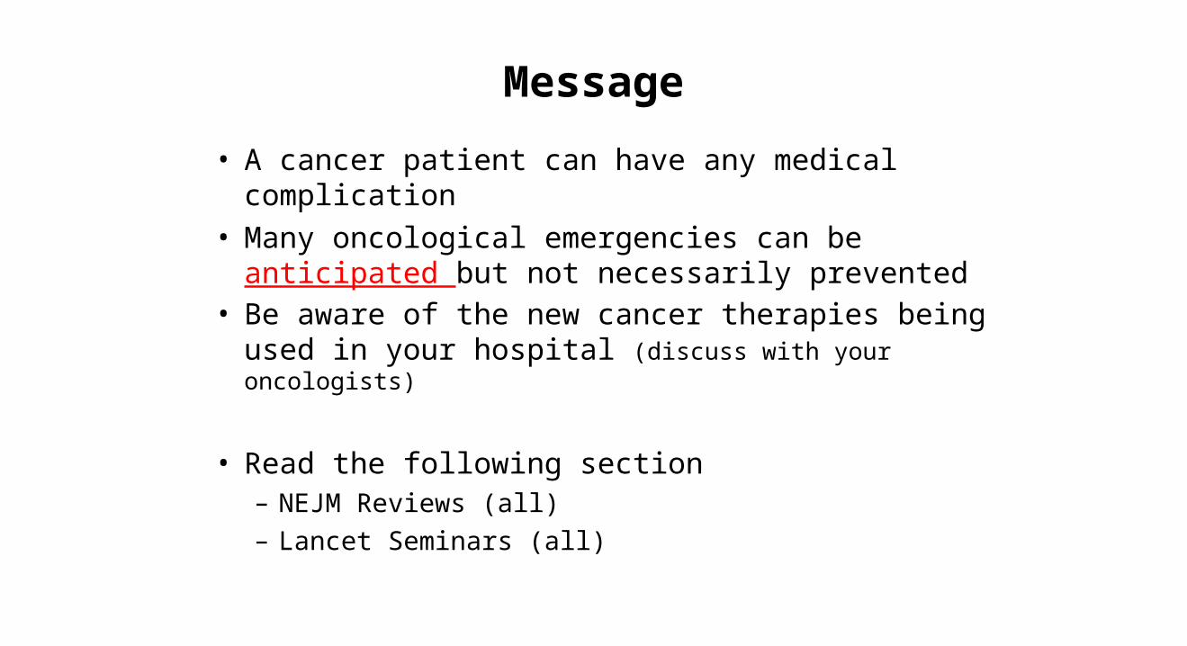
• A cancer patient can have any medical complication
• Many oncological emergencies can be anticipated but not necessarily prevented
• Be aware of the new cancer therapies being used in your hospital (discuss with your oncologists)
• Read the following section– NEJM Reviews (all) – Lancet Seminars (all)

NCI-NIH Clinical Research Center


