Bartlett et al. Molecular Cancer 2013, 12:103http://www.molecular-cancer.com/content/12/1/103
REVIEW Open Access
Oncolytic viruses as therapeutic cancer vaccinesDavid L Bartlett1, Zuqiang Liu1, Magesh Sathaiah1, Roshni Ravindranathan1, Zongbi Guo2, Yukai He3
and Zong Sheng Guo1*
Abstract
Oncolytic viruses (OVs) are tumor-selective, multi-mechanistic antitumor agents. They kill infected cancer andassociated endothelial cells via direct oncolysis, and uninfected cells via tumor vasculature targeting and bystandereffect. Multimodal immunogenic cell death (ICD) together with autophagy often induced by OVs not only presentspotent danger signals to dendritic cells but also efficiently cross-present tumor-associated antigens from cancercells to dendritic cells to T cells to induce adaptive antitumor immunity. With this favorable immune backdrop,genetic engineering of OVs and rational combinations further potentiate OVs as cancer vaccines. OVs armed withGM-CSF (such as T-VEC and Pexa-Vec) or other immunostimulatory genes, induce potent anti-tumor immunity inboth animal models and human patients. Combination with other immunotherapy regimens improve overalltherapeutic efficacy. Coadministration with a HDAC inhibitor inhibits innate immunity transiently to promoteinfection and spread of OVs, and significantly enhances anti-tumor immunity and improves the therapeutic index.Local administration or OV mediated-expression of ligands for Toll-like receptors can rescue the function of tumor-infiltrating CD8+ T cells inhibited by the immunosuppressive tumor microenvironment and thus enhances theantitumor effect. Combination with cyclophosphamide further induces ICD, depletes Treg, and thus potentiatesantitumor immunity. In summary, OVs properly armed or in rational combinations are potent therapeutic cancervaccines.
IntroductionIn the last few years, there is mounting evidence that OVsare effective in treating cancer in both preclinical modelsand clinical trials with human patients [1-3]. The antican-cer activities of OVs are derived from multimodal cancerkilling mechanisms. The first is the direct oncolysis of can-cer cells by the virus, which is, in most cases a mixture ofapoptosis, necrosis, pyroptosis and autophagic cell death,often with one as predominant for a particular OV. Thesecond is apoptotic and necrotic death of uninfected cellsinduced by anti-angiogenesis and anti-vasculature of theOVs as shown in animals and humans [4-6]. The last iscytotoxicity to cancer and stromal cells by activated innateand tumor-specific immune cells [7-9]. The antitumor im-munity helps eliminate the uninfected cancer cells
* Correspondence: [email protected] of Pittsburgh Cancer Institute and Department of Surgery,University of Pittsburgh, Pittsburgh, PA 15213, USAFull list of author information is available at the end of the article
in primary and metastatic nodules, and enforcemicrometastases in dormant state.Cancer vaccines are designed to boost the body’s immune
system to protect itself from carcinogenesis and progressionof cancer. The Food and Drug Administration of the USAhas approved both prophylactic and therapeutic vaccinesfor cancer in the last few years. The prophylactic vaccinesare against the hepatitis B virus, which can cause liver can-cer, and against human papillomavirus types 16 and 18,which are responsible for about 70 percent of cervical can-cer cases. These anti-viral vaccines are highly effective incurbing virus infections and onset of cancer. In contrast,therapeutic cancer vaccines are difficult to develop andmuch less effective. As a benchmark, Provenge, a cancervaccine designed to treat advanced prostate cancer, demon-strated an increase in survival and thus gained approvalfrom the FDA for use in the treatment of advanced prostatecancer patients in 2010 [10]. The approval of Provenge hasstimulated interest in development of other therapeuticcancer vaccines.
Ltd. This is an Open Access article distributed under the terms of the Creativeommons.org/licenses/by/2.0), which permits unrestricted use, distribution, andiginal work is properly cited.
Bartlett et al. Molecular Cancer 2013, 12:103 Page 2 of 16http://www.molecular-cancer.com/content/12/1/103
OVs provide a number of potential advantages as can-cer vaccines over conventional therapies. First, OVs aretumor-selective, thus in situ cancer vaccines, providinghigher cancer specificity and better safety margin. Sec-ond, immunogenic/inflammatory types of cell death, in-cluding recently characterized “immunogenic cell death”(ICD) of cancer and stromal cells induced by OVs pro-vides a natural repertoire of tumor-associated antigens(TAAs) in conjunction with danger signals [damage-asso-ciated molecular pattern (DAMP) and OV-derivedpathogen-associated molecular pattern (PAMP) mole-cules, and inflammatory cytokines] [11-13], to elicit anti-tumor immunity. However, just like other immunothera-peutic regimens, a number of challenges remain for OVs-mediated cancer vaccines. For example, the relative ineffi-ciency of delivering OVs to tumor nodules, selective viralreplication inside tumor nodules and spread to distantmicrometastases limits its overall efficacy. In order tomake up this deficiency, it often requires combinationswith conventional treatments for cytoreduction to de-crease the tumor burden. Most TAAs are self-antigensand thus weakly immunogenic. In addition, a highly im-munosuppressive tumor microenvironment (TME) oftensuppresses the activities of tumor-infiltrated lymphocytes(TILs) generated spontaneously, by adoptive cell transferor by active immunization such as cancer vaccines. There-fore, the balance between tumor growth and the status ofthe TME, versus the magnitude and avidity of antitumorimmune response elicited by a therapeutic vaccine inaddition to oncolytic potency by an OV ultimately deter-mines the therapeutic efficacy by this approach [9,14-17].In this review, we briefly introduce oncolytic virotherapy
and cancer immunotherapy, then focus on the rationalesand strategies of utilizing replicating OVs as therapeuticcancer vaccines, and combination strategies that have ledto potent antitumor immunity in preclinical models anddemonstration of the effectiveness of two OVs in clinicaltrials.
OVs and cancer immunotherapyOVs possess the ability to selectively infect and replicate incancer and associated endothelial cells and kill these cellsin cancerous tissues while leaving normal tissues un-harmed [1,3]. Many naturally occurring OVs have a prefer-ential tropism for tumor and/or associated endothelialcells. Others are genetically engineered to change theircellular or organ tropism to cancer. The mechanisms oftumor targeting by OVs, which include selectivity to can-cer cells and/or associated endothelial cells with alteredsignaling pathways of RB/E2F/p16, p53, PKR, EGFR, Ras,Wnt, anti-apoptosis, hypoxia conditions, or defects in IFNand other cellular innate immune signaling pathways havebeen reviewed [1,3,18]. The altered signaling pathways fos-ter favorable cellular environments for specific OVs to
replicate sufficiently in cancer cells and/or associated endo-thelial cells, leading to direct oncolysis of the infected cells.Viruses often display specificity for a cell type, tissue or
species, collectively known as viral tropism. Cytokines,particularly interferons and tumor necrosis factors, playkey roles in dictating the viral tropism [19,20]. Comple-ment system seems to play certain roles, as shown forNewcastle disease virus [21]. OVs also displayed speciesspecificity even though they broaden their tropism to can-cer cells from non-permissive species to various degrees.Myxoma virus, a poxvirus previously considered rabbitspecific, can replicate productively in a variety of humantumor cells [22]. Bovine herpes virus type 1 is a species-specific virus that fails to induce cytopathic effects in hu-man normal cells, yet is capable of infecting and killing avariety of immortalized and transformed human cell types[23]. However, human Ad can infect murine cancer cellsyet the production of infectious virus progeny is often lim-ited. One reason is the failure of translation of viralmRNAs and this could be overcome partially by expres-sion of L4-100 K in trans [24]. It is important to note thatOVs show aberrant, non-productive infection in non-native hosts such as mouse cells. In this case, the resultingmode of cell death can considerably differ from oncolysisin human cancer cells. As we will discuss later, the modeof cell death dictates to a large degree the subsequentantitumor immunity. Consequently, the antitumor immun-ity determined by studies in immunocompetent animalmodels with syngeneic tumors might not be relevant to thesituation in human cancer patients.OVs mediate multimodal killing of cancer and stromal
cells ranging from direct virus-mediated cytotoxicity[25-28], cell death due to anti-angiogenesis and vascula-ture targeting by OVs, to cytotoxic immune effector-induced cytotoxicity. The types of cell death, as classi-fied by morphological and ultrastructural changes dur-ing cell death, are apoptosis, necrosis, and autophagiccell death. With the exception of apoptosis, all othertypes of cell death have been considered to be inflam-matory and immunogenic. However, recent studies byinvestigators working on chemotherapy and radiationfor cancer therapy have led to new concepts, that apop-totic cell death can be divided into “immunogenic celldeath” (ICD) and “non-immunogenic cell death” (NICD)[29-31]. Based on this new classification, apoptotic celldeath caused by some OVs are ICD. Together, immuno-genic apoptosis, necrosis, autophagic cell death andpyroptosis of cancer and associated endothelial cells causedby OVs release and present danger signals (DAMPs andPAMPs as signal 0) and TAAs (as signal 1) to dendritic cells(DCs) for antitumor and antiviral immune responses.Immunotherapy has been a bright spot in the field of
novel therapeutics for cancer in the last few years [32-34].Tumor cells and associated stromal cells such as
Bartlett et al. Molecular Cancer 2013, 12:103 Page 3 of 16http://www.molecular-cancer.com/content/12/1/103
endothelial cells express a wide variety of proteins that canfunction as antigens including mutated proteins, fusionproteins, developmentally and tissue-restricted proteins, aswell as tumor-selectively over-expressed proteins, termedas TAAs [17]. These TAAs are direct targets for most im-munotherapeutic regimens, either active immunization oradoptive transfer of activated immune cells [14,33,34]. TheTME, in which cancer cells, stromal cells and infiltrated im-mune cells, as well as soluble molecules interact with eachother and dictate its properties, is immune tolerangenic ormore likely immunosuppressive [35]. However, the TMEand associated signaling pathways can be manipulated toactivate antitumor immunity in a therapeutic regimen.Thus, a number of immunotherapeutic strategies are aimedto disrupt the immune-regulatory circuits that are criticalfor maintaining tumor tolerance, such as CTLA-4 and PD-1, and augment protective antitumor immunity [15,35-37].
OV-induced ICD and autophagy elicit antitumorimmune responsesThe cell death can be classified according to morpho-logic and ultrastructural changes of dying cells, intoapoptosis, necrosis, autophagic cell death, pyroptosisand a few other types of death [38,39]. As stated, necrosis,pyroptosis and autophagic cell death are proinflammatoryand immunogenic. Necrosis release DAMPs from dyingcells. Autophagic cell death also releases many DAMPs.Pyroptosis, triggered by pathogens [40], is highly inflam-matory. The only exception is apoptosis. Apoptotic celldeath was considered to be non-immunogenic and non-inflammatory by nature (Table 1). However, recent studiessuggest that, under certain circumstances, apoptosis canbe ICD [29,30,41,42]. ICD involves changes in the com-position of the cell surface as well as the release of solublemediators, occurring in a defined temporal sequence. Atthe early phase of immunogenic apoptosis, surface-
Table 1 Types of cell death and their immunological consequ
Type of cell death
Apoptosis (type 1 cell death). This is accompanied by a rounding up of thecell, retraction of pseudopods, reduction of cellular volume, chromatincondensation, nuclear fragmentation, few or no ultrastructural modificationsof cytoplasmic organelles, and plasma membrane blebbing, but theintegrity of the cell is maintained until the final stages of the process.
Autophagic cell death (ACD; type 2 cell death). Occurs without chromatincondensation but is accompanied by massive autophagic vacuolization ofthe cytoplasm. The term ACD simply describes cell death with autophagy.
Necrosis (type 3 cell death). Characterized by a gain in cell volume,swelling of organelles and rupture of plasma membrane, and subsequentloss of intracellular contents, including HMGB1, ATP, etc.
Pyroptosis (or caspase 1-dependent cell death). It is a highlyinflammatory form of cell death mediated by the inflammasome andcaspase-1 activation, and triggered by various pathological stimuli,such as microbial infection, or stroke, heart attack and cancer.
Secondary necrosis. This is the dissolution of the cell followingapoptosis. Some remaining cellular contents are released.
exposed calreticulin (ecto-CRT) and secreted ATP arecrucial DAMPs [43]. While calreticulin (CRT) exposureon the cell surface prior to apoptosis dictates the immuno-genicity of cancer cell death [29,30,41,42], ERP57 is a keyprotein that controls immunogenicity by controlling CRTexposure [44,45]. In response to ICD inducers, activation ofendoplasmic reticulum (ER) stress is indispensable to con-fer the immunogenic character of cancer cell death, becauseER stress can coordinate the danger signaling pathways re-sponsible for the trafficking of vital DAMPs and subsequentanti-cancer immune responses. Other pathways such as au-tophagy (discussed later) have the ability to influence dan-ger signaling and thus antitumor immune response [46]. Atlater stages, other DAMPs such as HMGB1 are releasedfrom dying cancer cells and secreted from activated infil-trated immune cells [13,43,47-49]. Kroemer, Zitvogel andothers believed that ICD constitutes a prominent pathwayfor the activation of the immune system against cancer,which in turn determines the long-term success of antican-cer therapies [43,48,50]. The immunogenic characteristicsof ICD are mainly mediated by DAMPs that include ecto-CRT, secreted ATP and released HMGB1. Thus, the revisedconcept ICD would include not only immunogenic apop-tosis, but also necrosis, pyroptosis, and autophagic celldeath [29,30,42,43,46,51-53].Cancer cell death induced by OVs is mostly immuno-
genic (Table 2). For example, an oncolytic hTERT-Ad in-duced autophagic cell death in tumor cells and insubcutaneous gliomas, which is immunogenic [54]. Mea-sles virus causes ICD in human melanoma cells [55].Interestingly, a significant portion of the in vivo tumorkilling activity by OVs, e.g., vesicular stomatitis virus(VSV) and vaccinia virus (VV), is caused by indirect kill-ing of uninfected tumor cells [4]. OVs also target endo-thelial cells and tumor vasculature, leading to infectionand lysis of endothelial cells, and more necrotic death of
ence
Immunogenicity
Some forms of apoptosis are non-immunologic, while others areimmunogenic. The pre-apoptotic surface exposure of CRT andHSP70/HSP90 may have a profound impact on the immune response. Inaddition, the release of HMGB1 during late apoptosis promotes antigenprocessing by DCs and hence contributes to cytotoxic T-cell activation.
High. It may release DAMPs (HMGB1, ATP, and others) and elicitsubstantial inflammation.
High. This causes release of DAMPs and elicits substantial inflammationand affects local environment.
High. It is a highly inflammatory form of cell death due to cytokinerelease and escape of cytoplasmic contents (DAMPs). However, somepathogens encode immunosuppressive proteins.
High. It is quite immunogenic due to necrosis occurring inapoptotic cells at the late stage.
Table 2 OVs induce ICD and/or promote antitumor immunity in animal models or human patients (*)
Virus Modifications ICD and DAMPs (in vitro) Antitumor Immunity (in vivo) Reference
Enhanced autophagy; ecto- CRT;released ATP and HMGB1
Tumor-specific T cell responses and antitumor efficacy insome patients [clinical trial]
[62]
HSV
G207 R34.5-; ICP6- NA Systemic antitumor immunity (CD8+ T cells) [66]
HSV-1716 ICP 34.5 gene mutant Induction of IFN-γ, CXCL9 andCXCL10
Intratumoral injection increased NK and CD8+ T cells [67]
T-VEC ICP47-γ34.5 - GM-CSF + Necrosis/apoptosis (in vivo) Antigen-specific T cell responses and decreases in Treg, Ts,and MDSC in human melanoma patients [clinical trials]
Allowing DC to mature, produce high level of IFN-α, and cross-presentTAAs and production of tumor-specific CD8 T cells
[75,76]
*Notes:(1). data for T-VEC and Pexa-Vec are from human patients;(2). NA, not assessed.
Bartlettet
al.Molecular
Cancer2013,12:103
Page4of
16http://w
ww.m
olecular-cancer.com/content/12/1/103
Bartlett et al. Molecular Cancer 2013, 12:103 Page 5 of 16http://www.molecular-cancer.com/content/12/1/103
cancer cell cells due to disruption of tumor vasculature[4-6,56,57]. As for the release of DAMPs from dyingcancer cells, we first reported that cancer cells infectedby an oncolytic virus, led to necrotic/apoptotic deathpathways and HMGB1 was released into the extracellularmilieu [58]. As it turns out, HMGB1 release is a universalphenomenon for OVs, as shown in cancer cells infectedwith an Ad [59], a measles virus [55], an HSV-2 [60], anda coxsackievirus B3 [61]. Extracellular ATP is another po-tent danger signal released from OV-infected cancer cells[59,61,62]. Together, tumor cell death and ATP releasemay prime DC and lead to efficient antitumor immunity[63]. Finally, activated innate immune cells and elicitedadaptive anti-cancer immunity as well as inflammatory cy-tokines kill additional cancer cells and stromal cells, lead-ing to release of DAMPs such as HMGB1 [64]. Insummary, these studies strengthen the notion that OVs in-duce immunogenic types of cell death and present/releasea number of danger signals, and TAAs to DCs and
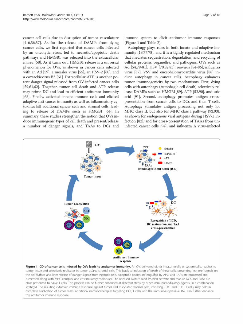
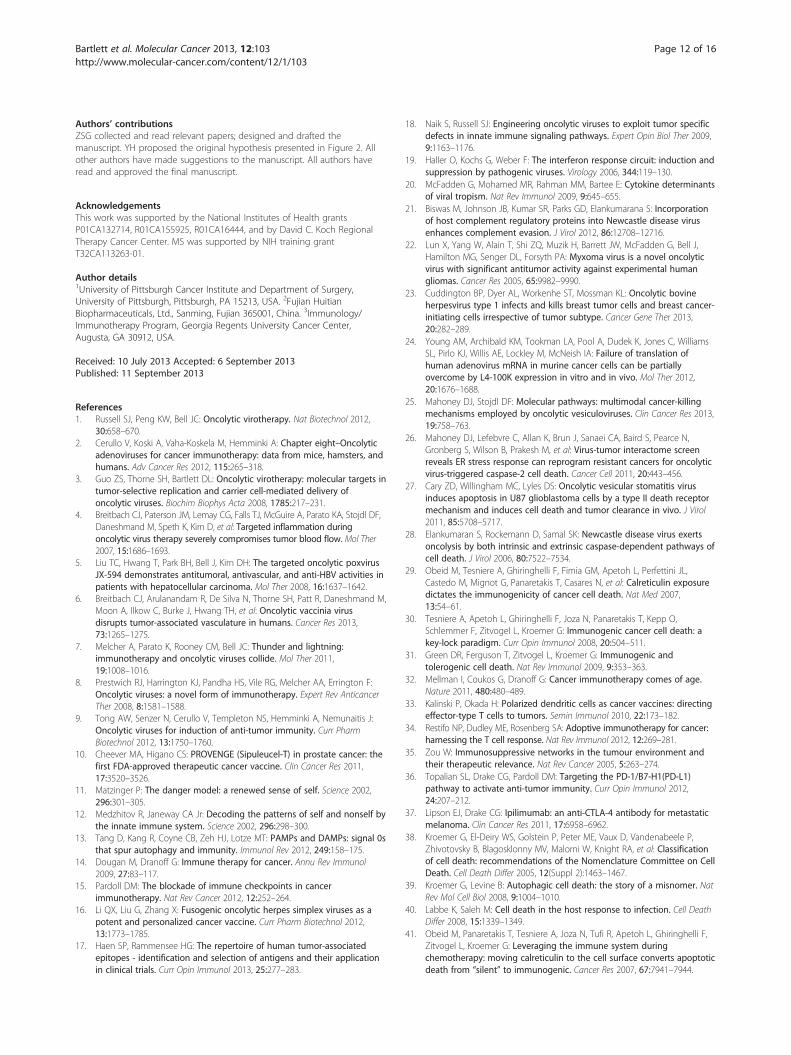
Figure 1 ICD of cancer cells induced by OVs leads to antitumor immutumor tissue and selectively replicates in tumor or/and stromal cells. This lethe cell surface and later release of danger signals from necrotic cells. Apoppresented along with MHC complex and costimulatory molecules. The relecross-presented to naive T cells. This process can be further enhanced at dstrategy). The resulting cytotoxic immune response against tumor and assocomplete eradication of tumor mass. Additional immunotherapies targetingthis antitumor immune response.
immune system to elicit antitumor immune responses(Figure 1 and Table 2).Autophagy plays roles in both innate and adaptive im-
munity [13,77,78], and it is a tightly regulated mechanismthat mediates sequestration, degradation, and recycling ofcellular proteins, organelles, and pathogens. OVs such asAd [54,79-81], HSV [70,82,83], reovirus [84-86], influenzavirus [87], VSV and encephalomyocarditis virus [88] in-duce autophagy in cancer cells. Autophagy enhancestumor immunogenicity by two mechanisms. First, dyingcells with autophagy (autophagic cell death) selectively re-lease DAMPs such as HMGB1[89], ATP [52,90], and uricacid [91]. Second, autophagy promotes antigen cross-presentation from cancer cells to DCs and then T cells.Autophagy stimulates antigen processing not only forMHC class II, but also for MHC class I pathway [92,93],as shown for endogenous viral antigens during HSV-1 in-fection [82], and for cross-presentation of TAAs from un-infected cancer cells [94], and influenza A virus-infected
nity. An OV, delivered either intratumorally or systemically, reaches toads to induction of death of these cells, presenting “eat me” signals ontotic bodies are engulfed by APC, and TAAs are processed andased DAMPs (and PAMPs) activate and mature DCs, and TAAs areifferent steps by other immunomodulatory agents (in a combinationciated stromal cells, involving CD4+ and CD8+ T cells, may help inDCs, T cells, and the immunosuppressive TME can further enhance
Bartlett et al. Molecular Cancer 2013, 12:103 Page 6 of 16http://www.molecular-cancer.com/content/12/1/103
tumor cells [95]. Inhibition of autophagy abolished cross-presentation almost completely, whereas induction of au-tophagy dramatically enhanced the cross-presentation ofTAAs. Interestingly, purified autophagosomes could func-tion as efficient antigen carriers for cross-presentation.These studies demonstrated that autophagy within theantigen donor cells facilitates antigen cross-priming togenerate TAA-specific or virus-specific CD8+ T cells[75,94,96], which could be further explored as a new strat-egy to enhance OV-mediated antitumor effects in the fu-ture [97].In summary, ICD and autophagy triggered by a num-
ber of OVs provide a highly favorable backdrop for theimmune system to respond and generate a potent adap-tive antitumor immunity (Table 2).
Oncolytic viruses as therapeutic cancer vaccinesOVs have been explored as therapeutic cancer vaccinesfor quite a few decades. Pioneering work done byLindenmann and Klein in 1967 demonstrated that viraloncolysis of tumor cells by influenza virus increases im-munogenicity of tumor cell antigens [98]. A few decadeslater, Martuza, Toda and others demonstrated that a gen-etically engineered oncolytic HSV G207 functions as an insitu cancer vaccine for induction of specific anti-tumorimmunity in CT26 colon cancer model [66]. When thisvirus is armed with IL-12, the virally expressed IL-12 canwork with the OV synergistically to elicit local and sys-temic anti-tumor immunity [99].In order to make OVs better therapeutic cancer vaccines,
investigators have recently come up with a number of gen-etically engineered and armed OVs and combination strat-egies with other anti-cancer agents that may work eitheradditively or synergistically to produce potent oncolysisand antitumor immunity. A number of studies lead us tonote two interesting findings. One is that adaptive antiviralimmunity may not be all bad. In fact, adaptive antiviral im-munity contributes to oncolytic virotherapy by an oncolyticHSV [100], even though it may not be the case for all OVs.The second is that selectivity of oncolytic viral replicationmay reduce antiviral immunity and toxicity, but it does notimprove antitumor immunity [101]. The therapeutic effi-cacy of an OV is a delicate balance of forces, between ef-fective viral replication and oncolysis, viral clearance byantiviral immunity, and antitumor immunity and factorspromoting tumor growth [102,103]. Thus, any combin-ation with immunotherapy should take antiviral immunityinto account.
A. Genetic modifications of OVs for enhanced immuneresponses
Genetic modifications of OVs aim to relieve the inhib-ition of immune responses by the OVs with deletion of
viral immune evasion genes, and to enhance antitumorimmune responses by inserting immune-enhancingtransgenes into the OV vectors. Clearly, no-armed OVscan elicit antitumor immunity in certain tumor models,as demonstrated with HSV-1 G207, H-1716, MV-EGFPand Coxackievirus B3 [55,61,66,67,104]. However, manystudies have shown that immunological effects can varydepending on a number of factors including tumor im-munogenicity, stage of the tumor and specifics of theparticular OV used. To gain better immunological ef-fects, a number of steps in immune response and mul-tiple cell types can be targeted by armed OVs or bycombination strategies. We will discuss some recentstudies to illustrate these points.
(1) Modulating the innate immunity
Toll-like receptors (TLRs) are a family of pattern rec-ognition receptors that recognize PAMPs and DAMPs,and trigger the activation and maturation of DCs. Asan example, TLR9 responds to viral dsDNA by recog-nizing unmethylated CpG sequences; thus CpG richoligodeoxynucleotides have been used as vaccine adju-vants. Along the same logic, oncolytic DNA virusesenriched with CpG motifs are believed to be strongerimmunogens. Raykov et al. have tested this idea in arat lung hepatoma metastasis model by using autolo-gous tumor cells that were infected with CpG enrichedparvovirus and then irradiated. They showed a signifi-cant reduction in metastatic rate compared with con-trols [105]. Cerullo et al. have also tested the anti-tumor effects of an oncolytic Ad enriched with CpGmotifs (Ad5D24-CpG) in cancer models [106]. In asyngeneic mouse model with B16-OVA melanoma,Ad5D24-CpG significantly improved tumor control,associated a significant increase in tumor and spleenanti-OVA specific T-cells and a decrease in both num-ber and activation of MDSCs in the tumor.
(2) Enhancing the cross-presentation and priming ofTAAs
Heat shock proteins (HSPs) are a family of proteins thatact as molecular chaperones and can be induced or re-leased during cellular stress or necrosis. Once they are ex-posed on the cell surface or released, they become activeDAMPs. Due to their mechanistic abilities to catalyze thefolding of proteins and their intracellular translocation,HSPs can bind potential antigens at a necrotic scene anddeliver them to a variety of antigen presenting cells [107].Oncolytic Ads expressing several HSPs, including HSP70,HSP90 and HSF1, a heat shock transcription factor, havebeen constructed and investigated in tumor models. In-deed, they can function as oncolytic cancer vaccines and
Bartlett et al. Molecular Cancer 2013, 12:103 Page 7 of 16http://www.molecular-cancer.com/content/12/1/103
can induce an MHC restricted tumor antigen-specificCD8+ T cell response in syngeneic melanoma, colorectaland prostate cancer models in immunocompetent mice[108,109]. In fact, an HSP70-overexpressing oncolytic Adhas been tested in a phase I clinical trial [110].As we have discussed earlier, autophagy induced in can-
cer cells has been shown to promote cross-presentation ofTAAs.
(3) Viruses engineered to express cytokines,chemokines and co-stimulatory molecules
Many OVs expressing cytokines (such as IL-2, IL-12,IL-18); chemokines (such as CCL5), or costimulatorymolecules (such as B7.1 and CD40L) have been studiedand some exciting antitumor immunity and therapeuticresults have been documented in animal models and inhuman cancer patients. Due to space limit, we will focuson the GM-CSF armed OVs in this section.Viruses have evolved with genes to suppress the im-
mune system in order to survive and gain maximumreplication in the hosts [111]. In the context of OVs,they may play yin-yang roles. On one hand, they may in-crease viral persistence in the tumor leading to betteroncolysis; while on the other hand, they may inhibit theimmune response to both the virus and cancer, and thusreduce the potency of antitumor immunity. The balan-cing act between the two is not only a science, but alsoan art [102,112].Talimogene laherparevec (T-VEC; formerly JS1/ICP34.5-/
ICP47-/GM-CSF or OncoVex), represents a good develop-ment to realize the potential as an oncolytic vaccine [113].First, the authors started to build oHSV-1 from a more po-tent oncolytic strain JS1 instead of a regular laboratorystrain. Then the authors made a number of mutations ofviral genes based on previous findings. Deletion of theICP34.5 gene would result in enhanced tumor cell killing.Mutation in ICP47 serves two functions. One is to increasethe expression of the HSV US11 gene, which enhances rep-lication of HSV ICP34.5 mutants in tumors. As ICP47 alsofunctions to block antigen processing in HSV infected cells,this mutation was also anticipated to improve the immunestimulating properties of the virus. Finally, in order to pro-vide viruses with maximum immune stimulating properties,the human GM-CSF-encoding gene was inserted into theJS1/34.5-/47- backbone. The data collected at the time indi-cated that the resulting virus T-VEC acts as a powerfuloncolytic agent. The continued work in multiple clinical tri-als confirmed and extended the original findings.Genetically engineered vaccinia virus (VV) is another
good example. The deletion of viral genes encoding thymi-dine kinase (tk) and vaccinia growth factor (vgf) makes it ahighly tumor-selective one, called vvDD [114]. These mu-tations restrict virus replication to cells that overexpress
E2F (positively regulate cellular TK expression) and haveconstitutively activated epithelial growth factor receptorpathway. When it is armed with GM-CSF gene, itsantitumor immunity and cytotoxicity were further en-hanced [115].GM-CSF mediates antitumor effects by recruiting NK
cells and by induction of tumor antigen-specific cytotoxicT cells through the action of APCs. Some of most promis-ing OVs are Ad, HSV or VV armed with the human GM-CSF gene. All three have been tested in multiple clinicaltrials. One of the Ad versions, Ad5-D24-GMCSF, inducesantitumor immunity in cancer patients. Of the 16 patientsevaluable, two had complete response, and 5 stable disease[116]. Another version, a serotype 5/3 chimeric Ad ex-pressing GM-CSF, has achieved similar immune and clin-ical responses in cancer patients [117].The HSV version is T-VEC. In animal models, this virus
acts as a powerful agent with enhanced oncolytic, immunestimulating, and anti-tumor properties [113]. In a phase Itrial, the virus was generally well tolerated. Virus replica-tion, local reactions, GM-CSF expression, and HSVantigen-associated tumor necrosis were observed. Aftertreatment, most patient biopsies contained residual tumorof which 14 showed tumor necrosis or apoptosis [68]. In aphase II trial, patients’ unresectable metastatic melanomaswere treated with multiple intratumoral injections of thevirus, then clinical responses, survival and safety weremonitored. The overall response rate by RECIST was 26%,with complete response in 8 out of 50 patients [118]. Dir-ect injection of this virus induced local and systemicantigen-specific T cell responses and decreased CD4+FoxP3+ regulatory T cells (Treg), CD8+FoxP3+ suppressorT cells, and myeloid-derived suppressive cells (MDSC) inpatients exhibiting therapeutic responses [69]. T-VEC hasan approximately 30% response rate against systemic dis-ease, following local injection into accessible tumors. Apivotal phase III trial for T-VEC has just been completedin melanoma, and a phase III trial in head and neck canceris also underway [119].The main findings of the phase III trial were presented
orally at the 2013 American Society of Clinical OncologyAnnual Meeting (Abstract no. LBA9008) [120]. In theOPTiM trial, 436 patients with unresectable stage IIIB-IVmelanoma were randomized 2:1 to receive either T-VECinjected into the lesions directly or by ultrasound guid-ance, or GM-CSF administered subcutaneously. Therewere 295 patients in the T-VEC group and 141 partici-pants in the GM-CSF arm. The overall durable responserate (DRR) was 16.3% for patients who took T-VEC, com-pared with 2.1% among participants who received justGM-CSF. The objective overall response (ORR) rate was26.4% among the T-VEC group, including 10.8% with acomplete response, compared with an ORR of 5.7% and acomplete response of 0.7% in the GM-CSF group. This is
Bartlett et al. Molecular Cancer 2013, 12:103 Page 8 of 16http://www.molecular-cancer.com/content/12/1/103
the first phase III trial demonstrating the efficacy of anOV immunotherapy.Pexa-Vec (pexastimogene devacirepvec; JX-594; TG6006)
is an oncolytic poxvirus armed with the GM-CSF gene andit has undergone multiple phase I/II clinical trials andobtained exciting clinical responses in liver cancer patients[5,72]. Viral replication and expression of GM-CSF and in-duction of antitumor immunity were all detected. Interest-ingly, survival duration of patients was significantly relatedto viral dosage, with median survival of 14.1 months com-pared to 6.7 months on the high and low dose, respectively[72]. In a related study, Pexa-Vec has been shown to induceantibody-mediated complement-dependent cancer celllysis in humans. The authors have identified about adozen of TAAs using serological expression cloningapproach [121].
B. Combination with other immunotherapy regimens
As a form of immunotherapy [7-9], OVs in combinationwith other immunotherapy regimens would make sense ifthey function additively or synergistically to exert potentand sustained antitumor immunity. Investigators havecombined OVs with DC-mediated active immunization,adoptive T cell transfer, or other immune-modulators toregulate other immune components in order to generatepotent antitumor immunity and improve overall thera-peutic efficacy.OVs and DC-mediated cancer vaccines can be com-
bined to improve the efficacy. A recent study hasshowed that intratumoral OV-induced inflammation is aprecondition for effective antitumor DC vaccination inmice [122]. This regimen combining tumor-targeted DCvaccine with ongoing OV-induced tumor inflammationelicited potent antitumoral CD8+ T cell responses andmarked tumor regression and successful eradication ofpre-established lung colonies, a model for tumor metas-tases. One unexpected finding has been that depletion ofTregs abrogated antitumor cytotoxicity. As such, Tregsare essential for the therapeutic success of multimodaland temporally fine-adjusted vaccination strategies.These results highlight tumor-targeting OVs as attractivetools for eliciting effective antitumor responses upon DCvaccination [122].CD8+ T cells are critical for the efficacy of VSV
virotherapy, and yet these cytotoxic T cells are inducedagainst both virally encoded and tumor-associatedimmunodominant epitopes [123]. Vile group and othershave tested various immune interventions to increasethe frequency/activity of activated antitumoral T cells inthe context of OVs. Treg depletion had a negative thera-peutic effect because it relieved suppression of the anti-viral immune response, leading to early viral clearance.In contrast, increasing the circulating levels of tumor
antigen–specific T cells using adoptive T cell transfertherapy, in combination with intratumoral virotherapy,generated significantly improved therapy over eitheradoptive therapy or virotherapy alone [123]. In addition,incorporation of a TAA within an OV increased thelevels of activation of naïve T cells against the antigen,which translated into increased therapeutic efficacy[123-125]. Therefore, these studies have demonstratedthat combination strategies that enhance immune acti-vation against TAAs can be integrated to enhance theefficacy of virotherapy [123-125].A number of studies have utilized a heterogeneous
“prime-boost” regimen in oncolytic immunotherapy. VSVengineered to express chicken ovalbumin (OVA) could ef-ficiently treat mice bearing B16 melanomas expressingOVA as a model tumor antigen [123,126]. Mice treatedwith VSVova developed potent anti-ova immunity andmany of their B16-ova tumors completely regressed. Inanother study, a similar regimen using Semliki Forest virus(SFV) followed by VV, or vice versa, leads to enhancedantitumor effect against a murine ovarian cancer model[127]. Infection with SFV-OVA followed with VV-OVAleads to enhanced antitumor effects through a combin-ation of viral oncolysis and antigen-specific immunity.The more clinically relevant strategy has been to developOVs that express self-tumor antigens and utilize syngeneictumor models with self-tumor antigens. This is muchmore challenging, yet investigators have come up with in-novative approaches. One strategy was to use replicatingOVs to boost antitumor immunity primed by anonreplicating Ad-based vaccine [128,129]. Bridle andcolleagues took a heterologous “prime boost” approachusing non-replicating Ad expressing self-antigen hDCT(Ad-hDCT) as prime intramuscularly, then boostedwith replicating VSV-hDCT by intravenous administra-tion in a metastatic B16 melanoma model. The im-munological results are very intriguing but consistentwith other prime-boost regimens. While VSV-hDCTtreatment alone elicited a strong T-cell response to-wards viral antigens, the prime boost regimen com-pletely polarized the adaptive immune response towardsthe hDCT tumor antigen. Using such a prime-boostregimen, a large percentage of mice were cured oftumors.T and NK cells express several members of the TNF
receptor (TNFR) family specialized in delivering acostimulatory signal. Engagement of these receptors istypically associated with proliferation, elevated effectorfunctions, resistance to apoptosis, and differentiationinto memory cells. Therefore, agonist monoclonal anti-bodies (mAb) against these molecules have been used tostimulate antitumor T and NK cells in cancer therapysettings [130]. It makes sense to combine an OV withsuch a mAb for therapeutic purpose. Combining an OV
Bartlett et al. Molecular Cancer 2013, 12:103 Page 9 of 16http://www.molecular-cancer.com/content/12/1/103
with a potent agonist antibody specific for thecostimulatory molecule 4-1BB showed improved thera-peutic outcomes [71]. Combination of an OV with animmunomodulatory mAb that blocks T-cell checkpointblockade receptors such as CTLA4 has also generatedpromising results [131].To overcome the heterogeneity nature of tumor, a group
of investigators have combined complementary OVs to at-tack cancers in distinct ways to improve therapeutic out-come [132]. Two genetically distinct viruses, VSV and VV,were used to eliminate the risk of recombination. Theyfound that VV synergistically enhanced VSV antitumor ac-tivity, dependent in large part on the activity of the VVB18R protein [132]. Recently, another combination of twoOVs applied at multiple low doses to tumor models of theSyrian hamster as an immune-competent model enhanceantitumor efficacy through the induction of tumor-specificimmunity and circumvention or mitigation of antiviral im-mune responses [133].In most cases, combinations with other immunother-
apy regimens have generated enhanced antitumor im-munity and better therapeutic outcomes. However, someof these studies lead to some unexpected conclusions inthe context of OVs. First, adaptive antiviral immunitycontributes to oncolytic virotherapy in the context ofoHSV [100], but high levels of VSV-associated immuno-genicity distracted immune response away from primingfor tumor-specific T cells [134]. Second, two studiesshowed that Treg cells are needed for optimal therapeuticresults, due to either prevention of early viral clearance ordue to the compensatory induction of MDSCs in Treg-depleted and thus vigorously inflamed tumors which pre-vent oncolysis-assisted DC vaccination [122,123]. Third, inprime-boost strategies using two different OVs, the im-munological outcomes depend upon the order of vaccin-ation – Ad followed by VV was not only better than eithervirus alone but better than VV followed by Ad [133]. Thisis not too surprising as similar observations have beenmade previously with classic replication-deficient viralvectors. However, this means that investigators will needto assess their scheduling carefully in all combination re-gimes with OVs.
C. Modulation of the TME to promote viral replicationand antitumor immunity
The TME can be modulated not only to promote OVviral replication and oncolysis, but also the activation,persistence and activities of antitumor immune cells. Wewill discuss only a few such strategies that have been ap-plied to OV regimens. Innate immune cell recruitmentand activation have been shown to be deleterious to theefficacy of OVs [135-138]. As an example, NK cells im-pede glioblastoma virotherapy through NKp30 and
NKp46 natural cytotoxicity receptors [139]. One majortrigger for the activation of innate immune cells is theinterferon (IFN) response induced by viral infection.Quite surprisingly, one class of small molecules that
inhibit the IFN responses is the inhibitors of histonedeacetylases (HDACi) [140,141]. HDACs can influenceepigenetic modifications of histones and chromatin, anda number of other cellular regulatory proteins, leadingto inhibition of the cellular antiviral response. In onestudy, the authors showed that two HDACi, MS-275 andvorinostat, markedly enhance the infection and spread ofVSV and VV in cancer cells and primary human tumortissue explants in vitro, and in multiple animal models.The authors found that reduced cellular IFN responsesand enhanced virus-induced apoptosis may explain theincreased viral replication and oncolytic activity [142]. Ithas been shown that HDACi valproic acid (VPA) aug-mented antitumor efficacy of oncolytic HSVs [143]. VPAlessens NK cell action against OV-infected glioblastomacells by inhibition of STAT5/T-BET signaling and gener-ation of IFN-γ [144]. When administered prior to HSVinoculation in an orthotopic glioblastoma mouse model,VPA resulted in a reduced recruitment of NK and mac-rophages into tumor-bearing brains at early time post-HSV infection. Interestingly, the recruitment of thesecells rebound robustly at a later time point. The authorscorroborate these findings in vitro by demonstrating thatVPA reduces NK cell-mediated cytotoxicity and produc-tion of gamma interferon. VPA has a profound suppressiveeffect on human NK cells by inhibiting NK cell cytotox-icity via downregulation of cytotoxic proteins granzyme Band perforin. In addition, suppression of gamma IFN(IFN-γ) production was associated with decreased STAT5phosphorylation and dampened T-BET expression. Theseresults demonstrate that HSV virotherapy of glioblastomais limited partially by an antiviral NK cell response, whichcan be modulated by VPA or other agents to enhance can-cer virotherapy [139].A recent study revealed an unexpected property of
HDACi on adaptive immunity [145]. A class I-specificHDAC inhibitor, MS-275, induced lymphopenia whichled to selective depletion of bystander lymphocytes andregulatory T cells while allowing expansion of antigen-specific secondary responses. Coadministration of vac-cine (oncolytic VSV) with the drug during the boostingphase focuses the immune response on the tumor bysuppressing the primary immune response against thevaccine vector and enhancing the secondary responseagainst the tumor antigen. Evidence suggests that MS-275 can orchestrate a complex array of effects thatsynergize immunotherapy and viral oncolysis. Overall,MS-275 enhanced efficacy, suppressed autoimmunityand thus improved the therapeutic index [145]. Inaddition, it is tempting to point out that such as HDACi
Bartlett et al. Molecular Cancer 2013, 12:103 Page 10 of 16http://www.molecular-cancer.com/content/12/1/103
or inhibitors of DNA methylation have been used to en-hance the immunogenicity of tumor cells by upregulationof TAAs [146,147] , and HMC class I antigens and antigenpresentation machinery [148,149], and thus enhance can-cer immunotherapy [146].The TME is characterized as chronic indolent inflam-
mation in which the effector function of tumor-infiltrating lymphocytes (TILs) is severely impaired.This TME makes the effector cells generated by cancervaccines malfunctional and impotent. Recent studieshave shown that costimulation with TLR ligands maygreatly enhance the efficacy of immunotherapy includ-ing cancer vaccines [150]. Injection of oncolytic VSVleads to tumor regression in established B16ova melan-oma model. This effect is in part due to the induction ofinnate immunity against the viral infection that is medi-ated by MyD88- and type III IFN-, but not TLR4-, sig-naling pathway [151]. Strikingly, intratumoral injectionof lipopolysaccharide (LPS), a TLR-4 agonist, leads toactivation of different innate immune pathways and sig-nificantly enhances the local oncolytic therapy by VSV.This antitumor activity is further enhanced by co-recruiting a potent antitumor, adaptive T-cell responseby using a VSV engineered to express ova, the artificialtumor antigen, in combination with LPS [152]. How-ever, this study also highlights unforeseen dangers ofcombination therapies in which an immunotherapy maysystemically sensitize the host (potentially a human
A
Tum
T
Acute Inflammation
OV-activatedLN-derived T cells
OV
TLR-L
CD8 CD4
DAMPAM
DC
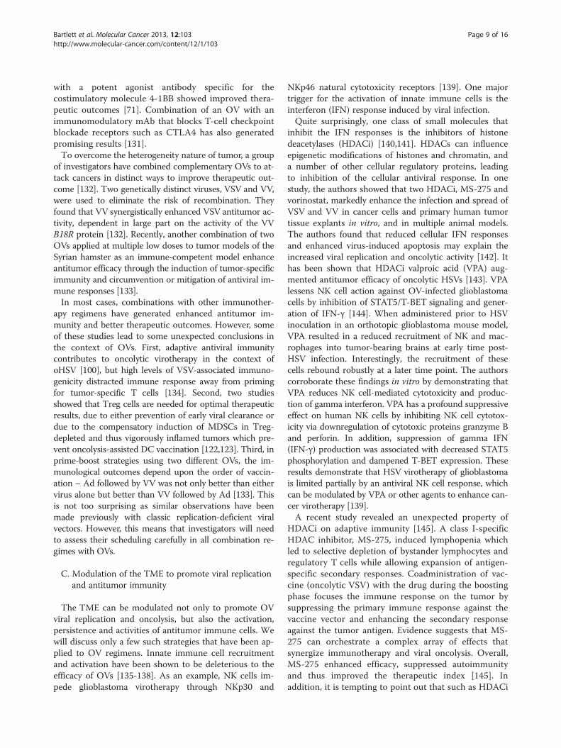
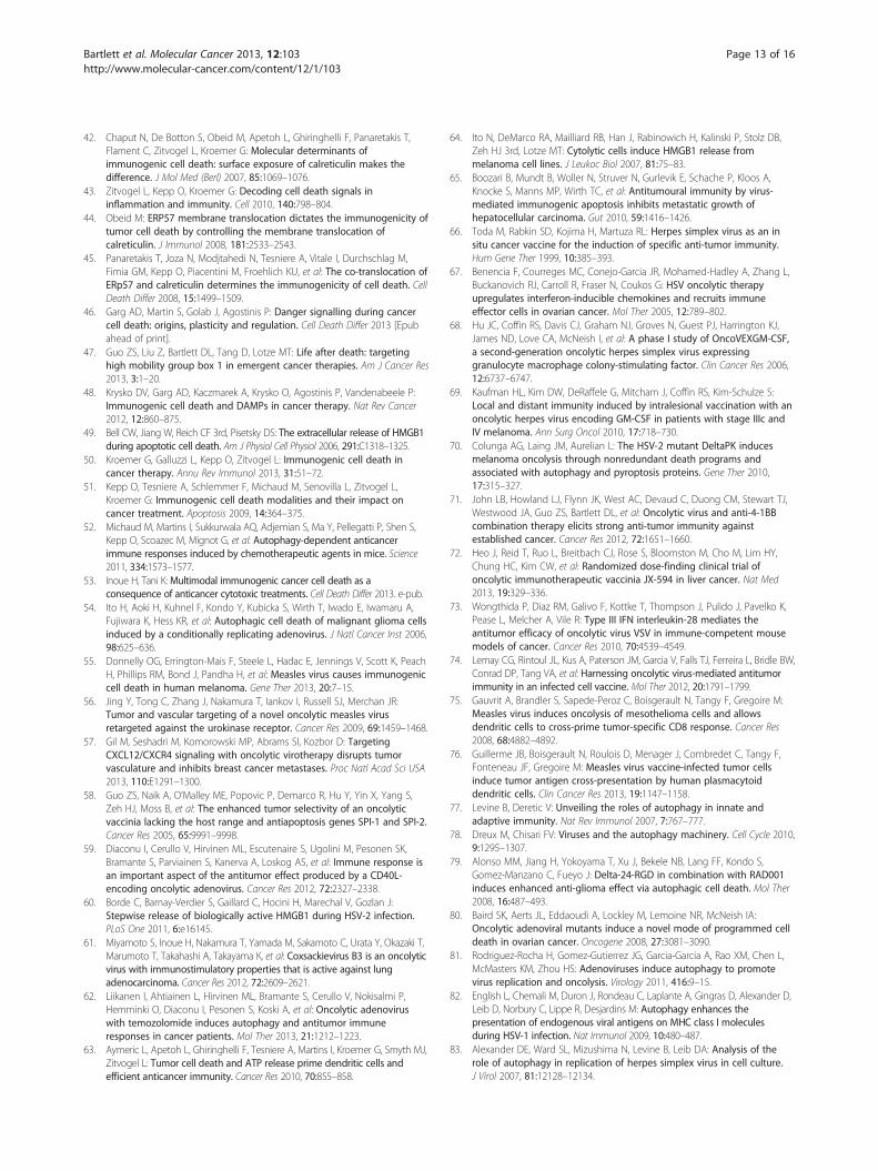
Figure 2 A model of how TILs in the TME are rescued to exert their eactivated by oncolytic virotherapy or other cancer vaccines migrated fromtumor–infiltrated DCs. However, the tumor-infiltrated DCs (TIDCs) are immuligands or/and other TLR3/9 ligands (TLR ligands) through type I IFN-dependsRNA) can function as TLR ligands. The functionally reactivated TIDCs canfunctions. This model is modified from Xiao H et al., 2013 [153].
patient) to a cytokine shock-like response triggered bysystemic delivery of an OV.The effector function of CD8+ TILs could be rescued
by converting the chronic inflammation milieu to acuteinflammation within tumors. Injection of TLR3/9 ligands(polyI:C/CpG) into a tumor during the effector phase oflentivector (lv) immunization effectively rescued thefunction of lv-activated CD8+ TILs and decreased thepercentage of Treg within the tumor, resulting in amarked improvement in the antitumor efficacy of theimmunization [153]. We provided a working mechanismby showing that rescue of the effector function is mostlikely dependent on production of type-1 IFN in thetumor that can mature and activate tumor-infiltratingDCs. It is worth noting that many OVs or their productscan be recognized as PAMPs by TLRs or other patternrecognition receptors (PRRs) expressed by DCs, thusstimulating DCs [154]. For example, oncolytic parvo-virus H-1 activates DCs partially through TLR3 andTLR9 [155]. Reovirus can escape the endosomes of DCand viral dsRNA triggers non-TLR3 receptor (other PRRreceptor) to induce IFN-γ production, and prime adap-tive antitumor immunity [156]. Based on these studies,we have presented a model how TLR ligands rescue theimmunological function of the TILs (Figure 2). In thismodel, type I IFN, produced via TLR-TLR ligand signal-ing and activation of the gene, plays some key roles inreactivating tumor-infiltrating DCs (TIDCs), which
ctivation
or Tissue
T
T
T Tumor cell killing
Cytokines
Ps & Ps
ffector functions by TLR ligands-reactivated DCs in the TME. TILslymph nodes to the tumor tissues may require in situ activation bynologically suppressed in the TME, but can be activated by TLRdent signaling. Some OVs themselves or their products (such asacquire, process, and present TAAs to reactivate TILs to exert their
Bartlett et al. Molecular Cancer 2013, 12:103 Page 11 of 16http://www.molecular-cancer.com/content/12/1/103
reactivate TILs. Some OVs can function well as ligandsfor TLRs.
D. Combination with cyclophosphamide for enhancedantitumor immunity.
The immune system makes a crucial contribution to theantitumor effects of conventional chemotherapy- andradiotherapy-based cancer regimens [157]. Cyclophospha-mide (CPA) is an alkylating agent and a classic chemothera-peutic compound. It induces genotoxic stress, apoptosisand/or cell cycle arrest. Recent studies indicate that it canenhance viral replication of OVs and adaptive antitumorimmunity in vivo, thus resulting in better efficacy. CPAfunctions to promote oncolytic virotherapy mainly via twomechanisms. (1). CPA enhances viral replication by sup-pressing antiviral innate immunity. Chiocca and associateshave first applied CPA in combination with oHSVvirotherapy based on the rationale that CPA would reduceantiviral immunity and thus augment viral replication toenhance oHSV efficacy [135-137,158]. They discovered thatpretreatment with a single dose of CPA could enhance boththe level and duration of viral replication of HSV withintreated tumors [137,158]. Similar enhancement was alsoreported for oncolytic Ad [159]. The CPA-enhanced viralreplication is well correlated to the significantly enhancedantitumor activities [137,158,160]. Nevertheless, it is worthnoting that not all combinations of an OV with CPA willwork. In fact, VSV-induced immune suppressor cells gener-ate antagonism between intratumoral OV and CPA [161].(2). CPA enhances adaptive antitumor immunity inducedby OVs. This is most likely through selective depletion andinhibition of Treg cells by low dose of CPA [162-164]. CPApretreatment followed with virotherapy leads to signifi-cantly enhanced antitumor immunity in tumor models ofimmunocompetent mice, as demonstrated with oncolyticHSV [160], Ad expressing gp96 [165], and VV expressingHPV E7 [166]. In addition, CPA can enhance antitumor ac-tivity of adoptively transferred immune cells through the in-duction of “cytokine storms” [167]. In the last few years,CPA in combination with OV has been tested in humancancer patients. Oncolytic Ad given together with metro-nomic CP increased cytotoxic T cells and induced Th1 typeimmunity on a systemic level in most cancer patients tested[62,168]. In summary, CPA has emerged as a clinically feas-ible agent that can suppress Tregs and allow more effectiveinduction of antitumor responses , in the settings of cancervaccines and other immunotherapy strategies [169].
ConclusionsIn the capacity of cancer vaccines, OVs exert two of themost important functions: (1). They kill cancer cells andassociated stromal cells directly by oncolysis or indir-ectly by anti-angiogenesis, vascular-targeting and by-
stander effect; and (2). They efficiently present/releaseDAMPs and PAMPs (signal 0) and present TAAs (signal1) to DCs in order to trigger a TAA-specific antitumorimmunity. However, OVs by themselves may not beenough because the immunosuppressive TME often im-pairs the functions of both innate and adaptive immunecells. Therefore, investigators have designed a number ofcombination strategies to overcome the TME and po-tentiate the antitumor immunity initiated by the OVs.We have discussed a variety of combination strategies
with OVs to boost the antitumor immunity and sustaintheir cytotoxic activity against cancer in the TME. Thesestrategies are targeted at the stages of immunogenicity of(dying) cancer cells, the process of antigen presentation,the potency of immune cells, and the overall immuno-logical status of the TME, the latter of which can be mod-ulated via blockade of immune checkpoints, depletion ofimmunosuppressive cells, and/or further activation of im-mune effector cells by either active immunization, or/andby adoptive T cell transfer. We envision that antitumorimmunity elicited by OVs properly armed or rationallycombined would kill not only residual cancer stem cellsand “differentiated” cancer cells in primary cancer andmetastases, but also maintain micrometastases in dormantstatus. This is a key for treating metastatic cancer.In phases I-II clinical trials, several OVs armed with ei-
ther GM-CSF or CD40L showed specific antitumor im-munity, significant antitumor activity and clinical responsesin a significant fraction of cancer patients. T-VEC has dem-onstrated efficacy in a phase III trial for melanoma patientswhile Pexa-Vec has been tested in a phase IIb trial for pa-tients with hepatocellular carcinoma. It is likely that one orboth of them may be approved by FDA in the near future.Looking forward, this new class of therapeutic cancer vac-cines is promising and more efforts should be invested inboth preclinical and clinical investigations.
Competing interestsDLB serves as a scientific advisor for and has financial interest with JennerexBiotherapeutics, a biotech company developing oncolytic viruses. The otherauthors declare no conflict of interest.
Bartlett et al. Molecular Cancer 2013, 12:103 Page 12 of 16http://www.molecular-cancer.com/content/12/1/103
Authors’ contributionsZSG collected and read relevant papers; designed and drafted themanuscript. YH proposed the original hypothesis presented in Figure 2. Allother authors have made suggestions to the manuscript. All authors haveread and approved the final manuscript.
AcknowledgementsThis work was supported by the National Institutes of Health grantsP01CA132714, R01CA155925, R01CA16444, and by David C. Koch RegionalTherapy Cancer Center. MS was supported by NIH training grantT32CA113263-01.
Author details1University of Pittsburgh Cancer Institute and Department of Surgery,University of Pittsburgh, Pittsburgh, PA 15213, USA. 2Fujian HuitianBiopharmaceuticals, Ltd., Sanming, Fujian 365001, China. 3Immunology/Immunotherapy Program, Georgia Regents University Cancer Center,Augusta, GA 30912, USA.
Received: 10 July 2013 Accepted: 6 September 2013Published: 11 September 2013
References1. Russell SJ, Peng KW, Bell JC: Oncolytic virotherapy. Nat Biotechnol 2012,
30:658–670.2. Cerullo V, Koski A, Vaha-Koskela M, Hemminki A: Chapter eight–Oncolytic
adenoviruses for cancer immunotherapy: data from mice, hamsters, andhumans. Adv Cancer Res 2012, 115:265–318.
4. Breitbach CJ, Paterson JM, Lemay CG, Falls TJ, McGuire A, Parato KA, Stojdl DF,Daneshmand M, Speth K, Kirn D, et al: Targeted inflammation duringoncolytic virus therapy severely compromises tumor blood flow. Mol Ther2007, 15:1686–1693.
5. Liu TC, Hwang T, Park BH, Bell J, Kirn DH: The targeted oncolytic poxvirusJX-594 demonstrates antitumoral, antivascular, and anti-HBV activities inpatients with hepatocellular carcinoma. Mol Ther 2008, 16:1637–1642.
6. Breitbach CJ, Arulanandam R, De Silva N, Thorne SH, Patt R, Daneshmand M,Moon A, Ilkow C, Burke J, Hwang TH, et al: Oncolytic vaccinia virusdisrupts tumor-associated vasculature in humans. Cancer Res 2013,73:1265–1275.
7. Melcher A, Parato K, Rooney CM, Bell JC: Thunder and lightning:immunotherapy and oncolytic viruses collide. Mol Ther 2011,19:1008–1016.
8. Prestwich RJ, Harrington KJ, Pandha HS, Vile RG, Melcher AA, Errington F:Oncolytic viruses: a novel form of immunotherapy. Expert Rev AnticancerTher 2008, 8:1581–1588.
9. Tong AW, Senzer N, Cerullo V, Templeton NS, Hemminki A, Nemunaitis J:Oncolytic viruses for induction of anti-tumor immunity. Curr PharmBiotechnol 2012, 13:1750–1760.
10. Cheever MA, Higano CS: PROVENGE (Sipuleucel-T) in prostate cancer: thefirst FDA-approved therapeutic cancer vaccine. Clin Cancer Res 2011,17:3520–3526.
11. Matzinger P: The danger model: a renewed sense of self. Science 2002,296:301–305.
12. Medzhitov R, Janeway CA Jr: Decoding the patterns of self and nonself bythe innate immune system. Science 2002, 296:298–300.
13. Tang D, Kang R, Coyne CB, Zeh HJ, Lotze MT: PAMPs and DAMPs: signal 0sthat spur autophagy and immunity. Immunol Rev 2012, 249:158–175.
14. Dougan M, Dranoff G: Immune therapy for cancer. Annu Rev Immunol2009, 27:83–117.
15. Pardoll DM: The blockade of immune checkpoints in cancerimmunotherapy. Nat Rev Cancer 2012, 12:252–264.
16. Li QX, Liu G, Zhang X: Fusogenic oncolytic herpes simplex viruses as apotent and personalized cancer vaccine. Curr Pharm Biotechnol 2012,13:1773–1785.
17. Haen SP, Rammensee HG: The repertoire of human tumor-associatedepitopes - identification and selection of antigens and their applicationin clinical trials. Curr Opin Immunol 2013, 25:277–283.
18. Naik S, Russell SJ: Engineering oncolytic viruses to exploit tumor specificdefects in innate immune signaling pathways. Expert Opin Biol Ther 2009,9:1163–1176.
19. Haller O, Kochs G, Weber F: The interferon response circuit: induction andsuppression by pathogenic viruses. Virology 2006, 344:119–130.
21. Biswas M, Johnson JB, Kumar SR, Parks GD, Elankumarana S: Incorporationof host complement regulatory proteins into Newcastle disease virusenhances complement evasion. J Virol 2012, 86:12708–12716.
22. Lun X, Yang W, Alain T, Shi ZQ, Muzik H, Barrett JW, McFadden G, Bell J,Hamilton MG, Senger DL, Forsyth PA: Myxoma virus is a novel oncolyticvirus with significant antitumor activity against experimental humangliomas. Cancer Res 2005, 65:9982–9990.
23. Cuddington BP, Dyer AL, Workenhe ST, Mossman KL: Oncolytic bovineherpesvirus type 1 infects and kills breast tumor cells and breast cancer-initiating cells irrespective of tumor subtype. Cancer Gene Ther 2013,20:282–289.
24. Young AM, Archibald KM, Tookman LA, Pool A, Dudek K, Jones C, WilliamsSL, Pirlo KJ, Willis AE, Lockley M, McNeish IA: Failure of translation ofhuman adenovirus mRNA in murine cancer cells can be partiallyovercome by L4-100K expression in vitro and in vivo. Mol Ther 2012,20:1676–1688.
25. Mahoney DJ, Stojdl DF: Molecular pathways: multimodal cancer-killingmechanisms employed by oncolytic vesiculoviruses. Clin Cancer Res 2013,19:758–763.
26. Mahoney DJ, Lefebvre C, Allan K, Brun J, Sanaei CA, Baird S, Pearce N,Gronberg S, Wilson B, Prakesh M, et al: Virus-tumor interactome screenreveals ER stress response can reprogram resistant cancers for oncolyticvirus-triggered caspase-2 cell death. Cancer Cell 2011, 20:443–456.
27. Cary ZD, Willingham MC, Lyles DS: Oncolytic vesicular stomatitis virusinduces apoptosis in U87 glioblastoma cells by a type II death receptormechanism and induces cell death and tumor clearance in vivo. J Virol2011, 85:5708–5717.
28. Elankumaran S, Rockemann D, Samal SK: Newcastle disease virus exertsoncolysis by both intrinsic and extrinsic caspase-dependent pathways ofcell death. J Virol 2006, 80:7522–7534.
29. Obeid M, Tesniere A, Ghiringhelli F, Fimia GM, Apetoh L, Perfettini JL,Castedo M, Mignot G, Panaretakis T, Casares N, et al: Calreticulin exposuredictates the immunogenicity of cancer cell death. Nat Med 2007,13:54–61.
30. Tesniere A, Apetoh L, Ghiringhelli F, Joza N, Panaretakis T, Kepp O,Schlemmer F, Zitvogel L, Kroemer G: Immunogenic cancer cell death: akey-lock paradigm. Curr Opin Immunol 2008, 20:504–511.
32. Mellman I, Coukos G, Dranoff G: Cancer immunotherapy comes of age.Nature 2011, 480:480–489.
33. Kalinski P, Okada H: Polarized dendritic cells as cancer vaccines: directingeffector-type T cells to tumors. Semin Immunol 2010, 22:173–182.
34. Restifo NP, Dudley ME, Rosenberg SA: Adoptive immunotherapy for cancer:harnessing the T cell response. Nat Rev Immunol 2012, 12:269–281.
35. Zou W: Immunosuppressive networks in the tumour environment andtheir therapeutic relevance. Nat Rev Cancer 2005, 5:263–274.
36. Topalian SL, Drake CG, Pardoll DM: Targeting the PD-1/B7-H1(PD-L1)pathway to activate anti-tumor immunity. Curr Opin Immunol 2012,24:207–212.
37. Lipson EJ, Drake CG: Ipilimumab: an anti-CTLA-4 antibody for metastaticmelanoma. Clin Cancer Res 2011, 17:6958–6962.
38. Kroemer G, El-Deiry WS, Golstein P, Peter ME, Vaux D, Vandenabeele P,Zhivotovsky B, Blagosklonny MV, Malorni W, Knight RA, et al: Classificationof cell death: recommendations of the Nomenclature Committee on CellDeath. Cell Death Differ 2005, 12(Suppl 2):1463–1467.
39. Kroemer G, Levine B: Autophagic cell death: the story of a misnomer. NatRev Mol Cell Biol 2008, 9:1004–1010.
40. Labbe K, Saleh M: Cell death in the host response to infection. Cell DeathDiffer 2008, 15:1339–1349.
41. Obeid M, Panaretakis T, Tesniere A, Joza N, Tufi R, Apetoh L, Ghiringhelli F,Zitvogel L, Kroemer G: Leveraging the immune system duringchemotherapy: moving calreticulin to the cell surface converts apoptoticdeath from “silent” to immunogenic. Cancer Res 2007, 67:7941–7944.
Bartlett et al. Molecular Cancer 2013, 12:103 Page 13 of 16http://www.molecular-cancer.com/content/12/1/103
42. Chaput N, De Botton S, Obeid M, Apetoh L, Ghiringhelli F, Panaretakis T,Flament C, Zitvogel L, Kroemer G: Molecular determinants ofimmunogenic cell death: surface exposure of calreticulin makes thedifference. J Mol Med (Berl) 2007, 85:1069–1076.
43. Zitvogel L, Kepp O, Kroemer G: Decoding cell death signals ininflammation and immunity. Cell 2010, 140:798–804.
44. Obeid M: ERP57 membrane translocation dictates the immunogenicity oftumor cell death by controlling the membrane translocation ofcalreticulin. J Immunol 2008, 181:2533–2543.
45. Panaretakis T, Joza N, Modjtahedi N, Tesniere A, Vitale I, Durchschlag M,Fimia GM, Kepp O, Piacentini M, Froehlich KU, et al: The co-translocation ofERp57 and calreticulin determines the immunogenicity of cell death. CellDeath Differ 2008, 15:1499–1509.
46. Garg AD, Martin S, Golab J, Agostinis P: Danger signalling during cancercell death: origins, plasticity and regulation. Cell Death Differ 2013 [Epubahead of print].
47. Guo ZS, Liu Z, Bartlett DL, Tang D, Lotze MT: Life after death: targetinghigh mobility group box 1 in emergent cancer therapies. Am J Cancer Res2013, 3:1–20.
48. Krysko DV, Garg AD, Kaczmarek A, Krysko O, Agostinis P, Vandenabeele P:Immunogenic cell death and DAMPs in cancer therapy. Nat Rev Cancer2012, 12:860–875.
49. Bell CW, Jiang W, Reich CF 3rd, Pisetsky DS: The extracellular release of HMGB1during apoptotic cell death. Am J Physiol Cell Physiol 2006, 291:C1318–1325.
50. Kroemer G, Galluzzi L, Kepp O, Zitvogel L: Immunogenic cell death incancer therapy. Annu Rev Immunol 2013, 31:51–72.
51. Kepp O, Tesniere A, Schlemmer F, Michaud M, Senovilla L, Zitvogel L,Kroemer G: Immunogenic cell death modalities and their impact oncancer treatment. Apoptosis 2009, 14:364–375.
52. Michaud M, Martins I, Sukkurwala AQ, Adjemian S, Ma Y, Pellegatti P, Shen S,Kepp O, Scoazec M, Mignot G, et al: Autophagy-dependent anticancerimmune responses induced by chemotherapeutic agents in mice. Science2011, 334:1573–1577.
53. Inoue H, Tani K: Multimodal immunogenic cancer cell death as aconsequence of anticancer cytotoxic treatments. Cell Death Differ 2013. e-pub.
54. Ito H, Aoki H, Kuhnel F, Kondo Y, Kubicka S, Wirth T, Iwado E, Iwamaru A,Fujiwara K, Hess KR, et al: Autophagic cell death of malignant glioma cellsinduced by a conditionally replicating adenovirus. J Natl Cancer Inst 2006,98:625–636.
55. Donnelly OG, Errington-Mais F, Steele L, Hadac E, Jennings V, Scott K, PeachH, Phillips RM, Bond J, Pandha H, et al: Measles virus causes immunogeniccell death in human melanoma. Gene Ther 2013, 20:7–15.
56. Jing Y, Tong C, Zhang J, Nakamura T, Iankov I, Russell SJ, Merchan JR:Tumor and vascular targeting of a novel oncolytic measles virusretargeted against the urokinase receptor. Cancer Res 2009, 69:1459–1468.
57. Gil M, Seshadri M, Komorowski MP, Abrams SI, Kozbor D: TargetingCXCL12/CXCR4 signaling with oncolytic virotherapy disrupts tumorvasculature and inhibits breast cancer metastases. Proc Natl Acad Sci USA2013, 110:E1291–1300.
58. Guo ZS, Naik A, O’Malley ME, Popovic P, Demarco R, Hu Y, Yin X, Yang S,Zeh HJ, Moss B, et al: The enhanced tumor selectivity of an oncolyticvaccinia lacking the host range and antiapoptosis genes SPI-1 and SPI-2.Cancer Res 2005, 65:9991–9998.
59. Diaconu I, Cerullo V, Hirvinen ML, Escutenaire S, Ugolini M, Pesonen SK,Bramante S, Parviainen S, Kanerva A, Loskog AS, et al: Immune response isan important aspect of the antitumor effect produced by a CD40L-encoding oncolytic adenovirus. Cancer Res 2012, 72:2327–2338.
60. Borde C, Barnay-Verdier S, Gaillard C, Hocini H, Marechal V, Gozlan J:Stepwise release of biologically active HMGB1 during HSV-2 infection.PLoS One 2011, 6:e16145.
61. Miyamoto S, Inoue H, Nakamura T, Yamada M, Sakamoto C, Urata Y, Okazaki T,Marumoto T, Takahashi A, Takayama K, et al: Coxsackievirus B3 is an oncolyticvirus with immunostimulatory properties that is active against lungadenocarcinoma. Cancer Res 2012, 72:2609–2621.
62. Liikanen I, Ahtiainen L, Hirvinen ML, Bramante S, Cerullo V, Nokisalmi P,Hemminki O, Diaconu I, Pesonen S, Koski A, et al: Oncolytic adenoviruswith temozolomide induces autophagy and antitumor immuneresponses in cancer patients. Mol Ther 2013, 21:1212–1223.
63. Aymeric L, Apetoh L, Ghiringhelli F, Tesniere A, Martins I, Kroemer G, Smyth MJ,Zitvogel L: Tumor cell death and ATP release prime dendritic cells andefficient anticancer immunity. Cancer Res 2010, 70:855–858.
65. Boozari B, Mundt B, Woller N, Struver N, Gurlevik E, Schache P, Kloos A,Knocke S, Manns MP, Wirth TC, et al: Antitumoural immunity by virus-mediated immunogenic apoptosis inhibits metastatic growth ofhepatocellular carcinoma. Gut 2010, 59:1416–1426.
66. Toda M, Rabkin SD, Kojima H, Martuza RL: Herpes simplex virus as an insitu cancer vaccine for the induction of specific anti-tumor immunity.Hum Gene Ther 1999, 10:385–393.
67. Benencia F, Courreges MC, Conejo-Garcia JR, Mohamed-Hadley A, Zhang L,Buckanovich RJ, Carroll R, Fraser N, Coukos G: HSV oncolytic therapyupregulates interferon-inducible chemokines and recruits immuneeffector cells in ovarian cancer. Mol Ther 2005, 12:789–802.
68. Hu JC, Coffin RS, Davis CJ, Graham NJ, Groves N, Guest PJ, Harrington KJ,James ND, Love CA, McNeish I, et al: A phase I study of OncoVEXGM-CSF,a second-generation oncolytic herpes simplex virus expressinggranulocyte macrophage colony-stimulating factor. Clin Cancer Res 2006,12:6737–6747.
69. Kaufman HL, Kim DW, DeRaffele G, Mitcham J, Coffin RS, Kim-Schulze S:Local and distant immunity induced by intralesional vaccination with anoncolytic herpes virus encoding GM-CSF in patients with stage IIIc andIV melanoma. Ann Surg Oncol 2010, 17:718–730.
70. Colunga AG, Laing JM, Aurelian L: The HSV-2 mutant DeltaPK inducesmelanoma oncolysis through nonredundant death programs andassociated with autophagy and pyroptosis proteins. Gene Ther 2010,17:315–327.
71. John LB, Howland LJ, Flynn JK, West AC, Devaud C, Duong CM, Stewart TJ,Westwood JA, Guo ZS, Bartlett DL, et al: Oncolytic virus and anti-4-1BBcombination therapy elicits strong anti-tumor immunity againstestablished cancer. Cancer Res 2012, 72:1651–1660.
72. Heo J, Reid T, Ruo L, Breitbach CJ, Rose S, Bloomston M, Cho M, Lim HY,Chung HC, Kim CW, et al: Randomized dose-finding clinical trial ofoncolytic immunotherapeutic vaccinia JX-594 in liver cancer. Nat Med2013, 19:329–336.
73. Wongthida P, Diaz RM, Galivo F, Kottke T, Thompson J, Pulido J, Pavelko K,Pease L, Melcher A, Vile R: Type III IFN interleukin-28 mediates theantitumor efficacy of oncolytic virus VSV in immune-competent mousemodels of cancer. Cancer Res 2010, 70:4539–4549.
74. Lemay CG, Rintoul JL, Kus A, Paterson JM, Garcia V, Falls TJ, Ferreira L, Bridle BW,Conrad DP, Tang VA, et al: Harnessing oncolytic virus-mediated antitumorimmunity in an infected cell vaccine. Mol Ther 2012, 20:1791–1799.
75. Gauvrit A, Brandler S, Sapede-Peroz C, Boisgerault N, Tangy F, Gregoire M:Measles virus induces oncolysis of mesothelioma cells and allowsdendritic cells to cross-prime tumor-specific CD8 response. Cancer Res2008, 68:4882–4892.
76. Guillerme JB, Boisgerault N, Roulois D, Menager J, Combredet C, Tangy F,Fonteneau JF, Gregoire M: Measles virus vaccine-infected tumor cellsinduce tumor antigen cross-presentation by human plasmacytoiddendritic cells. Clin Cancer Res 2013, 19:1147–1158.
77. Levine B, Deretic V: Unveiling the roles of autophagy in innate andadaptive immunity. Nat Rev Immunol 2007, 7:767–777.
78. Dreux M, Chisari FV: Viruses and the autophagy machinery. Cell Cycle 2010,9:1295–1307.
79. Alonso MM, Jiang H, Yokoyama T, Xu J, Bekele NB, Lang FF, Kondo S,Gomez-Manzano C, Fueyo J: Delta-24-RGD in combination with RAD001induces enhanced anti-glioma effect via autophagic cell death. Mol Ther2008, 16:487–493.
80. Baird SK, Aerts JL, Eddaoudi A, Lockley M, Lemoine NR, McNeish IA:Oncolytic adenoviral mutants induce a novel mode of programmed celldeath in ovarian cancer. Oncogene 2008, 27:3081–3090.
81. Rodriguez-Rocha H, Gomez-Gutierrez JG, Garcia-Garcia A, Rao XM, Chen L,McMasters KM, Zhou HS: Adenoviruses induce autophagy to promotevirus replication and oncolysis. Virology 2011, 416:9–15.
82. English L, Chemali M, Duron J, Rondeau C, Laplante A, Gingras D, Alexander D,Leib D, Norbury C, Lippe R, Desjardins M: Autophagy enhances thepresentation of endogenous viral antigens on MHC class I moleculesduring HSV-1 infection. Nat Immunol 2009, 10:480–487.
83. Alexander DE, Ward SL, Mizushima N, Levine B, Leib DA: Analysis of therole of autophagy in replication of herpes simplex virus in cell culture.J Virol 2007, 81:12128–12134.
Bartlett et al. Molecular Cancer 2013, 12:103 Page 14 of 16http://www.molecular-cancer.com/content/12/1/103
84. Chi PI, Huang WR, Lai IH, Cheng CY, Liu HJ: The p17 nonstructural proteinof avian reovirus triggers autophagy enhancing virus replication viaactivation of phosphatase and tensin deleted on chromosome 10 (PTEN)and AMP-activated protein kinase (AMPK), as well as dsRNA-dependentprotein kinase (PKR)/eIF2alpha signaling pathways. J Biol Chem 2012,288:3571–3584.
85. Meng S, Jiang K, Zhang X, Zhang M, Zhou Z, Hu M, Yang R, Sun C, Wu Y:Avian reovirus triggers autophagy in primary chicken fibroblast cells andVero cells to promote virus production. Arch Virol 2012, 157:661–668.
86. Thirukkumaran CM, Shi ZQ, Luider J, Kopciuk K, Gao H, Bahlis N, Neri P, Pho M,Stewart D, Mansoor A, Morris DG: Reovirus modulates autophagy duringoncolysis of multiple myeloma. Autophagy 2013, 9:413–414.
87. Comber JD, Robinson TM, Siciliano NA, Snook AE, Eisenlohr LC: Functionalmacroautophagy induction by influenza A virus without a contributionto major histocompatibility complex class II-restricted presentation.J Virol 2010, 85:6453–6463.
88. Chakrabarti A, Ghosh PK, Banerjee S, Gaughan C, Silverman RH: RNase Ltriggers autophagy in response to viral infections. J Virol 2012,86:11311–11321.
89. Thorburn J, Horita H, Redzic J, Hansen K, Frankel AE, Thorburn A:Autophagy regulates selective HMGB1 release in tumor cells that aredestined to die. Cell Death Differ 2009, 16:175–183.
90. Ayna G, Krysko DV, Kaczmarek A, Petrovski G, Vandenabeele P, Fesus L: ATPrelease from dying autophagic cells and their phagocytosis are crucialfor inflammasome activation in macrophages. PLoS One 2012, 7:e40069.
91. Endo Y, Sakai R, Ouchi M, Onimatsu H, Hioki M, Kagawa S, Uno F, Watanabe Y,Urata Y, Tanaka N, Fujiwara T: Virus-mediated oncolysis induces dangersignal and stimulates cytotoxic T-lymphocyte activity via proteasomeactivator upregulation. Oncogene 2008, 27:2375–2381.
92. van der Bruggen P, Van den Eynde BJ: Processing and presentation of tumorantigens and vaccination strategies. Curr Opin Immunol 2006, 18:98–104.
93. Crotzer VL, Blum JS: Autophagy and its role in MHC-mediated antigenpresentation. J Immunol 2009, 182:3335–3341.
94. Li Y, Wang LX, Yang G, Hao F, Urba WJ, Hu HM: Efficient cross-presentationdepends on autophagy in tumor cells. Cancer Res 2008, 68:6889–6895.
95. Wei J, Waithman J, Lata R, Mifsud NA, Cebon J, Kay T, Smyth MJ, Sadler AJ,Chen W: Influenza A infection enhances cross-priming of CD8+ T cells tocell-associated antigens in a TLR7- and type I IFN-dependent fashion.J Immunol 2010, 185:6013–6022.
96. Uhl M, Kepp O, Jusforgues-Saklani H, Vicencio JM, Kroemer G, Albert ML:Autophagy within the antigen donor cell facilitates efficient antigencross-priming of virus-specific CD8+ T cells. Cell Death Differ 2009,16:991–1005.
97. Meng S, Xu J, Wu Y, Ding C: Targeting autophagy to enhance oncolyticvirus-based cancer therapy. Expert Opin Biol Ther 2013, 13:863–873.
98. Lindenmann J, Klein PA: Viral oncolysis: increased immunogenicity of hostcell antigen associated with influenza virus. J Exp Med 1967, 126:93–108.
99. Toda M, Martuza RL, Kojima H, Rabkin SD: In situ cancer vaccination: an IL-12 defective vector/replication-competent herpes simplex viruscombination induces local and systemic antitumor activity. J Immunol1998, 160:4457–4464.
100. Sobol PT, Boudreau JE, Stephenson K, Wan Y, Lichty BD, Mossman KL:Adaptive antiviral immunity is a determinant of the therapeutic successof oncolytic virotherapy. Mol Ther 2011, 19:335–344.
101. Gurlevik E, Woller N, Struver N, Schache P, Kloos A, Manns MP, Zender L,Kuhnel F, Kubicka S: Selectivity of oncolytic viral replication preventsantiviral immune response and toxicity, but does not improveantitumoral immunity. Mol Ther 2010, 18:1972–1982.
102. Prestwich RJ, Errington F, Diaz RM, Pandha HS, Harrington KJ, Melcher AA,Vile RG: The case of oncolytic viruses versus the immune system: waitingon the judgment of Solomon. Hum Gene Ther 2009, 20:1119–1132.
103. Boisgerault N, Tangy F, Gregoire M: New perspectives in cancervirotherapy: bringing the immune system into play. Immunotherapy 2010,2:185–199.
104. Todo T, Rabkin SD, Sundaresan P, Wu A, Meehan KR, Herscowitz HB,Martuza RL: Systemic antitumor immunity in experimental brain tumortherapy using a multimutated, replication-competent herpes simplexvirus. Hum Gene Ther 1999, 10:2741–2755.
105. Raykov Z, Grekova S, Leuchs B, Aprahamian M, Rommelaere J: Armingparvoviruses with CpG motifs to improve their oncosuppressivecapacity. Int J Cancer 2008, 122:2880–2884.
106. Cerullo V, Diaconu I, Romano V, Hirvinen M, Ugolini M, Escutenaire S, Holm SL,Kipar A, Kanerva A, Hemminki A: An oncolytic adenovirus enhanced for toll-like receptor 9 stimulation increases antitumor immune responses andtumor clearance. Mol Ther 2012, 20:2076–2086.
107. Murshid A, Gong J, Stevenson MA, Calderwood SK: Heat shock proteinsand cancer vaccines: developments in the past decade and chaperoningin the decade to come. Expert Rev Vaccines 2011, 10:1553–1568.
108. Fan R, Wang C, Wang Y, Ren P, Gan P, Ji H, Xia Z, Hu S, Zeng Q, Huang W,et al: Enhanced antitumoral efficacy and immune response followingconditionally replicative adenovirus containing constitutive HSF1delivery to rodent tumors. J Transl Med 2012, 10:101.
109. Huang XF, Ren W, Rollins L, Pittman P, Shah M, Shen L, Gu Q, Strube R, Hu F,Chen SY: A broadly applicable, personalized heat shock protein-mediatedoncolytic tumor vaccine. Cancer Res 2003, 63:7321–7329.
110. Li JL, Liu HL, Zhang XR, Xu JP, Hu WK, Liang M, Chen SY, Hu F, Chu DT:A phase I trial of intratumoral administration of recombinant oncolyticadenovirus overexpressing HSP70 in advanced solid tumor patients.Gene Ther 2009, 16:376–382.
111. Alcami A, Koszinowski UH: Viral mechanisms of immune evasion. ImmunolToday 2000, 21:447–455.
112. Alemany R, Cascallo M: Oncolytic viruses from the perspective of theimmune system. Future Microbiol 2009, 4:527–536.
113. Liu BL, Robinson M, Han ZQ, Branston RH, English C, Reay P, McGrath Y,Thomas SK, Thornton M, Bullock P, et al: ICP34.5 deleted herpes simplexvirus with enhanced oncolytic, immune stimulating, and anti-tumourproperties. Gene Ther 2003, 10:292–303.
114. McCart JA, Ward JM, Lee J, Hu Y, Alexander HR, Libutti SK, Moss B, Bartlett DL:Systemic cancer therapy with a tumor-selective vaccinia virus mutantlacking thymidine kinase and vaccinia growth factor genes. Cancer Res 2001,61:8751–8757.
115. Thorne SH, Hwang TH, O’Gorman WE, Bartlett DL, Sei S, Kanji F, Brown C,Werier J, Cho JH, Lee DE, et al: Rational strain selection and engineeringcreates a broad-spectrum, systemically effective oncolytic poxvirus, JX-963. J Clin Invest 2007, 117:3350–3358.
116. Cerullo V, Pesonen S, Diaconu I, Escutenaire S, Arstila PT, Ugolini M,Nokisalmi P, Raki M, Laasonen L, Sarkioja M, et al: Oncolytic adenoviruscoding for granulocyte macrophage colony-stimulating factor inducesantitumoral immunity in cancer patients. Cancer Res 2010, 70:4297–4309.
117. Koski A, Kangasniemi L, Escutenaire S, Pesonen S, Cerullo V, Diaconu I,Nokisalmi P, Raki M, Rajecki M, Guse K, et al: Treatment of cancer patientswith a serotype 5/3 chimeric oncolytic adenovirus expressing GMCSF.Mol Ther 2010, 18:1874–1884.
118. Senzer NN, Kaufman HL, Amatruda T, Nemunaitis M, Reid T, Daniels G,Gonzalez R, Glaspy J, Whitman E, Harrington K, et al: Phase II clinical trial ofa granulocyte-macrophage colony-stimulating factor-encoding, second-generation oncolytic herpesvirus in patients with unresectablemetastatic melanoma. J Clin Oncol 2009, 27:5763–5771.
119. Kaufman HL, Bines SD: OPTIM trial: a Phase III trial of an oncolytic herpesvirus encoding GM-CSF for unresectable stage III or IV melanoma. FutureOncol 2010, 6:941–949.
120. Andtbacka RHI, Collichio FA, Amatruda T, et al: OPTiM: a randomizedphase III trial of talimogene laherparepvec (T-VEC) versus subcutaneous(SC) granulocyte-macrophage colony-stimulating factor (GM-CSF) for thetreatment (tx) of unresectable stage IIIB/C and IV melanoma. J Clin Oncol2013, 31. suppl; abstr LBA9008.
121. Kim MK, Breitbach CJ, Moon A, Heo J, Lee YK, Cho M, Lee JW, Kim SG, KangDH, Bell JC, et al: Oncolytic and immunotherapeutic vaccinia inducesantibody-mediated complement-dependent cancer cell lysis in humans.Sci Transl Med 2013, 5:185ra163.
122. Woller N, Knocke S, Mundt B, Gurlevik E, Struver N, Kloos A, Boozari B,Schache P, Manns MP, Malek NP, et al: Virus-induced tumor inflammationfacilitates effective DC cancer immunotherapy in a Treg-dependentmanner in mice. J Clin Invest 2011, 121:2570–2582.
123. Diaz RM, Galivo F, Kottke T, Wongthida P, Qiao J, Thompson J, Valdes M,Barber G, Vile RG: Oncolytic immunovirotherapy for melanoma usingvesicular stomatitis virus. Cancer Res 2007, 67:2840–2848.
124. Galanis E, Hartmann LC, Cliby WA, Long HJ, Peethambaram PP, Barrette BA,Kaur JS, Haluska PJ Jr, Aderca I, Zollman PJ, et al: Phase I trial ofintraperitoneal administration of an oncolytic measles virus strainengineered to express carcinoembryonic antigen for recurrent ovariancancer. Cancer Res 2010, 70:875–882.
Bartlett et al. Molecular Cancer 2013, 12:103 Page 15 of 16http://www.molecular-cancer.com/content/12/1/103
125. Rommelfanger DM, Wongthida P, Diaz RM, Kaluza KM, Thompson JM,Kottke TJ, Vile RG: Systemic combination virotherapy for melanoma withtumor antigen-expressing vesicular stomatitis virus and adoptive T-celltransfer. Cancer Res 2012, 72:4753–4764.
126. Wongthida P, Diaz RM, Pulido C, Rommelfanger D, Galivo F, Kaluza K,Kottke T, Thompson J, Melcher A, Vile R: Activating systemic T-cellimmunity against self tumor antigens to support oncolyticvirotherapy with vesicular stomatitis virus. Hum Gene Ther 2011,22:1343–1353.
127. Zhang YQ, Tsai YC, Monie A, Wu TC, Hung CF: Enhancing the therapeuticeffect against ovarian cancer through a combination of viral oncolysisand antigen-specific immunotherapy. Mol Ther 2010, 18:692–699.
128. Bridle BW, Boudreau JE, Lichty BD, Brunelliere J, Stephenson K, Koshy S,Bramson JL, Wan Y: Vesicular stomatitis virus as a novel cancer vaccinevector to prime antitumor immunity amenable to rapid boosting withadenovirus. Mol Ther 2009, 17:1814–1821.
129. Bridle BW, Stephenson KB, Boudreau JE, Koshy S, Kazdhan N, PullenayegumE, Brunelliere J, Bramson JL, Lichty BD, Wan Y: Potentiating cancerimmunotherapy using an oncolytic virus. Mol Ther 2010, 18:1430–1439.
130. Melero I, Hirschhorn-Cymerman D, Morales-Kastresana A, Sanmamed MF,Wolchok JD: Agonist antibodies to TNFR molecules that costimulate Tand NK cells. Clin Cancer Res 2013, 19:1044–1053.
131. Dias JD, Hemminki O, Diaconu I, Hirvinen M, Bonetti A, Guse K, EscutenaireS, Kanerva A, Pesonen S, Loskog A, et al: Targeted cancer immunotherapywith oncolytic adenovirus coding for a fully human monoclonalantibody specific for CTLA-4. Gene Ther 2012, 19:988–998.
132. Le Boeuf F, Diallo JS, McCart JA, Thorne S, Falls T, Stanford M, Kanji F, Auer R,Brown CW, Lichty BD, et al: Synergistic interaction between oncolytic virusesaugments tumor killing. Mol Ther 2010, 18:888–895.
133. Tysome JR, Li X, Wang S, Wang P, Gao D, Du P, Chen D, Gangeswaran R,Chard LS, Yuan M, et al: A novel therapeutic regimen to eradicateestablished solid tumors with an effective induction of tumor-specificimmunity. Clin Cancer Res 2012, 18:6679–6689.
134. Galivo F, Diaz RM, Thanarajasingam U, Jevremovic D, Wongthida P,Thompson J, Kottke T, Barber GN, Melcher A, Vile RG: Interference ofCD40L-mediated tumor immunotherapy by oncolytic vesicular stomatitisvirus. Hum Gene Ther 2010, 21:439–450.
135. Ikeda K, Ichikawa T, Wakimoto H, Silver JS, Deisboeck TS, Finkelstein D,Harsh GR 4th, Louis DN, Bartus RT, Hochberg FH, Chiocca EA: Oncolyticvirus therapy of multiple tumors in the brain requires suppression ofinnate and elicited antiviral responses. Nat Med 1999, 5:881–887.
136. Friedman A, Tian JP, Fulci G, Chiocca EA, Wang J: Glioma virotherapy:effects of innate immune suppression and increased viral replicationcapacity. Cancer Res 2006, 66:2314–2319.
138. Kurozumi K, Hardcastle J, Thakur R, Yang M, Christoforidis G, Fulci G,Hochberg FH, Weissleder R, Carson W, Chiocca EA, Kaur B: Effect of tumormicroenvironment modulation on the efficacy of oncolytic virus therapy.J Natl Cancer Inst 2007, 99:1768–1781.
139. Alvarez-Breckenridge CA, Yu J, Price R, Wojton J, Pradarelli J, Mao H, Wei M,Wang Y, He S, Hardcastle J, et al: NK cells impede glioblastomavirotherapy through NKp30 and NKp46 natural cytotoxicity receptors.Nat Med 2012, 18:1827–1834.
140. Grant S, Dai Y: Histone deacetylase inhibitors and rational combinationtherapies. Adv Cancer Res 2012, 116:199–237.
141. Yu XD, Guo ZS: Epigenetic drugs for cancer treatment and prevention:mechanisms of action. Biomol Concepts 2010, 1:239–252.
142. Nguyen TL, Abdelbary H, Arguello M, Breitbach C, Leveille S, Diallo JS,Yasmeen A, Bismar TA, Kirn D, Falls T, et al: Chemical targeting of theinnate antiviral response by histone deacetylase inhibitors rendersrefractory cancers sensitive to viral oncolysis. Proc Natl Acad Sci USA 2008,105:14981–14986.
143. Otsuki A, Patel A, Kasai K, Suzuki M, Kurozumi K, Chiocca EA, Saeki Y:Histone deacetylase inhibitors augment antitumor efficacy of herpes-based oncolytic viruses. Mol Ther 2008, 16:1546–1555.
144. Alvarez-Breckenridge CA, Yu J, Price R, Wei M, Wang Y, Nowicki MO, Ha YP,Bergin S, Hwang C, Fernandez SA, et al: The histone deacetylase inhibitorvalproic acid lessens NK cell action against oncolytic virus-infected
glioblastoma cells by inhibition of STAT5/T-BET signaling and generationof gamma interferon. J Virol 2012, 86:4566–4577.
145. Bridle BW, Chen L, Lemay CG, Diallo JS, Pol J, Nguyen A, Capretta A, He R,Bramson JL, Bell JC, et al: HDAC inhibition suppresses primary immuneresponses, enhances secondary immune responses, and abrogatesautoimmunity during tumor immunotherapy. Mol Ther 2013, 21:887–894.
146. Guo ZS, Hong JA, Irvine KR, Chen GA, Spiess PJ, Liu Y, Zeng G, WunderlichJR, Nguyen DM, Restifo NP, Schrump DS: De novo induction of a cancer/testis antigen by 5-aza-2’-deoxycytidine augments adoptiveimmunotherapy in a murine tumor model. Cancer Res 2006,66:1105–1113.
147. Karpf AR: A potential role for epigenetic modulatory drugs in theenhancement of cancer/germ-line antigen vaccine efficacy. Epigenetics2006, 1:116–120.
148. Khan AN, Gregorie CJ, Tomasi TB: Histone deacetylase inhibitors induceTAP, LMP, Tapasin genes and MHC class I antigen presentation bymelanoma cells. Cancer Immunol Immunother 2008, 57:647–654.
149. Kato N, Tanaka J, Sugita J, Toubai T, Miura Y, Ibata M, Syono Y, Ota S, Kondo T,Asaka M, Imamura M: Regulation of the expression of MHC class I-relatedchain A, B (MICA, MICB) via chromatin remodeling and its impact on thesusceptibility of leukemic cells to the cytotoxicity of NKG2D-expressingcells. Leukemia 2007, 21:2103–2108.
151. Wongthida P, Diaz RM, Galivo F, Kottke T, Thompson J, Melcher A, Vile R:VSV oncolytic virotherapy in the B16 model depends upon intact MyD88signaling. Mol Ther 2011, 19:150–158.
152. Rommelfanger DM, Grau MC, Diaz RM, Ilett E, Alvarez-Vallina L, ThompsonJM, Kottke TJ, Melcher A, Vile RG: The efficacy versus toxicity profile ofcombination virotherapy and TLR immunotherapy highlights the dangerof administering TLR agonists to oncolytic virus-treated mice. Mol Ther2013, 21:348–357.
153. Xiao H, Peng Y, Hong Y, Huang L, Guo ZS, Bartlett DL, Fu N, Munn DH,Mellor A, He Y: Local administration of TLR ligands rescues the functionof tumor-infiltrating CD8 T cells and enhances the antitumor effect oflentivector immunization. J Immunol 2013, 190:5866–5873.
154. Finberg RW, Wang JP, Kurt-Jones EA: Toll like receptors and viruses. RevMed Virol 2007, 17:35–43.
155. Sieben M, Schafer P, Dinsart C, Galle PR, Moehler M: Activation of thehuman immune system via toll-like receptors by the oncolyticparvovirus H-1. Int J Cancer 2013, 132:2548–2556.
156. Prestwich RJ, Errington F, Ilett EJ, Morgan RS, Scott KJ, Kottke T, Thompson J,Morrison EE, Harrington KJ, Pandha HS, et al: Tumor infection by oncolyticreovirus primes adaptive antitumor immunity. Clin Cancer Res 2008,14:7358–7366.
157. Apetoh L, Ghiringhelli F, Tesniere A, Obeid M, Ortiz C, Criollo A, Mignot G,Maiuri MC, Ullrich E, Saulnier P, et al: Toll-like receptor 4-dependentcontribution of the immune system to anticancer chemotherapy andradiotherapy. Nat Med 2007, 13:1050–1059.
158. Kambara H, Saeki Y, Chiocca EA: Cyclophosphamide allows for in vivodose reduction of a potent oncolytic virus. Cancer Res 2005,65:11255–11258.
159. Lamfers ML, Fulci G, Gianni D, Tang Y, Kurozumi K, Kaur B, Moeniralm S,Saeki Y, Carette JE, Weissleder R, et al: Cyclophosphamide increasestransgene expression mediated by an oncolytic adenovirus in glioma-bearing mice monitored by bioluminescence imaging. Mol Ther 2006,14:779–788.
160. Li H, Zeng Z, Fu X, Zhang X: Coadministration of a herpes simplex virus-2based oncolytic virus and cyclophosphamide produces a synergisticantitumor effect and enhances tumor-specific immune responses.Cancer Res 2007, 67:7850–7855.
161. Willmon C, Diaz RM, Wongthida P, Galivo F, Kottke T, Thompson J, AlbeldaS, Harrington K, Melcher A, Vile R: Vesicular stomatitis virus-inducedimmune suppressor cells generate antagonism between intratumoraloncolytic virus and cyclophosphamide. Mol Ther 2011, 19:140–149.
162. Ghiringhelli F, Larmonier N, Schmitt E, Parcellier A, Cathelin D, Garrido C,Chauffert B, Solary E, Bonnotte B, Martin F: CD4 + CD25+ regulatory T cellssuppress tumor immunity but are sensitive to cyclophosphamide whichallows immunotherapy of established tumors to be curative. Eur JImmunol 2004, 34:336–344.
Bartlett et al. Molecular Cancer 2013, 12:103 Page 16 of 16http://www.molecular-cancer.com/content/12/1/103
163. Lutsiak ME, Semnani RT, De Pascalis R, Kashmiri SV, Schlom J, Sabzevari H:Inhibition of CD4(+)25+ T regulatory cell function implicated inenhanced immune response by low-dose cyclophosphamide. Blood 2005,105:2862–2868.
164. Zhao J, Cao Y, Lei Z, Yang Z, Zhang B, Huang B: Selective depletion ofCD4 + CD25 + Foxp3+ regulatory T cells by low-dose cyclophosphamideis explained by reduced intracellular ATP levels. Cancer Res 2010,70:4850–4858.
165. Di Paolo NC, Tuve S, Ni S, Hellstrom KE, Hellstrom I, Lieber A: Effect ofadenovirus-mediated heat shock protein expression and oncolysis incombination with low-dose cyclophosphamide treatment on antitumorimmune responses. Cancer Res 2006, 66:960–969.
166. Song CK, Han HD, Noh KH, Kang TH, Park YS, Kim JH, Park ES, Shin BC,Kim TW: Chemotherapy enhances CD8(+) T cell-mediated antitumorimmunity induced by vaccination with vaccinia virus. Mol Ther 2007,15:1558–1563.
167. Bracci L, Moschella F, Sestili P, La Sorsa V, Valentini M, Canini I, Baccarini S,Maccari S, Ramoni C, Belardelli F, Proietti E: Cyclophosphamide enhancesthe antitumor efficacy of adoptively transferred immune cells throughthe induction of cytokine expression, B-cell and T-cell homeostaticproliferation, and specific tumor infiltration. Clin Cancer Res 2007,13:644–653.
168. Cerullo V, Diaconu I, Kangasniemi L, Rajecki M, Escutenaire S, Koski A,Romano V, Rouvinen N, Tuuminen T, Laasonen L, et al: Immunologicaleffects of low-dose cyclophosphamide in cancer patients treated withoncolytic adenovirus. Mol Ther 2011, 19:1737–1746.
169. Le DT, Jaffee EM: Regulatory T-cell modulation using cyclophosphamidein vaccine approaches: a current perspective. Cancer Res 2012,72:3439–3444.
doi:10.1186/1476-4598-12-103Cite this article as: Bartlett et al.: Oncolytic viruses as therapeutic cancervaccines. Molecular Cancer 2013 12:103.
Submit your next manuscript to BioMed Centraland take full advantage of:
• Convenient online submission
• Thorough peer review
• No space constraints or color figure charges
• Immediate publication on acceptance
• Inclusion in PubMed, CAS, Scopus and Google Scholar
• Research which is freely available for redistribution
Submit your manuscript at www.biomedcentral.com/submit