Oncostatin M Is Produced in Adipose Tissue and IsRegulated in Conditions of Obesity and Type 2Diabetes
David Sanchez-Infantes, Ursula A. White, Carrie M. Elks, Ron F. Morrison,Jeffrey M. Gimble, Robert V. Considine, Anthony W. Ferrante, Eric Ravussin,and Jacqueline M. Stephens
Pennington Biomedical Research Center (D.S.-I., U.A.W., C.M.E., J.M.G., E.R., J.M.S.) and Department ofBiological Sciences (J.M.S.), Louisiana State University, Baton Rouge, Louisiana 70808; Department ofNutrition (R.F.M.), UNC-Greensboro, Greensboro, North Carolina 24702; Indiana University School ofMedicine (R.V.C.), Indianapolis, Indiana 46202; Department of Medicine (A.W.F.), Columbia University,New York, New York 10032; and Endocrinology Department (D.S.-I.), St Joan de Deu, 08950 Barcelona,Spain
Context: Adipose tissue is a highly active endocrine organ that secretes many factors that affectother tissues and whole-body metabolism. Adipocytes are responsive to several glycoprotein 130(gp130) cytokines, some of which have been targeted as potential antiobesity therapeutics.
Objective: Oncostatin M (OSM) is a gp130 family member known to inhibit adipocyte differenti-ation in vitro, but its effects on other adipocyte properties are not characterized. The expressionof OSM in white adipose tissue (WAT) has not been evaluated in the context of obesity. Thus, ourobjective was to examine the expression of adipose tissue OSM in obese animals and humans.
Design: OSM expression was examined in adipose tissues from mice with diet-induced and geneticobesity and in obese humans as well as in fractionated adipose tissue from mice. Murine adipocyteswere used to examine OSM receptor expression and the effects of OSM on adipocytes, includingthe secretion of factors such as plasminogen activator inhibitor 1 and IL-6, which are implicated inmetabolic diseases.
Results: OSM expression is increased in rodent and human obesity/type 2 diabetes mellitus. Inhumans, OSM levels correlate with body weight and insulin and are inversely correlated withglucose disposal rate as measured by hyperinsulinemic-euglycemic clamp. OSM is not producedfrom the adipocytes in WAT but derives from cells in the stromovascular fraction, including F4/80�
macrophages. The specific receptor of OSM, OSM receptor-�, is expressed in adipocytes and adi-pose tissue and increased in both rodent models of obesity examined. OSM acts on adipocytes toinduce the expression and secretion of plasminogen activator inhibitor 1 and IL-6.
Conclusions: These data indicate that WAT macrophages are a source of OSM and that OSM levelsare significantly induced in murine and human obesity/type 2 diabetes mellitus. These studiessuggest that OSM produced from immune cells in WAT acts in a paracrine manner on adipocytesto promote a proinflammatory phenotype in adipose tissue. (J Clin Endocrinol Metab 99:0000–0000, 2013)
J Clin Endocrin Metab. First published ahead of print December 2, 2013 as doi:10.1210/jc.2013-3555
Copyright (C) 2013 by The Endocrine Society
The IL-6 family is a group of functionally and structur-ally related cytokines that include IL-6, IL-11, IL-27,
leukemia inhibitory factor (LIF), oncostatin M (OSM),ciliary neurotrophic factor, cardiotrophin-1 (CT-1), novelneurotrophin-1/B cell stimulating factor-3 or cardiotro-phin-like cytokine, and neuropoietin (NP) (reviewed inRef. 1). Because all members of this family use glycopro-tein 130 (gp130) as a common signal transducer that isrequired within their receptor complexes, the IL-6 familymembers are referred to as the gp130 cytokines. The pri-mary signal transduction pathway that mediates responseto gp130 cytokines is the Janus kinase/signal transducerand activator of transcription (STAT) pathway, which pri-marily activates STAT3.
The gp130 cytokines regulate a variety of biologicalprocesses, including hematopoiesis, immune responses,inflammation, stem cell potency, cardiovascular action,and neuronal survival (reviewed in Ref. 2). Circulatinglevels of many gp130 cytokines, namely IL-6, CT-1, LIF,and OSM, have been observed in humans (3–9). Althoughthe actions of this cytokine family in white adipose tissue(WAT) have not been fully elucidated, some gp130 cyto-kines, including ciliary neurotrophic factor and IL-6, havebeen targeted as potential therapeutic strategies in thetreatment of obesity (10). Some gp130 cytokines can exertprofound effects on WAT, body weight, and glucose andlipid metabolism in rodents and humans (11–20). WATand its primary constituents, adipocytes, are highly re-sponsive to gp130 cytokines (19, 21–25), but the effects ofthese cytokines in adipose tissue have not been fullyelucidated.
An important function of adipose tissue includes theproduction and secretion of factors (adipokines) that me-diate whole-body metabolism. Several WAT-derivedfactors have been investigated and shown to modulatephysiological systems as well as insulin resistance, inflam-mation, and other pathological conditions (reviewed inRef. 26). WAT is a source of at least 2 gp130 cytokines,IL-6 (27, 28) and CT-1 (7). In obesity, the expansion ofadipose tissue can be accompanied by a dysregulation ofadipocyte function and alterations in adipokine secretionthat can contribute to the development of metabolic dis-eases (reviewed in Ref. 29). Obesity is often associatedwith a state of chronic, low-grade inflammation, mediatedin part by macrophage infiltration in adipose tissue (30),and increased expression and secretion of proinflamma-tory cytokines that contribute to insulin resistance andtype 2 diabetes mellitus (T2DM) (reviewed in Ref. 31). Toenhance our understanding of the potential role of cyto-kines in the pathogenesis of obesity and related disorders,we have focused on the expression and function of gp130cytokines in adipose tissue.
OSM is a gp130 cytokine that shares substantial se-quence identity with LIF (32, 33). Although originallyidentified for its ability to inhibit cancer growth in humans(34), OSM can modulate a variety of biological processes.However, unlike other gp130 cytokines, OSM has its ownspecific receptor that heterodimerizes with gp130 (35),and this receptor, OSM receptor-� (OSMR�), mediatesmost OSM effects. OSM can regulate inflammatory re-sponses and is produced by activated T cells and macro-phages (34, 36, 37). OSM can suppress inflammation inmurine models of chronic inflammatory disease such asrheumatoid arthritis and multiple sclerosis (reviewed inRef. 38). Elevated OSM levels are found in inflammatorydiseases in humans, including rheumatoid arthritis andatherosclerosis (39, 40). OSM can also regulate develop-mental and regenerative processes in the liver (41–43).
Previous studies have shown that murine adipocytes invitro and WAT in vivo are responsive to OSM (44), andimportantly, this cytokine can inhibit preadipocyte differ-entiation (25, 45). Cytokines that inhibit adipogenesis,such as TNF� and interferon-�, tend to have metabolicallyunfavorable effects such as the induction of insulin resis-tance (reviewed in Ref. 46). One study suggests OSM canattenuate adiponectin expression in human adipocytes(47). Overall, the role of OSM in the pathogenesis of obe-sity and related metabolic diseases is not understood. Nev-ertheless, recent studies by Komori et al (48) suggest apotential metabolically protective role for the OSM re-ceptor, because mice with a global depletion of OSMR�
exhibited obesity, insulin resistance, and adipose tissueinflammation. However, our studies suggest that OSMhas negative effects on adipocytes.
Our results demonstrate for the first time that OSMmRNA and protein levels are elevated in obese/T2DMmice and humans. In addition, we observed a potentialrelationship between OSM expression and body mass in-dex (BMI) in human WAT. Interestingly, OSM is presentin nonadipocyte cells of human adipose tissue. In mice,OSM is present in both Cd11c� and Cd11c� macrophagesthat are F4/80� and found in adipose tissue of wild-typeand ob/ob mice. OSMR� is highly expressed in adi-pocytes, and its levels are increased in ob/ob mice andhigh-fat diet (HFD)-fed mice compared with appropriatecontrols. We also observed that OSM induces PAI-1 se-cretion. In addition to body weight, increased OSM levelscorrelate with insulin levels and have a negative associa-tion with glucose disposal rates in a small human study.Collectively, our data suggest that adipose tissue-derivedOSM may be a factor in metabolic disease states.
2 Sanchez-Infantes et al Adipose Oncostatin M in Obesity and Diabetes J Clin Endocrinol Metab
Materials and Methods
MaterialsDMEM was purchased from Sigma. Bovine and fetal bovine
sera were purchased from Hyclone. Mouse recombinant OSM,CT-1, NP, and TNF� and mouse PAI-1, OSM, and OSMR�antibodies were purchased from R&D Systems. Antibodies di-rected against human OSM, STAT5A, peroxisome proliferator-activated receptor-�, F4/80, and the ERK antibodies were allpurchased from Santa Cruz Biotechnology. Mouse recombinantLIF was purchased from BD Transduction. Adiponectin poly-clonal antibody was from Affinity Bioreagents. Monocyte che-moattractant protein-1 (MCP-1) rabbit polyclonal antibody wasfrom Cell Signaling. CT-1 antibody was from Calbiochem. Tri-zol was from Invitrogen. Nitrocellulose was purchased from Bio-Rad. The BCA kit and the enhanced chemiluminescence kit werefrom Pierce. Horseradish peroxidase-conjugated secondary an-tibodies were from Jackson ImmunoResearch Laboratories.
MiceAnimals used for this study included genetically obese male
B6.V-Lepob/J (B6-ob/ob) mice and their lean littermates as wellas C57BL/6J mice rendered obese by dietary intervention andtheir lean controls. All mice were housed by the supplier (TheJackson Laboratory) until shipment 1 to 2 weeks before tissueharvest. B6-ob/ob mice and lean littermates were purchased forexperimentation at 6 weeks of age and given free access to stan-dard laboratory chow. C57BL/6J mice subjected to diet-inducedobesity were fed an HFD consisting of 60% kcal from fat (Re-search Diets Inc; D12492) from 6 weeks of age. Lean C57BL/6Jcontrol mice were fed a control diet consisting of 10% kcal fromfat (Research Diet Inc; D12450B) from 6 weeks of age. Both dietscontained 10% kcal from protein with the balance in caloricintake provided by differences in carbohydrate content. Micereceiving both diets were given free access to food and shippedfor experimentation at 11 weeks of age. All animals were eutha-nized by CO2 gas asphyxiation, and tissues were collected fortotal RNA or whole-tissue extract isolation. Animal care and usewas approved by the Institutional Animal Care and Use Com-mittee at Pennington Biomedical Research Center (PBRC). Allanimal studies were conducted with 4 to 7 mice in each group.
Cell cultureMurine 3T3-L1 preadipocytes were grown to 2 days after
confluence in DMEM with 10% bovine serum. Medium waschanged every 48 hours. A cocktail containing 0.5 mmol/L3-isobutyl-methylxanthine, 1 �mol/L dexamethasone, and 1.7�mol/L insulin was used to induce preadipocytes differentiationin DMEM containing 10% fetal bovine serum. After 48 hours,the medium was replaced by DMEM with 10% fetal bovineserum.
Human subjects and adipose tissue biopsiesAll procedures and protocols were reviewed and approved by
PBRC or Indiana University Institutional Review Boards. Sub-cutaneous adipose tissues were obtained with informed writtenconsent from healthy nondiabetic subjects undergoing electiveliposuction surgery. For fractionation, lipoaspirates werewashed in PBS, digested in PBS supplemented with 1% BSA,0.1% collagenase type II (Worthington, Lakewood, NJ), and2mM calcium chloride for 60 minutes with rocking at 37°C. The
tissue digest was centrifuged at 300g for 5 minutes at room tem-perature to separate the floating mature adipocytes from thestromovascular fraction (SVF) cell pellet. For RNA analysis, scadipose tissue was collected from the abdomen (5 cm to left/rightof the umbilicus) using a Bergstrom needle in compliance withstandard clinical practice at PBRC. Subjects undergoing bariatricsurgery had approximately 20 g of visceral adipose tissue ex-tracted laparoscopically while under anesthesia.
RNA isolation from cells and tissueRNA from �100 mg of tissue was isolated by column puri-
fication (QIAGEN) and yield determined by spectrophotometry(NanoDrop Technologies). From each RNA sample, 200 ng wasreverse transcribed to cDNA using the High Capacity cDNAReverse Transcription kit (Applied Biosystems). Relative quan-tification of mRNA expression was analyzed using ABI PRISM7900 (Applied Biosystems). Mouse primers came from Inte-grated DNA Technologies, and the sequences were as follow:PAI-1 forward, CGGCACAACCCGACAGAGACA; PAI-1reverse, TCCGAGGTCTGGGATGCTGGT; OSM forward,AGGCACGGGCCAGAGTACCA; OSM reverse, GGCG-GATATAGGGCTCCAAGAGTG; OSMR� forward, CGTTC-CCCTGTGAGGCCGAG; OSMR� reverse, TCCTCCAA-GACTTCGCTTCGGG; MCP1 forward, GCAGAGAGCCAGACGGGAGGA; MCP1 reverse, TGGGGCGTTAACTG-CATCTGG; IL6 forward, TCCTCTCTGCAAGAGACTTC-CATCC; IL6 reverse, AAGCCTCCGACTTGTGAAGTGGT;ADPN forward, AAAAGGGCTCAGGATGCTACTG; ADPNreverse, TGGGCAGGATTAAGAGGAACA; PPIA (cyclo-philin A) forward, CCACTGTCGCTTTTCGCCGC; PPIA (cy-clophilin A) reverse, TGCAAACAGCTCGAAGGAGACGC;and GAPDH forward, TTCCAGGAGCGAGACCCCAC;GAPDH reverse, TTCAAGTGGGCCCCGGCCTT. For hu-man RNA, custom TaqMan gene expression microfluidic cardsfor OSM (Hs00968300_g1) were used. Samples were run intriplicate, and expression levels were normalized to cyclophilinB (Hs00168719_m1).
Adipose tissue macrophage isolationSVF isolated from mouse adipose tissue samples were cooled
on ice, centrifuged at 500g for 5 minutes, and resuspended influorescence-activated cell sorting (FACS) buffer at a concentra-tion of 7 � 106 cells/mL. Cells were incubated in the dark at 4°Con a bidirectional shaker for 30 minutes in FcBlock (20 �g/mL)(BD Pharmingen) and then for an additional 50 minutes withfluorophore-conjugated primary antibodies or isotype controlantibodies. After incubation with primary antibodies, 1 mLFACS buffer was added to the cells. Cells were centrifuged at500g for 5 minutes and resuspended in 1 mL FACS buffer. Thewash was repeated twice. Cells were analyzed on a FACSCaliburand analysis was performed using CellQuest software (Becton,Dickinson and Co).
Gel electrophoresis and immunoblottingProteins were separated in 7.5% or 10% polyacrylamide gels
containing sodium dodecyl sulfate and transferred to nitrocel-lulose membrane (Bio-Rad) in 25 mmol/L Tris, 192 mmol/L gly-cine, and 20% methanol. After transfer, the membrane wasblocked in 4% milk for 1 hour at room temperature and incu-bated with primary antibody overnight at 4°C. Results werevisualized with horseradish peroxidase-conjugated secondary
doi: 10.1210/jc.2013-3555 jcem.endojournals.org 3
antibodies (Jackson ImmunoResearch Laboratory) and en-hanced chemiluminescence (Pierce).
Statistical analysisResults are expressed as mean � SEM. Differences between
specified groups were analyzed using the Student’s t test (two-tailed) for comparing 2 groups with P � .05 considered statis-tically significant.
Results
Epididymal adipose tissue from 6-week-old ob/� andob/ob mice was used to isolate total RNA or to obtainwhole-tissue extracts for Western blot analysis. As shownin Figure 1A, the levels of OSM and OSMR� mRNA wereincreased in adipose tissue from ob/ob mice comparedwith lean littermates. We also observed a modest decreasein CT-1 mRNA in the obese mice. As expected, we ob-served statistically significant increases in IL-6,TNF�, andMCP-1 and a decline in adipsin mRNA in ob/ob mice. Ananalysis of OSM protein levels revealed a notable increasein adipose tissue from ob/ob mice compared with leanlittermates. The expression of STAT5A is shown as a pro-tein loading control. Similar data were observed in epi-didymal adipose tissue obtained from 18-week-old mice
fed an HFD for 12 weeks (Figure 1C). In this study,C57BL/6J mice fed an HFD had a significant increase inOSM and OSMR� mRNA levels compared with mice feda low-fat diet over the same period. There was no changein LIF mRNA levels and a modest decline in CT-1 mRNAlevels. As expected, we observed a statistically significantincrease in mRNA of IL-6, TNF�, and MCP-1 and a de-cline in adipsin mRNA in samples from the HFD mice.Because we observed some nonspecific bands when per-forming Western blot analysis on adipose tissue extractsfrom the feeding study, we performed an immunoprecipi-tation with an OSM antibody and examined the immu-noprecipitated extract with a different OSM antibody. Asshown in both lighter and darker exposures in Figure 1D,we observed a substantial increase in OSM protein levelsin HFD. Western blot analysis of these extracts also re-vealed an expected increase in MCP-1 protein levels.
To determine whether the induction in OSM expres-sion also occurred in human fat tissue in conditions ofobesity, we examined the expression of OSM protein fromsubjects with BMI ranging from 19.5 to 80.3. As shown inFigure 2, there were very low levels of OSM protein in BMIranging from 19.5 to 25.3. However, over a BMI range of40.8 to 80.3, there was a significant increase in the ex-
Figure 1. OSM mRNA and protein levels are increased in adipose tissue of leptin-deficient mice and HFD-fed mice. A, Relative mRNA abundancewas assessed in epididymal fat pads of male 6-week-old lean and leptin-deficient (ob/ob) mice. Data were normalized to 18S, and differencesbetween lean and obese animals were determined via Student’s t test where a P value of � .05 was considered significant. B, Whole-tissueextracts were prepared from epididymal adipose tissue, and 100 �g of protein was used for Western blot analysis to examine OSM protein levels.C, Relative mRNA abundance was assessed in epididymal fat pads of male mice fed a low-fat diet (LFD) or HFD for 12 weeks. Data werenormalized to 18S, and differences between lean and obese animals were determined via Student’s t test where a P value of �.05 was consideredsignificant. D, Whole-tissue extracts were prepared from epididymal adipose tissue, and 100 �g of protein was used for Western blot analysis toexamine OSM protein levels. Abbreviations: IP, immunoprecipitation; WB, Western blot.
4 Sanchez-Infantes et al Adipose Oncostatin M in Obesity and Diabetes J Clin Endocrinol Metab
pression of OSM protein. To follow up on these intriguingresults, we examined OSM expression in adipose tissuesamples from morbidly obese individuals that were can-didates for gastric bypass surgery. We had access only tosamples taken at the time of surgery. As shown in Figure3A, we detected elevated OSM mRNA levels in omentaland mesenteric fat. We also observed an increase in OSMmRNA levels from sc adipose tissue in the obese groupwhen compared with lean. Clinical data from these pa-
tients were used to search for potential correlations, and inour limited study of 8 female patients, we observed that scOSM mRNA levels correlated with insulin levels (Figure3C) and body weight (Figure 3D). The results in Figure 3Balso indicate OSM mRNA was inversely correlated withglucose disposal rate. We did not observe any significantcorrelations with OSMR� expression and body weight,insulin levels, or GDR in these subjects (data not shown).
Adipose tissue is composed of a variety of cell types, andmany cytokines produced in adipose tissue are made inimmune cells and not in adipocytes. We fractionated hu-man adipose tissue from 2 obese subjects into adipocytesand the other cells present, commonly referred to as theSVF. As shown in Figure 4A, we observed that OSM wasprimarily expressed in the SVF. In contrast, CT-1, anothergp130 cytokine, was expressed in both adipocytes andSVF cells. Adiponectin was examined as a positive controlrestricted solely to adipocyte expression, and STAT5Awas examined to demonstrate protein loading in both theadipocytes and SVF cells. We also examined the expres-sion of OSM in cultured murine adipocytes and found itwas undetectable (data not shown). We also looked for thepresence of OSM in cultured adipocytes and were unableto detect OSMsecretion fromadipocytes (SupplementalFig-
ure 1, published on The Endocrine So-ciety’s Journals Online website athttp://jcem.endojournals.org). OSMis known to be expressed in hemato-poietic organs and is found in vari-ous immune cell types, includingmacrophages (49, 50). Hence, we ex-amined the expression of OSMmRNA in adipose tissue macro-phages (ATMs) from 4 independentcohorts of male C57BL/6 lean andob/ob mice at 14 weeks of age. FBCrepresents ATMs that are F4/80�,CD11b�, and CD11c�. FB repre-sents ATMs that are F4/80�,CD11b�, and CD11c�. These re-sults clearly demonstrate the expres-sion of OSM in both types of ATMs.Also, there was a modest decrease inOSM mRNA in FBC compared withFB in ob/ob mice (Figure 4B).
Because adipocytes are highly re-sponsive to OSM and this cytokine isknown to primarily use the OSMR�,we examined the expression of thishighly specific OSM receptor over atime course of adipocyte differentia-tion in the 3T3-L1 cells. As shown in
Figure 2. OSM levels are increased with higher BMI in sc humanadipose tissue. Frozen human adipose tissue samples were obtainedand used to prepare whole-tissue extracts. These were subjected toWestern blot analysis for human OSM. The expression of STAT5 wasexamined to demonstrate equivalent protein loading for each sample.The results were visualized with horseradish peroxidase-conjugatedsecondary antibodies and chemiluminescence.
Figure 3. OSM mRNA in adipose tissue depots and correlations with body weight, insulin levels,and glucose disposal rate GDR in humans. A, Different depots of human adipose tissue wereobtained before gastric bypass surgery, and RNA was extracted and processed for OSM mRNAanalysis. The results are expressed as mean values � SEM. Lowercase letters represent statisticaldifferences of P � .05. B–D, Subcutaneous adipose tissue from different patients was processedfor mRNA analysis. Euglycemic clamp was used for assessment of glucose disposal rate (GDR) inthese patients. Glucose disposal rate (milligrams per minute per kilogram) was calculated fromthe high dose of a 5-hour 2-step euglycemic (120 mg/dL), hyperinsulinemic (last 30 minutes of2.5 hours of 400 mU/m2�min insulin infusion after 2.5 hours at 50 mU/m2�min). Body weight andblood insulin levels were also measured. Abbreviations: AT, adipose tissue; AU, arbitrary units;Mesent, mesenteric; Sub, sc.
doi: 10.1210/jc.2013-3555 jcem.endojournals.org 5
Supplemental Figure 2A, OSMR� mRNA and protein lev-els are nearly equivalent in both preadipocytes and matureadipocytes. The efficacy of adipogenesis is shown in Sup-plemental Figure 2B by examining peroxisome prolifera-tor-activated receptor-� expression. Of note, we did ob-serve an increase in OSMR� levels during the clonalexpansion of adipogenesis. We also examined expression
of OSMR� in a variety of murine tis-sues. OSMR� is highly expressed inbrown and white adipose tissue(Supplemental Figure 2C). These re-sults clearly indicate that expressionof OSMR� is present in adipocytes invitro and adipose tissue in vivo.
To date, few studies have focusedon the actions of OSM on adi-pocytes. Because many cell typespresent in adipose tissue, we exam-ined the effects of OSM on fully dif-ferentiated murine 3T3-L1 adi-pocytes. As shown in Figure 5, OSMand TNF� induce PAI-1 and IL-6mRNA in adipocytes. However, theeffects of both of these cytokines re-sults in an effect that is at least addi-tive, suggesting these cytokines me-
diate their effect via independent mechanisms. OSM alsoinduces MCP-1 mRNA levels in mature adipocytes, butthe induction is stronger with TNF�. TNF� also inhibitsadiponectin mRNA expression, but we did not observeany effects of OSM on adiponectin levels in the presenceor absence of TNF�. Next, we examined the effects of
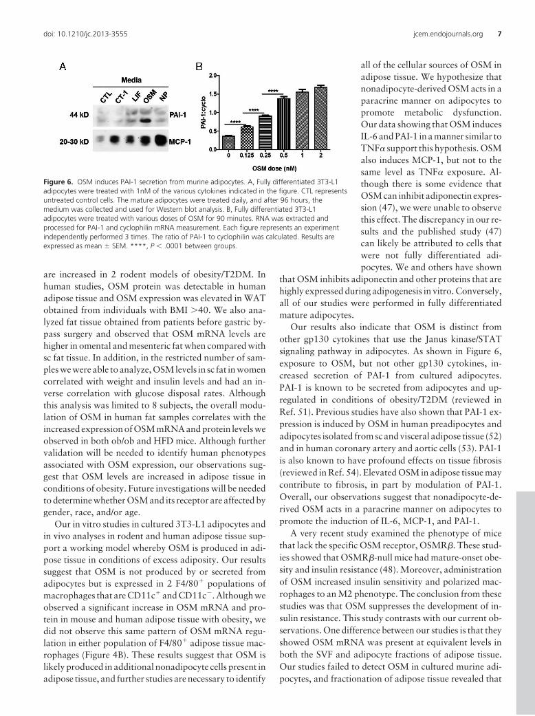
several gp130 cytokines on the secre-tion of 2 known adipokines, MCP-1and PAI-1, from murine adipocytes.As shown in Figure 6A, all of thegp130 cytokines examined (CT-1,LIF, OSM, and NP) resulted in aninduction of MCP-1 secretion. How-ever, only OSM resulted in a robustincrease in PAI-1 secretion. To assesswhether this response was directlyregulated by OSM, we treated ma-ture adipocytes for 90 minutes withvarious doses of OSM. Acute OSMtreatment resulted in a dose-depen-dent increase in PAI-1 mRNA levels(Figure 6B) and in the phosphoryla-tion of OSMR�, gp130, and STAT3(data not shown).
Discussion
Adipose tissue is highly sensitive toseveral gp130 cytokines, includingOSM (44). Also, OSM inhibits adi-pocyte differentiation in vitro (25,45). In these studies, we observedthat OSM mRNA and protein levels
Figure 4. OSM is present in nonadipocyte cells of human WAT. A, Human adipose tissue wasobtained and processed to separate adipocytes from the SVF as described in Materials andMethods. From each sample 100 �g was separated by SDS-PAGE, transferred to nitrocellulose,and subjected to Western blot analysis. B, ATMs from 4 independent cohorts of male C57/BL6lean and ob/ob mice at 14 weeks of age were isolated as indicated in Materials and Methods.FBC represents ATMs that are F4/80�, CD11b�, and CD11c�. FB represents ATMs that are F4/80�, CD11b�, and CD11c�.
Figure 5. OSM and TNF� induce PAI-1, MCP-1, and IL-6 mRNA in murine adipocytes. Fullydifferentiated 3T3-L1 adipocytes were treated with 1nM TNF�, 1nM OSM, or both cytokines for72 hours before harvesting. RNA was extracted and processed for PAI-1 (A), IL-6 (B), MCP-1 (C),and ADPN (D) mRNA measurement. Each figure represents an experiment independentlyperformed 3 times. The results are expressed as mean values � SEM. Lowercase letters representstatistical differences (P � .05).
6 Sanchez-Infantes et al Adipose Oncostatin M in Obesity and Diabetes J Clin Endocrinol Metab
are increased in 2 rodent models of obesity/T2DM. Inhuman studies, OSM protein was detectable in humanadipose tissue and OSM expression was elevated in WATobtained from individuals with BMI �40. We also ana-lyzed fat tissue obtained from patients before gastric by-pass surgery and observed that OSM mRNA levels arehigher in omental and mesenteric fat when compared withsc fat tissue. In addition, in the restricted number of sam-ples we were able to analyze, OSM levels in sc fat in womencorrelated with weight and insulin levels and had an in-verse correlation with glucose disposal rates. Althoughthis analysis was limited to 8 subjects, the overall modu-lation of OSM in human fat samples correlates with theincreased expression of OSM mRNA and protein levels weobserved in both ob/ob and HFD mice. Although furthervalidation will be needed to identify human phenotypesassociated with OSM expression, our observations sug-gest that OSM levels are increased in adipose tissue inconditions of obesity. Future investigations will be neededto determine whether OSM and its receptor are affected bygender, race, and/or age.
Our in vitro studies in cultured 3T3-L1 adipocytes andin vivo analyses in rodent and human adipose tissue sup-port a working model whereby OSM is produced in adi-pose tissue in conditions of excess adiposity. Our resultssuggest that OSM is not produced by or secreted fromadipocytes but is expressed in 2 F4/80� populations ofmacrophages that are CD11c� and CD11c�. Although weobserved a significant increase in OSM mRNA and pro-tein in mouse and human adipose tissue with obesity, wedid not observe this same pattern of OSM mRNA regu-lation in either population of F4/80� adipose tissue mac-rophages (Figure 4B). These results suggest that OSM islikely produced in additional nonadipocyte cells present inadipose tissue, and further studies are necessary to identify
all of the cellular sources of OSM inadipose tissue. We hypothesize thatnonadipocyte-derived OSM acts in aparacrine manner on adipocytes topromote metabolic dysfunction.Our data showing that OSM inducesIL-6 and PAI-1 in a manner similar toTNF� support this hypothesis. OSMalso induces MCP-1, but not to thesame level as TNF� exposure. Al-though there is some evidence thatOSM can inhibit adiponectin expres-sion (47), we were unable to observethis effect. The discrepancy in our re-sults and the published study (47)can likely be attributed to cells thatwere not fully differentiated adi-pocytes. We and others have shown
that OSM inhibits adiponectin and other proteins that arehighly expressed during adipogenesis in vitro. Conversely,all of our studies were performed in fully differentiatedmature adipocytes.
Our results also indicate that OSM is distinct fromother gp130 cytokines that use the Janus kinase/STATsignaling pathway in adipocytes. As shown in Figure 6,exposure to OSM, but not other gp130 cytokines, in-creased secretion of PAI-1 from cultured adipocytes.PAI-1 is known to be secreted from adipocytes and up-regulated in conditions of obesity/T2DM (reviewed inRef. 51). Previous studies have also shown that PAI-1 ex-pression is induced by OSM in human preadipocytes andadipocytes isolated from sc and visceral adipose tissue (52)and in human coronary artery and aortic cells (53). PAI-1is also known to have profound effects on tissue fibrosis(reviewed in Ref. 54). Elevated OSM in adipose tissue maycontribute to fibrosis, in part by modulation of PAI-1.Overall, our observations suggest that nonadipocyte-de-rived OSM acts in a paracrine manner on adipocytes topromote the induction of IL-6, MCP-1, and PAI-1.
A very recent study examined the phenotype of micethat lack the specific OSM receptor, OSMR�. These stud-ies showed that OSMR�-null mice had mature-onset obe-sity and insulin resistance (48). Moreover, administrationof OSM increased insulin sensitivity and polarized mac-rophages to an M2 phenotype. The conclusion from thesestudies was that OSM suppresses the development of in-sulin resistance. This study contrasts with our current ob-servations. One difference between our studies is that theyshowed OSM mRNA was present at equivalent levels inboth the SVF and adipocyte fractions of adipose tissue.Our studies failed to detect OSM in cultured murine adi-pocytes, and fractionation of adipose tissue revealed that
Figure 6. OSM induces PAI-1 secretion from murine adipocytes. A, Fully differentiated 3T3-L1adipocytes were treated with 1nM of the various cytokines indicated in the figure. CTL representsuntreated control cells. The mature adipocytes were treated daily, and after 96 hours, themedium was collected and used for Western blot analysis. B, Fully differentiated 3T3-L1adipocytes were treated with various doses of OSM for 90 minutes. RNA was extracted andprocessed for PAI-1 and cyclophilin mRNA measurement. Each figure represents an experimentindependently performed 3 times. The ratio of PAI-1 to cyclophilin was calculated. Results areexpressed as mean � SEM. ****, P � .0001 between groups.
doi: 10.1210/jc.2013-3555 jcem.endojournals.org 7
most OSM protein was present in the SVF (Figure 4A). Theother study did not observe OSMR� in adipocytes, al-though we have substantial data documenting the pres-ence of OSMR� in mature adipocytes (Supplemental Fig-ure 2B). Moreover, rodent adipose tissue in vivo is highlyresponsive to OSM exposure (44), indicating that theOSMR� is present in adipocytes. We also observed anincrease in OSMR� in the 2 rodent models of obesity weexamined (Figure 1), raising the possibility that adipocytesmight be more sensitive to OSM under these conditions.Our model supports a role of OSM in inducing the ex-pression of genes associated with inflammation and insu-lin resistance. Therefore, we hypothesize that adipose tis-sue-derived OSM promotes a metabolically unfavorablephenotype. We predict that OSMR� signaling in adi-pocytes is likely involved in the pathogenesis of T2DM butdoes not serve in a protective capacity as recently sug-gested in mice that have a global knockout of the OSMreceptor (48). Studies in liver indicate that OSM, producedin Kuppfer cells, can promote hepatic insulin resistance byinhibiting insulin-induced Akt phosphorylation (55).There is little question that many additional studies will benecessary to identify all the adipose tissue sources of OSMand to determine the overall function of this cytokine inT2DM. Nonetheless, our results clearly demonstrate thatOSM levels are induced in murine and human obesity, andOSM can modulate adipocyte gene expression in a mannerthat is similar, but distinct, from TNF�.
Acknowledgments
We thank Anik Boudreau for cell culture support and RyanGrant for comments regarding manuscript preparation.
Address all correspondence and requests for reprints to: Jac-queline M. Stephens, Louisiana State University, Department ofBiological Sciences, 202 Life Sciences Building, Baton Rouge, LA70803. E-mail: [email protected].
This work was supported by Grant R01DK052968-15 fromthe National Institutes of Health (to J.M.S.) and a Sara Borrellcontract from ISCIII (Madrid, Spain) (to D.S.-I.).
D.S.-I. and U.A.W. conducted most of the experiments.D.S.-I., U.A.W., and C.M.E. wrote and J.M.S. edited the man-uscript to produce the final version. C.M.E. performed studies onreceptor expression. A.W.F. provided the data on OSM expres-sion in macrophages. R.V.C. and J.G. provided human tissue ortissue fractions for Western blot analysis that was conducted byD.S.-I., R.F.M. performed RNA analysis on rodent tissues pre-pared by J.M.S., E.R. provided human tissue that was used byD.S.-I. for RNA analysis. E.R. also provided additional infor-mation on study participants. All of the authors contributed toediting the manuscript before submission.
Disclosure Summary: D.S.-I., U.A.W., C.M.E., R.F.M., E.R.,and J.M.S. have nothing to disclose. R.V.C. consults for Merck
Research Laboratories and receives grant funding from MerckResearch Laboratories and Eli Lilly and Company. A.W.F. re-ceives grant support from AstraZeneca Plc. J.M.G. is the co-founder and Chief Scientific Officer of LaCell LLC, a for-profitbiotechnology company focusing on the use of adipose stromal/stem cells for research and regenerative medical applications.
References
1. Fasnacht N, Müller W. Conditional gp130 deficient mouse mutants.Semin Cell Dev Biol. 2008;19:379–384.
2. Heinrich PC, Behrmann I, Haan S, Hermanns HM, Müller-NewenG, Schaper F. Principles of interleukin (IL)-6-type cytokine signal-ling and its regulation. Biochem J. 2003;374:1–20.
3. Hansen D, Dendale P, Beelen M, et al. Plasma adipokine and in-flammatory marker concentrations are altered in obese, as opposedto non-obese, type 2 diabetes patients. Eur J Appl Physiol. 2010;109:397–404.
4. Kern PA, Ranganathan S, Li C, Wood L, Ranganathan G. Adiposetissue tumor necrosis factor and interleukin-6 expression in humanobesity and insulin resistance. Am J Physiol Endocrinol Metab.2001;280:E745–E751.
5. Celik A, Sahin S, Koc F, et al. Cardiotrophin-1 plasma levels areincreased in patients with diastolic heart failure. Med Sci Monit.2012;18:CR25–CR31.
6. López B, Gonzalez A, Querejeta R, Barba J, Diez J. Association ofplasma cardiotrophin-1 with stage C heart failure in hypertensivepatients: potential diagnostic implications. J Hypertens. 2009;27:418–424.
7. Natal C, Fortuño MA, Restituto P, et al. Cardiotrophin-1 is ex-pressed in adipose tissue and upregulated in the metabolic syndrome.Am J Physiol Endocrinol Metab. 2008;294:E52–E60.
8. Slevin M, Krupinski J, Mitsios N, et al. Leukaemia inhibitory factoris over-expressed by ischaemic brain tissue concomitant with re-duced plasma expression following acute stroke. Eur J Neurol.2008;15:29–37.
9. Pradeep AR, S TM, Garima G, Raju A. Serum levels of oncostatinM (a gp 130 cytokine): an inflammatory biomarker in periodontaldisease. Biomarkers. 2010;15:277–282.
10. Febbraio MA. gp130 receptor ligands as potential therapeutic tar-gets for obesity. J Clin Invest. 2007;117:841–849.
12. Di Gregorio GB, Hensley L, Lu T, Ranganathan G, Kern PA. Lipidand carbohydrate metabolism in mice with a targeted mutation inthe IL-6 gene: absence of development of age-related obesity. Am JPhysiol Endocrinol Metab. 2004;287:E182–E187.
13. Rotter V, Nagaev I, Smith U. Interleukin-6 (IL-6) induces insulinresistance in 3T3-L1 adipocytes and is, like IL-8 and tumor necrosisfactor-alpha, overexpressed in human fat cells from insulin-resistantsubjects. J Biol Chem. 2003;278:45777–45784.
14. Gloaguen I, Costa P, Demartis A, et al. Ciliary neurotrophic factorcorrects obesity and diabetes associated with leptin deficiency andresistance. Proc Natl Acad Sci U S A. 1997;94:6456–6461.
15. Lambert PD, Anderson KD, Sleeman MW, et al. Ciliary neu-rotrophic factor activates leptin-like pathways and reduces body fat,without cachexia or rebound weight gain, even in leptin-resistantobesity. Proc Natl Acad Sci U S A. 2001;98:4652–4657.
16. Sleeman MW, Garcia K, Liu R, et al. Ciliary neurotrophic factorimproves diabetic parameters and hepatic steatosis and increasesbasal metabolic rate in db/db mice. Proc Natl Acad Sci U S A. 2003;100:14297–14302.
17. Ettinger MP, Littlejohn TW, Schwartz SL, et al. Recombinant vari-ant of ciliary neurotrophic factor for weight loss in obese adults: arandomized, dose-ranging study. JAMA. 2003;289:1826–1832.
8 Sanchez-Infantes et al Adipose Oncostatin M in Obesity and Diabetes J Clin Endocrinol Metab
18. Blüher S, Moschos S, Bullen J Jr, et al. Ciliary neurotrophic fac-torAx15 alters energy homeostasis, decreases body weight, and im-proves metabolic control in diet-induced obese and UCP1-DTAmice. Diabetes. 2004;53:2787–2796.
19. Zvonic S, Cornelius P, Stewart WC, Mynatt RL, Stephens JM. Theregulation and activation of ciliary neurotrophic factor signalingproteins in adipocytes. J Biol Chem. 2003;278:2228–2235.
20. Crowe S, Turpin SM, Ke F, Kemp BE, Watt MJ. Metabolic remod-eling in adipocytes promotes ciliary neurotrophic factor-mediatedfat loss in obesity. Endocrinology. 2008;149:2546–2556.
21. Balhoff JP, Stephens JM. Highly specific and quantitative activationof STATs in 3T3-L1 adipocytes. Biochem Biophys Res Commun.1998;247:894–900.
23. Stephens JM, Lumpkin SJ, Fishman JB. Activation of signal trans-ducers and activators of transcription 1 and 3 by leukemia inhibitoryfactor, oncostatin-M, and interferon-gamma in adipocytes. J BiolChem. 1998;273:31408–31416.
24. Zvonic S, Hogan JC, Arbour-Reily P, Mynatt RL, Stephens JM.Effects of cardiotrophin on adipocytes. J Biol Chem. 2004;279:47572–47579.
25. White UA, Stewart WC, Mynatt RL, Stephens JM. Neuropoietinattenuates adipogenesis and induces insulin resistance in adipocytes.J Biol Chem. 2008;283:22505–22512.
26. Lehr S, Hartwig S, Sell H. Adipokines: a treasure trove for the dis-covery of biomarkers for metabolic disorders. Proteomics ClinAppl. 2012;6:91–101.
27. Mohamed-Ali V, Goodrick S, Rawesh A, et al. Subcutaneous adi-pose tissue releases interleukin-6, but not tumor necrosis factor-�, invivo. J Clin Endocrinol Metab. 1997;82:4196–4200.
28. Fried SK, Bunkin DA, Greenberg AS. Omental and subcutaneousadipose tissues of obese subjects release interleukin-6: depot differ-ence and regulation by glucocorticoid. J Clin Endocrinol Metab.1998;83:847–850.
29. Blüher M. Clinical relevance of adipokines. Diabetes Metab J. 2012;36:317–327.
30. Weisberg SP, McCann D, Desai M, Rosenbaum M, Leibel RL, Fer-rante AW Jr. Obesity is associated with macrophage accumulationin adipose tissue. J Clin Invest. 2003;112:1796–1808.
31. Virtue S, Vidal-Puig A. Adipose tissue expandability, lipotoxicityand the metabolic syndrome–an allostatic perspective. Biochim Bio-phys Acta. 2010;1801:338–349.
32. Rose TM, Lagrou MJ, Fransson I, et al. The genes for oncostatin M(OSM) and leukemia inhibitory factor (LIF) are tightly linked onhuman chromosome 22. Genomics. 1993;17:136–140.
33. Rose TM, Bruce AG. Oncostatin M is a member of a cytokine familythat includes leukemia-inhibitory factor, granulocyte colony-stim-ulating factor, and interleukin 6. Proc Natl Acad Sci U S A. 1991;88:8641–8645.
34. Zarling JM, Shoyab M, Marquardt H, Hanson MB, Lioubin MN,Todaro GJ. Oncostatin M: a growth regulator produced by differ-entiated histiocytic lymphoma cells. Proc Natl Acad Sci U S A. 1986;83:9739–9743.
35. Mosley B, De Imus C, Friend D, et al. Dual oncostatin M (OSM)receptors. Cloning and characterization of an alternative signalingsubunit conferring OSM-specific receptor activation. J Biol Chem.1996;271:32635–32643.
36. Brown TJ, Lioubin MN, Marquardt H. Purification and character-ization of cytostatic lymphokines produced by activated human Tlymphocytes. Synergistic antiproliferative activity of transforming
growth factor �1, interferon-�, and oncostatin M for human mel-anoma cells. J Immunol. 1987;139:2977–2983.
37. Suda T, Chida K, Todate A, et al. Oncostatin M production byhuman dendritic cells in response to bacterial products. Cytokine.2002;17:335–340.
38. Wahl AF, Wallace PM. Oncostatin M in the anti-inflammatory re-sponse. Ann Rheum Dis. 2001;60(Suppl 3):iii75-iii80.
39. Hui W, Bell M, Carroll G. Detection of oncostatin M in synovialfluid from patients with rheumatoid arthritis. Ann Rheum Dis.1997;56:184–187.
40. Albasanz-Puig A, Murray J, Preusch M, et al. Oncostatin M is ex-pressed in atherosclerotic lesions: a role for Oncostatin M in thepathogenesis of atherosclerosis. Atherosclerosis. 2011;216:292–298.
41. Kamiya A, Kinoshita T, Ito Y, et al. Fetal liver development requiresa paracrine action of oncostatin M through the gp130 signal trans-ducer. EMBO J. 1999;18:2127–2136.
42. Okaya A, Kitanaka J, Kitanaka N, et al. Oncostatin M inhibitsproliferation of rat oval cells, OC15–5, inducing differentiation intohepatocytes. Am J Pathol. 2005;166:709–719.
43. Nakamura K, Nonaka H, Saito H, Tanaka M, Miyajima A. Hepa-tocyte proliferation and tissue remodeling is impaired after liverinjury in oncostatin M receptor knockout mice. Hepatology. 2004;39:635–644.
44. White UA, Stewart WC, Stephens JM. Gp130 cytokines exert dif-ferential patterns of crosstalk in adipocytes both in vitro and in vivo.Obesity (Silver Spring). 2011;19:903–910.
45. Miyaoka Y, Tanaka M, Naiki T, Miyajima A. Oncostatin M in-hibits adipogenesis through the RAS/ERK and STAT5 signalingpathways. J Biol Chem. 2006;281:37913–37920.
46. Ouchi N, Parker JL, Lugus JJ, Walsh K. Adipokines in inflammationand metabolic disease. Nat Rev Immunol. 2011;11:85–97.
47. Song HY, Kim MR, Lee MJ, et al. Oncostatin M decreases adi-ponectin expression and induces dedifferentiation of adipocytes byJAK3- and MEK-dependent pathways. Int J Biochem Cell Biol.2007;39:439–449.
48. Komori T, Tanaka M, Senba E, Miyajima A, Morikawa Y. 2013Lack of oncostatin M receptor beta leads to adipose tissue inflam-mation and insulin resistance by switching macrophage phenotype.J Biol Chem. 2013;288:21861–21875.
49. Wallace PM, MacMaster JF, Rouleau KA, et al. Regulation of in-flammatory responses by oncostatin M. J Immunol. 1999;162:5547–5555.
50. Tamura S, Morikawa Y, Miyajima A, Senba E. Expression of on-costatin M in hematopoietic organs. Dev Dynam. 2002;225:327–331.
51. Alessi MC, Poggi M, Juhan-Vague I. Plasminogen activator inhib-itor-1, adipose tissue and insulin resistance. Curr Opin Lipidol.2007;18:240–245.
52. Rega G, Kaun C, Weiss TW, et al. Inflammatory cytokines inter-leukin-6 and oncostatin m induce plasminogen activator inhibitor-1in human adipose tissue. Circulation. 2005;111:1938–1945.
53. Demyanets S, Kaun C, Rychli K, et al. The inflammatory cytokineoncostatin M induces PAI-1 in human vascular smooth muscle cellsin vitro via PI 3-kinase and ERK1/2-dependent pathways. Am JPhysiol Heart Circ Physiol. 2007;293:H1962–H1968.
54. Ghosh AK, Vaughan DE. PAI-1 in tissue fibrosis. J Cell Physiol.2012;227:493–507.
55. Henkel J, Gärtner D, Dorn C, et al. Oncostatin M produced inKupffer cells in response to PGE2: possible contributor to hepaticinsulin resistance and steatosis. Lab Invest. 2011;91:1107–1117.