Onderstepoort I ournal of Ve terinary Research, Volume 29, Number 1, March, 1962. The Government Print er, Pretoria. A PRELIMINARY REPORT ON THE OCCURRENCE OF SELENOSIS IN SOUTH AFRICA AND I TS POSSI BLE ROLE IN THE AET I OLOGY OF TRIBULOSIS ( GEEL- DIKKOP), ENZOOTIC I CTERUS AND SOME OTHER DISEASE CONDITIONS ENCOUNTERED IN THE KAROO AREAS J. M. M. BROWN and P. J. de WET, Onderstepoort laboratory INTRODUCTION Some of the symptoms of selenium .i ntoxication have been known for a very long time under diverse names. Probably the first written account we have is that of Marco Polo (1254-1327), who recognised a pecuJ.iar form of stock poisoning in Western China and assigned it correctly to the forage, stating: "a poisonous plant growi11g there, which if eaten . . . . has the effect of causing the hooves of the animal to drop off". The next authentic written record of selenium intoxication was a report written in 1856 by Madison (cit. Moxon & Rhian, 1943), an army surgeon stationed in Nebraska, who described a fatal disease, known locally as ''alkali disease" among the horses grazing in a certain area near Fort Randall. The disease remained an enigma until the discovery by Robinson (1933) of selenium in cereals originating from this area and proved to be toxic to laboratory animals by Franke and co-workers (Moxon & Rhian, 1943). Since then a tremendous amount of work was done in the United States, and selenium was proved beyond doubt to be the causal agent of two diseases known locally as " Blind Staggers" and " AJkali disease". These diseases were found to be present in a large area of the United States, and since then selenium in toxication has been reported from Canada (Davidson, 1940; Thorvaldson, 1951), Colombia, South America, the Southern States of South America (Roche Pamphlet, I 959), Mexico (Williams, Lakin & Byers, 1940), County Limerick in Ireland (Walsh, Fleming, O 'Connor & Sweeney, 1951), Israel and Australia (Roche Pamphlet, 1959), and probably South India and North Africa (Mahadevan, 1954) . Analysis by Robinson (1936) of cereals from various parts of the world showed that seleniferous areas are widespread over the surface of the earth. A sample of grain emanating from South Mrica examined by him, had the highest selenium content of all the imported grains, although it was nowhere near that of American grains. This is believed to be the first report incriminating selenium excess in the aetiology of certain diseases in South Africa. Three excellent reviews of the selenium problem are those by Russell & D uncan (1956), Moxon & Rhian (1943), and Painter (1 94 1). Received for publication on J6 August, 1961.- Editor. 111
Transcript
Onderstepoort I ournal of Veterinary Research,
Volume 29, Number 1, March, 1962. The Government Printer, Pretoria.
A PRELIMINARY REPORT ON THE OCCURRENCE OF SELENOSIS IN SOUTH AFRICA AND ITS POSSIBLE ROLE IN THE AETIOLOGY OF TRIBULOSIS (GEELDIKKOP), ENZOOTIC ICTERUS AND SOME OTHER DISEASE CONDITIONS
ENCOUNTERED IN THE KAROO AREAS
J. M. M. BROWN and P. J. de WET, Onderstepoort laboratory
INTRODUCTION
Some of the symptoms of selenium .intoxication have been known for a very long time under diverse names. Probably the first written account we have is that of Marco Polo (1254-1327), who recognised a pecuJ.iar form of stock poisoning in Western China and assigned it correctly to the forage, stating: "a poisonous plant growi11g there, which if eaten . . . . has the effect of causing the hooves of the animal to drop off". The next authentic written record of selenium intoxication was a report written in 1856 by Madison (cit. Moxon & Rhian, 1943), an army surgeon stationed in Nebraska, who described a fatal disease, known locally as ''alkali disease" among the horses grazing in a certain area near Fort Randall. The disease remained an enigma until the discovery by Robinson (1933) of selenium in cereals originating from this area and proved to be toxic to laboratory animals by Franke and co-workers (Moxon & Rhian, 1943). Since then a tremendous amount of work was done in the United States, and selenium was proved beyond doubt to be the causal agent of two diseases known locally as " Blind Staggers" and " AJkali disease". These diseases were found to be present in a large area of the United States, and since then selenium intoxication has been reported from Canada (Davidson, 1940; Thorvaldson, 1951), Colombia, South America, the Southern States of South America (Roche Pamphlet, I 959), Mexico (Williams, Lakin & Byers, 1940), County Limerick in Ireland (Walsh, Fleming, O'Connor & Sweeney, 1951), Israel and Australia (Roche Pamphlet, 1959), and probably South India and North Africa (Mahadevan, 1954) . Analysis by Robinson (1936) of cereals from various parts of the world showed that seleniferous areas are widespread over the surface of the earth. A sample of grain emanating from South Mrica examined by him, had the highest selenium content of all the imported grains, although it was nowhere near that of American grains.
This is believed to be the first report incriminating selenium excess in the aetiology of certain diseases in South Africa.
Three excellent reviews of the selenium problem are those by Russell & D uncan (1956), Moxon & Rhian (1943), and Painter (1941).
Received for publication on J6 August, 1961.- Editor.
111
SELENOSIS lN SOUTH AFRICA
The disease in nature, as recognised elsewhere in the world, may occur in different forms depending upon the amount, the form of and the period during which the selenium was ingested. The following forms are recognised:-
(a) Acute intoxication.- This is seen mainly in cattle and sheep, and sometimes also in pigs and horses. lt results from the ingestion of enough toxic plants, usually in a single feed to produce very severe symptoms of poisoning. It is incredible that this can occur, since highly seleniferous vegetation is most unpalatable to livestock. Jt seems, however, that in times when other forage is scarce or not available, animals are forced to eat it. Death follows within a few hours to a few days.
This form is characterised by a rapid weak pulse, dyspnoea, bloat, severe colic, polyuria, cyanosis, prostration and death due to respiratory failure. Post mortem signs include generalised haemorrhages, ascites and general atony of smooth muscle structures.
(b) Sub-acute intoxication (Blind Staggers, "Loco disease", Locoism).-This may develop after a short while in animals grazing on lands co11taining seleniferous weeds. Sometimes the onset of symptoms is delayed for several weeks or even months. Jn such cases death follows within a few days. It can be reproduced experimentally by feeding diets containing 10 to 20 p.p.m. of selenium for seven to eight weeks. In nature the plants causing this form of the disease may contain up to several thousands p.p.m. of selenium.
Both cattle and sheep are affected, but in general sheep seem to be more resistant to the effects of selenium. In the early stages the animal may stray from the herd; there is usually some slight impairment of vision and the animal has difficulty in judging nearness of objects in its path. As the disease progresses there is marked loss of weight, a poor staring coat and progressively increasing blindness.
The following nervous derangements may be seen: A tendency to wander which may progress to the stage where the animal goes round aimlessly in circles, a staggering gait and insistence on walking forwards. When solid objects are encountered the animal continues to press forward with its head. The front limbs become weak and unable to support the animal.
There is often lachrymation, salivation, inability to swallow, severe abdominal pain and ultimately varying degrees of paralysis. Death is due to respiratory failure.
There are some deviations from this typical pattern, notably: (a) Not all animals become blind, nor do they develop" staggers" in aU cases; (b) some animals develop a marked "pica ", i.e. depraved appetite manifested by a desire to chew wood, bones, metal objects, etc., One author (Davidson, 1940) attributes this to a disturbance of calcium and phosphorus metabolism, of which the symptom is typical.
Post mortem changes in this form of the disease include the following: Atony of smooth muscle structures such as the gastro-intestinal tract, gallbladder and urinary bladder is common. The gaUbladder may be markedly distended and there may be severe impaction of the rumen and omasum, especially in cases occurring during winter. The mucosa of the abomasum and small intestine shows irritation of varying degrees, petechiation and ulceration are seen in severe cases. Ascites and visceral venous congestion are common.
112
J. M. M. BROWN and P. J. DE WET
The appearance of the liver is noteworthy since similar lesions have been encountered during the course of recent field investigations into tribulosis and enzootic icterus amongst sheep and goats in the North-west Cape and Cape Midlands. This organ is often markedly congested and areas of focal necrosis are evident in the early stages of the intoxication. These necrotic areas appear as connective tissue scars which, on contraction , form pits upon the surface of the organ. Cirrhosis of an atrophic type occurs but is not as common in this form as it is in the following one.
The heart frequently shows endocarditis, myocarditis and petechial haemorrhages on the epicardium. Chronjc degenerative changes may be present in all the parenchymatous organs. The spleen is sometimes enlarged with localised haemorrhagic areas.
Erosion of the articular surfaces of the long bones, especially the tibia, is seen in the majo1ity of cases.
(c) Chronic intoxication ("Alkali disease ") .- This is caused by the daily ingestion of cereals, grasses and other forage plants which contain small amounts of selenium, e.g., 10 to 30 p.p.m. (5 p.p.m. of selenium is apparently the limit of tolerance for most livestock).
Dullness and lack of vitality are the first symptoms seen. The animals then become emaciated, stiff, lame and fail to respond to good care or to a selenium-free diet. Affected animals show a rough coat and marked "pica". A prominent symptom is the loss of hair-in horses from the mane and tail, in cattle from the switch of the tail and in pigs along the back.
There is swelling and marked tenderness of the coronary band of the hoof. Rings appear on the hoof just below the coronary band, cracks may appear in the hoof, and a gradual separation of the hoof from the underlying tissue occurs. Cracks may be sufficiently deep to separate the old horn growth from the new. Jn some cases the old hoof may be sloughed off, while in others it may remain attached until the growth of the new hoof has reached normal length. This is seen especially in horses. In cattle sloughing usually does not occur, but the old hoof usually remains attached to the new growth, and in cases where there have been several attacks, the old ragged hooves may be eight or ten inches long and tumed upwards at the end. Partial separation of the old hoof growth is sometimes seen giving the appearance of" slippers". During the time these hoof changes are occurring the animals are very lame and often in severe pain. Walking is difficult and the animals are often seen down on their knees while grazing. A well marked ring is frequently noted on the horns, just above the hair.
'the same hoof changes are noted in pigs, but do not seem to be prominent in sheep.
In cattle there is also some evidence of interference with the circulation of blood in the extremities, which results in freezing of the lower parts of the limbs and tail after exposure to the extreme cold of the northern plains of America during winter. This form of the disease is referred to by the ranchers of trus region as " Frozen Foot".
Very severe anaemia is a prominent symptom of the disease in all animals. There are also some reports of lowered fertility.
113
SELENOSIS IN SOUTH AFRICA
A very important point to bear in mind at this stage is that selenium may be transmitted via maternal blood to the developing foetus in a pregnant animal, and that the foetus may build up a level of selenium .in its tissues equal to that of the mother. Thus:-
(a) Colts may be born with deformed hooves and exhibit other evidence of selenium poisoning (c.f. " boxy foot "-discussed later).
(b) Calves born of affected cows may show evidence of the disease at birth or the cow may excrete sufficient selenium in her milk to produce definite symptoms in her calf later.
(c) Young pigs may be born hairless or ingest sufficient selenium via the maternal milk to cause loss of hair in a very short time (c. f. " Kaalsiekte " discussed later).
(d) Weak lambs a1e born of affected ewes; some of these are too weak to stand and die of starvation. Others able to stand show an immediate "pica". ln extreme cases such lambs are born with deformities of the limbs or hooves.
In general poultry seem very resistant to selenium intoxication. When they do become affected, the main effect seen is poor hatchability of eggs, and in chickens which do hatch there is either l ow vitality or a high proportion of "monsters", showing various limb deformities.
Chronic selenosis of dietary origin in man, has never been seriously considered as a health hazard. Mao is apparently as susceptible as his animals, but as he seldom eats plants of high selenium content, the danger of .ingestion of sufficient amounts of selenium is not as great. However, the margin of safety in the consumption of animal products from seleniferous areas is not very large. The following symptoms have been recorded in man (Mahadevan, 1954) :-
(a) Gastro-intestinal disorders. (b) Nervous or mental disorders often terminating io dementia. (c) On exposure to bright sunlight, skin manifestations present themselves
in the form of vesicu lations and actinic dermatitis. Symptoms (a) and (b) may then become aggravated and mortality may be high.
The various post mortem lesions described in animals include the fo1Jowing:-
A very severe degeneration of all the parenchymatous organs, especially the heart, liver and kidneys. The heart is invariably atrophied and shows evidence of decompensation, its bundles of muscle fibres appear to be separated by a serofibrinous exudate, while endocarditis and myocarditis are prevalent.
The Jiver is cirrhotic and often atrophied. The kidneys often show severe nephrosis, and glomerulo-nephritis is seen in many cases. Accord ing to one description (Davidson, 1940) these organs are clay-coloured, showing multiple peteclliae and infarcts. These liver and kidney changes are highly reminiscent of those seen in enzootic icterus.
The cortex and meduUa of the adrenals are described as being haemorrhagic (Moxon & Rhian, 1943), while the lesions of the gastro-intestinal tract are of the same general character as those seen in the ''sub-acute " form. Erosion of the articular surfaces of the joints io the long bones is common. Anaemia is particularly severe in this form of the disease. Hypoplastic changes in the bone marrow are described in one account (Davidson, 1940).
114
J. M. M. BROWN and P. J. DE WET
EXPERIMENTALLY J NDUCED SELENOSIS
(a) Acute intoxication
This has been studied in many species including frogs , dogs and herbivora. It is usually produced by intravenous or intraperitoneal injections of soluble selenium salts, e.g. selenite or selenate. The latter is far Jess toxic than the former. Doses used ranged from 2 to 5 mg.(Kg. of selenium depending on the species. Injection of selenite is rapidly followed by death due to respiratory failure. Various nervous symptoms are described. Dehydration 01 rather anhydraemia is encountered. Repeated small doses of selenium have a cumulative effect. There is no evidence of an acquired tolerance.
(b) Sub-acute and chronic intoxication
These conditions have been studied in most species, but mainly in small animals. and are reproduced by feeding selenium in the ration at levels of 5 to 25 p.p.m.
fn rats the following results ate seen:-
Marked anorexia , decreased growth rate, degeneration of the genitalia and impaired reproduction, a very marked and progressive anaemia, with. haemoglobin levels falling to as low as 2 gm. per cent, rough fur, in some cases paralysis of the hindquarters, intestinal haemorrhages, severe jaundice, and very severe liver lesions.
Both the anaemia and the liver lesions deserve special mention , since they are of importance to lhe problem as presented in South Africa. The anaemia is described (Smith, Stohlman & Lillie, 1937) as being typified by: "A relatively low haemoglobin compared with the red cell count. marked reticulocytosis sometimes up to 80 per cent, anisocytosis, polychromasia and normoblasts present in greater or lesser numbers " . The authors conclude that it is essentially microcytic and hypochromic in nature. Tn another paper (Schneider, 1936) the urine of rats developing this severe anaemia is described as " becoming highly pigmented··.
These descriptions and the interpretations based on them are not entirely sa tisfactory. In the first instance the account of the anaemia describes changes more indicative of the recovery phase of a post-haemorrhagic anaemia, or of a macrocytic type, rather than the microcytic hypochromic anaemias which are essentially states of iron deficiency. In the second place the description of the urine of these rats is not quite clear. For instance, was the highly pigmented appearance due to bilirubin or to the concentration of an oliguria? After careful consideration of the literature, this anaemia is believed by the authors to be haemolytic in nature with probable remissions and exacerbations and essentially similar to that encountered during the acute phases of enzootic icterus, or in the milder forms similar to the anaemia seen in geeldikkop. Severe icterus was seen in these rats. This seems inconsistent with the liver pathology described.
The livers of these rats are described as being atrophic, 11ecrotic, cirrhotic and haemorrhagic in varying degrees. It is often seen that the right lateral and/ or caudate Jobes are hypertrophied while the left lateral and central lobes are severely atropllied (c.f. Brown & de Boom, 1958, unpublished. data).
The heart and spleen are enlarged . Ascites is very common, hydrothorax less so.
Cats, rabbits and dogs in general show similar symptoms, some being more or less marked depending on the species.
115
SELENOSIS lN SOUTH AFRJCA
THE E FFECT OF DIETARY FACTORS ON SELENiUM P OISONTNG
The amount of protein in the diet is of great importance in alleviating or modifying the symptoms of selenosis. Selenium poisoning is much less severe on a diet of h.igh protein content. Smith & Stohl man (J 940) expressed the percentage of protein and the micrograms of selen ium present in 100 gm. of ration as a ratio and concluded that a ration with a protein: selenium ratio of 1: 30 or less is nontoxic, while a ration with the same selenium content but havi ng a protein: selenium ratio of J: 100 is dangerously toxic. The toxicity is even more apparent if methionine is deficient in such a ration. Addition of methionine to such rations was found to have beneficial effects (Moxon & Rhjan, 1943). Crude casein and linseed meal give full protection against selenium (Moxon & Rhian, 1943). There is much disagreement about the p rotective effect of other protein supplements. lt is of interest to the authors to note than zei n, a protein of poor biological value is reported to have a protective influence.
It would appear from the literature perused that certa in species differences, which have been observed in regard to the pathology of selenium intoxication , might be due, in part, not only to the protein content of the ration affected animals received but also the qualitative differences in the amino acid composition of the proteins present a11d species variations in the requirements of essential amino acids.
Cystine supplementation of the ration does not seem to be effective. It was found that thiamine and certa in elements potentiated the deleterious effects of selenium, e.g., fluorine, molybdenum, zinc and cobalt amongst others (Moxon & Rhian, 1943).
It was found that 5 p.p.m. of arsenjc administered as sodium ar$eni•e concurrently with selenium, completely prevented the symptoms of selenium poisoning (Moxon & Rhian , 1943). This fact has received a considerable amount of attention and the concensus of opinion is that arsenic undoubtedly has a beneficial effect on selenium intoxication. Its exact mode of action is unknown. lt is thought to prevent the inactivation of succinic dehydrogenase by selenium (see later), possibly by reacting with selenium compounds in s uch a way, in l'ivo, as to prevent selenium from attacking the sulphydryl compounds of the body. lt is impo1tant to bear in mind the beneficial effects of arsenic, especially when one considers the large amounts dosed annually to stock in this country, for various purposes.
At this stage it is pertinent to review briefly the various disease conditions which occur in this country, chiefly in the Karoo and of which the aetiology is not yet explained. They do, however, simulate various aspects of selenosis very closely.
DISEASE CONDITIONS OF ANIMALS lN SOUTH AFRICA AND THE KAROO IN PARTICULAR, WHICH RESEMBLE SOME FORMS OF S ELENOS LS
When considered individually any connection between these diseases and selenosis might at a first glance seem remote and perhaps even incredible. However, when they are considered together, with all the foregoing as a background, the gross resemblance is obvious. Some of these d iseases are as enigmatic as our ovine icterus complex, show as many epizootological vagaries and have defied all attempts to explain them properly.
116
J. M. M. BROWN and P. J. DE WET
(a) Enzootic idiopathic liver necrosis and atrophic cirrhosis
It is a very well known fact that sheep i n the Karoo are much more susceptible to the effects of certain anthelminthics, notably the chlorinated hydrocarbons, than sheep elsewhere in South Africa. Heavy mortality often attends the dosing of these anthehninthics even when the recommended doses are used. I t is obvious that there are present in the Karoo especially, one or more factors causing latent liver damage in sheep. Tltis may flare up when aggravated by conditions such as dosing with the drugs mentioned, certain virus infections, or even severe stress conditions.
De Boom & Brown (unpubbshed data, 1958) during a critical study of enzootic icterus, encountered 11umerous cases of unexplained necrosis of the liver and a peculiar form of atrophic cirrhosis which in extreme cases was seen as a virtual replaceme11t of the entire central area of the liver by .fibrous tissue, giving this organ a dumb-bell-shaped appearance. Earlier stages took the form of marked softening or pitting of the centrai area of t11e liver. Such livers were seen even in clinically normal sheep. Subsequently the present authors have encountered a very large number of these cases during i nvestigations into geeldikkop in the Victoria West area and work on Angora goat abortions in the Cape Midlands. They have found a large percentage of these cases to be outwardly 110rmal, yet to show some profound clh.emical pathological disturbances. The condition is prevalent over a wide area of the Cape Province, with markedly different vegetat ion from part to part. The liver lesions observed are indeed very similar to those seen in chronic selenosis.
(b) " Waterpens ", "Enzootic Ascites"
This condition is extremely prevalent in certain parts of the Calvinia, Fraserburg and Sutherland distJ icts and is said to be caused by the plant Galenia c!fi·icana (" Kraalbos "). lt is characterised by a severe atrophic cirrhosis and marked ascites.
Although the bush assumed to cause the disease is fairly widespread in the Karoo, the disease is usually confined to the areas mentioned. No active principle has as yet been isolated from this plant, nor has any work been published, dealing with the disease.
(c) "Loose Wool" (Unpublished observations, Brown, 1961 b)
On numerous farms in the north-west Cape sheep are encountered in which handfu ls of wool can be plucked from the skin with ease. The skin may not be damaged by the plucking, but in extreme cases where the skin appears to be very thin, it may be torn badly. The condition is a serious problem on certain farms and its cause is quite unknown.
(d) •· Stywesiekte" in cattle, sheep, goats and equines caused by Crotalaria spp. , e.g. Crotalaria burkeana (Steyn, 1934).
These plants are widespread over the drier parts of the Republic of South Africa. The disease occurs in the Karoo, Griqualand West, Western Transvaal and parts of South West Africa. The toxicity of the plant varies considerably. The. disease is. characterised by a painful inflammation of the soft tissues of the hooves (laminitis), followed by acute lameness. Affected animals are often unable to graze. The a•cute phase is followed by excessive growth of the hooves, the old hoof tissue growing out and curling up at the point while remaining attached to the new growth. There is. a concomitant severe necrosis and atrophic cirrhosis of the liver.
117
SELENOS1S 1N SOUTH AFRICA
"Jaagsiekte" in horses caused by Crotalaria dura, C. globifera and C. juncea. These plants are seldom eaten voluntarily in nature, but are often accidentally ingested. C. juncea is a cultivated plant, "Sunnhemp ", and is often incorporated into rations. The distribution of these plants is as for the preceding plant, except that they also occur in Natal. T he symptoms seen, fall into three general classes:-
(i) Respiratory and cardio-vascular symptoms. A critical study of these symptoms seems to indicate that the basic condition is cardio-vascular, followed by pulmonary oedema, secondary bronchiolitis and ultimately pulmonary emphysema.
(ii) Icterus in horses and very severe cirrhosis of the liver. (iii) In some cases "staggers".
ln sheep the liver lesions resulting frorr, Crotalaria poisoning are prominent and there is also marked loss of wool. There is often a long latent period of feeding the plants, e.g. 20 days, before symptoms appear (Steyn, 1934).
Quite a few alkaloids have been isola ted from various Crotalaria species, e.g. monocrotaline and dicrotaline. Marais (1944) isolated dicrotaline from C. dura and C. globifera, while Louw (J 952) isolated a third alkaloid from C. damarensis. Louw states that his preparation was non-toxic, but Marais gave no details of any pharmacological tests performed with his dicrotaline specimens. It is very tempting to suggest that the cardio-vascular symptoms observed especially in horses are possibly due to the alkaloids but that the very severe liver lesions, "staggers", loss of wool in sheep and the hoof abnormalities which are described, are in effect due to selenium. The rather long latent period before the appearance of symptoms and the large quantities of tb.e plants which have to be fed to produce them, add some weight to the idea. It must be remembered, however, U1at the same conditions pertain in the production of chronic seneciosis. The alkaloids or the Senecio spp. are well known hepato-toxins, of the same chemical class as the Crotalaria compounds, but do not produce loss of hair, wool or hoof abnormalities in poisoned animals.
(e) " Staggers ·• o r "pushing disease " of callle (stoorsiekte, brain staggers, brain disease).
Tbis disease has been demonstrated to be caused by the plant Matricaria niKellaefo/ia. The disease occurs in atal and the eastern Cape Province. An excellent description or the disease is that by Andrews (1923). Animals in the initial stages appear to develop impairment of vision, there is an ever-increasing tendency to wander aimlessly, often in circles away from the herd. As the disease progresses the tendency to push against solid objects becomes evident and gradually more marked. Marked weakness of the forelimbs appears and often myotonic and myoclonic spasms. There is salivation, lachrymation , anorexia and emaciation. Ultimately the animals develop varying degrees of paralysis and die within a few days to a few weeks.
On post mortem severe fatty degeneration of all the parenchymatous organs is seen. ln a large number of cases marked ulceration of the abomasum is seen.
I nteresting points in the epizootology of the disease are: the plant is not normally eaten except under compulsion; very large amounts have to be consumed before symptoms appear; there is a latent period of 20 to 40 days after ingestion of large amounts of the plant before symptoms appear.
118
J. M. M. BROWN and P. J. DE WET
A considerable amount of work has been done on the chemistry of the organic constituents of different Matricaria species. Some species have been used medicinally. All of them contain volatile oils in which numerous terpenes and some azulenes are present. The symptoms described are inconsistent with poisoning by any of these compounds.
(f) " Enzootic alopecia of kids and lambs " (" kaalsiekte ") (Steyn, 1934)
This disease is believed to be caused by the plant Chrysocoma tenuifolia, and occurs in the Karoo, southern Cape M_id.lands and southern Free State. lt is not very prevalent at the present time. The disease occurs in sheep and goats of all breeds. In its typical form it is seen in lambs and kids and appears as shedding of the fleece or haircoat witbin a few days after birth and a profuse diarrhoea. Jo adult sheep, goats and cattle the plant causes a severe gastro-enteritis. Various nervous symptoms and paresis of the hindquarters have been noticed in Jambs and kids. Animals which survive the initial attacks show poor growth and ultimately die within a few weeks to a few months. The post mortem signs of the disease are not very enlightening.
It has been shown that the pregnant mother must consume large amounts of the plant, over a fairly long period, before the lambs or kids can be born with the disease. A point of great interest is that a similar condition has been described United States and Mexico (Reko, 1928) in various ungulates and it occurs only in areas where the loco-weed, Astragalus species (United States) or Tamarindus sp. (Mexico) are present. The Astragalus species are amongst the most highly seleniferous plants known and are referred to as "indicator" or "converter" plants.
Later in this report some figures are given of analyses of foetal livers for selenium, the material having been collected from pregnant goats or sheep on farms in the Cape Midlands. These figures show that considerable amounts of selenium can pass into the foetus via the maternal blood. After the birth of young animals from affected ewes these amounts could probably be boosted further by selenium excreted in the mother's milk. Although the authors had no opportunity to study any genuine cases of " kaalsiekte ", tJ1ey think that selenosis is a factor which deserves serious consideration in this regard.
(g) " Boxy-foot " in foals
" Boxy-foot" is a peculiar and troublesome condition frequently encountered in foals in the Cape Midlands and elsewhere. These foals are either born with deformed hooves of the " club foot" type, or develop them shortly after birth. There is also marked contraction of the flexor tendons. Various factors have been put forward as to the cause of the condition , e.g. heredity, management or deficiences or excesses of either calcium, or phosphorus in the dieL No doubt a fair percentage of these cases has a hereditary background, but it is likely that it may be found that an even larger percentage is possibly due to the intake of excessive amounts of selenium by the mother animal d·uring gestation.
Toms (1960) describes the appearance of a mild coronitis in foals grazing on lush winter pastures on a large horse stud farm near Kimberley. If grazing on these pastures is at all prolonged, marked laminitic rings develop on the hooves, followed by various hoof abnormalities.
ll9
SELENOSIS IN SOUTH AFRICA
(h) Enzootic icterus
The retationship of this disease to geeldikkop has been established (Brown, Le R oux & Tustin, 1960). It has been extensively studied by de Boom, Brown, Grosskopf and Malherbe (unpublished information). In its typical form it appears as an acute haemolytic crisis, retention icterus and glomerulo-nephritis. ln chronic cases a marked atrophic cirrhosis frequently of the type described under (a) above is encountered, together with ch.ronic nephritis, atony of a ll smooth muscle organs, atrophy of the lymphatic tissue and spleen, degeneration of the myocard and an extremely severe anaemia. Haemoglobin levels may drop to as low as 2 gm. per cent. Ulceration of the abomasum, hypoplasia of the red bone marrow and erosion of the articular surfaces of the long bones have been observed by de Boom and Brown (unpublished data, 1957). Shedding of parts of the fleece or breaks in the wool are fairly common. Laminitis and coronitis are seen i n both this disease and in geeldikkop (unpublished data de Boom & Brown, 1957; Brown eta!., 1960). Later in this report more will be said about this condition and the possible interrelationships between it, selenosis and geeldikkop.
THE ABSORPTION AND EXCRETION OF SELENIUM nY THE BODY
Selenium in a soluble and toxic form is apparently absorbed directly ftom the gut, and rapidly distributed by the blood stream throughout the body.
In cases of chronic poisoning it may be found in all tissues, but the highest concentrations are encou11tered in liver, kidneys, spleen and heart muscle. Appreciable amounts may be found in hoof, hair and wool. lt is present in the blood, chiefly in the erythrocytes.
A small amount of selenium may be excreted via the lungs, probably in the form of the volatile dimethyl selenide. The main excretory pathway is through the urine, probably in the form of selenium homologues of mercapturic acid. A small amount is excreted in the faeces, probably gaining access to these via the bile. Appreciable amounts are excreted in the eggs of poultry, and in the milk, probably being protein bound in these media.
THE ACTION OF SELENIUM JN THE A NIMAL BODY
As yet little is known about the action of selenium in the living animal. It is known to be necessary for the germination and growth of certain plants, and can thus be regarded as a trace element essential for certain plant life. Recent work, however, indicates that it must also be regarded as a necessary trace element for animals. It is in some way connected with vitamin E metabolism and forms a part of the so-called F actor 3 in this regard (Roche Pamphlet, 1959). Where it is deficient in the diet of mammals or birds, conditions such as "white muscle disease", other myodystrophias or exudative diathesis may appear. Jt appears that from 0·1 to 0·2 p.p.m. is an adequate nutritional level (Roche Pamphlet, 1959). However, the margin of safety is indeed very low, since at concentrations only ten times higher than its nutritionally active level it may be toxic for many animal species such as the horse, bovine, sheep, dog, pig and poultry. Young animals are especially susceptible to its toxic effects.
ln spite of the fact that selenosis l1as been recognised as such for the last three decades, work on the mechanism of selenium poisoning is still in its infancy. The basis of the toxic effect of selenium is its relation to sulphur. lt readily displaces
120
J. M. M. BROWN and P. J. DE WET
sulphur in some chemical combinations and the resulting compounds lack the biological activity of the original sulphur containing progenitors. The metabolism of selenium and sulphur by plants and animals seems to be similar but not identical (Russel & Duncan, 1956).
The toxic effects of selenium fall roughly into two main classes: (a) There seems to be Jittle doubt that it becomes partially incorporated into protein molecul.es at the expense of sulphur, resulting in biologically useless compounds. There was at one time much speculation regarding the possibility of it replacing sulphur in amino acids such as cystine and methionine. That it does so seems to be generally accepted. The selenium analogue of cystine has been prepared and its toxicity is comparable to that of selenite or the 01ganic selenium present in toxic grains. Horn & Jones (1941) have isolated a crystalline selenium containing an amino acid complex from the highly seleniferous plant Astragalus pectinatus, and believe the selenium in toxic wheat to be in a simi lar form. The compound which they isolated is very similar in stmcture to lanthionine, a sulphur containing amino acid present in wool, hair, feathers and lactalbumin. The loss of hair and hooves in chronic selenosis may be due to the replacement of sulphur by selenium in the cystine molecule, which is then rendered useless for keratin formation. Indeed hair, WO:)l and hooves from such animals contain large amO\JOts of selenium.
(b) Many authors have shown selenium to be an inhibitor of numerous enzymes and notably those depending on sulphydryl groups for their activity. These include some of the very important dehydrogenases of the body, for instance succinic dehydrogenase. Klug, Moxon, Petersen & Potter (1950) stress that its toxic action on the body is due in part to the inactivation of succinic dehydrogenase. It may well be said that selenium is a general dehydrogenase inhibitor. Selenite is known to oxidise sulphydryl compounds forming a disulphide and an unstable RS-Se-SR1
compound.
Besides succinic dehydrogenase, other enzymes which are particularly sensitive to toxic amounts of selenium are pyruvic oxidase, triose phosphate dehydrogenase, choline oxidase, d-proline oxidase, !-proline oxidase, tyramine oxidase and glutathione. In all cases it combines with or destroys the active group of the enzyme.
Other biochemical effects seen in selenium intoxication include a gradual decrease in the blood levels of vitamins A and C, a slowly developing hypo-proteinaemia, reduction in the rate of conversion of carotene to vitamin A and low liver storage levels of vitamins Bl2 and C.
TRE GEOGRAPH ICAL D ISTRIBUTION OF SELENIUM AND SELENOSIS
Selenium is widespread over the surface of the earth. The areas of the world in which it occurs in appreciable amounts are so far known to be the foUowing:-
(a) The United States of America: in Colorado, Nebraska, Kansas, Oregon, Idaho, Wyoming, The Dakotas, Utah, Montana and Nevada.
(b) Canada: in Manitoba, Saskatchewan and Alberta. (c) Mexico. (d) South America: in Columbia and the Southern States. (e) Puerto Rico. (f) Hawaii.
121
SELENOSJS TN SOUTH AFRICA
(g) Australia. (h) Israel. (i) County Limerick in Ireland, and possibly m (j) South India, and (k) North Africa.
Marco Polo described selenosis in Western China, while Robinson (1936) examining cereal samples from all over the world, found the highest amounts in wheat imported from South Africa.
Seleniferous areas occur in strips in the abovementioned regions. These strips of country have been designated " Poison Strips ".
In general the seleniferous areas of the world are arid or semi-arid areas with a rainfall of less than 20 inches per annum. Heavy rainfall, irrigation or percolating water tends to leach out soluble selenium from the soil, especially if the drainage of the soil is good. However, this is not a general rule ; County Limerick in Ireland for instance has a fairly high rainfall. The affected areas in Limerick consist of low-lying valleys, overlying upper carboniferous limestone. Drainage of these valleys is poor. Above these valleys is an area of pyrites-bearing shale, which is probably the source of selenium for these valley soils. These contain 30 to 324 p.p.m. of selenium. The selenium in the Hawaiian soils is deposited by rain and the higher the rainfall, the higher is the selenium content. Up to 10 or 15 p.p.m. have been recorded in these soils.
THE GEOLOGY OF SELENIFEROUS AREAS
Selenium is present in all soils in minute amounts, but very high concentrations are encountered in certain soils derived from Cretaceous shales and lime stones. However, it has been found that highly selenised soils may arise from geological structures much older than the Cretaceous series, e.g. those of the Permian and Triassic age. The formations from which toxic soils are derived are usually shales, which often contain less than lO p.p.m. of selenium. The soils derived from these shales are of diverse types. The highest amounts of selenium are present in fine raw shale, chalky soil and clay loam in that order. Tn Canada clay soils and not black loams are particularly seleniferous, also fine sandy loams emanating from shales. Black soils contain on the whole very li ttle selenium.
The selenium content of any soil varies considerably according to the depth of soil. In general the top soil in seleniferous areas contains one to six p.p.m. of selenium, but under special circumstances this may rise as high as 82 p.p.m. All glacial deposits conta in some selenium, usually less than 1 · 5 p.p.m. , although they may under certain circumstances contain up to 8 · 9 p.p.m. It has been estimated that from 60 to 80 per cent of the selenium present in the original geological formation may be lost in the soil-forming process through leaching, e.g. soils containing two to four p.p.m. of selenium may have been derived from parent formations containing 10 p.p.m. selenium.
Irrigation waters remove soluble selenium from soils and irrigated soils may have a dangerously low selenium content if drainage is good (e.g. the Yaalhartz Scheme, where white muscle disease is encountered amongst sheep). Seleniferous lands may be improved by irrigation and drainage. The ooze, mud and seepage deposit in the drainage areas of such lands may, however, contain dangerously high amounts of selenium.
122
J. M. M. BROWN and P. J. DE WET
Seleniferous soils containing more than 0· 5 to 1·0 p.p.m. selenium can produce toxic vegetation. Most of the available selenium is present in the second or third foot of soil profile. Ferruginous soils, rich in iron oxide, bind selenium as insoluble basic ferrous selenite, making it unavailable to plants. This is the case io Hawaii, where selenosis is no problem despite the highly seleniferous soils.
Selenium occurs in soils in the following forms: (a) elemental; (b) pyritic or selenide; (c) selenite; (d) selenate; and (e) organic. Selenates are readily available to many plants, selenites are unavailable to crop plants but readily available to the so-called converter plants, while organic selenium, being usually readily soluble is available to most plants especially crop plants.
Selenium may be incorporated into the parent formations in two general ways: (a) a lot of it is of volcanic origin; (b) depending upon the geological structure of the area concerned, a large proportion or all of it may be of marine origin. It is found in much the same mineral deposits as those where sulphur abounds, e.g. in deep sea deposits, sea water or glacial drift occurring in the form of 1arge glacial silt lakes. Small amounts of selenium are continually being removed from seleniferous soils by leaching. The percolating water eventually joins rivers which may ·ultimately flow into seas and lakes, carrying the element far out into these. It is then precipitated from suspension by colloidal ferric hydroxide for instance and then settles out into the mud or ooze. As these stretches of water dry up, the mud or silt becomes shale. Sedimentary rocks like the shales may then undergo considerable changes, caused by molten masses intruding into fissures, or the movements of the folds of the earth generating great lateral pressure. In ways like this shales may give rise to slates or micaschists.
THE ABSORPTION OF SELENfUM BY P LANTS
Although selenium is toxic to many pl_ants, some species tolerate it well and concentrate it to a marked degree, apparently utilising it in their normal metabolism.
These plants are known as " indicator " or " converter " plants and are usually found only on seleniferous soils. Other plants which normally contain very low amounts of selenium seem to absorb more of it under adverse conditions, e.g. drought, and in the absence of sufficient sulphur as sulphate. Sulphate in sufficient amounts in the soil can inhibit the absorption of selenate by plants, but not selen ite or organic selenium.
Plants which normally contain large amounts of sulphur, e.g. the Brassica spp. (cabbages, cauliflowers, broccoli, mustard, etc.) and the Allium spp. (onions, garlic, etc.) and also some common cereals like wheat or the sunflower may absorb from moderate to large quantities of selenium (Mahadevan, 1954). Grasses absorb the least and have a limited tolerance to it. (For instance, on a soil containing two p.p.m. of selenium, cabbage may contain 758 p.p.m., onions 18 p.p.m., maize ten p.p.m., wheat 30 to 60 p.p.m., and range grasses two to three p.p.m.) In favourable circumstances the selenium content of range grasses may rise as high as 80 p.p.m.
The selenium content of plants varies considerably with the stage of growth and the part of the plant examined. The stage of maximum absorption also differs from one plant to another. The selenium content increases with increasing size of foliage and roots.
Young plants contain more selenium in their leaves than old plants (e.g. wheat on a 30 p.p.m. selenium soil contains 1,120 p.p.m. in the young stages and 220 p.p.m. at maturity).
123
SELENOSiS JN SOUTH AFRICA
The grain may contain from 0 to 30 p.p.m. The amount of selenium present in plants is also dependent on the quantity
and character of selenium in the soil, and on seasonal variations relative to rainfall. Drought and overgrazing contribute markedly to the appearance of seleniferous weeds on the range. This point is of considerable importance.
The so-called " indicator" or " converter plants " are in general leguminous weeds, which usually occur only on selen iferous soils. Inorganic selenium, especially as selenite is practically non-available to most plants including crop plants but is readily taken up by these" converter" plants and stored in amazing amounts relative to the actual quantity available in the soil. The following is a short list of such plants, with the amount of selenium which they may contain:-
Certain range grasses like Oryzopsis hymenoides or Poa fendleria may also conlain 60 to 120 p.p.m. of selenium.
These plants are able to take up these large amounts of selenium even from raw shale. They are indeed never free from the element. They are usually avoided by native stock, whjch may, however, consume them in times of drought or under conditions of overstocking.
These plants convert the inorganic selenium which they absorb mainly into organic selenium and when they decay this soluble organic selenium is readily absorbed by most other plants, especially cereals, hence their name "converter plants". There exists a very real danger if these plants are ploughed into fields which are subsequently sown with wheat, oats or barley. Mature wheat on such converter soils may contain up to 220 p.p.m. of selenium.
ft is generally held that plants which contain more than five p.p.m. of selenium are dangerous to stock. The toxicity of organic selenium present in the grains of wheat and barley is far greater than that of selenite.
THE FORM OF SELENIUM IN PLANTS
Metallic selenium has been found in some seleniferous plants, and is apparently the only form of inorganic selenium which is ever present .in plants. In any case it is relatively non-toxic and .its presence in plants is infrequent, being sometimes deposited in growing plants, particularly in the root system.
Almost all the selenium present in plants is present as organic selenium. The nature of the compounds varies from plant type to type. For instance much of the selenium present in indicator plants is water soluble and not reducible to metallic selenium. [t is in a different fom1 to that present in cereals, constitutes some 20 per cent of the total selenium, and is a non-protein fraction. There is some very strong chemical evidence that this selenium may be present as a selenide of the general type R-Se-R or as diselenide of the structure R-Se-Se-R. These indicator plants also contain some volatile selenium compounds.
With the foregoing review of what is known of selenium intoxication in animals to date as a background, the suggestive a11d definite evidence upon which the case for the existence of selenosis in South Africa is based may now be further considered.
124
J. M. M. BROWN and P. J. DE WET
Some of this has already been discussed under an earlier heading dealing with disease conditions of animals which simulate various forms of selenosis, in this country and the Karoo in particular. In the opinion of the authors these constitute a body of highly suggestive evidence which merits serious consideration.
THE G EOLOGY OF THE KAROO SYSTEM WITH REGARD TO SELENOSIS
The Karoo system is the dominant and most widely spread geological system in South Africa, occupying about half of the whole territory. It occurs as an enormous very shallow basin occupying the great central area of the Cape Province, almost aU of the Orange Free State, the western portion of Natal and large tracts of the south-eastern Transvaal. The type area is the Karoo proper in which the maximum development is attained. Broadly speaking the system may be regarded as a huge thickness of shales and sandstones having as a base a thick glacial tillite and a roof of basaltic lavas.
H'p ttlowint the Jppro wl,...act Jrt:" in whkh ceeldikkop an<l el'l:zooc.ic kter~o~s, O<c~r ai'Mf thoe1r r~uion to th4 Owyh •~ &uurort Ceolotfal s. ....
0
125
0
REPUBUC OF SOUTH AFIUCA
SELENOSIS IN SOUTH AFRICA
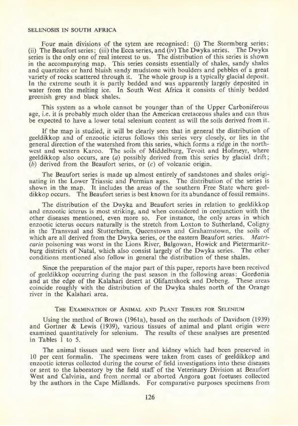
Four main divisions of the sytem are recognised: (i) The Stormberg series ; (ii) The Beaufort series; (iii) the Ecca series, and (iv) The Dwyka series. The Dwyka series is the only one of real interest to us. The distribution of this series is shown in the accompanying map. This series consists essentially of shales, sandy shales and quartzites or hard bluish sandy mudstone with boulders and pebbles of a great variety of rocks scattered through it. The whole group is a typically glacial deposit. In the extreme south .it .is partly bedded and was apparently largely deposited in water from the melting ice. In South West Africa it consists of thinly bedded greenish grey and black shales.
This system as a whole cannot be younger than of the Upper Carboniferous age, i.e. it is probably much older than the American cretaceous shales and can thus be expected to have a lower total selenium content as will the soils derived from it.
1f the map is studied, it will be clearly seen that in general the distribution of geeldikkop and of enzootic icterus follows this series very closely, or lies in the general direction of the watershed from this series, which forms a ridge .in the northwest and western Karoo. The soils of Middelburg, Tevoit and Hofmeyr, where geeldikkop also occurs, are (a) possibly derived from this series by glacial drift; (b) derived from the Beaufort series, or (c) of volcanic origin.
The Beaufort series is made up almost entirely of sandstones and shales originating in the Lower Triassic and Permian ages. The distribution of the series is shown in the map. I t includes the areas of the southern Free State where geeldikkop occurs. The Beaufort series is best known for its abundance of fossil remains.
The distribution of the Dwyka and Beaufort series in relation to geeldikkop and enzootic icterus is most striking, and when considered in conjunction with the other diseases mentioned, even more so. For instance, the only areas in which enzootic icterus occurs naturally is the stretch from Loxton to Sutherland, Coligny in the Transvaal and Stutterheim, Queenstown and Grahamstown, the soils of which are all derived from the Dwyka series, or the eastern Beaufort series. Matricaria poisoning was worst in the Lions River, Balgowan, Howick and Pietermaritzburg districts of Natal, which also consist largely of the Dwyka series. The other conditions mentioned also follow in general the distribution of these shales.
Since the preparation of the major part of this paper, reports have been received of geeldikkop occurring during the past season in the following areas: Gordonia and at the edge of the Kalahari desert at Olifantshoek and Debeng. These areas coincide roughly with the distribution of the Dwyka shales north of the Orange river in the Kalahari area.
THE EXAMINATION OF ANIMAL AND PLANT TISSUES FOR SELENIUM
Using the method of Brown (196la), based on the methods of Davidson (1939) and Gortner & Lewis (1939), various tissues of animal and plant origin were examined quantitatively for selenium. The results of these analyses are presented in Tables 1 to 5.
The animal tissues used were liver and kidney which had been preserved in 10 per cent formalin. The specimens were taken from cases of geeldikkop and enzootic icterus collected during the course of field investigations into these diseases or sent to the laboratory by the field staff of the Veterinary Division at Beaufort West and Calvinia, and from normal or aborted Angora goat foetuses collected by the authors in the Cape Midlands. For comparative pw·poses specimens from
126
J. M. M. BROWN and P. J. DE WET
normal Karoo sheep used by the authors for experimental work at Victoria West and normal Onderstepoort sheep, were analysed. Specimensfromafewmiscellaneous cases were also included for general interest.
The plant tissues used were the vegetative portions of plants collected at random from Calvinia, Fraserburg, Beaufort West and Fauresmith, by the authors or by members of the field staff. For comparative purposes specimens from a variety of plants growing on the heavy black clay soils at Onderstepoort were selected. A few odd plants of interest from elsewhere were also included.
TABLE 1.- Selenium analyses on tissues from cases of Enzootic Icterus (E.!.) and Geeldikkop (G.D.K.)
(All results are expressed as micrograms (meg.) selenium per gram of tissue on a wet basis. A dash in the columns below indicates that no material was available for analysis. The letters G DK/EI indicate a mixed Geeldikkop/Enzootic Icterus Syndrome as established by h istopathological examination.]
E.I. E.l. E.I. E.I. E.I. E. I. E.I. E. I. E. I. E. I. E.l. E.l. E.I. E. I. E.l. E.I. E.l. E.l. E. I. E.l. E.T. E.I.
GD K GDK GDK GDK GDK
GDK/EI GDK/EI
GD K GDK GDK GDK GDK GDK GDK
Normal Ewe? Aged normal
ewe
127
Liver meg. Se
11 ·5 14·0 11·0 5·5
11·5 4· 0 4·0
10·0 3·0 8·5
11 .5 20·0
0 3·0 7 ·0
17·0
8·5 10·0
3·0 0
0 0
3·0 0 0
11·0 8·5 6·0 3·0
0 0
8·5 5·5 8·5 8·5 8·5
Kidney meg. Se
3·0 14·0 14·0
0
11·0
7·0 8·5
10·0 34·0 8·5
0 4·5
11·5 0
11.5
0 1·0
25·5 14·0
0 0 0 0 0
SELENOSIS lN SOUTH AFRICA
The results in Table I show beyond doubt the presence of dangerously high levels of selenium in the tissues of sheep from farms where enzootic icterus and geeldikkop are prevalent. It will be seen that in the case of enzootic icterus the selenium content of the livers and kidneys of the affected sheep is in general much higher than in the cases of geeldikkop. Moreover the presence of selenium is virtually a constant :finding in enzootic icterus. The authors stated elsewhere (Brown et a/. , J 960) that geeldikkop and enzootic icterus are the two extreme forms of a single basic disease entity, each with its own set of special precipitating influences. This has been borne out by a critical study of the epizootology, pathology and chemical pathology of these conditions. Enzootic icterus is the more violently acute and often fatal syndrome, so the results obtained with these selenium analyses are in excellent accord with the known facts.
An interesting point with regard to Table 1 concerns the last two sheep designated " Celeryfontein numbers 1 and 2 ". These sheep were apparently normal before being slaughtered for fa rm rations. The flock is owned by one of the leading sheep breeders in the Karoo, and is situated in the Jnidst of the worst enzootic icterus area in the country. The disease is seldom seen on this farm although the adjoining farms are severely affected every year. The low incidence of the disease on this farm may possibly be attributed to one or both of the following very important factors: (a) the sheep on this farm receive ad lib. throughout the year the best proteLn supplements available (there are very few farms in this part of the Karoo where the nutritional level of the sheep is so high); (b) the management and grazing control of this farm leave very little to be desired. The result is that overstocking with all its attendant evils has been avoided aod the animals are seldom, if ever, forced to eat inferior and possibly highly seleniferous plants. ln spite of appreciable amounts of selenium present in their livers, these sheep seem to have suffered few ill effects. The same conditions pertain in the case of two sheep shown in Table 2, designated "Sheep Minnaar 1 and 2 ". The owner is one of the leading ram breeders in this country, and the sheep were in perfect condition . Once more, nutrition and management on this farm are of an exceptionally high order.
TABLE 2.- Se/enium analyses on tissue from miscellaneous cases [All results are expressed as micrograms (meg.) selenium per gram of tissue on a wet basis.
A dash in the columns below indicates that oo material was available for analysis.)
I Disease Condition I Liver meg. Se I Kidney meg. Se
--------Normal foetus .. . Aborted foetus .. . Normal foetus .. . Normal foetus '? . . Aboned foetus'! .. Aborted foetus ... . Normal foet us .. . Lupinosis ....... . Lup[nosis ....... .
31 ·0 6·0 I · I
14·0 5·5
0 17 ·0
l · 5 5·5
----- ---------- --- ------'---
Consider now the animals presented in Table 2. These were with the exception of the Minnaar sheep, foetuses taken from normal, healthy Angora or sh.eep ewes, or aborted foetuses from such ewes in the Cape Midlands. As will be seen most of these livers contain appreciable amounts of selenium. This illustrates the point made earlier under "kaalsiektc ", that large amounts of selenium may cross the placenta into the foetus. There are, however, some important poi.ots to bear in
128
J. M. M. BROWN and P. J. DE WET
mind regarding these foetuses, viz. (a) the one gm. of liver used for the analysis in many cases represents the greater part of these livers; (b) the various enzyme systems of the foetuses are by no means fully developed until just before or just after birth ; (c) the metabolic functions of the foetuses are to a large extent dependent on those of the mothers. The tolerance of the foetus to selenium, almost up to birth, might, therefore, be high, and in these cases the selenium content of the livers bears n o relationship to whether the foetuses were normal or pathological. They were in general only about six weeks old having thus about three to four months growth ahead before reachjng full term.
The sheep presented in Tables 3 and 4 are of considerable interest. These can be regarded as controls. Those in Table 3 were used for the authors' experimental work on Tribulus saponins or sapogenins in Victoria West. They were purchased from one of the best farmers in the area. They were matntained on a diet of high protein content, viz. green lucerne hay and yellow maize ad lib. Some of these cases contain large amounts of selenium in their tissues, yet appeared clinically normal.
T ABLE 3.- Se/enium analyses on tissues from sheep used for experimental work in Victoria West
[All results are expressed as micrograms (meg.) selenium per gram of tissue on a wet basis.]
The sheep in Table 4 were normal healthy Onderstepoort sheep, which were slaughtered for staff rations. With the exception of three sheep, none of the others appeared to have any selenium in their livers. These three sheep were fairly recent introductions from the Karoo (7303 from Laingsburg on 2nd May, 1959; 9814 from Laingsburg on 5th November, 1960, and 8634 from Grootfontein on 7th January, 1960). It appears that selenium may persist in active tissues like the liver for some length of time, indicating that selenium is a cumulative poison. Its rate of excretion from the body will depend to a large extent on the sulphur turnover, and the amount of sulphur ingested. In order to displace selenium the sulphur probably has to be in organic form, e.g. as methionine. Therefore, the duration of retention of selenium in the body will depend largely upon the amount of methionine or biologically ideal proteins in the diet.
129
SELENOSIS IN SOUTH AFRICA
TABLE 4.- Selenium analyses on tissues from normal healthy Onderstepoort sheep [AJI results are expressed as micrograms (meg.) selenium per gram of tissue on a wet basis.]
The details of plant analyses are presented in Table 5. Only the vegetative portions of the plants were used. The plants were air-dried for about one month prior to analysis. The leaves were stripped off and then coarsely powdered before the required amount was weighed out.
TABLE 5.-Selenium analyses on plant tissues [All results are expressed as micrograms (meg.) selenium per gram of dry plant material.]
It wi ll be seen that a fair number of these plants, even some grown at Onderstepoort, contain significant amounts of selenium.
At this stage it would be most unwise to try and draw any conclusions from these figures. This work was merely a preliminary survey to establish the presence of selenium in South African plants, especially Karoo vegetation. It is, however, interesting to note that selenium was constantly present in plants from Fauresmith, a1t times a bad geeldikkop area. Plants such as Eriocephalus glaber, Tetragonia a.rbuscula and Chrysocoma contain dangerous amounts of selenium. I t is further of interest only to note at this stage that locally-grown Lippia and Lantana both C•ontain fair amounts of the element. No conclusions have been drawn from this other than that both are fast-growing encroacrung weeds .
THE P OSSIBLE RELATIONSHIP OF SELENOSIS TO ENZOOTIC ICTERUS AND G EELDIKKOP
A description of these two diseases and an account of much of the work done on them to date has been presented elsewhere (Theiler, 1918; Brown, 1959a, 1959b, 1'959c; Brown et al. , 1960; Brown & de Kock, 1959; de Kock & Enslin, 1958 ; Enslin & Wells, 1956). The relationsrup between the two diseases has been disClussed (Brown, 1959c; Brown et a/. , 1960). Geeldikkop and enzootic icterus in their typical forms can be considered as the two extremes of a single basic condition. The former is characterised by a severe photo-sensitisation, a severe icterus in the nature of an intra-hepatic cholestasis and a severe nephrosis, while in the ]atter a severe acute haemolytic icterus and extremely severe nephrosis are considered the typical disease pattern . Each syndrome seems to be precipitated by a different set of circumstances or factors. In the case of geeldikkop for instance the plant Tribulus terrestris is in some way concerned, while in the case of enzootic icterus some severe stress condition plays the major role, for example, adverse environmental conditions, transport of the animals any long distance by rail or a virus infection such as Wesselsbron virus disease (Le Roux, 1959). Between these two extremes various symptom complexes are encountered wruch are typical of neither major syndrome.
131
SELENOSIS IN SOUTH AFRTCA
At a first glance any resemblance between these two syndromes and selenosis may seem remote. However, if they are viewed as a single disease entity against the background of the foregoing text, an attractive working hypothesis regarding their aetiology may be evolved. Consider for the moment the geeldikkop syndrome only. That Tribulus terrestris plays a role in the aetiology seems certain. However, the authors have been consistently unable to produce the typical disease picture with the saponins or steroidal sapogenins isolated from it. Their contention has, therefore, been that although Tribulus is concerned in the genesis of the disease, it is by no means the only factor involved. They postulated the existence of one or more co-factors which acted synergistically with the active agents present in Tribulus. These factors might be other plant or mineral toxins or else deficiencies of some essential nutritional factors. The existence of widespread unexplained liver necrosis throughout the Karoo certainly pointed to such factors being present.
The discovery by New Zealand workers that a fungus growing on pastures under certain conditions was the agent responsible for facial eczema, a disease which in some respects resembles our ovine icterus complex, opened new avenues of thought. However, up to the present the authors have been unable to establish that fungal toxins are in any way concerned with the production of geeldikkop.
A serious consideration of all the disease conditions prevalent in the Karoo showed that there are some which co1Iectively bear a gross resemblance to selenosis in its various forms. These conditions which have been discussed earlier, suggested a fresh line of approach to the problem and stimulated the present work. The presence of dangerously high levels of selenium in plant and animal tissues from tbe Karoo areas is in itself a significant .finding, but as yet the authors can only speculate as to its precise role in the aetiology of geeldikkop and enzootic icterus.
Geeldikkop is in general a much less severe condition than enzootic icterus. If selenium intoxication, therefore plays any role in the cause of these conditions, one would expect to find relatively less selenium in the tissues of cases of the former syndrome. This is borne out by a critical study of the analytical data presented in Table 1. A tempting hypothesis as to the pathogenesis of geeldikkop is the following: The continual ingestion of small amounts of selenium, greater than the safe level of intake, yet not enough to cause frank selenosis has a cumulative damaging effect on the enzymes mentioned earlier and possibly others as well. Systems particularly affected are those responsible for the conjugation of bile pigments and their subsequent excretion. The bile pigment conjugation mechanisms of the sheep seem to exist at their best rather precariously (Brown 196la). If the conjugation of bilirubin proceeds through medium of a uridine nucleotide cycle as proposed by Kalckar & Maxwell (1958) and others, and it seems likely that it does, then such conjugation would depend not only upon the availability of sufficient glucose and hence the integrity of the enzymes of the glycolytic cycle, but also upon the correct functioning of the tricarboxylic acid cycle, gluco-neogenetic mechanisms and various other energy producing systems. The sapogenins present in Tribulus or possibly other compounds as yet unidentified might now overwhelm the already weakened conjugation mechanisms by acting either as competitive aglycones or by exerting an additional strongly inhibitory influence somewhere in the conjugation system.
Tribulus terrestris itself apparently contains very little selenium, as will be seen from Table 5. However, the presence of significant amounts of the element in plants such as Eriocephalus glaber, Tetragonia arbuscula and Chrysocoma spp. selected at random indicates that there might be many plants amongst the Karoo vegetation
132
J. M. M. BROWN and P. J. DE WET
which contain dangerous amounts of selenium. It is probably unlikely that plants will be found in the Karoo containing as much of the element as for instance the Astragalus spp. or the other " converter " plants which have been mentioned.
Enzootic icterus appears to be a much more frank expression of selenosis than geeldikkop, but here, as before, a number of factors may have to act simultaneously t•) produce the typical clinical syndrome. Jt is known that prominent amongst tl~ese is any form of severe non-specific stress. As will be seen from Table 1, selenium is constantly present in the tissues of cases of this disease. Severe atrophic liver c.irrhosis, acute glomerulo-nephritis, bone marrow hypoplasia, an extremely severe anaemia, and sometimes loss of wool, erosion of the articular surfaces of the long hones or coronitis are prominent features of this condition. The anaemia seen in typical cases is of considerable interest. It is the direct result of one or more acute attacks of intravascular haemolysis. Haemoglobin values as low as 2 gm. per cent are common during such episodes. ln the more chronic cases the anaemia is difficult to classify. There seems to be an almost continuous low grade intravascular haemolysis, with periodic acute exacerbations, and since these are often associated with haemoglobinuria, considerable loss of body iron must occur. The red bone marrow on the other hand is fequently severely hypoplastic. Grosskopf (unpublished observations, 1958) demonstrated that the erythrocytes of typical cases of the disease slb.owed a very pronounced fragility, haemolysis sometimes even occurring in isotonic saline.
This finding is of more than passing interest, as is also the fact that methaenwglobin-cythaemia is sometimes encountered during acute attacks of the condition. An analogous condition is to be found in the human disease designated" Primaquinesensitivity". This appears as an acute intravascular haemolysis following the a~dministration of drugs such as the 8-aminoquinolines and many others, and is essentially an inborn error of metabolism due to a deficiency of glucose-6-phosphate dehydrogenase in the erythrocytes (Carson, Flanagan, Ickes & Alving, 1956; Brewer, Tarlov & Alving, 1960). Individuals who inherit this defect experience a:n acute haemolytic episode after the ingestion of certain drugs and some vegetables, e .. g. fava beans. It has been shown that the erythrocytic lifespan is shortened even in the absence of drug administration. The decreased activity of glucose-6-phosphate dehydrogenase in the erythrocytes of these cases results in a diminished g1eneration of T.P.N.H. (reduced triphosphopyridine nucleotide) from T.P.N. , and a:s a consequence leads to defective reduction of methaemoglobin by T.P. N.H. Met. Bb. reductase, even in the presence of suitable hydrogen carriers (Brewer, et a/. , 1960).
lt is indeed possible that similar conditions pertain in the erythrocytes of enzootic icterus cases, and that the essential lesion is an inactivation of glucose-6-phosphate dtehydrogenase or other enzymes in the pentose phosphate pathway due to selenium. Work is proceeding on this aspect of the disease at the moment.
In conclusion the authors would like to reiterate a point which they have frequently stressed in their earlier papers on geeldikkop, and one which was repeatedly made by Quin and Rimington, earlier workers on the disease. This important p•oint is that even when the full sequence of events in the pathogenesis of geelctikkop and enzootic icterus is ultimately elucidated, we will still be faced with the fundamental fact that the high annual incidence of both diseases is due in no small measure to overstocking, and other grazing malpractices which are forcing Karoo stock to e>tist on inferior and potentially dangerous vegetation. This is even more true nQw than ever before.
133
SELENOSIS TN SOUTH AFRICA
RESUME
Selen()sis is described, it is believed for the first time, as being a potential grazing hazard in many parts of this country, especially in the Karoo areas. A review of the symptomatology, pathology and epizootology of the condition as it is known elsewhere in the world is presented together with a short account of experimental selenosis and some of the known actions or toxic effects of the element on the body. A brief description of some disease conditions which are believed to be essentially chronic selenosis syndromes is given, and some thoughts are advanced as to the possible role of selenium intoxication in the two conditions known as tribulosis ovis (geeldikkop), and enzootic icterus. In conclusion the results of numerous analyses of animal and plant tissues for selenium are presented, which indicate the presence of dangerous amounts of this element in the material examined.
A CKNOWLEDGEMENTS
The permission of the Director of Veterinary Services for publishing this paper is gratefully acknowledged. We are indebted to PJOfessor R. Clark for his most valuable assistance and advice throughout the course of this work. We would also like to thank Professors H. P. A. de B()orn and K. van der Walt for much useful advice, and help, which they placed at our disposal, and also doctors R. C. Tustin and L. W. van der Reever for material supplied to us. A special word of thanks is due to State VetetinaJ ians Truter, Badenhorst and van der Riet, and Stock Inspector Hendricks for the collection of much valuable material for us in the field; and also to numerous farmers who so willingly placed material at our disposal.
REFERENCES
ANDREWS, W. H. (1923). The so·called "staggers" or "pushing disease" of cattle in Natal: An intoxication due to the ingestion of Matricaria nigel/aefo/ia. 9th and 10th Rept. Dir. Vet. Educ. Res., Pretoria, 123.
BREWER, G. J., TARLOV, A. R. & ALVJNG, A. S. (1960). Methaemoglobin Reduction Test. A new simple in vitro test for identifying Primaquine-sensitivity. Buff. Wid. Hfth. Org. No. 22. p. 633.
BROWN, J. M. M. (L959a). Advances in "Geeldikkop " (Tribulosis Ovis) Research. I. The history of ·'Geeldikkop " Research. J. S. Afr. Ver. Med. Ass., Vol. 30, p. 97.
BROWN, J. M. M. (J959b). Advances in" Geeldikkop" (Tribulosis Ovis) Research. H. Field investigations- the mobile laboratory and experimental facilities. J. S. Afr. Vet. Med. Ass., Vol. 30, p. 395.
BROWN, J. M. M. (1959c). Advances in "Geeldikkop" (Tribulosis ovis) Research. ill. The epizootology of Geeldikkop. J. S. Afr. Vet. Med. Ass., p. 403.
BROWN, J. M. M. (196Ja). Method to be published in the next number of this journal. BROWN, J. M. M. & DE KOCK, W. T. (1959). A chemical and physiological investigation of
Geeldikkop in sheep in South Africa. S. Afr. Indust. Chemist, Vol. 13, p. 189. BROWN, J. M. M., LEROUX, J. M. W. & TUSTIN, R. C. (1960). Advances in Geeldikkop
(Tribulosis ovis) Research. IV. The pathology of Geeldikkop-Part 1. J. S. Afr. Vet. Med. Ass., Vol. 31, p. 179.
CARSON, P., FLANAGAN, C. l...,ICKES, C. E . & ALVfNG, A. S. (1956). Enzymatic deficiency in Primaquine-sensitive erythrocytes. Science, Vol. 124, p. 484.
DAVIDSON, J. (1939). The quantitative adaptation of the codeine test to the colorimetric determination of selenium in plant materials. J. Ass. Off. Agr. Chem., Vol. 22, p. 450,
DAVIDSON, W. B. (1940). Selenium poisoning. Canad. J. Comp. Med. Vet. Sci., Vol. 4, p. 19. DE KOCK, W. T . & ENSLIN, P. R. (1958). Chemical investigations of photosensitisation diseases
of domestic animals, Part [- [solation and characterisation of steroidal sapogenins from Tribulus terrestris. J. S. Afr. Chem. Inst., Vol. 11, p. 33.
134
J. M. M. BROWN and P. J . DE WET
ENSLIN, P. R. & WELLS, R. J. (1956). Chemical investigations of photosensitisation diseases of domestic animals. S. A[r. Indust. Chem., Vol. 10, p . 96.
GORTNER, R . A. & LEWJS, H. B. (1939). Quantitative determina.tion of selenium in tissues and faeces. A photometric method. Ind. Eng. Chem. Anal., Ed. 11, p. 198.
HORN, M. J . & JONES, D. B. (1941). Isolation from Astragalus pectinatus of a crystalline amino-acid complex containing selenium and sulphur. J. Bioi. Chem., Vol. 139, p. 649.
KALCK.AR, H. M. & MAXWELL, E. S. (1958). Biosynthesis and metabolic function of uridine diphospho-glucose in mammalian organisms and its relevance to cerltain inborn errors. Physiol. Rev., Vol. 38, p. 77.
KLUG, H. L., MOXON, A. L .. PETERSEN, D. F. & POTTER, VAN R . (1950). The in vivo inhibition of succinic dehydrogenase by selenium and its release by arsenic. Arch. Biochem., Vol. 28, p. 253.
LEROUX, J. M. W. (1959). The histopathology of Wesselsbron disease in sheep. Onderstepoort J. Vet. Res., Vol. 28, p. 237.
LOUW, P. G. J. (1952). A note on the a lka loid of Crotalaria damarensis. Onderstepoort J. Vet. Res., Vol. 25, p. I 1 J.
MAHADEVAN, V. (1954). Selenium poisoning in men and animals. Indian Vet. J., Vol. 31, p. 210.
MARAIS, J. S. C. (1944). Dicrotaline; the toxic alkaloid from Crotalaria dura and Crotalaria globifera. Onderstepoort J. Vet. Sci. Anim. Ind., Vol. 120, p. 61.
MOXON, A. L. & RHlAN, M. (1943). Selenium poisoning. Physiol. Rev., Vol. 23, p. 305.
PAINTER., E. P. (1941). The chemistry and toxicity of selenium compounds with special reference to the selenium problem. Chem. Rev., Vol. 28, p. 179.
REKO, V. A. (1928). Cited by Steyn, D. G., 1931. Investigations into the cause of a lopecia (k.aalsiekte) in kids and lambs. 17th Rept . .Dir. Vet. Serv. Anim. Ind. p. 729.
ROBINSON, W. 0. (1933). Determination of selenium in wheat and soils. J. Ass. Off. Agric' Clzem .. Vol. 16, p. 423.
ROBINSON, W. 0. (1936). Selenium content of wheat from variou!> parts of the world. Ind. Eng. Chem., Vol. 28, p. 736.
ROCHE INFORMATION SERVICE OF THE VITAMIN DEPARTMENT (1959). Pamphlet, Vitamin E and selenium in animal and poultry nutrition. F . Hooffman-La Roche and Co. Ltd., Basle, Switzerland.
RUSSEL, F. C. & DUNCAN, D. L. (1956). Minerals in pasture: Deficiencies and excesses in relation to animal health. Commonweahh Bureau of Animal Nut rition. Tech. Comm. No. 15, Second Ed., C.A.B., Farnham Royal, Slough, Bucks.
SCHNEIDER, H. A. (1936). Selenium in nu trition. Science, Vol. 83, p. 32.
SMITH, M. I. & STOHLMAN, E. F. (1940). Furth er observations on the influence of dietary protein on the toxicity of selenium. J. Pharm. Exp. Therap., VoL 70, p. 270.
SMITH, M. I., STOHLMAN, E. F. & LlLLlE, R. D. ( 1937). The toxicity and pathology of selenium. J. Pharm. Exp. Therap. Vol. 60, p. 449.
STEYN, D. G. ( 1934). The toxicology of plants in South Africa. Central News Agency, Ltd., South Africa.
THBlLER, A. (1918). Geeldikkop in Sheep (" Tribulosis ovium "). 7th a nd 8th Rept. Dir. Yet Res., South Africa, p. 1-56.
THORVALDSEN, T . (1951). Proc. Specialist Conf. Agric., Australia, 1949, p. 310.
TOMS, T. (1960). Care and management of thoroughbred foals. J'. S. Af'r. Vet. Med. Ass., Vol. 31, p. 75.
WALSH, T., FLEMING, G. H., O'CONNOR, R. & SWEENEY, A. (1951). Selenium toxicity associated with an Irish soil series. Nature, Vol. 168, p. 881.
WILLIAMS, K. T., LAKIN, H. W . & BYERS, H. G. (1940). Selenium occurrence in certain soils in the United States with a discussion of related topics. Fourth Rept., U.S . .Dept. Agric. Tech. Bull. No. 702.