Page 1
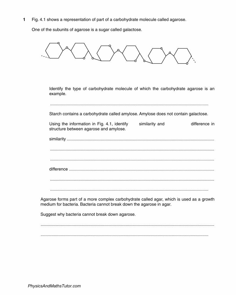
1 Fig. 4.1 shows a representation of part of a carbohydrate molecule called agarose.
One of the subunits of agarose is a sugar called galactose.
Fig. 4.1
(a) (i) Identify the type of carbohydrate molecule of which the carbohydrate agarose is anexample.
...................................................................................................................................... [1]
(ii) Starch contains a carbohydrate called amylose. Amylose does not contain galactose.
Using the information in Fig. 4.1, identify one similarity and one further difference instructure between agarose and amylose.
similarity .............................................................................................................................
...........................................................................................................................................
...........................................................................................................................................
difference ...........................................................................................................................
...........................................................................................................................................
...................................................................................................................................... [2]
(b) Agarose forms part of a more complex carbohydrate called agar, which is used as a growthmedium for bacteria. Bacteria cannot break down the agarose in agar.
Suggest why bacteria cannot break down agarose.
...................................................................................................................................................
.............................................................................................................................................. [1]
PhysicsAndMathsTutor.com
Page 2
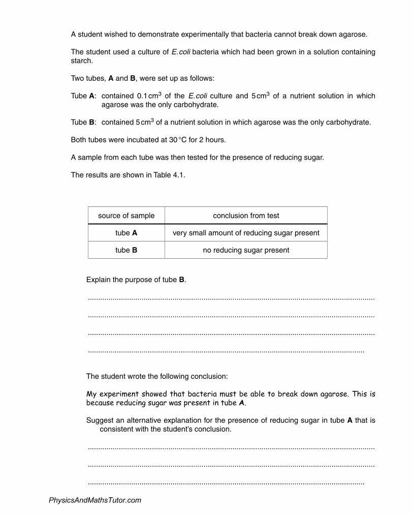
(c) A student wished to demonstrate experimentally that bacteria cannot break down agarose.
The student used a culture of E. coli bacteria which had been grown in a solution containingstarch.
Two tubes, A and B, were set up as follows:
Tube A: contained 0.1 cm3 of the E. coli culture and 5 cm3 of a nutrient solution in whichagarose was the only carbohydrate.
Tube B: contained 5 cm3 of a nutrient solution in which agarose was the only carbohydrate.
Both tubes were incubated at 30 °C for 2 hours.
A sample from each tube was then tested for the presence of reducing sugar.
The results are shown in Table 4.1.
Table 4.1
source of sample conclusion from test
tube A very small amount of reducing sugar present
tube B no reducing sugar present
(i) Explain the purpose of tube B.
...........................................................................................................................................
...........................................................................................................................................
...........................................................................................................................................
...................................................................................................................................... [2]
(ii) The student wrote the following conclusion:
My experiment showed that bacteria must be able to break down agarose. This isbecause reducing sugar was present in tube A.
Suggest an alternative explanation for the presence of reducing sugar in tube A that isnot consistent with the student’s conclusion.
...........................................................................................................................................
...........................................................................................................................................
...................................................................................................................................... [1]
PhysicsAndMathsTutor.com
Page 3
(iii) Suggest two ways in which the reliability of the experiment could be improved.
1 .........................................................................................................................................
...........................................................................................................................................
2 .........................................................................................................................................
...................................................................................................................................... [2]
(d) (i) The student did not have access to a colorimeter when testing solutions for the presenceof reducing sugar.
Describe how the student could carry out a chemical test for reducing sugar and suggest how he could estimate the amount of reducing sugar in the sample from tube A.
...........................................................................................................................................
...........................................................................................................................................
...........................................................................................................................................
...........................................................................................................................................
...........................................................................................................................................
...........................................................................................................................................
...........................................................................................................................................
...........................................................................................................................................
...........................................................................................................................................
...........................................................................................................................................
...........................................................................................................................................
...........................................................................................................................................
...........................................................................................................................................
...........................................................................................................................................
...................................................................................................................................... [5]
PhysicsAndMathsTutor.com
Page 4
(ii) Another student suggested that the agarose may have been broken down to anon-reducing sugar.
Describe how the test for reducing sugar could be modified to investigate this hypothesis.
...........................................................................................................................................
...........................................................................................................................................
...........................................................................................................................................
...........................................................................................................................................
...........................................................................................................................................
...................................................................................................................................... [3]
[Total: 17]
PhysicsAndMathsTutor.com
Page 5
The use of microscopy has greatly enhanced our knowledge of cell structure.
(a) Explain the difference between magnification and resolution.
...................................................................................................................................................
...................................................................................................................................................
...................................................................................................................................................
...................................................................................................................................................
...................................................................................................................................................
.............................................................................................................................................. [2]
(b) State the resolution that can be achieved by each of the following types of microscope.
light microscope .........................................................
transmission electron microscope ......................................................... [2]
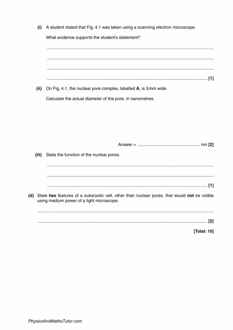
(c) Fig. 4.1 is an electron micrograph showing part of a nucleus.
Fig. 4.1
A
x 25000
2
PhysicsAndMathsTutor.com
Page 6
(i) A student stated that Fig. 4.1 was taken using a scanning electron microscope.
What evidence supports the student’s statement?
...........................................................................................................................................
...........................................................................................................................................
...........................................................................................................................................
...................................................................................................................................... [1]
(ii) On Fig. 4.1, the nuclear pore complex, labelled A, is 3 mm wide.
Calculate the actual diameter of the pore, in nanometres.
Answer = .................................................... nm [2]
(iii) State the function of the nuclear pores.
...........................................................................................................................................
...........................................................................................................................................
...................................................................................................................................... [1]
(d) State two features of a eukaryotic cell, other than nuclear pores, that would not be visibleusing medium power of a light microscope.
...................................................................................................................................................
.............................................................................................................................................. [2]
[Total: 10]
PhysicsAndMathsTutor.com
Page 7
3
20 μmA B
Fig. 5.1
PhysicsAndMathsTutor.comPhysicsAndMathsTutor.com
Page 8
(a) Fig. 5.1 is provided for you on the insert.
(i) State two features of the cell shown in Fig. 5.1 that indicate it is eukaryotic.
...........................................................................................................................................
...........................................................................................................................................
...........................................................................................................................................
..................................................................................................................................... [2]
(ii) The line A–B on Fig. 5.1 represents 20 μm.
Calculate the magnification of the cell shown in Fig. 5.1.
Show your working.
Answer = ...................................................... x [2]
(iii) Microtubules and microfilaments are part of the cytoskeleton.
Suggest two roles of the cytoskeleton in the type of cell shown in Fig. 5.1.
...........................................................................................................................................
...........................................................................................................................................
...........................................................................................................................................
...........................................................................................................................................
..................................................................................................................................... [2]
(b) The cells of a multicellular organism are usually specialised to perform a particular function.
(i) Name the process in which a cell becomes specialised.
..................................................................................................................................... [1]
3
PhysicsAndMathsTutor.com
Page 9
(ii) Neutrophils are phagocytic blood cells that can engulf and digest foreign cells found inthe blood.
Describe how the ultrastructure of a neutrophil is specialised to enable it to perform thisfunction.
In your answer, you should use appropriate technical terms, spelt correctly.
...........................................................................................................................................
...........................................................................................................................................
...........................................................................................................................................
...........................................................................................................................................
...........................................................................................................................................
...........................................................................................................................................
...........................................................................................................................................
...........................................................................................................................................
..................................................................................................................................... [4]
[Total: 11]
PhysicsAndMathsTutor.com
Page 10
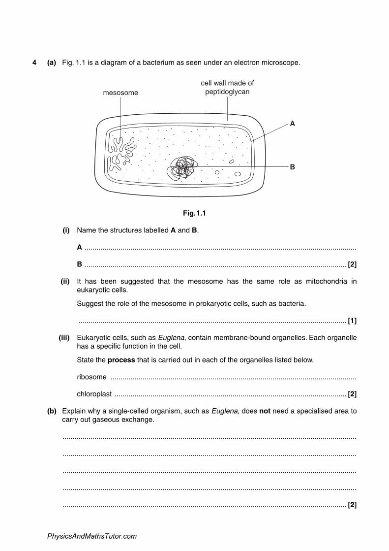
4 (a) Fig. 1.1 is a diagram of a bacterium as seen under an electron microscope.
mesosomecell wall made of
peptidoglycan
A
B
Fig. 1.1
(i) Name the structures labelled A and B.
A ........................................................................................................................................
B ................................................................................................................................... [2]
(ii) It has been suggested that the mesosome has the same role as mitochondria ineukaryotic cells.
Suggest the role of the mesosome in prokaryotic cells, such as bacteria.
...................................................................................................................................... [1]
(iii) Eukaryotic cells, such as Euglena, contain membrane-bound organelles. Each organellehas a specific function in the cell.
State the process that is carried out in each of the organelles listed below.
ribosome ...........................................................................................................................
chloroplast .................................................................................................................... [2]
(b) Explain why a single-celled organism, such as Euglena, does not need a specialised area tocarry out gaseous exchange.
...................................................................................................................................................
...................................................................................................................................................
...................................................................................................................................................
...................................................................................................................................................
.............................................................................................................................................. [2]
PhysicsAndMathsTutor.com
Page 11
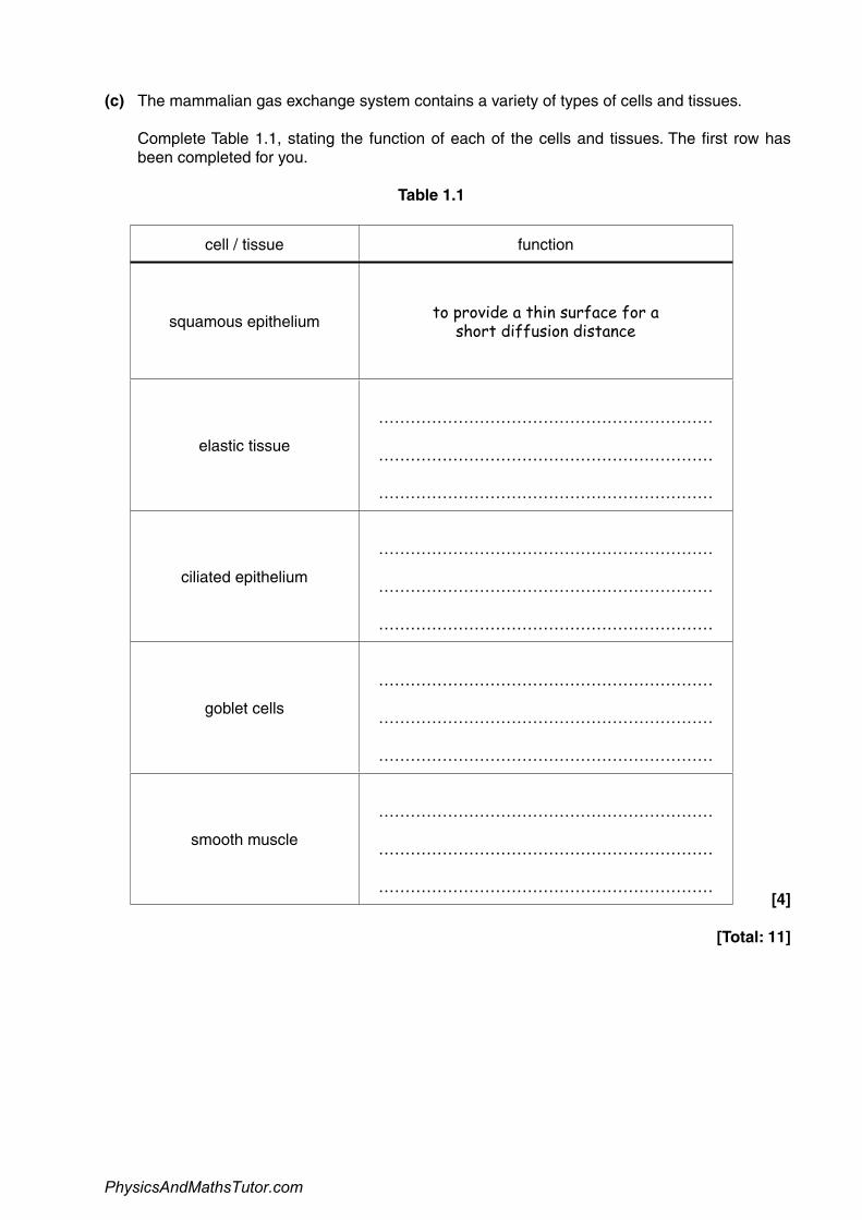
(c) The mammalian gas exchange system contains a variety of types of cells and tissues.
Complete Table 1.1, stating the function of each of the cells and tissues. The first row hasbeen completed for you.
Table 1.1
cell / tissue function
squamous epithelium to provide a thin surface for ashort diffusion distance
elastic tissue
………………………………………………………
………………………………………………………
………………………………………………………
ciliated epithelium
………………………………………………………
………………………………………………………
………………………………………………………
goblet cells
………………………………………………………
………………………………………………………
………………………………………………………
smooth muscle
………………………………………………………
………………………………………………………
………………………………………………………[4]
[Total: 11]
PhysicsAndMathsTutor.com
Page 12
5 Fig. 4.1 shows diagrams of two different types of cells, X and Y.
The cells are not drawn to scale.
X
Y
Fig. 4.1
(a) (i) State, using only the information in Fig. 4.1, two differences between plant cells andanimal cells.
1 ........................................................................................................................................
...........................................................................................................................................
2 ........................................................................................................................................
...................................................................................................................................... [2]
(ii) Cell Y is a guard cell.
State, using only the information in Fig. 4.1, one adaptation of this cell and explain howthe adaptation allows the cell to carry out its function.
adaptation .........................................................................................................................
explanation ........................................................................................................................
...........................................................................................................................................
...................................................................................................................................... [2]
PhysicsAndMathsTutor.com
Page 13
(b) Fig. 4.2 shows drawings of the six chromosomes inside an animal cell viewed during lateprophase of mitosis.
Fig. 4.2
(i) Identify one pair of homologous chromosomes in Fig. 4.2 by drawing around eachchromosome in the pair on the diagram. [1]
(ii) The nucleus of a sperm cell is produced by meiosis.
Draw a diagram in the space below to represent the chromosomes that are present inthe nucleus of a sperm cell from the same animal.
[2]
[Total: 7]
PhysicsAndMathsTutor.com