+ All Categories
Home > Documents > Open Access Full Text Article Spectral domain optical ...

Open Access Full Text Article Spectral domain optical ...

Date post: 22-Jan-2022
Category:
Upload: others
View: 7 times
Download: 0 times
Share this document with a friend
4
© 2012 Chalam et al, publisher and licensee Dove Medical Press Ltd. This is an Open Access article which permits unrestricted noncommercial use, provided the original work is properly cited. Clinical Ophthalmology 2012:6 155–158 Clinical Ophthalmology Spectral domain optical coherence tomography documented rapid resolution of pseudophakic cystoid macular edema with topical difluprednate KV Chalam Vijay Khetpal Chirag J Patel Department of Ophthalmology, University of Florida Jacksonville, FL, USA Correspondence: KV Chalam Department of Ophthalmology, University of Florida Jacksonville, 580 W 8th Street, Tower 2, 3rd Floor, Jacksonville, FL 32209, USA Tel +1 904 244 9361 Fax +1 904 244 9391 Email [email protected]fl.edu Introduction: Pseudophakic cystoid macular edema is a common cause of poor vision after cataract surgery, and topical corticosteroids and nonsteroidal anti-inflammatory drugs are used for its treatment. We investigated the effectiveness of difluprednate (Durezol ® , recently approved by the US Food and Drug Administration) in the treatment of cystoid macular edema, assisted with spectral domain optical coherence tomography (SD-OCT). Case report: A 63-year-old African-American woman presented 6 weeks after uneventful cataract surgery in her left eye with decreased vision and associated distortion of the central visual field. Fluorescein angiogram and SD-OCT confirmed pseudophakic cystoid macular edema. Difluprednate was topically administered twice daily and monitored with serial imaging. Resolution was noted after 1 month of topical therapy, with improvement in visual acuity and resolution of distortion. Conclusion: Difluprednate is an effective treatment for patients with severe pseudophakic cystoid macular edema. SD-OCT allows the physician to monitor resolution of the macular edema easily. Keywords: cystoid macular edema, difluprednate, spectral domain optical coherence tomography, pseudophakic Introduction Cystoid macular edema (CME) is a common cause of visual loss after complicated cataract surgery, but may also occur in uncomplicated cases. 1 Intraoperative vitreous loss, placement of an anterior chamber intraocular lens, and prolonged postoperative inflammation are some of the risk factors associated with CME. 1 Patients experience diminished vision approximately 6–8 weeks after cataract surgery. Diagnosis is based on clinical findings, characteristic appearance on fundus fluorescein angiography, and optical coherence tomography. Common modalities of treatment currently include topical nonsteroidal anti-inflammatory drugs alone or in combination with topical corticosteroids for 3–4 months or until resolution of the macular edema. In patients with persistent CME, intravitreal injections of bevacizumab or triamcinolone have been used with success. 1,2 In this case report, we describe rapid resolution of pseudophakic CME documented with spectral domain optical coherence tomography (SD-OCT, Spectralis ® , Heidelberg Engineering Inc, Carlsbad, CA) after topical administration of difluprednate 0.05% (Durezol ® , Sirion Therapeutics, Tampa, FL), a fluorinated corticosteroid with reportedly greater intraocular penetration. 3–6 Dovepress submit your manuscript | www.dovepress.com Dovepress 155 CASE REPORT open access to scientific and medical research Open Access Full Text Article http://dx.doi.org/10.2147/OPTH.S28151
Transcript

© 2012 Chalam et al, publisher and licensee Dove Medical Press Ltd. This is an Open Access article which permits unrestricted noncommercial use, provided the original work is properly cited.

Clinical Ophthalmology 2012:6 155–158

Clinical Ophthalmology

Spectral domain optical coherence tomography documented rapid resolution of pseudophakic cystoid macular edema with topical difluprednate

KV ChalamVijay KhetpalChirag J PatelDepartment of Ophthalmology, University of Florida Jacksonville, FL, USA

Correspondence: KV Chalam Department of Ophthalmology, University of Florida Jacksonville, 580 W 8th Street, Tower 2, 3rd Floor, Jacksonville, FL 32209, USA Tel +1 904 244 9361 Fax +1 904 244 9391 Email [email protected]

Introduction: Pseudophakic cystoid macular edema is a common cause of poor vision after

cataract surgery, and topical corticosteroids and nonsteroidal anti-inflammatory drugs are used for

its treatment. We investigated the effectiveness of difluprednate (Durezol®, recently approved by

the US Food and Drug Administration) in the treatment of cystoid macular edema, assisted with

spectral domain optical coherence tomography (SD-OCT).

Case report: A 63-year-old African-American woman presented 6 weeks after uneventful

cataract surgery in her left eye with decreased vision and associated distortion of the central

visual field. Fluorescein angiogram and SD-OCT confirmed pseudophakic cystoid macular

edema. Difluprednate was topically administered twice daily and monitored with serial imaging.

Resolution was noted after 1 month of topical therapy, with improvement in visual acuity and

resolution of distortion.

Conclusion: Difluprednate is an effective treatment for patients with severe pseudophakic

cystoid macular edema. SD-OCT allows the physician to monitor resolution of the macular

edema easily.

Keywords: cystoid macular edema, difluprednate, spectral domain optical coherence

tomography, pseudophakic

IntroductionCystoid macular edema (CME) is a common cause of visual loss after complicated

cataract surgery, but may also occur in uncomplicated cases.1 Intraoperative

vitreous loss, placement of an anterior chamber intraocular lens, and prolonged

postoperative inflammation are some of the risk factors associated with CME.1

Patients experience diminished vision approximately 6–8 weeks after cataract

surgery. Diagnosis is based on clinical findings, characteristic appearance on

fundus fluorescein angiography, and optical coherence tomography. Common

modalities of treatment currently include topical nonsteroidal anti-inflammatory

drugs alone or in combination with topical corticosteroids for 3–4 months or until

resolution of the macular edema. In patients with persistent CME, intravitreal

injections of bevacizumab or triamcinolone have been used with success.1,2 In this

case report, we describe rapid resolution of pseudophakic CME documented with

spectral domain optical coherence tomography (SD-OCT, Spectralis®, Heidelberg

Engineering Inc, Carlsbad, CA) after topical administration of difluprednate 0.05%

(Durezol®, Sirion Therapeutics, Tampa, FL), a fluorinated corticosteroid with

reportedly greater intraocular penetration.3–6

Dovepress

submit your manuscript | www.dovepress.com

Dovepress 155

C A S E r E P O rT

open access to scientific and medical research

Open Access Full Text Article

http://dx.doi.org/10.2147/OPTH.S28151

Clinical Ophthalmology 2012:6

Case presentationA 63-year-old African-American woman with a history

of hypertension, asthma, gastroesophageal reflux disease,

anemia, and hyperlipidemia, was referred for evaluation of

poor vision. Her past ocular history was unremarkable. Her

best corrected visual acuity was 20/30 in each eye, but on

glare testing decreased to 20/40 in the right eye and 20/60 in

the left eye. Near vision examination revealed J1+ in each

eye and color vision was full. Amsler and visual fields were

normal. Motility and pupillary examinations were normal.

Intraocular pressures were 16 mmHg in the right eye and

18 mmHg in the left eye. Anterior segment examination

was notable for a nuclear sclerotic cataract in both eyes (left

greater than right). Dilated fundus examination revealed

minimal diabetic retinopathy with microaneurysms in the

posterior pole.

Uneventful phacoemulsification was performed to her

left eye and a B and L LI61AO + 21.5 diopter lens was

placed into the capsular bag. On postoperative day 1, the

patient’s visual acuity was 20/20 and intraocular pressure was

16 mmHg in the left eye. A routine postoperative regimen

of tobramycin and dexamethasone was started and tapered

off over the next 4 weeks. Six weeks later, she presented

with central visual field distortion and decreased vision in

her left eye. Best corrected visual acuity in her left eye was

20/50 + 2, with refraction of −0.50 + 0.25 × 005. Slit lamp

examination revealed a well centered intraocular lens without

anterior chamber inflammation. Dilated fundus examination

revealed diffuse macular edema.

SD-OCT revealed CME with intraretinal thickening and

a central macular thickness of 819 µm (0.64 mm3) in her

left eye (Figure 1A and C). Fundus fluorescein angiography

confirmed severe CME in the late phase of the angiogram

(Figure 1B).

Although pseudophakic CME may resolve spontaneously,

topical difluprednate was initiated in the left eye twice daily

given the severe cystic macular thickening. After 4 weeks of

therapy, her best corrected visual acuity improved to 20/20 in

the left eye and the CME resolved clinically. The intraocular

pressure remained at 16 mmHg in the left eye after 4 weeks

of therapy. SD-OCT imaging showed complete resolution

of CME with a central macular thickness of 310 (0.24 mm3,

Figure 2A and C) Repeat fundus fluorescein angiography

showed resolution of CME (Figure 2B).

857 µm

800

700

600

500

400

300

200

100

0

857 µm

745 µm752819

727

675

668509

503

610

584

864 µm

200 µm

200 µm

10/26/2010 OS

C

BA

T

Central max

Center

MarkerAverage thickness [µm] Retina thickness [µm] Examination: 10/20/2010

Vol [mm3]4.47

0.50 0.39 0.64 0.44

0.40

0.49

0.43

0.57

0.60

Central min

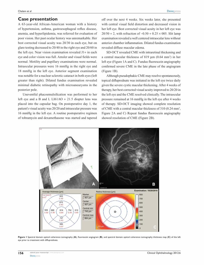
Figure 1 Spectral domain optical coherence tomography (A), fluorescein angiogram (B), and spectral domain optical coherence tomography thickness map (C) of the left eye prior to treatment with difluprednate.

submit your manuscript | www.dovepress.com

Dovepress

Dovepress

156

Chalam et al

Clinical Ophthalmology 2012:6

DiscussionPseudophakic CME is the most common cause of unexpected

visual loss after cataract surgery.7,8 The incidence is estimated

to range from 4% to 11% in the modern era. Because large

randomized controlled trials with long-term follow-up are

lacking, no standardized treatment or prophylactic regimen

exists.7,8 Currently, topical nonsteroidal anti-inflammatory

drugs and topical corticosteroids are first-line treatment

modalities. Refractory cases of CME may be treated

intravitreally with corticosteroids, nonsteroidal anti-

inflammatory drugs, or antivascular endothelial growth factor

agents.2,7–10 We chose to administer topical difluprednate

in view of its effectiveness, potency, and bioavailability.

Furthermore, topical treatment modalities avoid the risks of

intravitreal injections which include increased intraocular

pressure and endophthalmitis.

Difluprednate (difluoroprednisolone butyrate acetate) is

effective in the treatment of anterior uveitis, postoperative

inflammation, and pain. However, its efficacy in postopera-

tive CME is not described. Difluprednate, recently approved

for inflammation and pain associated with ocular surgery

in 2008, penetrates the corneal epithelium rapidly and

effectively.3 Difluoroprednisolone butyrate, its active metabo-

lite, has strong corticosteroid receptor agonist activity and

is six times stronger than prednisolone in animal studies.3 It

is preserved with sorbic acid that is nontoxic to the corneal

surface compared with benzalkonium chloride.3 Fluorina-

tion at the C-6 and C-9 positions increases glucocorticoid

receptor binding affinity and the addition of butyrate and

acetate esters at positions C-17 and C-21 improve corneal

penetration compared with topical betamethasone.4,5 In

addition, difluprednate is administered in an emulsion, thus

increasing its bioavailability.4 The dose uniformity of diflu-

prednate ophthalmic emulsion 0.05% was highly predict-

able compared with both branded and generic prednisolone

(shaken or unshaken) acetate suspensions.6 Similar to other

corticosteroids, difluprednate may cause a steroid-responsive

intraocular pressure rise; however, its effect is minimal com-

pared with other drugs in its class.10

We present a patient with severe CME with a central

macular thickness of 819 µm (0.64 mm3) and the presence

of subretinal fluid on OCT. Although pseudophakic CME

may spontaneously resolve, severe cystic thickening warrants

therapy.7,8 Our patient’s visual acuity returned to normal after

252 µm800

700

600

500

400

300

200

100

0

252 µm

246 µm378310

375

369

370366

357

380

369

371 µm

200 µm

200 µm

C

BA

T

Central max

Center

MarkerAverage thickness [µm] Retina thickness [µm] Examination: 12/1/2010

Vol [mm3]2.59

0.36 0.22 0.24 0.22

0.22

0.37

0.22

0.36

0.37

Central min

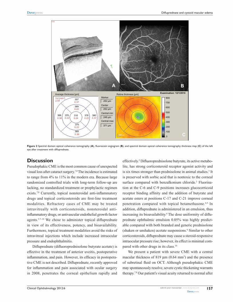
Figure 2 Spectral domain optical coherence tomography (A), fluorescein angiogram (B), and spectral domain optical coherence tomography thickness map (C) of the left eye after treatment with difluprednate.

submit your manuscript | www.dovepress.com

Dovepress

Dovepress

157

Difluprednate and cystoid macular edema

Clinical Ophthalmology

Publish your work in this journal

Submit your manuscript here: http://www.dovepress.com/clinical-ophthalmology-journal

Clinical Ophthalmology is an international, peer-reviewed journal covering all subspecialties within ophthalmology. Key topics include: Optometry; Visual science; Pharmacology and drug therapy in eye diseases; Basic Sciences; Primary and Secondary eye care; Patient Safety and Quality of Care Improvements. This journal is indexed on

PubMed Central and CAS, and is the official journal of The Society of Clinical Ophthalmology (SCO). The manuscript management system is completely online and includes a very quick and fair peer-review system, which is all easy to use. Visit http://www.dovepress.com/ testimonials.php to read real quotes from published authors.

Clinical Ophthalmology 2012:6

using difluprednate for 4 weeks and the ultrastructural changes

in the fovea resolved with recovery of normal anatomy.

ConclusionIn summary, severe pseudophakic CME resolves rapidly

after topical administration of difluprednate. SD-OCT was

helpful both in identifying CME and monitoring of treatment

with serial scans.

DisclosureThe authors report no conflicts of interest in this work.

References1. Hariprasad SM, Akduman L, Clever JA, Ober M, Recchia FM, Mieler WF.

Treatment of cystoid macular edema with the new-generation NSAID nepafenac 0.1%. Clin Ophthalmol. 2009;3:147–154.

2. Chalam KV, Keshavamurthy R, Brar VS. Spectral domain OCT documented resolution of recalcitrant macular edema after intravitreal bevacizumab in branch retinal vein occlusion. Eur J Ophthalmol. 2008;18:831–833.

3. Korenfeld MS, Silverstein SM, Cooke DL, Vogel R, Crockett RS; Difluprednate Ophthalmic Emulsion 0.05% (Durezol) Study Group. Difluprednate ophthalmic emulsion 0.05% for postoperative inflamma-tion and pain. J Cataract Refract Surg. 2009;35:26–34.

4. Foster CS, Davanzo R, Flynn TE, et al. Durezol (difluprednate oph-thalmic emulsion 0.05%) compared with Pred Forte 1% ophthalmic suspension in the treatment of endogenous anterior uveitis. J Ocul Pharmacol Ther. 2010;2:475–483.

5. Tajika T, Waki M, Tsuzuki M, Kida T, Sakaki H. Pharmacokinetic features of difluprednate ophthalmic emulsion in rabbits as determined by glucocorticoid receptor-binding bioassay. J Ocul Pharmacol Ther. 2011;27:29–34.

6. Nakano S, Yamamoto T, Kirii E, Abe S, Yamashita H. Steroid eye drop treatment (difluprednate ophthalmic emulsion) is effective in reducing refractory diabetic macular edema. Graefes Arch Clin Exp Ophthalmol. 2010;248:805–810.

7. Shelsta HN, Jampol LM. Pharmacologic therapy of pseudophakic cystoid macular edema: 2010 update. Retina. 2011;31:4–12.

8. Flach AJ. The incidence, pathogenesis and treatment of cystoid macular edema following cataract surgery. Trans Am Ophthalmol Soc. 1998;96:557–634.

9. Spitzer MS, Ziemssen F, Yoeruek E, et al. Efficacy of intravitreal bevacizumab in treating postoperative pseudophakic cystoid macular edema. J Cataract Refract Surg. 2008;34:70–75.

10. Ahmadabadi HF, Mohammadi M, Beheshtnejad H, Mirshahi A. Effect of intravitreal triamcinolone acetonide injection on central macular thickness in diabetic patients having phacoemulsification. J Cataract Refract Surg. 2010;36:917–922.

submit your manuscript | www.dovepress.com

Dovepress

Dovepress

Dovepress

158

Chalam et al


Recommended