* Corresponding Author: Marjan Esmaeili, Ali asghar Children Hospital, Zafar Street, Iran university of Medical Sciences, Tehran-Iran. Tel: 0915 512 94 88; E-mail: [email protected]Original Article Open Access Head Circumference in Iranian Infants Mohammad Esmaeili 1 , Marjan Esmaeili 2* , Reza Saeidi 3 , Fatemeh Ghane Sharbaf 4 1. Associate Professor of Pediatrics, Ghaem Medical Center, Mashhad University of Medical Sciences, Mashhad, Iran 2. Resident of Pediatrics, Iran university of Medical Sciences, Tehran, Iran 3. Neonatal Research Center, Imam Reza Hospital, Faculty of Medicine, Mashhad University of Medical Sciences, Mashhad, Iran 4. Assistant Professor of Pediatrics, Dr. Sheikh Children Hospital, Mashhad University of Medical Sciences, Mashhad, Iran ABSTRACT Background: Head circumference (HC) measurement is an important parameter in the diagnosis of neurological and developmental disorders as well as dysmorphic syndromes. Recognition of different disorders requires an understanding of the normal variation of the HC size, especially during infancy which is a period of rapid brain growth. Regarding the differences in the international and interracial standard charts of anthropometric indices, local and generational diversities, changes in the ethnic mix of the population and socioeconomic factors, periodic re-evaluations need to be done on the size of HC. This study aimed to represent a local HC standard for an Iranian infant population in comparison with the charts of the American National Center of Health Statistics (NCHS) approved by the World Health Organization (WHO). Methods: About 1003 neonates were randomly enrolled in this cross-sectional study. They were ageing from 3 days to 24 months and were apparently in normal condition. The HC size of these infants was measured and recorded. Tables and graphs were diagramed by Excel Microsoft Office 2007 software and two-tailed Student’s t-test was used for statistical analysis of data. Results: According to the findings of this study, the mean of the HC size in male infants was larger than the female ones. Based on a visual comparison, the curves followed a pattern similar to that of the NCHS’s. Overall, our subjects in both genders were found to have a smaller HC size at birth compared to the NCHS charts. However, infants of other ages had a larger HC size compared to that of the NCHS’s. Conclusion: With respect to the international and interracial differences in the HC size, it is recommended that local anthropometric indices be constructed and used clinically across the world. In addition, extensive and longitudinally designed studies are required in this regard. Key Words: Children, Head circumference; Iranian; Infants Introduction Head circumference (HC) measurement is an important parameter in the routine physical and neurological examination of children. HC may be an early indicator of intracranial pathology essential to the diagnosis of neurological and developmental disorders as well as dysmorphic syndromes associated with abnormal size and/or shape of the HC (1-3). Standard HC charts are of foremost significance because, unlike other somatic growth parameters, even small deviations of the HC from the normal range could be associated with various diseases (3-8). Thus, it is suggested that HC measurement in neonates be performed and recorded with care and regularity. As an indirect measure of brain growth, the most significant increase in the HC size occurs within the first few years of life. It grows as much as 80% of the adult size by two years with the pace slowing down afterwards. Due to the differences in the international and interracial standard charts of anthropometric indices, several countries around the world have attempted to provide alternative standard charts for measuring the HC size at different ages (2, 9-15). Since only a few studies have been conducted about HC on Iranian infants (4, 16, 17) and due to the local and generational diversities, changes in the ethnic mix of the population and socioeconomic factors, periodic re-evaluations of growth standards are recommended (1, 4, 9, 16). This study aimed to represent a local HC standard of an Iranian infant population within the first two years of life during which the most significant increase in the HC size occurs. Moreover, we compared the results of this study with the charts of the American National Center of Health Statistics (NCHS) approved by the World Health
Transcript
* Corresponding Author: Marjan Esmaeili, Ali asghar Children Hospital, Zafar Street, Iran university of Medical Sciences, Tehran-Iran.
Original Article Open Access Head Circumference in Iranian Infants
Mohammad Esmaeili1, Marjan Esmaeili2*, Reza Saeidi 3, Fatemeh Ghane Sharbaf4
1. Associate Professor of Pediatrics, Ghaem Medical Center, Mashhad University of Medical Sciences, Mashhad, Iran 2. Resident of Pediatrics, Iran university of Medical Sciences, Tehran, Iran 3. Neonatal Research Center, Imam Reza Hospital, Faculty of Medicine, Mashhad University of Medical Sciences, Mashhad, Iran 4. Assistant Professor of Pediatrics, Dr. Sheikh Children Hospital, Mashhad University of Medical Sciences, Mashhad, Iran
ABSTRACT
Background: Head circumference (HC) measurement is an important parameter in the diagnosis of neurological and developmental disorders as well as dysmorphic syndromes. Recognition of different disorders requires an understanding of the normal variation of the HC size, especially during infancy which is a period of rapid brain growth. Regarding the differences in the international and interracial standard charts of anthropometric indices, local and generational diversities, changes in the ethnic mix of the population and socioeconomic factors, periodic re-evaluations need to be done on the size of HC. This study aimed to represent a local HC standard for an Iranian infant population in comparison with the charts of the American National Center of Health Statistics (NCHS) approved by the World Health Organization (WHO). Methods: About 1003 neonates were randomly enrolled in this cross-sectional study. They were ageing from 3 days to 24 months and were apparently in normal condition. The HC size of these infants was measured and recorded. Tables and graphs were diagramed by Excel Microsoft Office 2007 software and two-tailed Student’s t-test was used for statistical analysis of data. Results: According to the findings of this study, the mean of the HC size in male infants was larger than the female ones. Based on a visual comparison, the curves followed a pattern similar to that of the NCHS’s. Overall, our subjects in both genders were found to have a smaller HC size at birth compared to the NCHS charts. However, infants of other ages had a larger HC size compared to that of the NCHS’s. Conclusion: With respect to the international and interracial differences in the HC size, it is recommended that local anthropometric indices be constructed and used clinically across the world. In addition, extensive and longitudinally designed studies are required in this regard.
Key Words: Children, Head circumference; Iranian; Infants
Introduction Head circumference (HC) measurement is an
important parameter in the routine physical and neurological examination of children. HC may be an early indicator of intracranial pathology essential to the diagnosis of neurological and developmental disorders as well as dysmorphic syndromes associated with abnormal size and/or shape of the HC (1-3).
Standard HC charts are of foremost significance because, unlike other somatic growth parameters, even small deviations of the HC from the normal range could be associated with various diseases (3-8). Thus, it is suggested that HC measurement in neonates be performed and recorded with care and regularity.
As an indirect measure of brain growth, the most significant increase in the HC size occurs within the first few years of life. It grows as much as 80% of the adult size by two years with the
pace slowing down afterwards. Due to the differences in the international and interracial standard charts of anthropometric indices, several countries around the world have attempted to provide alternative standard charts for measuring the HC size at different ages (2, 9-15).
Since only a few studies have been conducted about HC on Iranian infants (4, 16, 17) and due to the local and generational diversities, changes in the ethnic mix of the population and socioeconomic factors, periodic re-evaluations of growth standards are recommended (1, 4, 9, 16).
This study aimed to represent a local HC standard of an Iranian infant population within the first two years of life during which the most significant increase in the HC size occurs. Moreover, we compared the results of this study with the charts of the American National Center of Health Statistics (NCHS) approved by the World Health
Head Circumference in Iranian Infants Esmaeili M et al
Iranian Journal of Neonatology 2015; 6(1): 29
Organization (WHO) as the international standard of growth in the first years of infant’s life (18).
Methods This cross–sectional study was conducted on
1003 infants (505 male, 498 female) ageing from 3 days to 24 months. The subjects consisted of 104 full-term neonates and 899 infants of 1-24 months coming from a diverse socioeconomic background (Table 1) in an urban population in Mashhad, a nearly developed city located in the Northeast of Iran.
Measurements were provided from the neonates in the maternity ward and medical and vaccination centers affiliated with Mashhad University of Medical Sciences during a six-month period.
Sampling was carried out randomly via active observational methods once a week. The inclusion criteria for this study were as follows: 1) healthy neonates with the gestational age of 37-42 weeks as determined by maternal dates and the Dubowitz score; 2) birth weight of more than 2500 grams.
Among our subjects, the infants of less than two years of age were apparently in normal, healthy condition without any obvious signs of physical or neurological disorders.
The measurements were provided from the neonates after reaching 48 hours of age in order to allow head molding resolution. The neonates’ head was measured at the largest area, at a level
passing the supraorbital protuberance anteriorly and occipital protuberance posteriorly, using a fresh, non-elastic, non-stretchable, accurately scaled standard tape. Afterwards, tables and graphs were diagramed and depicted by the Excel Microsoft Office software 2007.
Statistical analysis for the comparison of data was performed by two-tailed Student’s t-test.
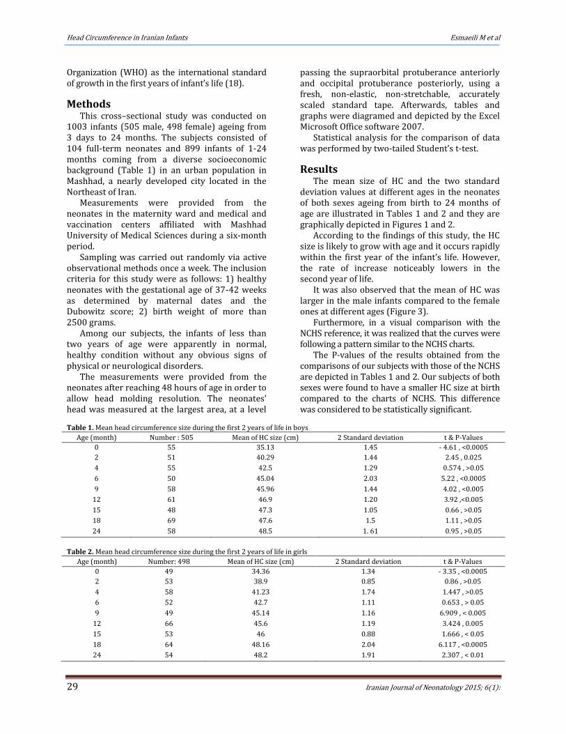
Results The mean size of HC and the two standard
deviation values at different ages in the neonates of both sexes ageing from birth to 24 months of age are illustrated in Tables 1 and 2 and they are graphically depicted in Figures 1 and 2.
According to the findings of this study, the HC size is likely to grow with age and it occurs rapidly within the first year of the infant’s life. However, the rate of increase noticeably lowers in the second year of life.
It was also observed that the mean of HC was larger in the male infants compared to the female ones at different ages (Figure 3).
Furthermore, in a visual comparison with the NCHS reference, it was realized that the curves were following a pattern similar to the NCHS charts.
The P-values of the results obtained from the comparisons of our subjects with those of the NCHS are depicted in Tables 1 and 2. Our subjects of both sexes were found to have a smaller HC size at birth compared to the charts of NCHS. This difference was considered to be statistically significant.
Table 1. Mean head circumference size during the first 2 years of life in boys
Age (month) Number : 505 Mean of HC size (cm) 2 Standard deviation t & P-Values
0 55 35.13 1.45 - 4.61 , <0.0005
2 51 40.29 1.44 2.45 , 0.025
4 55 42.5 1.29 0.574 , >0.05
6 50 45.04 2.03 5.22 , <0.0005
9 58 45.96 1.44 4.02 , <0.005
12 61 46.9 1.20 3.92 ,<0.005
15 48 47.3 1.05 0.66 , >0.05
18 69 47.6 1.5 1.11 , >0.05
24 58 48.5 1. 61 0.95 , >0.05
Table 2. Mean head circumference size during the first 2 years of life in girls
Age (month) Number: 498 Mean of HC size (cm) 2 Standard deviation t & P-Values
0 49 34.36 1.34 - 3.35 , <0.0005
2 53 38.9 0.85 0.86 , >0.05
4 58 41.23 1.74 1.447 , >0.05
6 52 42.7 1.11 0.653 , > 0.05
9 49 45.14 1.16 6.909 , < 0.005
12 66 45.6 1.19 3.424 , 0.005
15 53 46 0.88 1.666 , < 0.05
18 64 48.16 2.04 6.117 , <0.0005
24 54 48.2 1.91 2.307 , < 0.01
Esmaeili M et al Head Circumference in Iranian Infants
ranian Journal of Neonatology 2015; 6(1): 30
Figure 1. Mean ±2 standard deviation of head circumference size during first 2 years of life in boys
It is also noteworthy that at other ages, our subjects were found to have a larger HC size compared to the charts of NCHS.
Discussion Adequate knowledge of the normal growth of
HC as one of the most important anthropometric indices is essential to detecting and preventing pathological conditions.
Although a large head might be a result of various other factors such as the increased intracranial pressure and/or Hydrocephalus and a small size head (Microcephaly) could be associated with the undergrowth of the brain, such defects may not always be pathological. By one aspect, they could be explained through a familial observation of the size and shape of the parents’ and the siblings’ head (1). Additionally, maternal reproductive history, prematurity, maternal diet and season of birth are among the factors influencing the HC size (5-8, 19-22).
Figure 3. Mean of head circumference size in infant boys and girls
Figure 2. Mean ±2 standard deviation of head circumference size during first 2 years of life in girls
The formation of the skull is a complex
interaction reflecting the growth of both the bones and the brain as the major stimulus for skull growth. Head growth is a dynamic process and plotting serial measurements of HC over a period of time could provide useful information in this regard (1).
Several recent studies have attempted to demonstrate the differences in the size of HC in various countries as well as between successive generations within the same country (9, 23).
For the most part, their results emphasize the need for every population to perform periodical re-evaluations with respect to the HC size standards.
The standard growth charts are based on the data collected from 1963 to 1975 by the NCHS. The NCHS charts have been approved by WHO as the international standard over the past decades. Nevertheless, these references have been falling out of date, and so, the universal use of NCHS/WHO references for all populations is a matter of controversy nowadays (16, 24-26).
While recommending the NCHS/WHO international reference in their study, Sullivan et al. also suggested that in developing countries, resources are required in order to produce focal growth (27). On the other hand, Goldstein and Tanner strongly argued that developing countries in particular need to provide their own standards for clinical use (28).
In another study, Hoey et al. claimed that the mean of HC size in Irish children was larger than the British standard produced by Tanner and Nellhaus while it was smaller than Ounsted data from Oxford. They also outlined that the children of non-manual workers had a larger head compared to the children of the manual workers (1, 29).
0
10
20
30
40
50
60
0 2 4 6 9 12 15 18 24
2sd
Mean
2SD
0
10
20
30
40
50
60
0 2 4 6 9 12 15 18 24
Boys
Girls
0
10
20
30
40
50
60
0 2 4 6 9 12 15 18 24
2sd
Mean
2SD
Head Circumference in Iranian Infants Esmaeili M et al
Iranian Journal of Neonatology 2015; 6(1): 31
Another study conducted in Japan indicated a significant difference in the HC size between Japanese and Caucasian children which was correlated with the overall smaller stature of the Japanese (14).
Moreover, the HC values in Turkish children were similar to that of the Irish children (1, 2).
The data obtained in the present study could replace the Nellhaus criteria in clinical use (2).
A study conducted in Shiraz, Iran demonstrated that the HC in the Iranian children was larger compared to that of their Turkish peers whereas it was smaller than Irish, Japanese and American children (4, 17).
In another study performed by Vazirian et al, no significant differences of HC were observed between their subjects as a population of Iranian children and those of the NCHS/WHO charts (16).
Furthermore, Mohammadi et al. and Talebean et al. in two different studies conducted in Iran compared the growth indices of a group of Iranian children with those of the NCHS charts and realized that the HC of their subjects was similar to that of the NCHS charts (30, 31).
In another research, Hams P et al. concluded that there were no significant differences in the HC percentiles in different regions of the world (32).
According to the findings of the current research, which are different from previous studies in Iran, it is suggested that the HC standards of western countries (NCHS, Nellhaus) may not thoroughly apply to Iranian children. However, due to the inconsistencies between the results of various studies, it is recommended that extensive and longitudinally designated studies be conducted in order to determine standard HC charts for the urban and rural Iranian children. Essentially, such studies need to be performed periodically in every population in different regions of the world.
Acknowledgements The authors would like to thank the Vice-
Chancellory for Research of Mashhad University of Medical Sciences
References 1. Hoey HMCV, Cox LA. Head circumference standards
for Irish children. Acta Paediatr Scand. 1990; 79(2):162-7.
2. Karabiber H, Durmaz Y, Yakinci C, Kutlu O, Gumusalan Y, Yologlu S, et al. Head circumference measurement of urban children aged between 6 and 12 in Malatya, Turkey. Brain & Development. 2001; 23(8): 801–4
3. Nilsson D, Svensson J, Korkmaz BA, Nelvig H, Tisell M. Decreased head circumference in shunt-treated compared with healthy children. J Neurosurg Pediatr. 2013;12(5):483-90.
4. Ayatollahi SMT, Shayan Z. Head Circumference Standards for School Children of Shiraz, Iran. J Trop Pediatr. 2006; 52(6): 406-10
5. Morgan C, McGowan P, Herwitker S, Hart AE, Turner MA. Postnatal head growth in preterm infants: a randomized controlled parenteral nutrition study. Pediatrics. 2014; 133(1):e120-8.
6. Pedersen M, von Stedingk H, Botsivali M, Agramunt S, Alexander J, Brunborg G, Chatzi L, et al. Birth weight, head circumference, and prenatal exposure to acrylamide from maternal diet: the European prospective mother-child study (NewGeneris). Environ Health Perspect. 2012; 120(12):1739-45.
7. Ortega-García JA, Gutierrez-Churango JE, Sánchez-Sauco MF, Martínez-Aroca M, Delgado-Marín JL, Sánchez-Solis M, et al. Head circumference at birth and exposure to tobacco, alcohol and illegal drugs during early pregnancy. Childs Nerv Syst. 2012; 28(3):433-9.
8. Ilves P, Lintrop M, Talvik I, Muug K, Maipuu L, Metsvaht T. Low cerebral blood flow velocity and head circumference in infants with severe hypoxic ischemic encephalopathy and poor outcome. Acta Paediatr. 2009; 98(3):459-65.
9. Ishikawa T, Furuyama M, Ishikawa M, Ogawa J, Wada Y. Growth in head circumference from birth to fifteen years of age in Japan. Acta Paediatr Scand. 1987; 76(5):824-8.
10. Kuczmarski RJ, Ogden CL, Grummer-Strawn LM, Flegal KM, Guo SS, Wei R, et al. CDC growth charts: United States. Adv Data. 2000; 8(314):1-27.
11. Palti H, Peritz E, Flug D, Gitlin M, Adler B. Comparison of head circumference in an Israeli child population with United States and British standards. Ann Hum Biol. 1983; 10(2):195-8.
12. Chen ST. Growth of head circumference of Malaysian infants and pre-school children. J Singapore Paediatr Soc. 1990; 32(3-4):81-6.
13. Werner B, Bodin L. Head circumference from birth to age 48 months for infants in Sweden. Acta Paediatr. 2006; 95(12):1601-7.
14. Tsuzaki S, Matsuo N, Saito M, Osano M. The head circumference growth curve for Japanese children between 0–4 years of age: comparison with Caucasian children and correlation with stature. Ann Hum Biol. 1990; 17(4): 297-303.
15. Malina RM, Habicht JP, Martorell R, Lechtig A, Yarbrough C, Klein RE. Head and chest circumferences in rural Guatemalan Ladino children, birth to seven years of age. Am J Clinl Nutr. I 975; 28(9):106 1-70.
16. Vazirian S, Sedighnezhad A. Update of Growth Percentiles for Children of an Iranian Population. Arch Iranian Med 2003; 6(3):163-9.
17. Ayatollahi SM. Reference charts for arm, chest and head circumferences of south Iranian infants. J Trop Pediatr. 2001; 47(6):376-8.
Esmaeili M et al Head Circumference in Iranian Infants
ranian Journal of Neonatology 2015; 6(1): 32
18. Feigelman S. Growth, development and behavior. In: Kleigman RM, Behrman RE, Jenson HB, Stanton BF, editors. Nelson Textbook of Pediatrics. 19th ed. Philadelphia: Saunders; 2011. P. 48-54
19. Pomeroy E, Wells JC, Stanojevic S, Jaime Miranda J, Cole TJ, Stock JT. Birth month associations with height, head circumference, and limb lengths among peruvian children. Am J Phys Anthropol. 2014; 154(1):115-24.
20. Brantsæter AL, Birgisdottir BE, Meltzer HM, Kvalem HE, Alexander J, Magnus P, et al. Maternal seafood consumption and infant birth weight, length and head circumference in the Norwegian Mother and Child Cohort Study. Br J Nutr. 2012; 107(3):436-44.
21. Xu T, Zhang ZX, Han SM, Hu HT, Xiao XH, Gong XM, et al. Relationship between birth head circumference and adulthood quality of life in Chinese people. J Paediatr Child Health. 2010; 46(11):642-6.
22. Leviton A, Kuban K, Allred EN, Hecht JL, Onderdonk A, O'Shea TM, et al. Antenatal antecedents of a small head circumference at age 24-months post-term equivalent in a sample of infants born before the 28th post-menstrual week. Early Hum Dev. 2010; 86(8):515-21.
23. Dale J, Maurer PK. Abnormal head. In: Ziai M, editor. Bedside pediatrics: diagnostic evaluation of the child. Boston: Little, Brown; 1983.P.17-31.
24. De Onis M, Garza C, Onyango AW, Rolland-Cachera MF; le Comité de nutrition de la Société française de
pédiatrie. WHO growth standards for infants and young children. Arch Pediatr. 2009; 16(1):47-53.
25. Daymont C, Hwang WT, Feudtner C, Rubin D. Head-circumference distribution in a large primary care network differs from CDC and WHO curves. Pediatrics. 2010; 126(4):e836-42.
26. Júlíusson PB, Roelants M, Nordal E, Furevik L, Eide GE, Moster D, et al. Growth references for 0-19 year-old Norwegian children for length/height, weight, body mass index and head circumference. Ann Hum Biol. 2013; 40(3):220-7.
28. Goldstein H, Tanner JM. Ecological considerations in the creation and the use of child standards. Lancet. 1980; 1(8168-Pt1): 582–5.
29. Ounsted M, Moar VA, Scott A. Head circumference charts updated. Arch Dis Child. 1985; 60: 936–939.
30. Mohammadi M. Do the Iranian growth charts are similar to NCHS curves?. Arch Dis Child. 1997; 60(10): 17-23.
31. Sharif M, Azimi A, Talebian A, Mousavi GA, Azimi R. Growth rate of head circumference in first year of life in breastfed infants in Kashan, Iran. Feiz J Sci Res. 1998; 4(2): 47-53.
32. Hams P. The state of world’s children. United Nation Children's Fund (UNICEF). New York: WHO Bull; 1994.


![EON 13W16C F v2 [轉換] - manualszoom.com Size Circumference Nunber ... CIRCUMFERENCE REFERENCE TABLE ... Enter this value to set the wheel circumference. Quick Table ...](https://static.documents.pub/doc/80x56/5af038c77f8b9abc788ce64f/eon-13w16c-f-v2-size-circumference-nunber-circumference-reference.jpg)


