Opening the black box of carbon degradation pathways in marine sediments through single cell genomics and metagenomics Karen G. Lloyd C-DEBI network talk July 25, 2013 niversity of Tennessee, Knoxville
Transcript
Opening the black box of carbon degradation pathways in marine sediments through single
cell genomics and metagenomics
Karen G. LloydC-DEBI network talk July 25, 2013
University of Tennessee, Knoxville
Q: What drives microbial diversity in the vast marine subsurface?
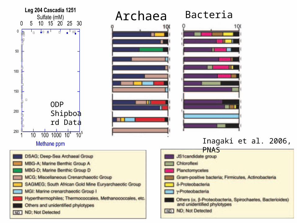
Inagaki et al. 2006, PNAS
Archaea Bacteria
ODP Shipboard Data
Sulfate reducers
Q: What drives microbial diversity in the marine subsurface?
A1. Not the terminal electron acceptor (sulfate, iron, manganese, CO2)
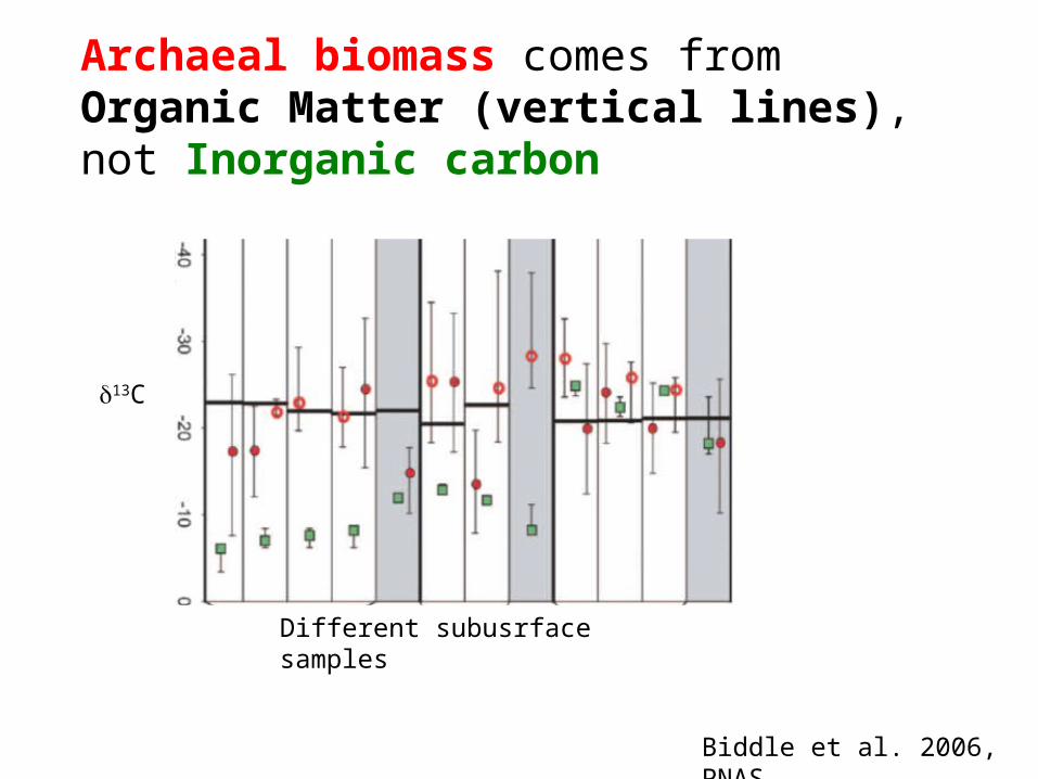
Biddle et al. 2006, PNAS
Archaeal biomass comes from Organic Matter (vertical lines), not Inorganic carbon
d13C
Different subusrface samples
Q: What drives microbial diversity in the marine subsurface?
A1. Not the terminal electron acceptor (sulfate, iron, manganese, CO2)
A2. Most likely organic matter
Inagaki et al. 2006, PNAS
Archaea Bacteria
ODP Shipboard Data
Q: What types of organic matter are available as substrates?
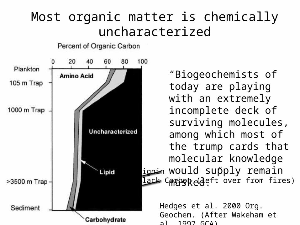
Hedges et al. 2000 Org. Geochem. (After Wakeham et al. 1997 GCA)
“Biogeochemists of today are playing with an extremely incomplete deck of surviving molecules, among which most of the trump cards that molecular knowledge would supply remain masked.”
Most organic matter is chemically uncharacterized
LigninBlack Carbon (left over from fires)
Q: What types of organic matter are available as substrates?
A. Proteins, carbohydrates, lipids, lignin, and a bunch of mysterious compounds.
Q: How do protein, carbohydrate, and lipids create diverse physiological niches?
Centre for Ecological Sciences, IISc, Bangalore
Extracellular enzymes and abiotic processes
Primary intracellular fermentation
Fermentation of organic matter
Secondary intracellular fermentation
Terminal respiration
Q: How do protein, carbohydrate, and lipids create diverse physiological niches?
A: There are thousands of biochemically characterized extracellular and intracellular enzymes with different substrate specificities, acting at different points in the fermentation cascade, and requiring different chemical conditions. So, maybe by examining the range of enzymes available to an organism, we can discover its organic matter niche.
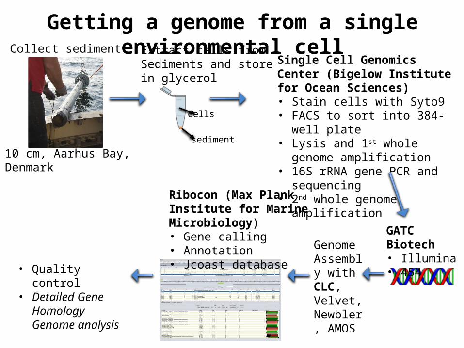
Single cell genomics: A way to put together large genomic fragments of
a single uncultured organism, to connect “who” to “what” they’re doing
Single Cell Genomics Center (Bigelow Institute for Ocean Sciences)• Stain cells with Syto9• FACS to sort into 384-well plate• Lysis and 1st whole genome
Ribocon (Max Plank Institute for Marine Microbiology)• Gene calling• Annotation• Jcoast database
Collect sediment Extract cells from Sediments and store in glycerol
cells
sediment
10 cm, Aarhus Bay, Denmark
Genome Assembly with CLC, Velvet, Newbler, AMOS
• Quality control• Detailed Gene
Homology Genome analysis
Getting a genome from a single environmental cell
We caught cells from archaea with worldwide distribution and can be the dominant cells (by FISH and qPCR) in some marine sediments (Kubo et al. 2012, ISME J).
We retrieved only 15-70% of each genome, but that’s a lot more than 0%!
MBG-D MCG MBG-D MBG-D
Lloyd et al. 2013, Nature
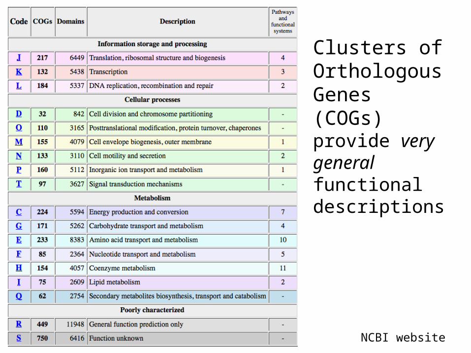
Q: How do we find genes relevant to the degradation of proteins, lipids, and carbohydrates in these genomes?
NCBI website
Clusters of Orthologous Genes (COGs) provide very general functional descriptions
Q: How do we find genes relavent to the degradation of proteins, lipids, and carbohydrates in these genomes?
A: Step 1. Use COGs, Pfams, Tigrfams, SEED, Swissprot/Uniprot, Genbank, Kegg to annotate predicted genes.
Step 2. Conduct “detailed gene homologue analysis”, where experimentally-determined
traits of nearest gene homologue are used to hypothesize functions in predicted genes.
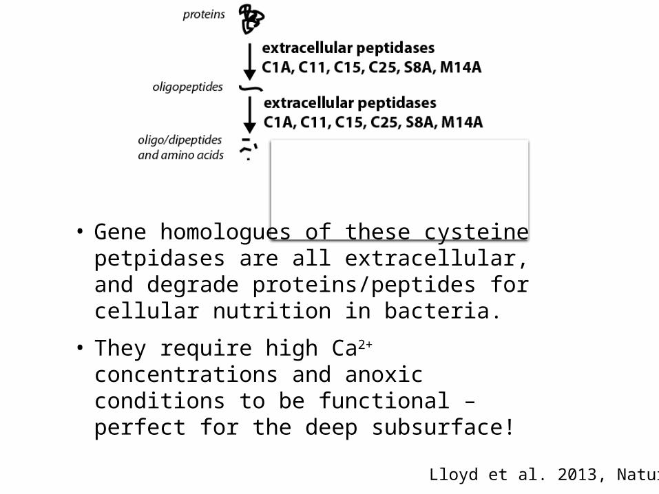
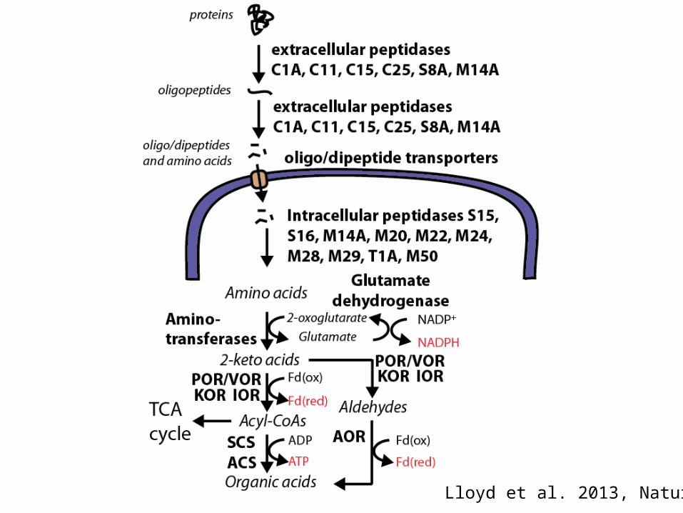
• Gene homologues of these cysteine petpidases are all extracellular, and degrade proteins/peptides for cellular nutrition in bacteria.
• They require high Ca2+ concentrations and anoxic conditions to be functional – perfect for the deep subsurface!
Lloyd et al. 2013, Nature
Lloyd et al. 2013, Nature
Lloyd et al. 2013, Nature
Lloyd et al. 2013, Nature
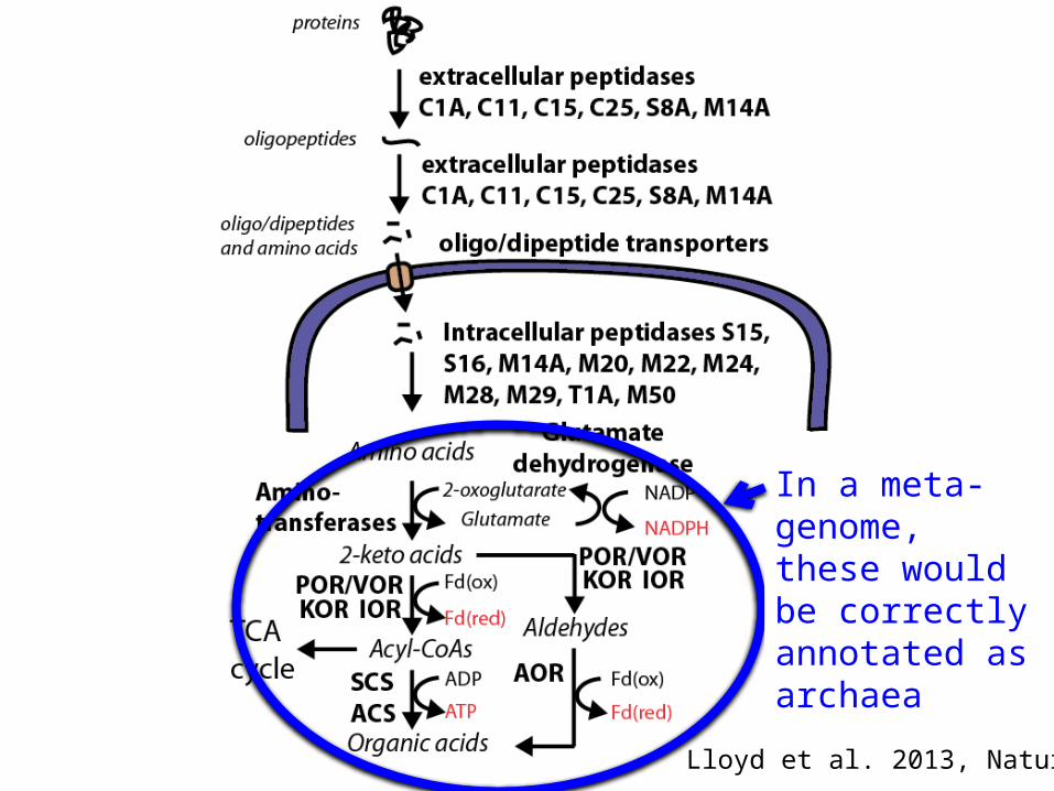
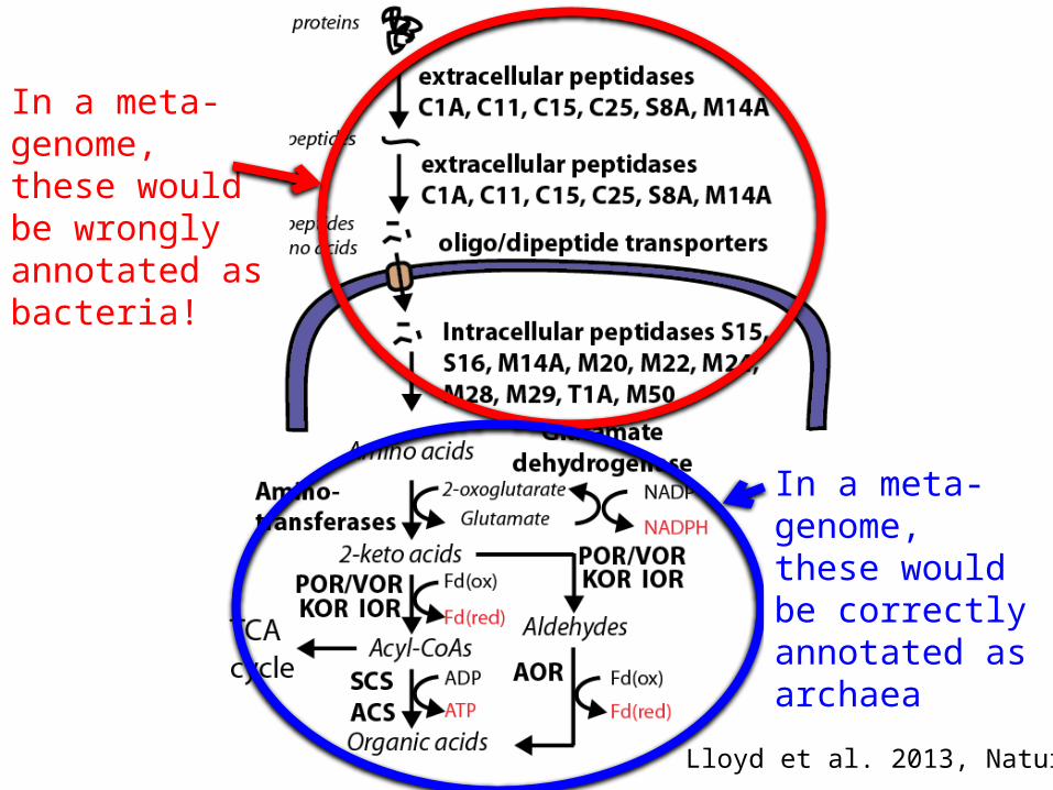
In a meta-genome, these would be correctly annotated as archaea
Lloyd et al. 2013, Nature
In a meta-genome, these would be wrongly annotated as bacteria!
In a meta-genome, these would be correctly annotated as archaea
Lloyd et al. 2013, Nature
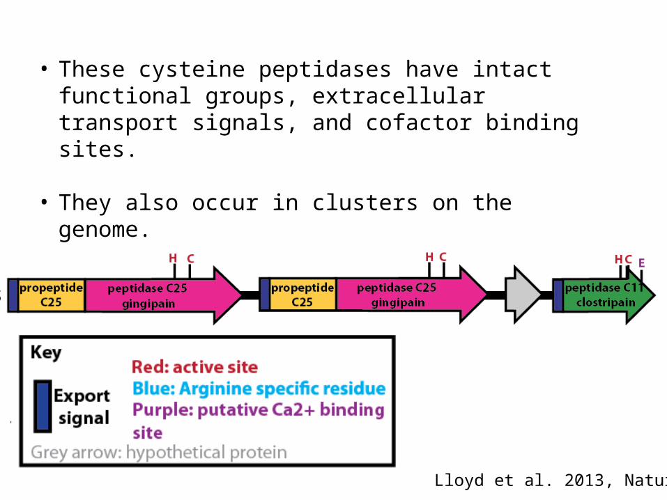
• These cysteine peptidases have intact functional groups, extracellular transport signals, and cofactor binding sites.
• They also occur in clusters on the genome.
Lloyd et al. 2013, Nature
Lloyd et al. 2013, Nature
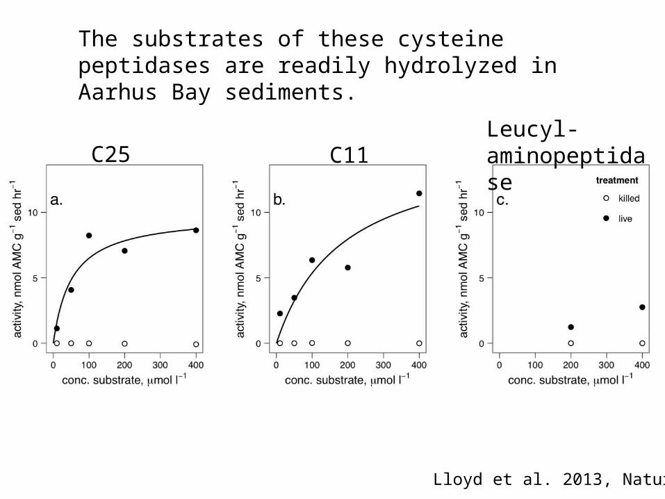
The substrates of these cysteine peptidases are readily hydrolyzed in Aarhus Bay sediments.
C25 C11Leucyl- aminopeptidase
Conclusions:
1. Some subsurface archaea degrade detrital proteins using extracellular enzymes that prefer cleaving at arginine and have special adaptations to the anoxic subsurface environment.
2. “Detailed gene homologue analysis” is an effective way to discover OM degrading gene pathways.
What about the rest of the subsurface microbial community?
Arch
aea
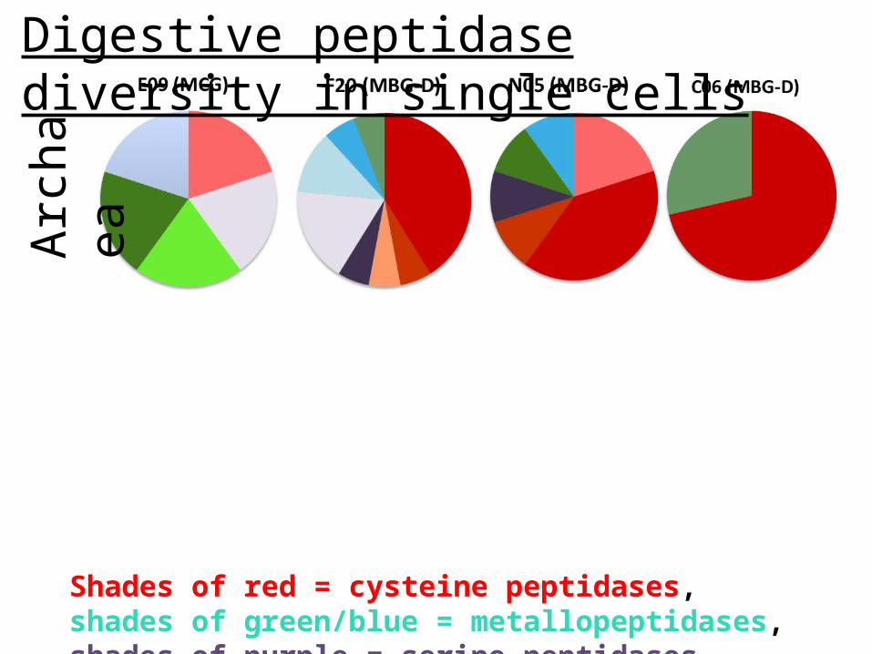
Digestive peptidase diversity in single cells
Shades of red = cysteine peptidases, shades of green/blue = metallopeptidases, shades of purple = serine peptidases
Arch
aea
Bact
eria
Digestive peptidase diversity in single cells
Shades of red = cysteine peptidases, shades of green/blue = metallopeptidases, shades of purple = serine peptidases
What about the rest of the subsurface microbial community?
A1: So far, archaea have more cysteine peptidases (cleave at arginine or proline, all require strict anoxic environment) and bacteria have more metallopeptidases (cleave at leucine or proline, or cell wall degradation for predation).
What about the rest of the subsurface microbial community?
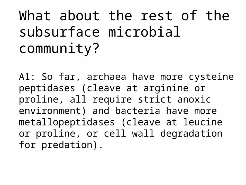
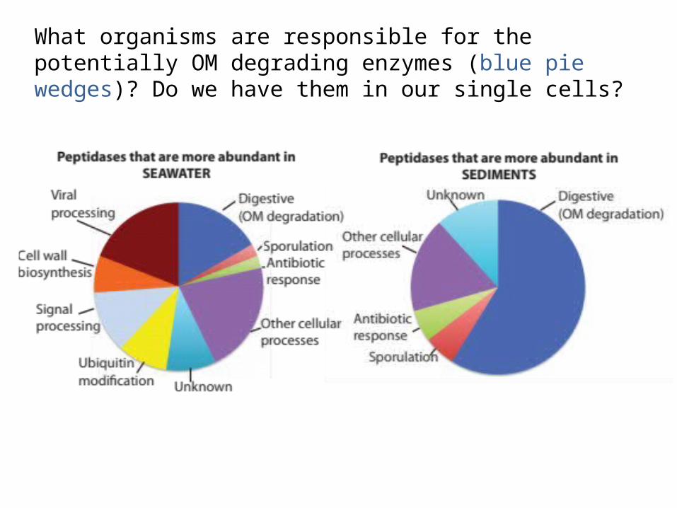
Analyzed the following from IMG database: • 86 water metagenomes (deep N. Atlantic and shallow Delaware Bay) • 12 sediment methane seep metagenomes (Santa Barbara Basin and Arctic Ocean)
Analyzed the following from IMG database: • 86 water metagenomes (deep N. Atlantic and shallow Delaware Bay) • 12 sediment methane seep metagenomes (Santa Barbara Basin and Arctic Ocean)
Seawater has a bunch of peptidases for viruses, eukaryotes, growth, intercellular communication, and digestion that are less represented in sediments
Analyzed the following from IMG database: • 86 water metagenomes (deep N. Atlantic and shallow Delaware Bay) • 12 sediment methane seep metagenomes (Santa Barbara Basin and Arctic Ocean)
Microbes in sediments and seawater seem to use very different peptidases for nutrition (OM degradation) as well as sporulation, antibiotic responses, and housekeeping.
What about the rest of the subsurface microbial community?
A2: They might be using different enzymes than seawater organisms to degrade organic matter. So, sediments may differ from seawater not just in speed of OM degradation, but in quality.
What organisms are responsible for the potentially OM degrading enzymes (blue pie wedges)? Do we have them in our single cells?
Arch
aea
Bact
eria
Digestive peptidase over-represented in sediments
Shades of red = cysteine peptidases, shades of green/blue = metallopeptidases, shades of purple = serine peptidases
What about the rest of the subsurface microbial community?
A3: Half of the potentially OM-degrading peptidases that were over-represented in sediment metagenomes were present in our single cells, and were found in either only archaea, or both archaea and bacteria. So, our single cells might actually be descriptive of the larger community, and archaea and bacteria both have sediment-specific peptidases.
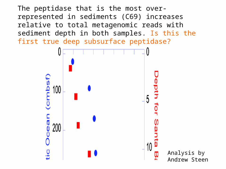
The peptidase that is the most over-represented in sediments (C69) increases relative to total metagenomic reads with sediment depth in both samples. Is this the first true deep subsurface peptidase?
Analysis by Andrew Steen
Directions for the immediate future:
• Deeper sediments
• More peptidase trends with depth and environments
• Carbohydrates and lipids
• Create an OM degradation database tool for other researchers to use
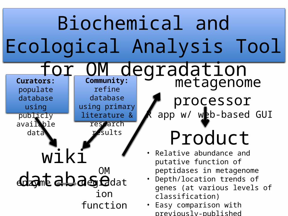
wiki databaseenzyme
OM degradation
function
Curators:populate database
using publicly available data
Community:refine database using primary literature &
research results
metagenome processorR app w/ web-based GUI
Product• Relative abundance and putative
function of peptidases in metagenome• Depth/location trends of genes (at
various levels of classification)• Easy comparison with previously-
published metagenomes
Biochemical and Ecological Analysis Tool for OM degradation
Aarhus University: Lars Schreiber, Dorthe Petersen, Kasper Kjeldsen, Mark Lever, Andreas Schramm, Bo Barker Jorgensen
Max Plank Institute for Marine Microbiology in Bremen, Germany: Michael Richter, Sara Kleindeinst, Sabine Lenk
Bigelow Institute for Ocean Sciences: Ramunas Stepanauskas, Wendy Bellows, Jochen Nuester
University of Tennessee: Andrew Steen, Jordan Bird