Operative Vaginal Delivery: The Art of Obstetrics EF “Pat” Magann MD FACOG FRANZCOG MFM Division and Fellowship Director University of Arkansas for the Medical Sciences Little Rock, Arkansas
Transcript
Operative Vaginal Delivery: The Art of Obstetrics
EF “Pat” Magann MD FACOG FRANZCOG MFM Division and Fellowship Director University of Arkansas for the Medical Sciences Little Rock, Arkansas
Objectives
Define the classification of operative vaginal delivery performed. Understand the indications for performing
operative vaginal delivery. Properly apply both forceps
No disclosures
Timing of Delivery
I am a fetus in the womb I fear it may become my tomb If only I could give a shout To make my doctor get me out Unknown medical student, Dublin Ireland BJOG
Hawks-Dennen Overlapping shanks: Elliott, Tucker-McLane Rotational (Kielland, Leff) Special (Piper)
Types of Forceps
Simpson forceps (1848) are the most commonly used among the types of forceps and has an elongated cephalic curve. These are used when there is substantial molding of the fetal head. , Elliot forceps (1860) are similar to Simpson forceps but with an adjustable pin in the end of the handles which can be drawn out as a means of regulating the lateral pressure on the handles when the instrument is positioned for use. They are used most often when there is minimal moulding
Types of Forceps
Kielland forceps (1915, Norwegian) are distinguished by an extremely small pelvic curve and a sliding lock. The most common forceps used for rotation. The sliding lock is helpful in asynclitic Kielland forceps lack traction because they have almost no pelvic curve Wrigley's forceps are used in low or outlet delivery
and in cesarean section delivery where manual traction is proving difficult. The short length results in a lower chance of uterine rupture. Piper's forceps have a perineal curve to allow
application to the after-coming head in breech delivery.
Presenter
Presentation Notes
Wrigley's forceps were designed for use by general practitioner obstetricians, having the safety feature of an inability to reach high into the pelvis
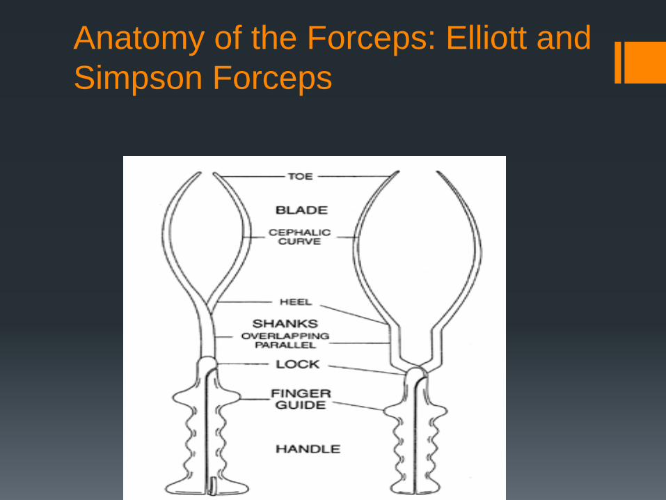
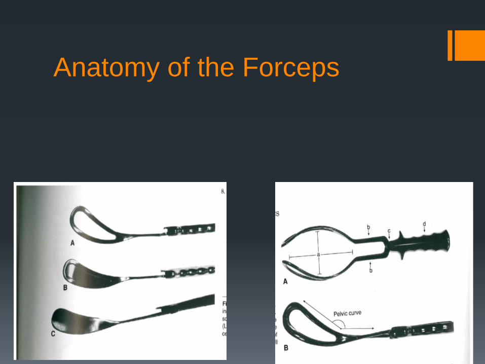
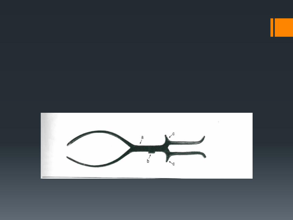
Anatomy of the Forceps: Elliott and Simpson Forceps
Anatomy of the Forceps
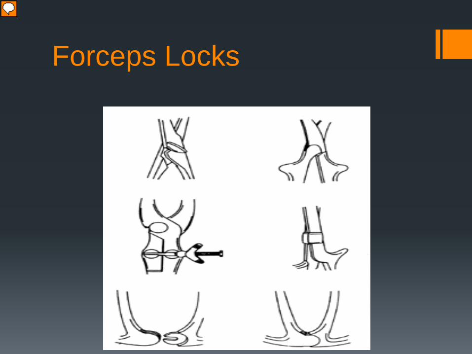
Forceps Locks
Presenter
Presentation Notes
French, English, German , sliding, pivot
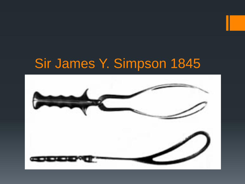
Sir James Y. Simpson 1845
Luikart-Simpson • Luikart R. A modification of the Kielland, Simpson, and Tucker-McLane forceps to simplify their use and improve function and safety. Am J Obstet Gynecol 1937;34:686
• pseudofenestrated blade
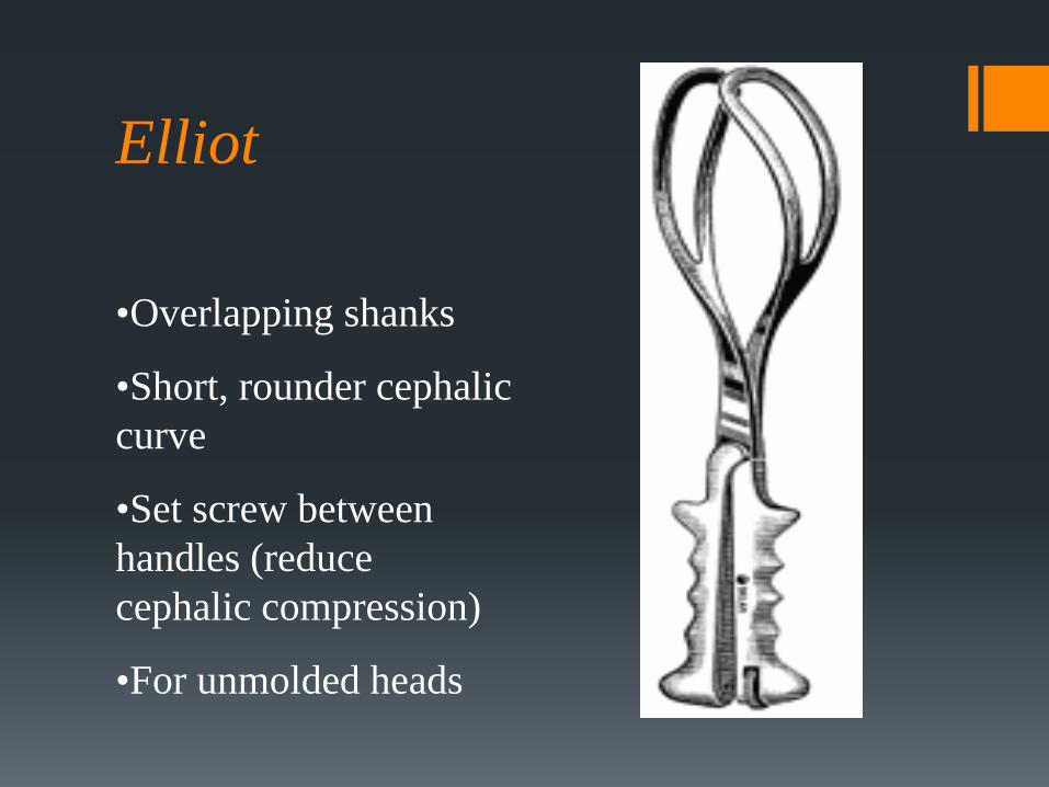
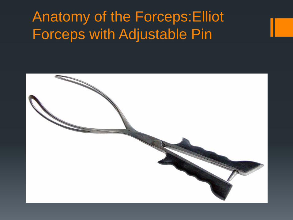
Elliot •Overlapping shanks
•Short, rounder cephalic curve
•Set screw between handles (reduce cephalic compression)
•For unmolded heads
Anatomy of the Forceps:Elliot Forceps with Adjustable Pin
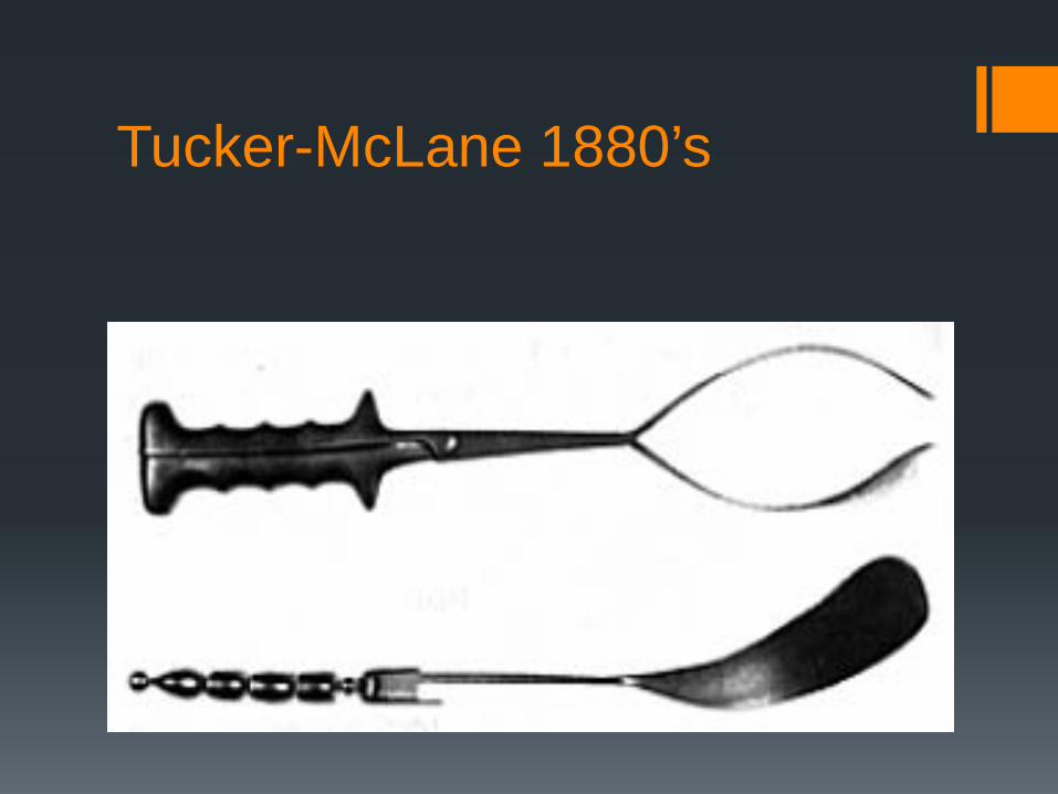
Tucker-McLane 1880’s
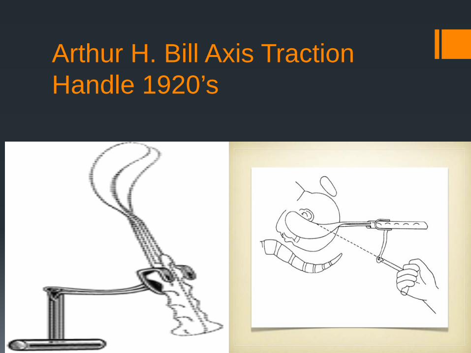
Arthur H. Bill Axis Traction Handle 1920’s
Christian Kielland 1915
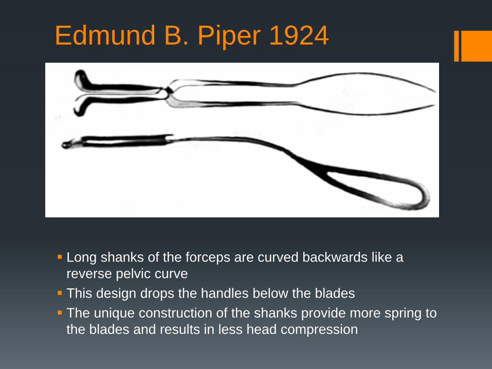
Edmund B. Piper 1924
Long shanks of the forceps are curved backwards like a reverse pelvic curve This design drops the handles below the blades The unique construction of the shanks provide more spring to
the blades and results in less head compression
Operative Vaginal Delivery
Applying direct traction to the fetal skull (forceps) or the fetal scalp (vacuum) along with maternal expulsive efforts to effect a vaginal delivery Incidence estimated at 8-15% Fetal head must be engaged Membranes ruptured Cervix completely dilated Bladder empty of urine
Operative Vaginal Delivery
Indications for operative vaginal delivery Prolonged second stage of labor (nulliparous 3
hours with regional anesthesia or 2 hours without) multiparous (2 hours with regional anesthesia and 1 hour without regional anesthesia) Fetal compromise Shorten of the second stage of labor for maternal
indications
Prerequisites for Forceps Delivery The head should be engaged and the station of the
head accurately known The cervix should be completely dilated.
The exact position of the head should be known.
Occasionally ultrasound may help if the degree of molding creates confusion. The type of pelvis should be known. Certain pelvic
types will not allow for rotation. For example, a fetus in a posterior position in an android or anthropoid pelvis is best delivered in the occipitoposterior (OP) position.
Prerequisites for Forceps Delivery The operator should be familiar with the
advantages and disadvantages of the different forceps. There should be adequate anesthesia for the
forceps delivery contemplated. A low or outlet forceps delivery can be performed under pudendal block ; a forceps rotation of greater than 45° or a midforceps procedure requires a good epidural The bladder should be empty.
This is an operative procedure, and it should be
accorded the same respect and care for aseptic technique as any other operative procedure.
Contraindications for Operative Vaginal Delivery
Unengaged fetal head In ability to determine fetal position Malpresentation (face or brow) CPD actual or suspected Prematurity (< 34 weeks vacuum) Repeated scalp pH (vacuum) Inability to apply instrument correctly Incompletely dilated cervix
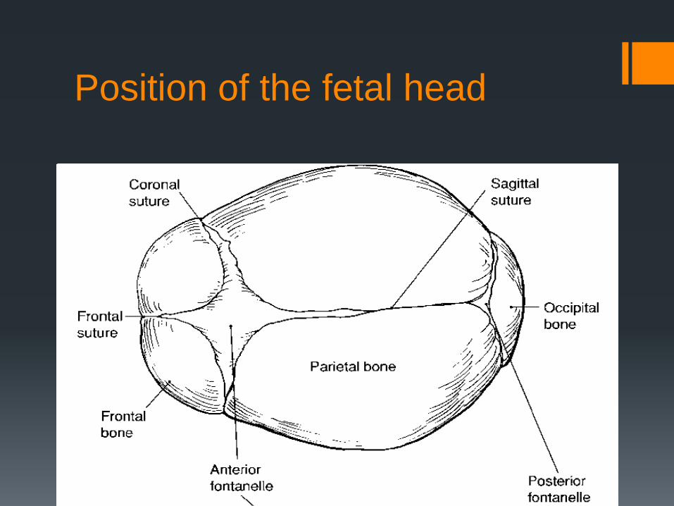
Position of the fetal head
Position of the Fetal Head
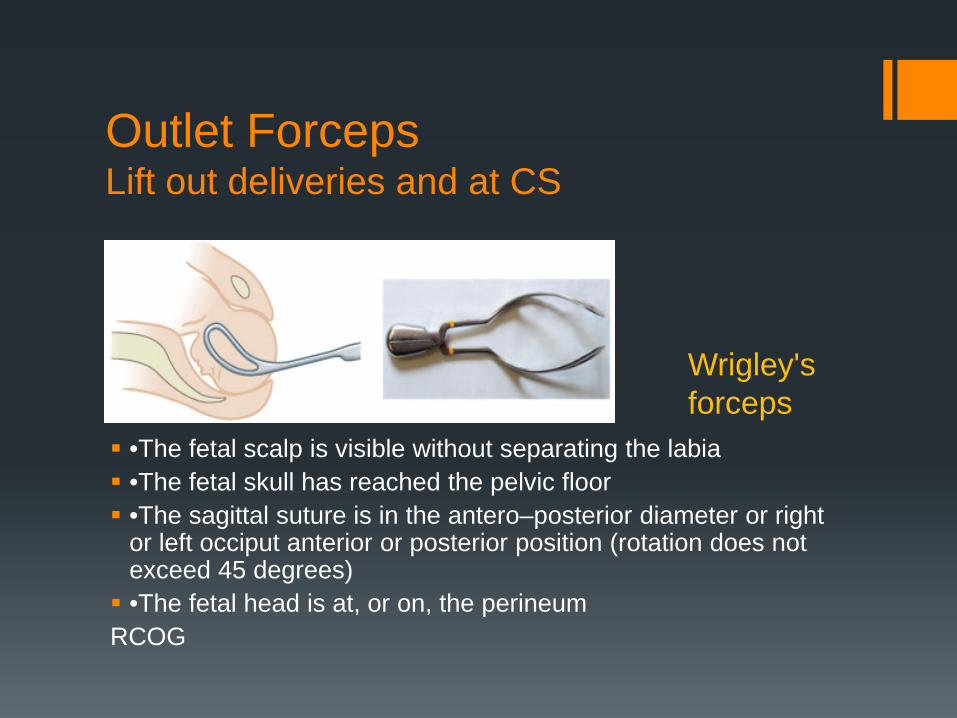
Outlet Forceps
Scalp is visible at the introitus without separating the labia Fetal skull has reached the pelvic floor Sagittal suture in A-P diameter, or Right or Left
anterior or posterior position Fetal head is at or near the perineum Rotation does not exceed 45 degrees ACOG
Outlet Forceps Lift out deliveries and at CS
•The fetal scalp is visible without separating the labia •The fetal skull has reached the pelvic floor •The sagittal suture is in the antero–posterior diameter or right
or left occiput anterior or posterior position (rotation does not exceed 45 degrees) •The fetal head is at, or on, the perineum RCOG
Wrigley's forceps
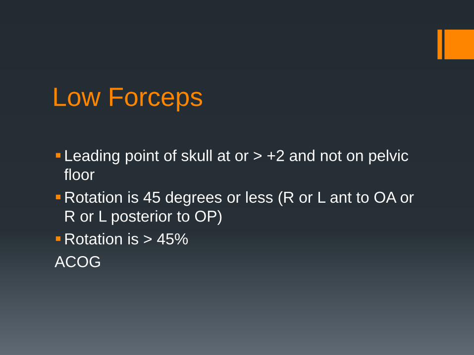
Low Forceps
Leading point of skull at or > +2 and not on pelvic floor Rotation is 45 degrees or less (R or L ant to OA or
R or L posterior to OP) Rotation is > 45% ACOG
Midforceps Stations is above +2 but the head is engaged High forceps are no longer included in the
classification
ACOG
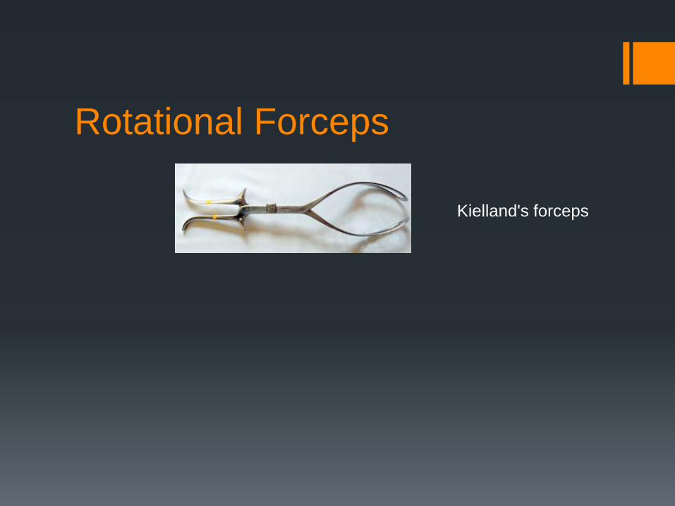
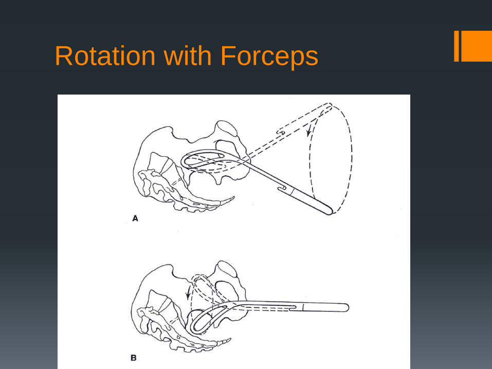
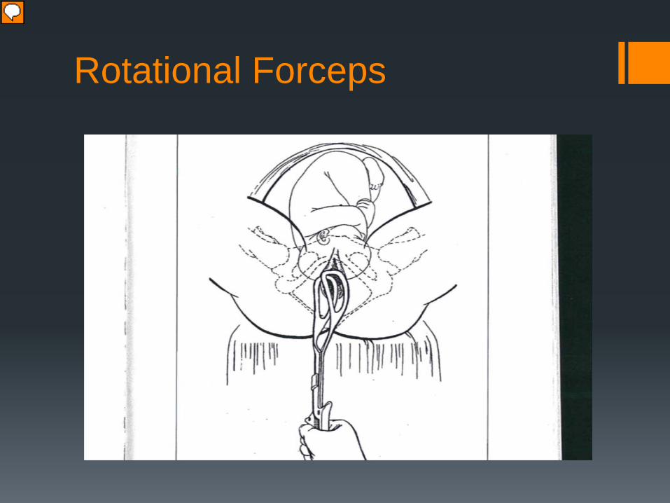
Rotational Forceps
Kielland's forceps
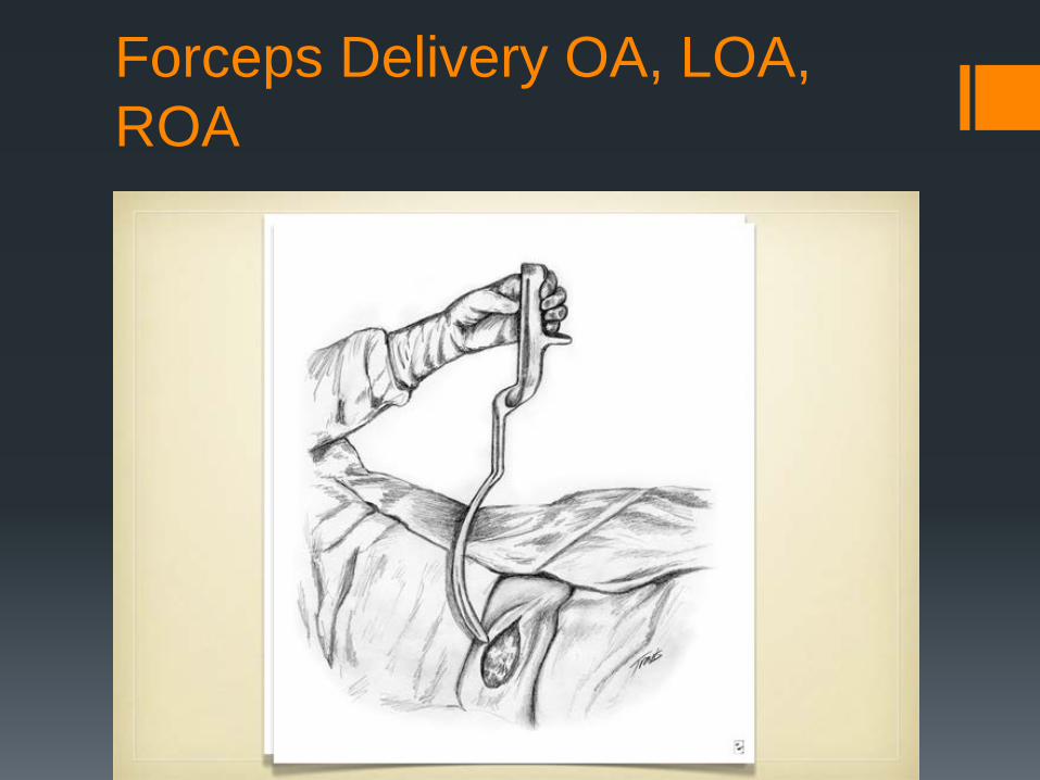
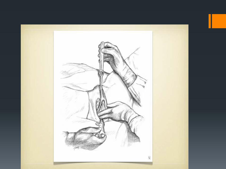
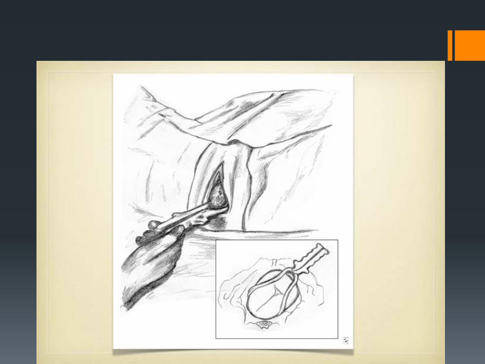
Forceps Delivery OA, LOA, ROA
Checks for proper placement
Sagittal suture lies in the midline of the shanks No more than one finger can be placed between the fetal head and the blades or fenestrations on either side Posterior fontanelle is not more than one finger’s breadth above the plane of the shanks (in OA position)
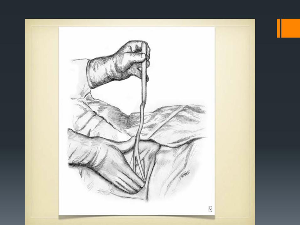
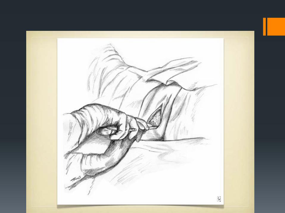
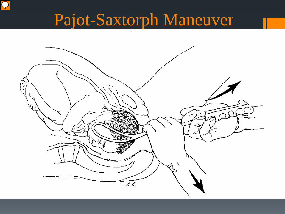
Pajot-Saxtorph Maneuver
Presenter
Presentation Notes
Apply force in plane perpendicular to plane of pelvis where BPD is moving Higher station requires traction lower from the horizontal Exert force in two directions: Down with hand on shanks Outward with hand on finger guards Apply least forces necessary for descent
Neonatatl and maternal complications are low following successful rotational forceps delivery. 873 over a period of 7 years in a single tertiary center in Scotland
Rotational Forceps
Presenter
Presentation Notes
First orientation for application of the Keilland forceps is done, in this case as you can see the fetus is ROT
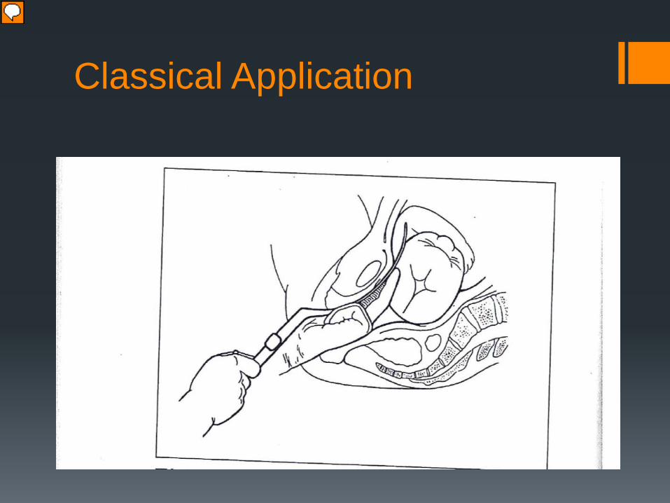
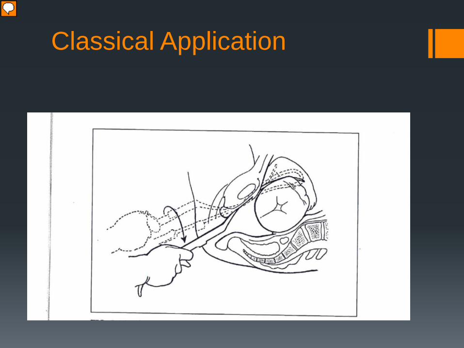
Classical Application
Presenter
Presentation Notes
Insertion of the anterior blade (L) blade to the anterior L ear of the fetus in the ROT If the classic approach is used the anterior blade is inserted first
Classical Application
Presenter
Presentation Notes
Clockwise rotation in R occiput transverse of the anterior L blade so that the cephalic curve will coincide with the curve of the head. The cephalic curve has been oriented away from the fetal head towards the anterior uterus, it must now be rotated away from the occiput toward the midline and toward the knob. , this is done with a twist of the wrist over 180 degrees until the know points towards 3 clock as the rotation is taking place the handle is depressed slightly to follow the plane of least resistence. If the toe of blade is rotated towards the occiput the the toe may lacerate the uterus.
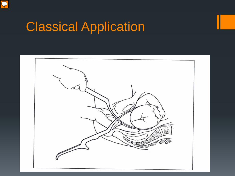
Classical Application
Presenter
Presentation Notes
The L blade is always introduced posterioly between the shnak of the anterior blad and the and the patients R thigh this obviates the crossing the handles to lock the blades.
Classical Application
Presenter
Presentation Notes
The top picture depicts the final application of the Kelliand forceps This is the keillan forceps after clockwise rotation from ROT to OA, since there us a reverse pelvic curve the instrument is rotated almost directly on its shanks and the handles are depressed at the completion of the turn.
Direct Application
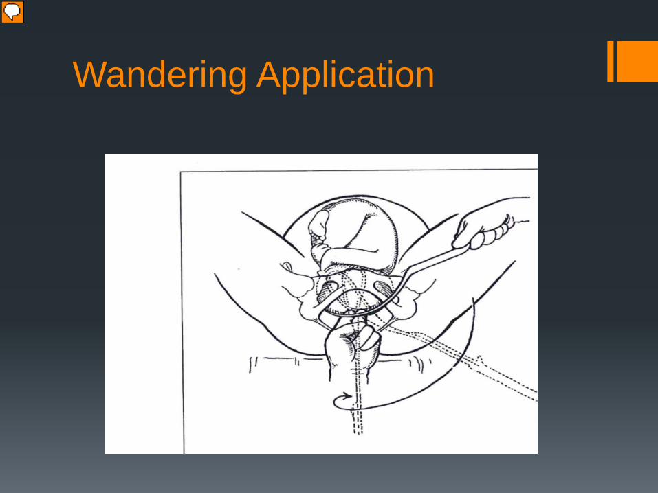
Wandering Application
Presenter
Presentation Notes
Anterior R blade of Keilland to anterior R ear in LOT presentation.
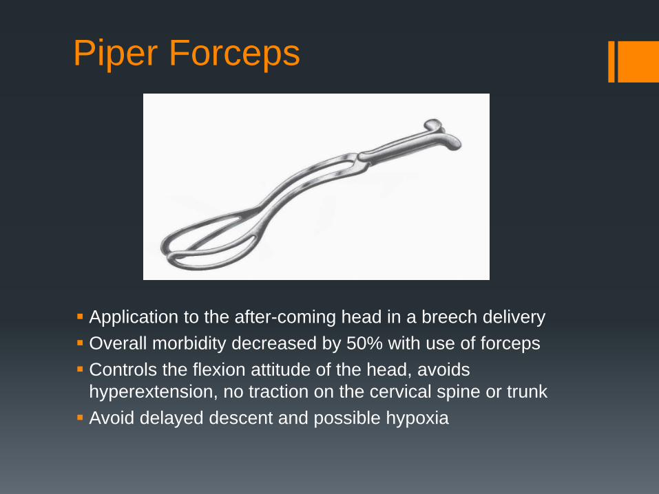
Piper Forceps
Application to the after-coming head in a breech delivery Overall morbidity decreased by 50% with use of forceps Controls the flexion attitude of the head, avoids
hyperextension, no traction on the cervical spine or trunk Avoid delayed descent and possible hypoxia
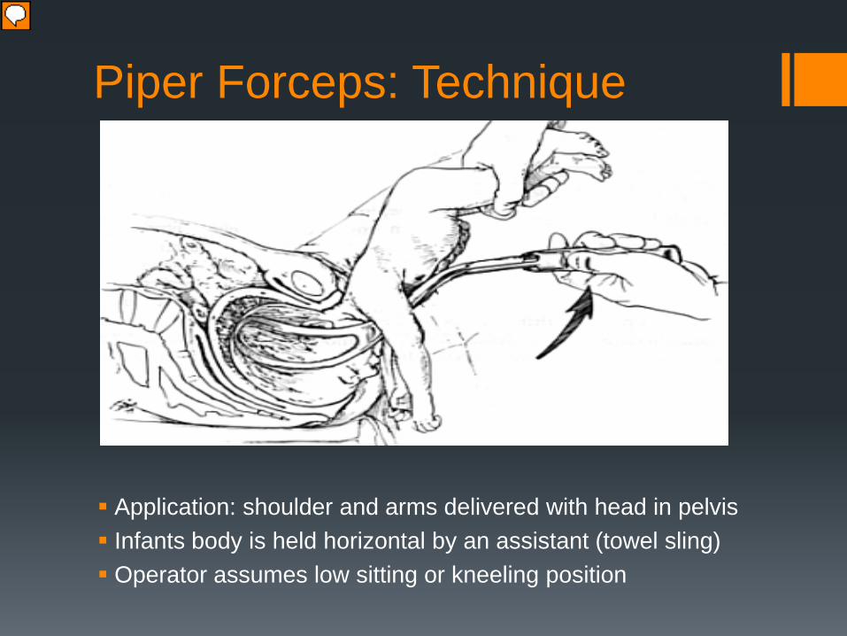
Piper Forceps: Technique
Application: shoulder and arms delivered with head in pelvis Infants body is held horizontal by an assistant (towel sling) Operator assumes low sitting or kneeling position
Presenter
Presentation Notes
Very important not to have assistant elevate the body of the fetus above the horizontal
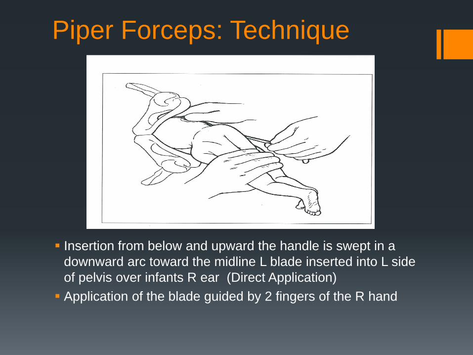
Piper Forceps: Technique
Insertion from below and upward the handle is swept in a
downward arc toward the midline L blade inserted into L side of pelvis over infants R ear (Direct Application) Application of the blade guided by 2 fingers of the R hand
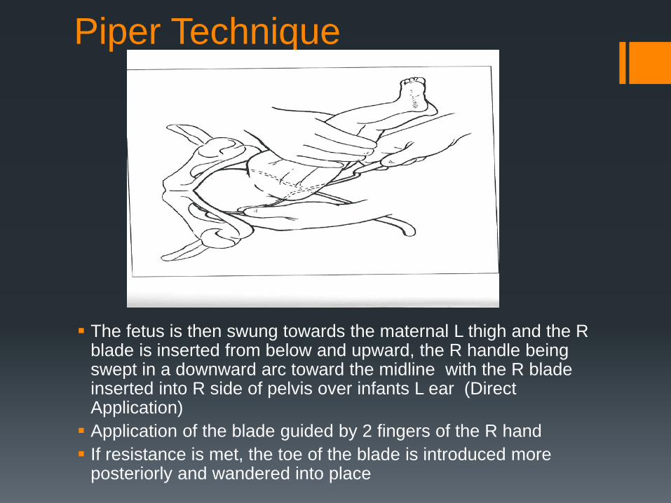
Piper Technique
The fetus is then swung towards the maternal L thigh and the R blade is inserted from below and upward, the R handle being swept in a downward arc toward the midline with the R blade inserted into R side of pelvis over infants L ear (Direct Application) Application of the blade guided by 2 fingers of the R hand If resistance is met, the toe of the blade is introduced more
posteriorly and wandered into place
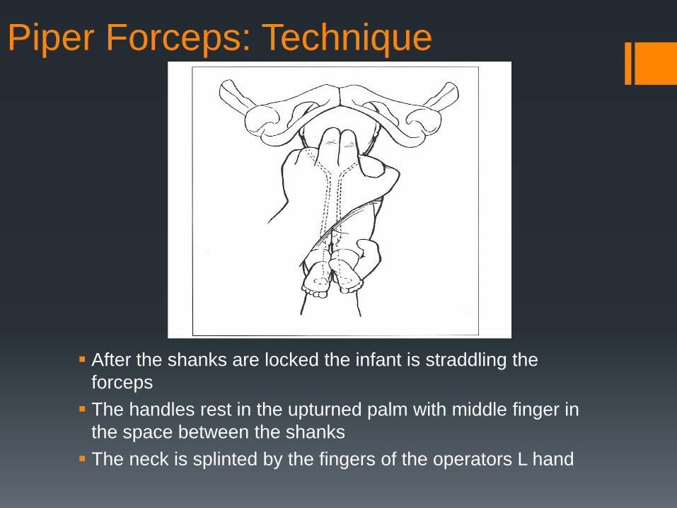
Piper Forceps: Technique
After the shanks are locked the infant is straddling the forceps The handles rest in the upturned palm with middle finger in
the space between the shanks The neck is splinted by the fingers of the operators L hand
Piper Forceps: Technique
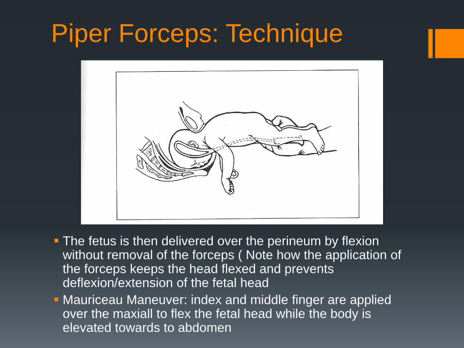
The fetus is then delivered over the perineum by flexion without removal of the forceps ( Note how the application of the forceps keeps the head flexed and prevents deflexion/extension of the fetal head Mauriceau Maneuver: index and middle finger are applied
over the maxiall to flex the fetal head while the body is elevated towards to abdomen
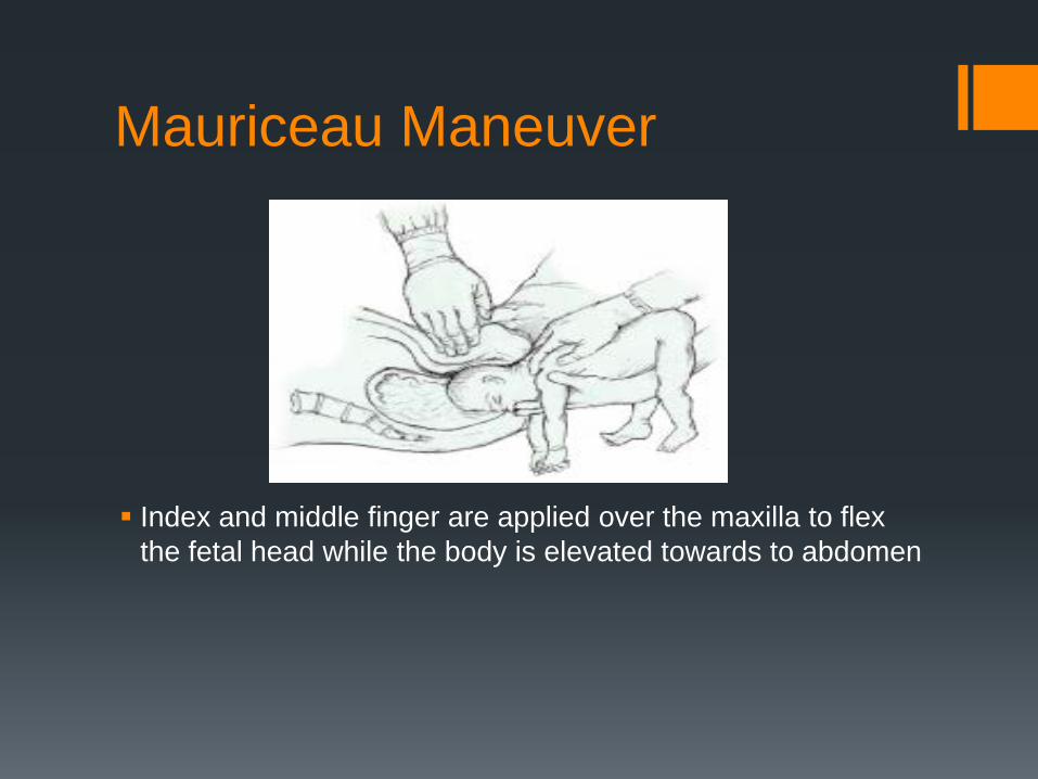
Mauriceau Maneuver
Index and middle finger are applied over the maxilla to flex the fetal head while the body is elevated towards to abdomen
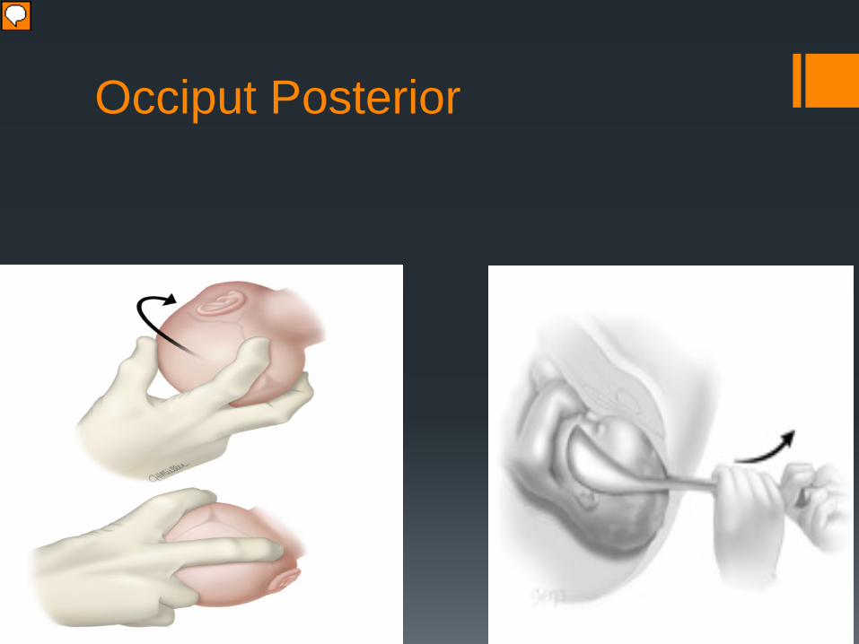
Occiput Posterior
Presenter
Presentation Notes
Process for applying the forceps is the same as in the OA Positon Axis of traction is horizontal and downward until the fetal nose clears the symphysis and then the axis turns upwards. Manual rotation — We avoid performing manual rotation in the first stage of labor because it may disengage the fetal head, which could lead to prolapse of the umbilical cord or small parts. Rotation before full dilatation is also less successful than rotation in the second stage [25]. The optimum time for rotation during the second stage has not been studied, but prophylactic manual rotation is more likely to be successful than a procedure performed after arrest of descent [25]. We define prophylactic rotation as rotation at the beginning of the second stage, regardless of the station of the fetal head. We do not routinely perform prophylactic rotations, but consider its use in two main scenarios: first, if there is a need for more expeditious delivery, and second, if there has been minimal or slow descent after a trial of pushing. The maternal bladder is emptied prior to the procedure. A hand is inserted into the vagina with the palm upward. Digital rotation is performed by placing the tips of the index and middle fingers in the anterior segment of the lambdoidal suture near the posterior fontanelle (figure 2). The fingers are used to flex and slightly dislodge the vertex, rotating the fetal head to the OA position via rotation of the operator's hand and forearm. The thumb may also be used with gentle downward pressure more anteriorly on the parietal bone to aid in this rotation. The fetal head may need to be held in place for a few contractions to prevent rotation back toward the posterior position [29,30]. Manual rotation can also be performed by placing the operator's four fingers behind the posterior parietal bone with the palm up and the thumb over the anterior parietal bone. The right hand is used for left OP position and the left hand is used for right OP position. The head is grasped with the tips of the fingers and thumb. During a contraction, the patient is encouraged to push and the operator attempts to flex and rotate the fetal head anteriorly (figure 3) [25,31]. Occasional, mild upward pressure may help to slightly displace the station and facilitate rotation. A study in which manual rotation from the OP or occiput transverse position was attempted in 796 patients reported 90 percent of fetuses were successfully rotated to an anterior position, although multiple attempts were sometimes required [25]. Manual rotation is more successful in multiparous women and women less than 35 years of age [32]. Cesarean delivery is common after a failed manual rotation (cesarean delivery rate 34 to 59 percent versus 2 to 4 percent if rotation is successful [25,32]). However, if prompt delivery is indicated, failed manual rotation may be followed by vacuum or forceps delivery from the OP position, in an appropriately assessed patient. Manual rotation performed prior to instrumental birth has little or no increase in risk to the pregnant woman or to the fetus [29,30]. Forceps rotation — The performance of rotational forceps (eg, Kielland rotation [33]) should be reserved to those experienced and skilled in their art, due to the high risk of potential complications. These procedures are beyond the scope of this topic review. Operative vaginal delivery from the OP position — Forceps or vacuum can be used to deliver the fetus from the direct OP position. Direct delivery from OP position rather than rotation is probably preferable in women who on clinical examination have ample room between the fetal occiput and maternal sacrum/coccyx and when the pelvis is too narrow to permit anterior rotation (women with an anthropoid pelvis with a narrow transverse diameter and women with an android pelvis with a narrow arch). However, in a retrospective study, forceps delivery from the OP position was associated with more third or fourth degree lacerations than rotation (manual or forceps) to OA and forceps delivery from OA position [34]. Regardless of whether rotation is performed, the usual criteria for operative vaginal delivery also apply to the OP position; we do not perform elective operative vaginal deliveries for fetuses above +2 station. OP position is associated with a significantly higher rate of failed operative vaginal delivery than OA position [14]. Because the flexion point on OP presentations is positioned more posterior and higher in the vagina than with OA presentations, certain adjustments must be employed when performing vacuum extraction. (See "Procedure for vacuum assisted operative vaginal delivery", section on 'Instrumentation'.) There is no demonstrated advantage to forceps over vacuum delivery for OP fetuses [14]. We make our choice based on individual patient factors. As an example, for a tired nulliparous woman with an edematous perineum and fetus at or near the outlet, we prefer a vacuum delivery. For a multiparous patient at +2 station with fetal bradycardia, we would perform a forceps delivery.