115 Chapter 7 CHEMICAL INJURIES OF THE EYE EDWARD W. TRUDO, JR, MD * ; AND WILLIAM RIMM, MD † INTRODUCTION INJURIES OF THE EYE CAUSED BY COMMON CHEMICALS Chemical Agents Acids and Alkalis Thermal Injury OCULAR RESPONSE TO CHEMICAL INJURY CLINICAL COURSE OF CHEMICAL OCULAR INJURY Immediate Phase Acute Phase Early Reparative Phase Late Reparative Phase PATIENT TREATMENT AND EVALUATION Immediate Action Patient Examination Acute Phase Treatment Early Reparative Phase Treatment Late Reparative Phase Treatment Rehabilitative Phase INJURIES OF THE EYE CAUSED BY CHEMICAL WARFARE AGENTS Blister Agents Nerve Agents Mycotoxins SUMMARY * Lieutenant Colonel, Medical Corps, US Army Reserve; Azar Eye Institute, 31519 Winter Place Parkway, Suite 1, Salisbury, Maryland 21804, and Assistant Professor of Surgery (Ophthalmology), Uniformed Services University of the Health Sciences, 4301 Jones Bridge Road, Bethesda, Maryland 20814-4799; formerly, Director, Corneal and External Disease, Walter Reed Army Medical Center, Washington, DC 20307-5001 † Colonel, Medical Corps, US Army; Ophthalmology Service, Walter Reed Army Medical Center, Washington, DC 20307-5001
Transcript
115
Chemical Injuries of the Eye
Chapter 7
CHEMICAL INJURIES OF THE EYE
EDWARD W. TRUDO, JR, MD*; AND WILLIAM RIMM, MD†
INTRODUCTION
INJURIES OF THE EYE CAUSED BY COMMON CHEMICALSChemical AgentsAcids and AlkalisThermal Injury
OCULAR RESPONSE TO CHEMICAL INJURY
CLINICAL COURSE OF CHEMICAL OCULAR INJURYImmediate PhaseAcute PhaseEarly Reparative PhaseLate Reparative Phase
INJURIES OF THE EYE CAUSED BY CHEMICAL WARFARE AGENTSBlister AgentsNerve AgentsMycotoxins
SUMMARY
*Lieutenant Colonel, Medical Corps, US Army Reserve; Azar Eye Institute, 31519 Winter Place Parkway, Suite 1, Salisbury, Maryland 21804,and Assistant Professor of Surgery (Ophthalmology), Uniformed Services University of the Health Sciences, 4301 Jones Bridge Road,Bethesda, Maryland 20814-4799; formerly, Director, Corneal and External Disease, Walter Reed Army Medical Center, Washington, DC20307-5001
†Colonel, Medical Corps, US Army; Ophthalmology Service, Walter Reed Army Medical Center, Washington, DC 20307-5001
116
Ophthalmic Care of the Combat Casualty
INTRODUCTION
Military forces face possible chemical injurycaused not only by hazards of the battlefield butalso by occupational hazards in the military’sindustrial base. Therefore, an understanding ofthe physiological damage and the treatmentof chemical eye injuries is required for both thebattlefield and the peacetime environment.1 Occu-pational hazards come in the form of commonchemicals that can injure the eye, including acids(eg, automobile battery acid, refrigerants, vinegar)and alkalis (eg, drain cleaners, fertilizers, buildingsupplies). Military forces run the risk of beingexposed to offensive chemical weapons that posespecific ocular risks. The principles of treatment aresimilar, however, whether the injury occurred as theresult of chemical weaponry or of occupationalhazards.
Chemical injuries of the eye are true emergen-cies requiring prompt recognition and treatment.Rapid dilution of the chemical agent is the imme-diate treatment necessary to reduce tissue damageand preserve vision. The extent of ocular injury isproportional to the departure of the corrosive sub-stance from the neutrality of pH 7.4, the time that it
remains in contact with the eye, and the quantityrequiring neutralization.
Other factors must be considered when treatingpatients with injuries caused by chemical warfareagents. Ocular chemical injuries can cause imme-diate loss of vision, combat ineffectiveness, andeven permanent blindness. Some effects are moresubtle: the mere threat of chemical agents on thebattlefield reduces unit morale and efficiency. Medi-cal personnel who discern that injuries might be theresult of chemical agents may be in a position toalert field command to the possible use of chemicalwarfare on the battlefield. In addition, medical per-sonnel must be aware of specific antidotes for treat-ing systemic and ocular effects stemming from ex-posure to chemical warfare agents.
Three other volumes in the Textbooks of MilitaryMedicine series contain additional information onchemical injuries to the eye, which interested read-ers can peruse: Occupational Health: The Soldier andthe Industrial Base2; Military Dermatology, particularlyChapter 5, Cutaneous Reactions to Nuclear, Biologi-cal, and Chemical Warfare3; and Medical Aspects ofChemical and Biological Warfare.4
INJURIES OF THE EYE CAUSED BY COMMON CHEMICALS
Chemical Agents
Chemical agents with the potential to cause ocu-lar injury are often found in the home, at workwithin the military’s industrial base, on the train-ing field, and on the battlefield. Industrial andhousehold cleaners often contain acidic or alkalineproducts in sufficient concentrations to cause eyeand skin injury. Building materials, such as mortarand plaster, and automobile batteries are the mostcommon sources of household chemical eye inju-ries today. Any of these agents can significantlydamage human tissue after contact (Table 7-1).
Some common examples of acid-containing prod-ucts are automobile batteries (sulfuric acid), refrig-erants, and vinegar (acetic acid). Common alkaliproducts include drain cleaners, fertilizers, refrig-erants, and building supplies. Another householdchemical injury can occur when an individual mixescleaning agents and unknowingly liberates chlorinegas. This event can precipitate acute respiratorydistress syndrome from chlorine gas inhalation andalso cause ocular surface damage.
Peacetime training exposes military personnel toadditional chemical hazards. Tear gas is often an
element of training scenarios. The term tear gas re-fers to several different agents that can cause lacri-mation. Ordinary tear gas (2-chloro-1-phenyl-ethanone, also called CN and Mace) is used in riotcontrol and civilian police activity. The most com-mon military tear gas is 2-chlorobenzalmalonitrile(CS). In addition to its lacrimatory effect, tear gasproduces a mild chemical keratitis, which is usu-ally self-limited.5 In its most concentrated form, teargas has the potential to rapidly cause significantdamage to the ocular surface.
In military field-training exercises, weapon andgrenade simulators, flares, and other incendiarydevices also are ocular hazards. These training de-vices may cause thermal injury in addition to chemi-cal injury from the magnesium hydroxide containedin them.6 Also, the projectile and explosive nature ofthese devices poses a risk of penetrating or perforat-ing foreign bodies in addition to their toxic effects.Open globe injury must always be suspected.
Acids and Alkalis
The normal pH of the human eye is approxi-mately pH 7.4. Acids (considered here as substances
Sulfurous acid BleachRefrigerantFruit and vegetable
preservative
Hydrofluoric acid Glass polishing andetching
Gasoline alkylationSilicone production
Alkalis
Ammonia FertilizerRefrigerantCleaning agent
Lye Drain cleaner
Lime PlasterMortarCementWhitewash
Potassium hydroxide Caustic potash
Magnesium hydroxide SparklersIncendiary devices
with lower-than-normal pH values) precipitate tis-sue proteins, creating a barrier to further ocularpenetration. The corneal epithelium offers someprotection against weaker acids. Very weak acidsmay cause only temporary loss of the corneal epi-thelium with minimal damage to the deeper struc-tures.
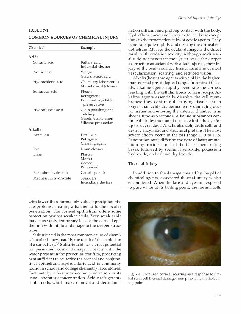
Sulfuric acid is the most common cause of chemi-cal ocular injury, usually the result of the explosionof a car battery.7,8 Sulfuric acid has a great potentialfor permanent ocular damage; it reacts with thewater present in the preocular tear film, producingheat sufficient to cauterize the corneal and conjunc-tival epithelium. Hydrochloric acid is commonlyfound in school and college chemistry laboratories.Fortunately, it has poor ocular penetration in itsusual laboratory concentration. Acidic refrigerantscontain oils, which make removal and decontami-
nation difficult and prolong contact with the body.Hydrofluoric acid and heavy metal acids are excep-tions to the penetration rules of acidic agents. Theypenetrate quite rapidly and destroy the corneal en-dothelium. Most of the ocular damage is the directresult of fluoride ion toxicity. Although acids usu-ally do not penetrate the eye to cause the deeperdestruction associated with alkali injuries, their in-jury of the ocular surface tissues results in cornealvascularization, scarring, and reduced vision.
Alkalis (bases) are agents with a pH in the higher-than-normal physiological range. In contrast to ac-ids, alkaline agents rapidly penetrate the cornea,reacting with the cellular lipids to form soaps. Al-kaline agents essentially dissolve the cell mem-branes; they continue destroying tissues muchlonger than acids do, permanently damaging ocu-lar tissues and entering the anterior chamber in asshort a time as 5 seconds. Alkaline substances con-tinue their destruction of tissues within the eye forup to several days. Alkalis also dehydrate cells anddestroy enzymatic and structural proteins. The mostsevere effects occur in the pH range 11.0 to 11.5.Penetration rates differ by the type of base; ammo-nium hydroxide is one of the fastest penetratingbases, followed by sodium hydroxide, potassiumhydroxide, and calcium hydroxide.
Thermal Injury
In addition to the damage created by the pH ofchemical agents, associated thermal injury is alsoencountered. When the face and eyes are exposedto pure water at its boiling point, the normal cells
Fig. 7-1. Localized corneal scarring as a response to lim-bal stem cell thermal damage from pure water at the boil-ing point.
118
Ophthalmic Care of the Combat Casualty
that populate the cornea (limbal stem cells) are de-stroyed, resulting in altered corneal surface heal-ing. Scarring and opacification ensue (Figure 7-1).Chemical munitions are often delivered at a hightemperature generated by explosions, or they may
generate heat due to exothermic chemical reactions.For these reasons, the extent of injury from a chemi-cal agent may have additional thermal damage notevident when the casualty presents to medical per-sonnel.8
OCULAR RESPONSE TO CHEMICAL INJURY
Each ocular structure responds uniquely to achemical insult. The conjunctival tissues are cauter-ized. The corneal epithelium sloughs; the cornealstroma swells and opacifies; endothelial cells die andare replaced by neighboring cells that stretch to coverthe resultant empty space. The angle structures scar,resulting in increased intraocular pressure (IOP). Cata-racts form as a result of insult to the lens.9–12
Weak acids and alkalis in the eye cause similar in-jurious effects, including injection, chemosis, mildcorneal clouding, and edema with minimal visibleinflammation. In severe acid burns, however, the cor-nea and conjunctiva rapidly turn white and opaque.Nitric and chromic acids turn tissue yellow-brown.
An initially deepithelialized cornea with clear stromamay belie the severity of the burn. The most severeacid burns produce corneal anesthesia, limbal pallor,and uveitis. Severe alkali burns can result in cornealmelting and perforation within 2 to 4 weeks.13
The extent of injury to the limbal area is critical indetermining the severity and prognosis of chemicalburns. The ocular reparative response to chem-ical injuries involves reepithelialization and vascular-ization. If the perilimbal blood supply is damaged,sterile necrosis of the peripheral cornea canensue. Injury to deep structures at the limbus candestroy the normal source (stem cells) for reepithe-lialization.
CLINICAL COURSE OF CHEMICAL OCULAR INJURY
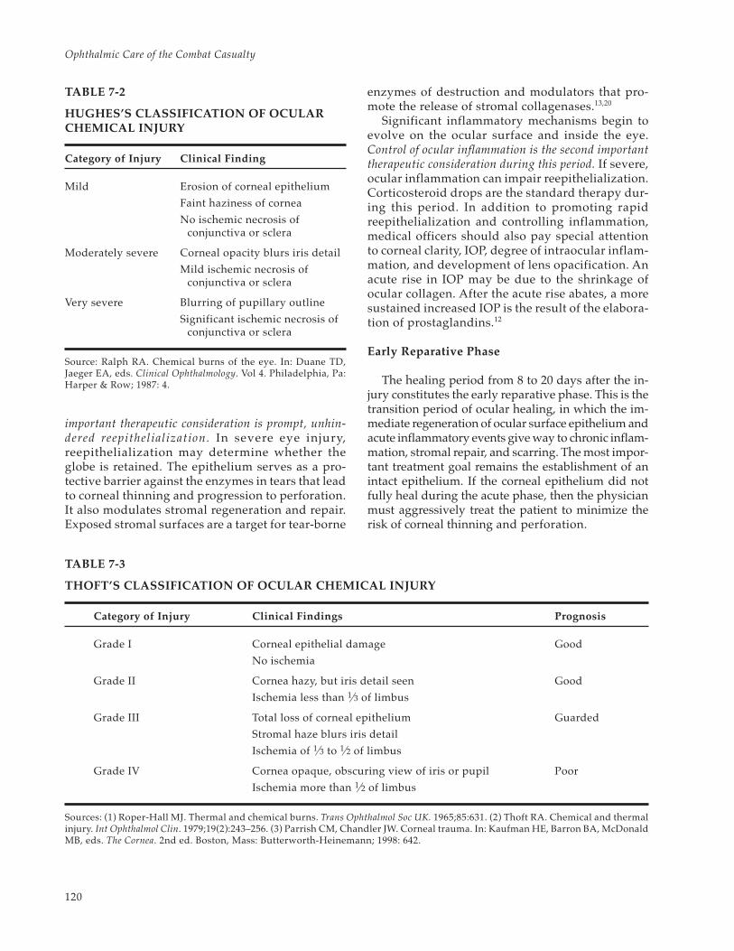
According to McCulley,14 the clinical course ofocular chemical injury can be divided into the fol-lowing four phases: immediate, acute, early repara-tive, and late reparative.
Immediate Phase
The immediate phase begins the moment achemical agent comes in contact with the ocularsurface. The major determinants of prognosis arebased on the initial clinical examination, althoughpredictors of ocular recovery after chemical injuryhave proven to be more accurate if the evaluationis made 24 to 48 hours after the injury. Animal mod-els demonstrate that the pH of the involved sub-stance, its concentration, and the length of time thesubstance is in contact with the tissue are the majordeterminants of the depth of penetration and dam-age to deeper ocular structures.15
Because these factors are often unknown to thepresenting physician and may not be recoverablefrom the patient’s history, classification schemesand prognostic data have been based on examina-tion findings. The clinical utility of classificationschemes is to better predict which patients will re-spond to conventional medical therapy and whichwill require extensive treatment or are at risk forloss of the eye. In a military scenario, this classifi-cation of chemical injury is useful during triage for
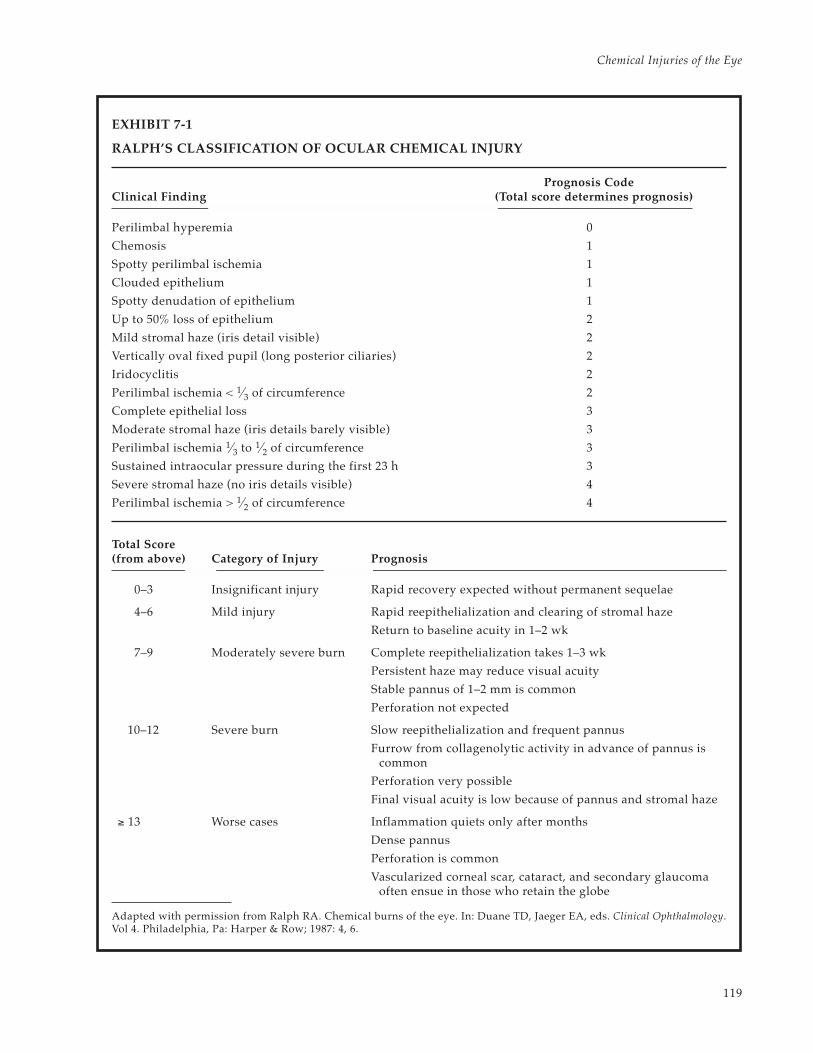
assigning patients for therapy on-site versus imme-diate evacuation. The most common classificationschemes of ocular chemical injury are those byRalph (Exhibit 7-1),16 Hughes (Table 7-2),17,18 and Thoft(Table 7-3).19 The key elements for determining theextent of chemical ocular injury and prognosis are
• the total area of the corneal epithelial defect;• the area of the conjunctival epithelial defect;• the number of clock hours or degrees of lim-
bal blanching (ischemia);• the area and degree of density of corneal
opacification;• evidence of increased IOP on presentation,
especially if resistant to treatment; and• any loss of lens clarity.
The last two elements imply deeper effects of thechemical agent and damage to the inner ocularstructures.
Acute Phase
The first 7 days after chemical eye injury consti-tute the acute phase of recovery. During this time,the tissues rid themselves of contaminants whilereestablishing the superficial protective layer of thecorneal epithelium. Reepithelialization is the most cru-cial factor in ultimate visual recovery; therefore, the first
Sustained intraocular pressure during the first 23 h 3
Severe stromal haze (no iris details visible) 4
Perilimbal ischemia > 1⁄2 of circumference 4
Total Score(from above) Category of Injury Prognosis
0–3 Insignificant injury Rapid recovery expected without permanent sequelae
4–6 Mild injury Rapid reepithelialization and clearing of stromal haze
Return to baseline acuity in 1–2 wk
7–9 Moderately severe burn Complete reepithelialization takes 1–3 wk
Persistent haze may reduce visual acuity
Stable pannus of 1–2 mm is common
Perforation not expected
10–12 Severe burn Slow reepithelialization and frequent pannus
Furrow from collagenolytic activity in advance of pannus iscommon
Perforation very possible
Final visual acuity is low because of pannus and stromal haze
≥ 13 Worse cases Inflammation quiets only after months
Dense pannus
Perforation is common
Vascularized corneal scar, cataract, and secondary glaucomaoften ensue in those who retain the globe
Adapted with permission from Ralph RA. Chemical burns of the eye. In: Duane TD, Jaeger EA, eds. Clinical Ophthalmology.Vol 4. Philadelphia, Pa: Harper & Row; 1987: 4, 6.
120
Ophthalmic Care of the Combat Casualty
TABLE 7-3
THOFT’S CLASSIFICATION OF OCULAR CHEMICAL INJURY
Category of Injury Clinical Findings Prognosis
Grade I Corneal epithelial damage Good
No ischemia
Grade II Cornea hazy, but iris detail seen Good
Ischemia less than 1⁄3 of limbus
Grade III Total loss of corneal epithelium Guarded
Stromal haze blurs iris detail
Ischemia of 1⁄3 to 1⁄2 of limbus
Grade IV Cornea opaque, obscuring view of iris or pupil Poor
Ischemia more than 1⁄2 of limbus
Sources: (1) Roper-Hall MJ. Thermal and chemical burns. Trans Ophthalmol Soc UK. 1965;85:631. (2) Thoft RA. Chemical and thermalinjury. Int Ophthalmol Clin. 1979;19(2):243–256. (3) Parrish CM, Chandler JW. Corneal trauma. In: Kaufman HE, Barron BA, McDonaldMB, eds. The Cornea. 2nd ed. Boston, Mass: Butterworth-Heinemann; 1998: 642.
TABLE 7-2
HUGHES’S CLASSIFICATION OF OCULARCHEMICAL INJURY
Category of Injury Clinical Finding
Mild Erosion of corneal epithelium
Faint haziness of cornea
No ischemic necrosis ofconjunctiva or sclera
Moderately severe Corneal opacity blurs iris detail
Mild ischemic necrosis ofconjunctiva or sclera
Very severe Blurring of pupillary outline
Significant ischemic necrosis ofconjunctiva or sclera
Source: Ralph RA. Chemical burns of the eye. In: Duane TD,Jaeger EA, eds. Clinical Ophthalmology. Vol 4. Philadelphia, Pa:Harper & Row; 1987: 4.
important therapeutic consideration is prompt, unhin-dered reepithelialization. In severe eye injury,reepithelialization may determine whether theglobe is retained. The epithelium serves as a pro-tective barrier against the enzymes in tears that leadto corneal thinning and progression to perforation.It also modulates stromal regeneration and repair.Exposed stromal surfaces are a target for tear-borne
enzymes of destruction and modulators that pro-mote the release of stromal collagenases.13,20
Significant inflammatory mechanisms begin toevolve on the ocular surface and inside the eye.Control of ocular inflammation is the second importanttherapeutic consideration during this period. If severe,ocular inflammation can impair reepithelialization.Corticosteroid drops are the standard therapy dur-ing this period. In addition to promoting rapidreepithelialization and controlling inflammation,medical officers should also pay special attentionto corneal clarity, IOP, degree of intraocular inflam-mation, and development of lens opacification. Anacute rise in IOP may be due to the shrinkage ofocular collagen. After the acute rise abates, a moresustained increased IOP is the result of the elabora-tion of prostaglandins.12
Early Reparative Phase
The healing period from 8 to 20 days after the in-jury constitutes the early reparative phase. This is thetransition period of ocular healing, in which the im-mediate regeneration of ocular surface epithelium andacute inflammatory events give way to chronic inflam-mation, stromal repair, and scarring. The most impor-tant treatment goal remains the establishment of anintact epithelium. If the corneal epithelium did notfully heal during the acute phase, then the physicianmust aggressively treat the patient to minimize therisk of corneal thinning and perforation.
121
Chemical Injuries of the Eye
Ocular inflammation must also be controlled dur-ing this stage because inflammation can continue toinhibit epithelial migration over the corneal defects.21
High-dose corticosteroids are usually required for thefirst 10 days of treatment, tapering at 14 days if anepithelial defect persists. The use of corticosteroidsfor more than 21 days in an eye without an intact epi-thelium risks collagenolysis and perforation.22
Late Reparative Phase
Three weeks after a chemical injury occurs, the
healing process begins the late reparative phase.Application of ocular lubricants and tear substitutesmust be continued to ensure a healthy epithelium.Chemical agents can cause loss of corneal sensation,decreasing the blink reflex and reducing the pro-duction of tears. Destruction of the associatedmucin and lipid-producing cells also leads to aninadequate corneal tear film. Severe injury canlead to pannus formation during this time. Persis-tent corneal epithelial defects or recurring epithe-lial breakdown can be surgically managed bytarsorrhaphy.
PATIENT TREATMENT AND EVALUATION
Immediate Action
At first glance, this section appears to be errone-ously titled. However, in any known or even suspectedchemical injury of the eye, immediate treatment withirrigation precedes patient evaluation. Prompt recog-nition and immediate treatment of a chemical injuryare the most important aspects in the preservation ofvision because only then can reepithelialization beoptimized. In a wartime scenario, maintaining a highdegree of suspicion that a chemical attack is immi-nent or has occurred is also important to ensure thatproper protective measures are taken and that medi-cal resources are preserved. Observing isolation anddecontamination regimens and recognizing the typeof agent involved may support intelligence gather-ing in the combat zone and preserve the lives of theevacuation and medical teams.
Immediately on suspecting a chemical injury,medical personnel should begin treatment. Themost readily available nontoxic liquid is used toflush the area of the face, eyes, and any other areasof contact. Tap water is usually the most readilyavailable, but using iced tea, milk, or any neutralliquid is better than delaying treatment. Researchindicates that attempts to chemically neutralize theoriginal agent (eg, using a dilute acidic solutionsuch as vinegar to neutralize an alkali injury) arecontraindicated because they may cause even moredamage. The exception is the use of prepared neu-tralization kits, specific for the known chemicalagent encountered.
Medical treatment begins by irrigating with 2 Lof normal saline or lactated Ringer’s solution over20 to 30 minutes. Because of its flexibility, an intra-venous line attached to the bottle is a useful meansof delivering the irrigation fluid. These injuries arepainful and result in blepharospasm and squeez-
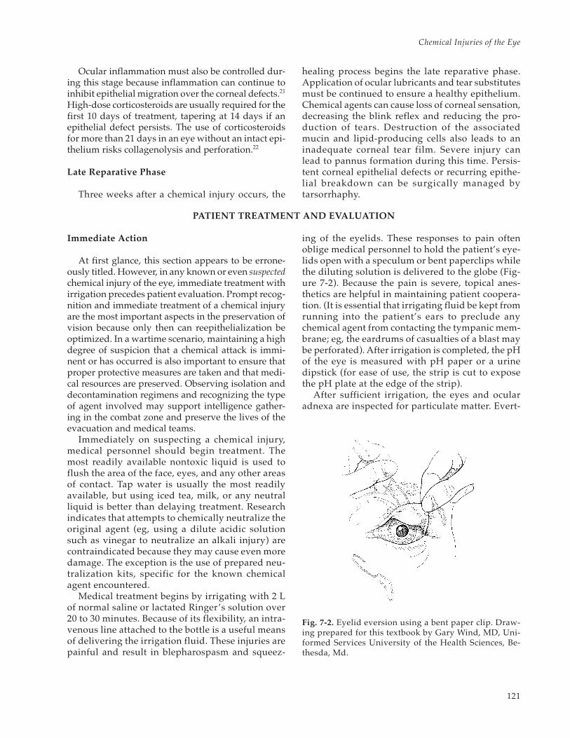
ing of the eyelids. These responses to pain oftenoblige medical personnel to hold the patient’s eye-lids open with a speculum or bent paperclips whilethe diluting solution is delivered to the globe (Fig-ure 7-2). Because the pain is severe, topical anes-thetics are helpful in maintaining patient coopera-tion. (It is essential that irrigating fluid be kept fromrunning into the patient’s ears to preclude anychemical agent from contacting the tympanic mem-brane; eg, the eardrums of casualties of a blast maybe perforated). After irrigation is completed, the pHof the eye is measured with pH paper or a urinedipstick (for ease of use, the strip is cut to exposethe pH plate at the edge of the strip).
After sufficient irrigation, the eyes and ocularadnexa are inspected for particulate matter. Evert-
Fig. 7-2. Eyelid eversion using a bent paper clip. Draw-ing prepared for this textbook by Gary Wind, MD, Uni-formed Services University of the Health Sciences, Be-thesda, Md.
122
Ophthalmic Care of the Combat Casualty
ing both the upper and lower eyelids is mandatoryto search for retained particles trapped in the con-junctival fornix or embedded in the tissues them-selves. Five minutes after the end of irrigation, thepH is again measured. If the pH is nearly normal,irrigation is temporarily suspended, particulatematter removed, and the examination portionbegun. If the pH is not normal, irrigation is againperformed with an additional 2 L of fluid. If the ob-served pH does not match the initial incident his-tory, the patient and others should be questionedagain.
Patient Examination
Once a normal pH level is attained and thepatient’s eye remains neutralized, a full ophthal-mological examination is performed. Visual acuitymeasurements are taken for each eye independently.If a Snellen chart is not available, recording the abil-ity of the patient to read newspaper headlines of“x” inches in height at a distance of “y” feet awayis helpful.
Pupil examination is performed, noting any ir-regular shape or sluggish response to light. Anirregular response may indicate iris ischemia dueto chemical coagulation of the blood vessels in theiris or in the ciliary vessels. The external examina-tion consists of the facial skin, eyelids, lashes, andlacrimal apparatus. Look for areas of lid burn,which can cause incomplete globe coverage, and forresidual chemical particulate matter.
Ask the patient to look at an object straight inthe line of view as the examiner looks at the eyelidposition. Then the examiner should talk to the pa-tient about other subjects while observing for amore relaxed lid position as well as blink reflex ex-cursion and frequency. The patient is then asked tolook up and down while the examiner observes theupper and lower eyelid position over the globeduring the motion. Finally, the patient is asked tosqueeze his or her eyelids tightly and then relax. Itis also important for the physician and the assist-ing staff to observe the eyelid position and expo-sure of the globe while the patient is sleeping. Cor-neal or conjunctival exposure can lead to epithelialbreakdown, infection, and corneal melting, and isa risk factor for loss of the eye.
Attention is then directed to the globe itself. Apenlight, or preferably a slitlamp, examination isperformed to detect epithelial loss, corneal opacifi-cation, and limbal ischemia. First, shine a light ontothe corneal surface and observe the luster of theepithelium. Injured epithelial cells lack their typi-
cal reflective luster, resulting in an irregular corneallight reflex from the ocular surface. Note any grayor white areas of stromal opacification by observ-ing whether iris detail or pupillary border is ap-parent when looking through the cornea. Limbalischemia is measured by the number of clock hoursof blood vessel loss of the conjunctival tissues whereit nears the peripheral edge of the cornea.
Once the previous examination details are noted,a fluorescein strip may be wetted and placed on theocular surface to delineate the extent of cornealepithelial loss (see also Chapter 3, Ocular Trauma:History and Examination). IOP is measured withone of the many instruments for this purpose orsimply by comparing the tactile IOP of an intactglobe by gently placing the fingertips on the closedeyelids. To facilitate evaluation of the posterior seg-ment of the eye, eye drops (tropicamide or cyclo-pentolate) are instilled to achieve pupillary dilation.Cycloplegic eye drops also help control pain by ef-fectively “splinting” pupillary reaction and reduc-ing the pain associated with pupil constriction inbright light. Pupil dilatory drops also enhance theoutflow of aqueous from the eye, helping to con-trol IOP. The use of phenylephrine is not recom-mended, because its vasoconstrictive propertiesmay lead to an increased risk of ocular ischemia.
A summary of evaluation and treatment is pro-vided for rapid reference (Exhibit 7-2).
Acute Phase Treatment
Once the emergency treatment and evaluationare completed, the challenging task of healing thechemically injured eye begins. The major treatmentgoals that are important throughout the healingphases are (a) the reestablishment and maintenanceof an intact and healthy corneal epithelium, (b) con-trol of the balance between collagen synthesis andcollagenolysis, and (c) minimizing the adverse se-quelae that often follow a chemical injury. This triadof care for casualties with ocular chemical injuriestakes place in the acute (urgent) phase of treatment.
The top priority in the acute phase is the rees-tablishment of an intact corneal epithelium. With-out an intact epithelium, the risks of corneal thin-ning and perforation (melt), infection, and othercomplications that follow a chemical injury are sig-nificantly higher. The source for healthy epithelialcells is the rim of corneal epithelial stem cells thatlie near the limbus.23 A severe ocular chemical in-jury may permanently damage all stem cells. Thecorneal surface is therefore lacking in progenitorepithelial cells, and the surface is replaced with
123
Chemical Injuries of the Eye
EXHIBIT 7-2
CHEMICAL INJURY: EVALUATION AND TREATMENT SUMMARY
A. History
1. Suspected or known chemical contact
2. Possible wartime/lethal chemical agenta. Observe mission-oriented protective posture (MOPP) gear or self-protectionb. Sound chemical alarm
B. Initiate Treatment
1. Irrigationa. 1–2 L normal saline or lactated Ringer’s solution (30 min)b. Intravenous tubingc. Speculumd. Topical anesthetic
3. Indicator test: test pH at end of irrigation and 5 min after completion of irrigationa. If pH = 7: Stop irrigation
Begin examination (see below)Recheck pH after 20 more min elapseDebride devitalized tissueInitiate medical therapy
b. If pH < 7 or > 7: Restart irrigation with another 2 Lc. If pH matches history (eg, low pH for acid injury), continue therapyd. If pH does not match history, obtain more details of injury while continuing treatment
4. Examinationa. Visual acuity
DISTANCE: Right LeftNEAR: Right Left
b. PupilsSHAPE: Round/IrregularREACTION: Fast/Sluggish
c. External examination(1) Facial skin(2) Eyelid skin(3) Lashes
(a) Loss(b) Eversion(c) Inversion
d. Slitlamp or pen light examination(1) Conjunctiva
(a) Number of clock hours of limbal ischemia(b) Areas of fluorescein staining
(iii) OpacityHaze, but iris detail visibleHaze blurs iris detailHaze obscures view of iris or pupil
(c) Descemet’s: Folds(3) Anterior chamber
(a) Depth(b) Foreign body(c) Red blood cells layered(d) White blood cells layered(e) Inflammation
C. Additional Information
1. Intraocular pressure: Low Normal High*
2. Lens: Clear Clouded*
*Elevated intraocular pressure or loss of lens clarity suggests significant intraocular penetration
Exhibit 7-2 continued
slower-growing conjunctival epithelial cells andfibrosed and vascularized tissue.24 In less-severeinjury, corneal epithelial stem cells that survive achemical injury act as progenitor cells for epithe-lial cell division and subsequent migration of thecells to cover the epithelial surface.25 Promotion ofa healthy microenvironment for these processes isthe mainstay of therapy.
The preocular tear film is normally rich in mois-ture, lipids, mucus, and mineral cofactors. Treat-ment is usually aimed at replacing the aqueousportion of the tear film with preservative-freeartificial tears, ointments, and antibiotics that arerelatively nontoxic to the epithelium. If the eye issignificantly damaged or eyelid closure is not suf-ficient to maintain a healthy tear surface, preven-tion of tear evaporation is aided by eyelid closurewith a patch, eyelid taping, or tarsorrhaphy. Again,control of inflammation is important because sig-nificant inflammation immediately after a chemi-cal injury inhibits reepithelialization. Corticoster-oids should be used if there is no evidence of coex-isting infection.
The second priority in the acute phase of heal-ing after a chemical injury is maintaining a posi-tive balance between collagen synthesis and colla-
genase activity. This balance is necessary for achiev-ing the removal of damaged collagen tissue whilerebuilding the stroma proper. Recent studies ofchemical eye injuries26–28 have indicated the impor-tance of the stroma and epithelial interactions in themodulation of corneal wound healing, and thus theachievement of an intact epithelium is vital. Thesesame studies have also shown that treatment withcitrate and ascorbate eye drops with oral supple-mentation of ascorbate (vitamin C) can enhancecollagen synthesis.
Limiting adverse sequelae is the third goal in thetriad of care for the ocular chemical injury. Reduc-ing the risk of infection is accomplished with anantibiotic that presents little toxicity to the epithe-lial cells. Control of IOP by suppression of aqueousproduction is often effective. Oral and intravenousmedication may be preferred in the acute setting tominimize the amount of drops administered to theeye. Control of pain should never be taken lightly,as chemical injuries are often severely painful.Long-acting cycloplegics help in pain management,because they prevent the repeated movement of thepupillary muscles. Again, oral, intramuscular, andintravenous pain control are preferred.
Limitation of conjunctival scarring can be one of
125
Chemical Injuries of the Eye
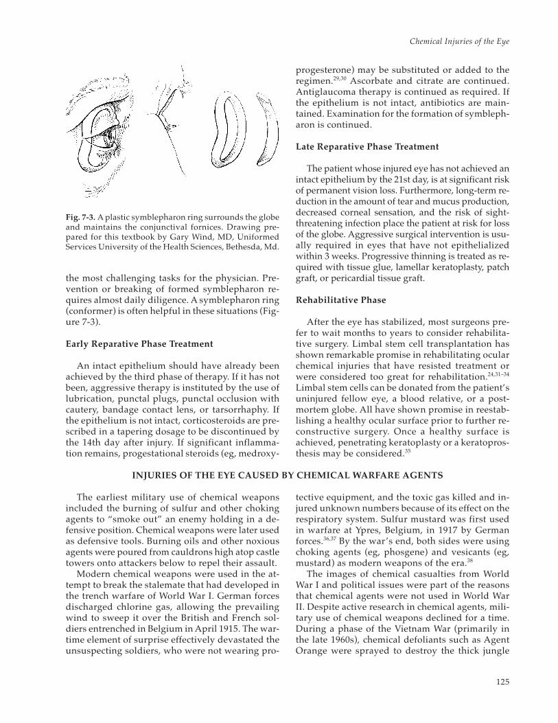
the most challenging tasks for the physician. Pre-vention or breaking of formed symblepharon re-quires almost daily diligence. A symblepharon ring(conformer) is often helpful in these situations (Fig-ure 7-3).
Early Reparative Phase Treatment
An intact epithelium should have already beenachieved by the third phase of therapy. If it has notbeen, aggressive therapy is instituted by the use oflubrication, punctal plugs, punctal occlusion withcautery, bandage contact lens, or tarsorrhaphy. Ifthe epithelium is not intact, corticosteroids are pre-scribed in a tapering dosage to be discontinued bythe 14th day after injury. If significant inflamma-tion remains, progestational steroids (eg, medroxy-
Fig. 7-3. A plastic symblepharon ring surrounds the globeand maintains the conjunctival fornices. Drawing pre-pared for this textbook by Gary Wind, MD, UniformedServices University of the Health Sciences, Bethesda, Md.
progesterone) may be substituted or added to theregimen.29,30 Ascorbate and citrate are continued.Antiglaucoma therapy is continued as required. Ifthe epithelium is not intact, antibiotics are main-tained. Examination for the formation of symbleph-aron is continued.
Late Reparative Phase Treatment
The patient whose injured eye has not achieved anintact epithelium by the 21st day, is at significant riskof permanent vision loss. Furthermore, long-term re-duction in the amount of tear and mucus production,decreased corneal sensation, and the risk of sight-threatening infection place the patient at risk for lossof the globe. Aggressive surgical intervention is usu-ally required in eyes that have not epithelializedwithin 3 weeks. Progressive thinning is treated as re-quired with tissue glue, lamellar keratoplasty, patchgraft, or pericardial tissue graft.
Rehabilitative Phase
After the eye has stabilized, most surgeons pre-fer to wait months to years to consider rehabilita-tive surgery. Limbal stem cell transplantation hasshown remarkable promise in rehabilitating ocularchemical injuries that have resisted treatment orwere considered too great for rehabilitation.24,31–34
Limbal stem cells can be donated from the patient’suninjured fellow eye, a blood relative, or a post-mortem globe. All have shown promise in reestab-lishing a healthy ocular surface prior to further re-constructive surgery. Once a healthy surface isachieved, penetrating keratoplasty or a keratopros-thesis may be considered.35
INJURIES OF THE EYE CAUSED BY CHEMICAL WARFARE AGENTS
The earliest military use of chemical weaponsincluded the burning of sulfur and other chokingagents to “smoke out” an enemy holding in a de-fensive position. Chemical weapons were later usedas defensive tools. Burning oils and other noxiousagents were poured from cauldrons high atop castletowers onto attackers below to repel their assault.
Modern chemical weapons were used in the at-tempt to break the stalemate that had developed inthe trench warfare of World War I. German forcesdischarged chlorine gas, allowing the prevailingwind to sweep it over the British and French sol-diers entrenched in Belgium in April 1915. The war-time element of surprise effectively devastated theunsuspecting soldiers, who were not wearing pro-
tective equipment, and the toxic gas killed and in-jured unknown numbers because of its effect on therespiratory system. Sulfur mustard was first usedin warfare at Ypres, Belgium, in 1917 by Germanforces.36,37 By the war’s end, both sides were usingchoking agents (eg, phosgene) and vesicants (eg,mustard) as modern weapons of the era.38
The images of chemical casualties from WorldWar I and political issues were part of the reasonsthat chemical agents were not used in World WarII. Despite active research in chemical agents, mili-tary use of chemical weapons declined for a time.During a phase of the Vietnam War (primarily inthe late 1960s), chemical defoliants such as AgentOrange were sprayed to destroy the thick jungle
126
Ophthalmic Care of the Combat Casualty
plants and thereby deny the enemy concealment.Whether antipersonnel chemical agents were usedduring the Vietnam War continues to be debated.
A sharp rise has been seen in the distribution and
a b
cFig. 7-4. (a) Although the vesicular effects of mustardagent on the skin of the casualty’s back are impressive(the casualty’s head is seen in the upper right of this pho-tograph), (b) the eyes are significantly more sensitive tomustard’s effects; the eye involvement seen here is rela-tively severe even though the skin is only minimally af-fected. (c) In 1918, the British prepared for the AmericanExpeditionary Force a series of color drawings and de-scriptions of injuries by chemical warfare agents. Thisdrawing depicts a severely burned eye in the acute stageafter exposure to mustard vapor. A portion of the origi-nal description follows:
[Severely burned eyes] may be recognized by certaincharacteristic features … . Whenever a dead whiteband crosses the exposed area of the conjunctiva,while the parts of this membrane covered by the up-per and lower lids are red and oedematous, seriousinjury from the burning is likely to have occurred.
In the case illustrated, the caustic effect of the vapour is seen chiefly in the interpalpebral aperture. On eachside of the cornea there is a dead white band due to coagulative oedema, which compresses the vessels, impairsthe circulation, and thus acts as a menace to the nutrition of the cornea. The swelling in the region of this whiteband is slight, while the protected conjunctiva above and below it is greatly swollen and injected and may evenbulge between the lids.
The exposed portion of the cornea is grey and hazy; it has lost its lustre, and when viewed with a bright lightand a magnifying glass it shows a blurred “window reflex” and a typical “orange skinned” surface. The hazegradually faces off above in the region of the protected part of the cornea where the surface is bright andsmooth. The pupil is at first contracted as a result of irritation and congestion. In this drawing it is shown asartificially dilated by atropine ointment, which should always be used early in severe cases or where there ismuch pain and blepharospasm.
Photograph b: Courtesy of Dr Luis Requena, Universidad Autónoma de Madrid, Spain. Reproduced from BennionSD, David-Jabar K. Cutaneous reactions to nuclear, biological, and chemical warfare. In: James WD, ed. MilitaryDermatology. In: Zajtchuk R, Bellamy RF, eds. Textbook of Military Medicine. Washington, DC: Department of the Army,Office of The Surgeon General, Borden Institute; 1994: 95. Drawing c: Reproduced from An Atlas of Gas Poisoning.1918: Plate 11A. Handout provided by the American Red Cross to the American Expeditionary Force. In: Joy RJT.Historical aspects of medical defense against chemical warfare. In: Sidell FR, Takafuji ET, Franz DR, eds. MedicalAspects of Chemical and Biological Warfare. In: Zajtchuk R, Bellamy RF, eds. Textbook of Military Medicine. Washington,DC: Department of the Army, Office of The Surgeon General, Borden Institute, 1997: 99.
use of chemical weapons in events around theworld. Chemical injuries have increased both onand off the battlefield in recent decades. For ex-ample, as many as 45,000 casualties may have oc-
127
Chemical Injuries of the Eye
curred when Iraq employed mustard agent duringits war (1982–1988) with Iran39,40; Iraq also deployedvesicants in its suppression of the 1988 rebellion inKurdistan (Figure 7-4).
Military-strength chemical injuries have also oc-curred outside the classically defined boundariesof warfare. In the United States, highly toxic agents,both industrial and weapons grade, are commonlytransported throughout the country. A significantindustrial toxic chemical spill from a tanker truckin the Washington, DC, area required almost 24hours for HAZMAT (hazardous material) team op-erations to completely decontaminate a major high-way and the surrounding residential area.
In addition to the immediate effects, chemicalinjuries produce long-term problems with reducedvision, as well as employment and rehabilitationissues.41 One patient who was exposed to mustardagent during the 1988 attack in Iraq presented 10years later with delayed mustard gas keratopathy.42
This and the recent examples above are just a fewof the readiness issues for chemical injury that the
military physician must be prepared to identify andtreat whether in combat or everyday life.
Chemical munitions currently available includeblister agents (vesicants), nerve agents, irritants,and blood agents (Tables 7-4 and 7-5). These agentscan affect different organ systems, producing tem-porary incapacitation, temporary illness, permanentdisability, or even death. The eyes can be very sen-sitive to many chemical agents (Table 7-6); perma-nent damage to the eye with loss of vision may oc-cur.
Blister Agents
The major agent in the vesicant, or blister agent,class is sulfur mustard. During World War I, therewere as many as 400,000 chemical casualties, butfewer than 3% died of their chemical wounds.43 Ofthe casualties of mustard agent, 86% had ocular in-volvement, and many had skin involvement, espe-cially in warm, moist areas such as the scrotum,buttocks, axillae, neck, face, and areas that were
TABLE 7-4
CHEMICAL AGENTS
US Army Code Name of Agent
Vesicants
H Sulfur mustard, munitions grade (30% impurities)
*Although HN has previously been used only as a chemotherapeutic agent, it might be used as a weapon in the future.
TABLE 7-6
OCULAR EFFECTS OF CATEGORIES OF CHEMICAL WARFARE AGENTS
Category of Agent Ocular Effects Category of Agent Ocular Effects
Vesicants(Blister Agents)
Nerve Agents
Mycotoxins
Conjunctivitis
Irritation blepharospasm
Photophobia
Corneal clouding andvascularization
Inflammation
Symblepharon
Lid burns
Corneal perforation
Miosis
Pain
Dimming of vision
Ocular pain
Tearing
Pain/burning
Decreased vision
Conjunctivitis
Keratitis
Riot Control Agents
Phosgene
Cyanide
Burning/irritation
Conjunctival injection/conjunctivitis
Lacrimation
Blepharospasm
Photophobia
Keratitis
Pain
Keratitis
Conjunctivitis
Irritation
Difficulty focusing (latemydriasis)
129
Chemical Injuries of the Eye
constricted by clothing, such as the waist.43 The Brit-ish reported many thousands of eye casualties, with75% mildly affected (2 wk on average before casu-alties were returned to duty); 15% were intermedi-ate (incapacitated, 4–6 wk); and 10% were severe(their ocular injuries remained active for 4–6 mobefore stabilizing). Fifty-one soldiers were blinded,and 180 were given vision-related pensions.43 Onthe other hand, the Americans reported 1,500 chemi-cal casualties, with 15% recovering in 10 to 14 daysand 80% recovering in 5 to 8 weeks. They also re-ported cases of panophthalmitis.44,45
The apparent discrepancy between the Britishand American experience in World War I is hard toexplain. The important fact remains that most pa-tients recovered without significant damage butwere incapacitated for a long time. The symptomsof photophobia, grittiness, pain, and blepharospasmeffectively immobilized those affected. Keep in mindthat 10% to 20% of those evacuated from the frontduring World War I for ocular injuries were sufferinga combat reaction (gas hysteria) and either did notsustain a physical injury or demonstrated symp-toms far in excess of their injuries. Today, our troopsare better trained and equipped to deal with thechemical threat, and medical therapy for these typesof exposures has been improved.
Between 75% and 90% of vesicant casualties canbe anticipated to have ocular involvement,44,46 withsymptoms usually peaking 6 to 12 hours after ex-posure. Of these, 90% should have no significantcorneal involvement.47,48 They may present with agritty sensation, conjunctivitis, chemosis, lid edema,blepharospasm, photophobia, blurred vision, tear-ing, and exudates.39,48 The 10% with corneal involve-ment may additionally demonstrate corneal edema,keratitis, ocular pain or headache, temporary blind-ness, tissue necrosis, iridocyclitis, glaucoma, vas-cularization, delayed keratopathy, and rarely ulcer-ation or perforation.39,49–52
Mechanisms of Injury
The vesicants are alkylating agents that have apronounced intracellular effect, especially on rep-lication of deoxyribonucleic acid (DNA).53 Irrevers-ible histological changes occur within 10 minutes,and these changes become pronounced at 30 min-utes. Additionally, the arsenical Lewisite liberateshydrochloric acid. The acid lowers the pH of theeye to 1.3, which causes superficial opacities, butarsine oxide is its main toxin.
Lewisite, developed in the United States at theend of World War I but never used, was neverthe-
less extensively studied in World War II, revealingthe following facts50,54:
• the corneal surface is free of toxins within2 to 4 minutes,
• toxins are in the stroma within 2 minutes,• toxins are in the anterior chamber within
1.5 minutes,• the anterior chamber is free of toxins within
30 minutes, and• some stromal toxins are present for 1 to 26
hours.
Research with mustard agent shows similar pen-etration, with the eye being free of toxins within 15minutes.50 The delay in symptoms with mustardthus makes timely decontamination difficult. Earlydetection and prevention of injury become critical.
Signs and Symptoms
Exposure to minute quantities (0.001 mg/L) ofmustard agent for periods up to 1 hour does notaffect the skin or the respiratory tract significantly.Yet, within 4 to 12 hours, lacrimation occurs, and asandy sensation in the eyes becomes manifest. Theconjunctivae and lids become swollen and edema-tous. Exposure to increased concentrations short-ens the latent period, causes more damage withcorneal involvement, and prolongs recovery to 2 to6 weeks. Hot, humid weather increases the rapid-ity of action but also decreases the persistence ofthe agent.
The skin shows erythema similar to a sunburnafter a latent period, and then large, thin-walledbullae usually form. Irritating these affected areas(eg, by scrubbing during decontamination), canpromote vesicle formation in casualties who mightnot have developed them otherwise. The fluid inthese vesicles is not contaminated and may safelybe drained when necessary. These lesions behavemuch like second-degree burns and heal in severalweeks, depending on the area affected.
Respiratory tract effects begin with hoarsenessand a persistent cough that can progress to bron-chopneumonia; these effects usually do not reachmaximum severity for several days. This delayshould prompt us to carefully observe those withocular or facial burns for subsequent signs of pul-monary damage.
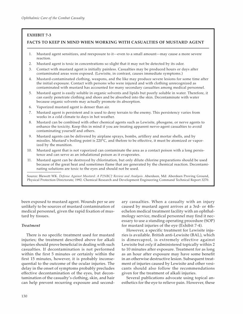
Working with casualties of mustard agent putsmedical personnel at risk. Exhibit 7-3 lists someproperties of mustard that healthcare personnelmust keep in mind while aiding casualties who have
130
Ophthalmic Care of the Combat Casualty
been exposed to mustard agent. Wounds per se areunlikely to be sources of mustard contamination ofmedical personnel, given the rapid fixation of mus-tard by tissues.
Treatment
There is no specific treatment used for mustardinjuries; the treatment described above for alkaliinjuries should prove beneficial in dealing with suchcasualties. If decontamination is not performedwithin the first 5 minutes or certainly within thefirst 15 minutes, however, it is probably inconse-quential to the outcome of the ocular injuries. Thedelay in the onset of symptoms probably precludeseffective decontamination of the eyes, but decon-tamination of the casualty’s clothing, skin, and haircan help prevent recurring exposure and second-
EXHIBIT 7-3
FACTS TO KEEP IN MIND WHEN WORKING WITH CASUALTIES OF MUSTARD AGENT
1. Mustard agent sensitizes, and reexposure to it—even to a small amount—may cause a more severereaction.
2. Mustard agent is toxic in concentrations so slight that it may not be detected by its odor.
3. Contact with mustard agent is initially painless. Casualties may be produced hours or days aftercontaminated areas were exposed. (Lewisite, in contrast, causes immediate symptoms.)
4. Mustard-contaminated clothing, weapons, and the like may produce severe lesions for some time afterthe initial exposure. Contact with persons who were injured and with clothing unrecognized ascontaminated with mustard has accounted for many secondary casualties among medical personnel.
5. Mustard agent is easily soluble in organic solvents and lipids but poorly soluble in water. Therefore, itcan easily penetrate clothing and shoes and be absorbed into the skin. Decontaminate with waterbecause organic solvents may actually promote its absorption.
6. Vaporized mustard agent is denser than air.
7. Mustard agent is persistent and is used to deny terrain to the enemy. This persistency varies fromweeks in a cold climate to days in hot weather.
8. Mustard can be combined with other chemical agents such as Lewisite, phosgene, or nerve agents toenhance the toxicity. Keep this in mind if you are treating apparent nerve-agent casualties to avoidcontaminating yourself and others.
9. Mustard agents can be delivered by airplane sprays, bombs, artillery and mortar shells, and bymissiles. Mustard’s boiling point is 220°C, and thefore to be effective, it must be atomized or vapor-ized by the munition.
10. Mustard agent that is not vaporized can contaminate the area as a contact poison with a long persis-tence and can serve as an inhalational poison as it evaporates.
11. Mustard agent can be destroyed by chlorination, but only dilute chlorine preparations should be usedbecause of the great heat and sometimes flame that are generated by the chemical reaction. Decontami-nating solutions are toxic to the eyes and should not be used.
Source: Blewett WK. Defense Against Mustard: A P2NBC2 Review and Analysis. Aberdeen, Md: Aberdeen Proving Ground,Physical Protection Directorate; 1992. Chemical Research and Development Engineering Command Technical Report 3270.
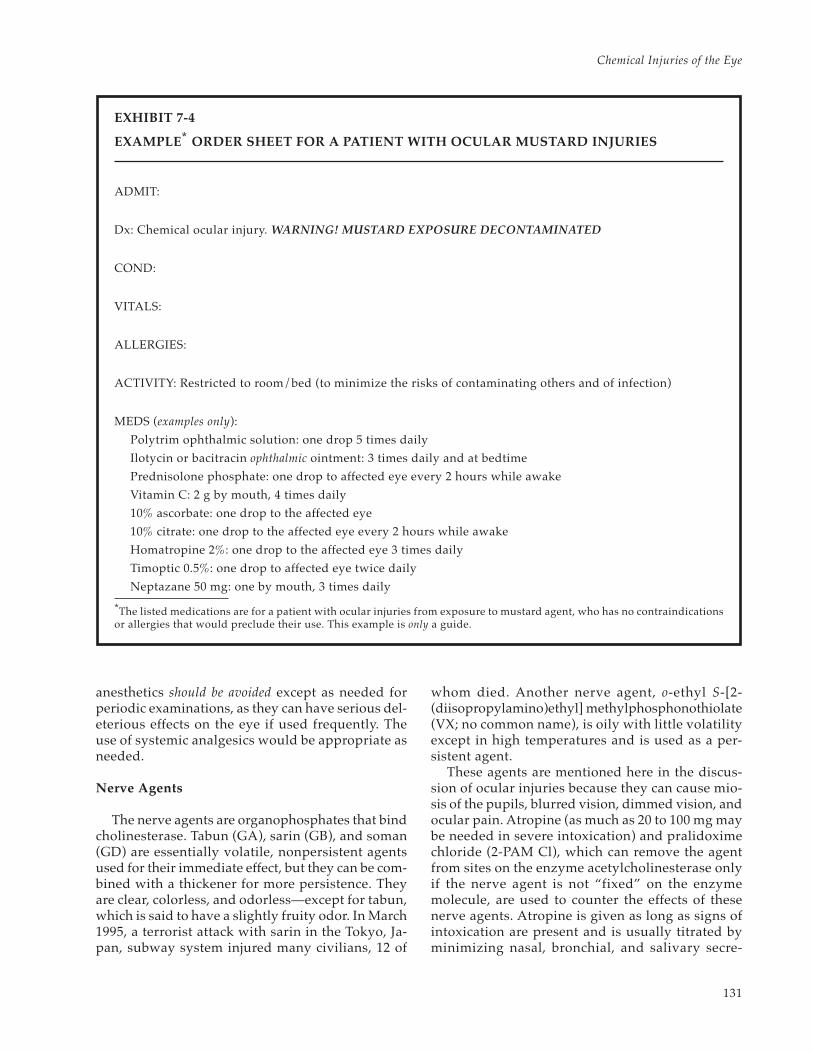
ary casualties. When a casualty with an injurycaused by mustard agent arrives at a 3rd- or 4th-echelon medical treatment facility with an ophthal-mology service, medical personnel may find it nec-essary to use a standing operating procedure (SOP)for mustard injuries of the eye (Exhibit 7-4).
However, a specific treatment for Lewisite inju-ries is available. British anti-Lewisite (BAL), whichis dimercaprol, is extremely effective againstLewisite but only if administered topically within 2to 10 minutes after exposure. Treatment for as longas an hour after exposure may have some benefitin an otherwise destructive lesion. Subsequent treat-ment of injuries caused by Lewisite and other vesi-cants should also follow the recommendationsgiven for the treatment of alkali injuries.
Several publications advocate using topical an-esthetics for the eye to relieve pain. However, these
131
Chemical Injuries of the Eye
anesthetics should be avoided except as needed forperiodic examinations, as they can have serious del-eterious effects on the eye if used frequently. Theuse of systemic analgesics would be appropriate asneeded.
Nerve Agents
The nerve agents are organophosphates that bindcholinesterase. Tabun (GA), sarin (GB), and soman(GD) are essentially volatile, nonpersistent agentsused for their immediate effect, but they can be com-bined with a thickener for more persistence. Theyare clear, colorless, and odorless—except for tabun,which is said to have a slightly fruity odor. In March1995, a terrorist attack with sarin in the Tokyo, Ja-pan, subway system injured many civilians, 12 of
whom died. Another nerve agent, o-ethyl S-[2-(diisopropylamino)ethyl] methylphosphonothiolate(VX; no common name), is oily with little volatilityexcept in high temperatures and is used as a per-sistent agent.
These agents are mentioned here in the discus-sion of ocular injuries because they can cause mio-sis of the pupils, blurred vision, dimmed vision, andocular pain. Atropine (as much as 20 to 100 mg maybe needed in severe intoxication) and pralidoximechloride (2-PAM Cl), which can remove the agentfrom sites on the enzyme acetylcholinesterase onlyif the nerve agent is not “fixed” on the enzymemolecule, are used to counter the effects of thesenerve agents. Atropine is given as long as signs ofintoxication are present and is usually titrated byminimizing nasal, bronchial, and salivary secre-
EXHIBIT 7-4
EXAMPLE* ORDER SHEET FOR A PATIENT WITH OCULAR MUSTARD INJURIES
ADMIT:
Dx: Chemical ocular injury. WARNING! MUSTARD EXPOSURE DECONTAMINATED
COND:
VITALS:
ALLERGIES:
ACTIVITY: Restricted to room/bed (to minimize the risks of contaminating others and of infection)
MEDS (examples only):
Polytrim ophthalmic solution: one drop 5 times daily
Ilotycin or bacitracin ophthalmic ointment: 3 times daily and at bedtime
Prednisolone phosphate: one drop to affected eye every 2 hours while awake
Vitamin C: 2 g by mouth, 4 times daily
10% ascorbate: one drop to the affected eye
10% citrate: one drop to the affected eye every 2 hours while awake
Homatropine 2%: one drop to the affected eye 3 times daily
Timoptic 0.5%: one drop to affected eye twice daily
Neptazane 50 mg: one by mouth, 3 times daily
*The listed medications are for a patient with ocular injuries from exposure to mustard agent, who has no contraindicationsor allergies that would preclude their use. This example is only a guide.
132
Ophthalmic Care of the Combat Casualty
tions. The miosis or pupil size should not be usedas an index of atropinization.
Mycotoxins
Trichothecene mycotoxins—the most memorableexample is the infamous “yellow rain” from the Viet-nam War era—are produced by certain strains of
Fusarium fungi. They seem to inhibit synthesis of pro-teins. Symptoms appear from 1 hour (pulmonaryroute) to 24 hours (cutaneous route) after exposureand include vomiting, weakness, hypotension, andburns in exposed areas including the cornea.55 Micro-gram quantities can cause irreversible corneal injury.Victims have complained of tearing, eye pain, con-junctivitis, a burning sensation, and blurred vision.56,57
SUMMARY
Despite advances in treatment for chemical in-jury, knowledge of risks and prevention remain thebest ways to avoid the often-long therapeutic coursefor recovery of vision. When a patient with a chemi-cal ocular injury presents to the treating physician,early recognition and prompt treatment remain thestandards to minimize ocular tissue damage andprovide hope for preservation of vision.
Military chemical agents are usually not part ofthe daily training environment of civilian physi-
cians, but chemical warfare agents remain a realthreat on the battlefield. Knowledge of battlefieldchemical munitions, their characteristics, and ex-pected treatment is important for patient care aswell as protection of medical personnel.
Chemical ocular injuries—whether they happenon the battlefield, in the military industrial base, orat home—will occur. When they do, knowledgeablephysicians can minimize tissue destruction andenhance healing throughout all phases of treatment.
REFERENCES
1. Bowen TE, Bellamy RF, eds. Emergency War Surgery NATO Handbook. 2nd rev US ed. Washington, DC: Depart-ment of Defense, Government Printing Office; 1988.
2. Deeter DP, Gaydos JC, eds. Occupational Health: The Soldier and the Industrial Base. In: Zajtchuk R, Bellamy RF,eds. Textbook of Military Medicine. Washington, DC: Department of the Army, Office of The Surgeon General,and Borden Institute; 1993.
3. James WD, ed. Military Dermatology. In: Zajtchuk R, Bellamy RF, eds. Textbook of Military Medicine. Washington,DC: Department of the Army, Office of The Surgeon General, and Borden Institute; 1994. Also available atwww.armymedicine.army.mil/history/borden/default.htm.
4. Sidell FR, Takafuji ET, Franz DR, eds. Medical Aspects of Chemical and Biological Warfare. In: Zajtchuk R, Bellamy RF,eds. Textbook of Military Medicine. Washington, DC: Washington, DC: Department of the Army, Office of The SurgeonGeneral, and Borden Institute; 1997. Also available at www.armymedicine.army.mil/history/borden/default.htm.
5. Laibson PR, Oconor J. Explosive tear gas injuries of the eye. Trans Am Acad Ophthalmol Otolaryngol. 1970:74:811–819.
6. Harris LH, Cohn K, Galin MA. Alkali injury from fireworks. Ann Ophthalmol. 1971;3:849–851.
7. Minatoya HY. Eye injuries from exploding car batteries. Arch Ophthalmol. 1978;96:477–481.
8. Wagoner MD. Chemical injuries of the eye: Current concepts in pathophysiology and therapy. Surv Ophthalmol.1997;41:275–313.
9. Pfister RR. The effects of chemical injury on the ocular surface. Ophthalmology. 1983;90:601–609.
10. Matsuda H, Smelser GK. Epithelium and stroma in alkali-burned corneas. Arch Ophthalmol. 1973;89:396–401.
12. Paterson CA, Pfister RR. Intraocular pressure changes after alkali burns. Arch Ophthalmol. 1974;91:211–218.
13. Kenyon KR. Inflammatory mechanisms in corneal ulceration. Trans Am Ophthalmol Soc. 1985;83:610–663.
14. McCulley JP. Chemical injuries. In: The Cornea: Scientific Foundation and Clinical Practice. Smolin G, Thoft RA,eds. Boston, Mass: Little, Brown & Co; 1987: 527–542.
15. Pfister RR. Chemical corneal burns. Int Ophthalmol Clin. 1984;24:157–168.
16. Ralph RA. Chemical burns of the eye. In: Duane TD, Jaeger EA, eds. Clinical Ophthalmology. Vol 4. Philadelphia,Pa: Harper & Row; 1987: 1–10.
17. Hughes WF Jr. Alkali burns of the eye, I: Review of the literature and summary of present knowledge. ArchOphthalmol. 1946;35:423–449.
18. Hughes WF Jr. Alkali burns of the eye, II: Clinical and pathologic course. Arch Ophthalmol. 1946;36:189–214.
19. Roper-Hall MJ. Thermal and chemical burns. Trans Ophthalmol Soc UK. 1965;85:631.
20. Kenyon KR, Berman MB, Rose J, Gage J. Prevention of stromal ulceration in the alkali-burned rabbit cornea byglued-on contact lens: Evidence for the role of polymorphonuclear leukocytes in collagen degradation. InvestOphthalmol Vis Sci. 1979;18:570–587.
22. Donshik PC, Berman MB, Dohlman CH, Gage J, Rose J. The effect of topical corticosteroids on corneal ulcer-ation in alkali-burned corneas. Arch Ophthalmol. 1978;96:2117–2120.
23. Pfister RR. Stem cell disease. CLAO J. 1993;20:64–72.
24. Huang AJW, Tseng SCG. Corneal epithelial wound healing in absence of limbal epithelium. Invest OphthalmolVis Sci. 1991;32:96–105.
25. Chen JJ, Tseng SCG. Corneal epithelial wound healing in partial limbal deficiency. Invest Ophthalmol Vis Sci.1990;31:1301–1314.
26. Pfister RR, Paterson CA, Hayes SA. Topical ascorbate decreases the incidence of corneal ulceration after ex-perimental alkali burns. Invest Ophthalmol Vis Sci. 1978;17:1019–1024.
27. Pfister RR, Nicolaro ML, Paterson CA. Sodium citrate reduces the incidence of corneal ulceration and perfora-tion in extreme alkali-burned eyes: Acetylcysteine and ascorbate have no favorable effect. Invest Ophthalmol VisSci. 1981;18:486–490.
28. Pfister RR, Haddox J, Barr D. The combined effect of citrate/ascorbate treatment in alkali-injured rabbit eye.Cornea. 1991;10:100–104.
29. Lass JH, Campbell KC, Rose J. Medroxyprednisone on corneal ulceration: Its effects after alkali burns on rab-bits. Arch Ophthalmol. 1981;99:673–676.
30. Newsome DA, Gross JA. Prevention by medroxyprogesterone of perforation of the alkali burned rabbit cornea:Inhibition of collagenolytic activity. Invest Ophthalmol Vis Sci. 1977;16:21–31.
31. Thoft RA. Conjunctival transplantation as an alternative to keratoplasty. Ophthalmology. 1979;86:1084–1092.
32. Herman WK, Doughman DJ, Lindstrom RL. Conjunctival autograft transplantation for unilateral ocular sur-face disease. Ophthalmology. 1983;90:1121–1126.
35. Dohlman CH, Schneider HA, Doane MG. Prosthokeratoplasty. Am J Ophthalmol. 1974;77:694–700.
36. Prentiss AM. Chemicals in War: A Treatise on Chemical War. New York, NY: McGraw-Hill; 1937.
37. Heller CE. Chemical Warfare in World War I: The American Experience, 1917–1918. Fort Leavenworth, Kan: USArmy Command and General Staff College, Combat Studies Institute; 1984. Leavenworth Papers 10.
38. National Research Council, Division of Medical Sciences, Committee on Treatment of Gas Casualties. Fascicu-lus on Chemical Warfare Medicine: Eye. Vol 1. Washington, DC: NRC; 1945.
39. Lashkari K, Lashkari MH, Kim AJ, Crane WG, Jalkh AE. Combat-related eye trauma: A review of 5320 cases. IntOphthalmol Clin. 1995;35:193–203.
40. Carus WS. Chemical Weapons in the Middle East. Washington, DC: Washington Institute for Near East Policy;1988. Research Memorandum 9.
41. The Johns Hopkins University Press. Studies on the Physiologic, Biochemistry, and Cytopathology of the Cornea inRelation to Injury by Mustard Gas and Allied Toxic Agents. Baltimore, Md: The Johns Hopkins University Press;1948: 82(2): n.p.
42. Pleyer U, Sherif Z, Baatz H, Hartman C. Delayed mustard gas keratopathy: Clinical findings and confocalmicroscopy. Am J Ophthalmol. 1999;128:506–507.
43. Duke-Elder J, MacFaul PA. Chemical injuries. In: Non-Mechanical Injuries. Part 2. In: Injuries. Vol 14. In: Duke-Elder J, ed. System of Ophthalmology. St Louis, Mo: CV Mosby; 1972: 1112–1153.
44. Gilchrist HL. A Comparative Study of WWI Casualties from Gas and Other Weapons. Edgewood, Md: US ChemicalWarfare School; 1928: 1–51.
45. Combat Casualty Care Office. Medical Management of Chemical Casualties Handbook. Aberdeen Proving Ground,Md: Combat Casualty Care Office, US Army Medical Research Institute of Chemical Defense; 1994.
46. Hughes WF Jr. Mustard gas injuries to the eyes. Arch Ophthalmol. 1942;27:582–609.
47. Greenmeaux P. Ocular lesions following the action of lacrymatory gases [abstract]. Br J Ophthalmol. 1917;1:512.
48. Teulieres M, Valois G. The action of asphyxiating or lacrymatory gases on the visual apparatus [abstract]. Br JOphthalmol. 1917;1:512–513.
49. Mann I, Pullinger BD. Experiments on effect of ascorbic acid in mustard gas burns of the eye. Br J Ophthalmol.1940;24:444–451.
50. Cordes FC. Nonsurgical aspects of ocular war injuries. Am J Ophthalmol. 1943;26:1062–1071.
51. Davis WT. Military ophthalmology. Am J Ophthalmol. 1944;27:26–44.
52. Zagora E. Specific Protein Denaturants and Selective Enzyme Inhibitors in Eye Injuries. Springfield, Ill: Charles CThomas; 1970: 308–309.
53. Papirmeister B, Feister AJ, Robinson SI, Ford RD. Medical Defense Against Mustard Gas: Toxic Mechanisms andPharmacologic Implications. Boca Raton, Fla: CRC Press; 1991.
135
Chemical Injuries of the Eye
54. Wiener M. The treatment of recent injuries to the eye and adnexa. Trans Am Acad Ophthalmol Otolaryngol. 1944;49:425–433.
55. Wannemacher RW Jr, Bunner DL, Neufeld HA. Toxicity of trichothecenes and other related mycotoxins in labo-ratory animals. In: Smith JE; Henderson RS, eds. Mycotoxins and Animal Foods. Boca Raton, Fla: CRC Press;1991: 499–552.
56. Haig AM Jr. Chemical Warfare in Southeast Asia and Afghanistan. Report to the Congress. Washington, DC: USGovernment Printing Office; 22 March 1982.
57. Watson SA, Mirocha CJ, Hayes AW. Analysis for trichothecenes in samples from Southeast Asia associatedwith “yellow rain.” Fundam Appl Toxicol. 1984;4:400–417.