28

REFRACTION • LENSMETERS • SLIT LAMPS • ACUITY CHARTS • RETINAL CAMERAS • EXAM LANES & FURNITURE • SURGICAL MICROSCOPE
OPHTHALMIC
www.CoburnTechnologies.com
Proud distributors of

3
COMPACT INSTRUMENT TABLE
ADVANCED REFRACTION TABLE
500(W) x 465(D) x 850(H)mm
EXAM CHAIRS & STANDS
COMPACT INSTRUMENT TABLE
ADVANCED REFRACTION TABLE
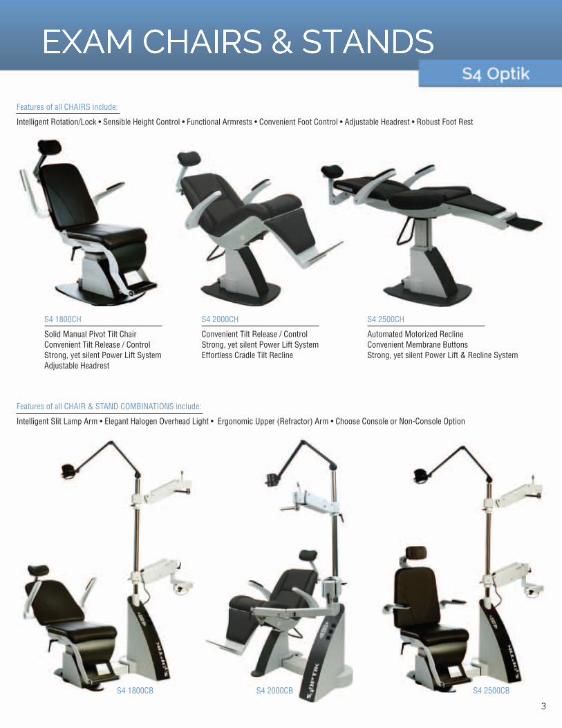
Features of all CHAIRS include:
Intelligent Rotation/Lock • Sensible Height Control • Functional Armrests • Convenient Foot Control • Adjustable Headrest • Robust Foot Rest
Features of all CHAIR & STAND COMBINATIONS include:
Intelligent Slit Lamp Arm • Elegant Halogen Overhead Light • Ergonomic Upper (Refractor) Arm • Choose Console or Non-Console Option
S4 2500CH
Automated Motorized ReclineConvenient Membrane ButtonsStrong, yet silent Power Lift & Recline System
S4 2500CB
S4 2000CH
Convenient Tilt Release / ControlStrong, yet silent Power Lift SystemEffortless Cradle Tilt Recline
S4 2000CB
S4 1800CH
Solid Manual Pivot Tilt ChairConvenient Tilt Release / ControlStrong, yet silent Power Lift SystemAdjustable Headrest
S4 1800CB
3

RETINAL CAMERA1 Shot Stereo Photography
Capturing a stereo pair un-dilated with one picture
24 Megapixel Resolution
Highest Resolution Camera on the Market
Anterior Segment Photography
Photography away from the Slit Lamp
4

RETINAL CAMERA
SPECIFICATIONS
Photography Modes
Stereoscopic photography method
Stereo photography parallax
Field angle
Working Distance
Minimum pupil diameter
Compensation range of examined eye
Focusing
Working distance adjustment
Camera
Monitor
Internal fixation target
External fixation target
Optical head base adjustment range
Chin rest adjustment range
Interface
Power supply
Dimensions
Weight
Normal/ SP/ Stereo (Electrically switched)
Simultaneous stereo photography
7.4° (at the 0 diopter eye)
Normal: 45°SP: 45°*Stereo: 34°(20°x27°)*Some eyes may cause a flare around the circumferences
30mm
Normal mode: ø4.0mmSP mode ø3.5mmStereo mode: ø4.0mm
Without compensation: -12D ~ +13DCompensation - : -32D ~ -10DCompensation + : +10D ~ +35D
Split luminous bars coincidence
2 luminous dots indication type
Specific Nikon digital SLR camera
5.7 ich LCD monitor
Central, Disc, Macula, mosaic 8 positions
Red light (option)
Movable 40mm forward/backwardMovable 98mm left/rightMovable 27mm vertically (electric)
Movable 55mm (electric)
USB
Input: AC100-240V 50 / 60HzPower Consumption: 150VA
310(W) x 540(D) x 548(H)mm
21kg / 46lbs(excluding the attached digital SLR camera)
2D - Normal & SP (Small Pupil)
Normal Field Angle: 45°Kowa’s exclusive optical design in combination with the 12 megapixel digital SLR camera delivers extremely detailed retinal images. The integrated 9-point internal fixation system allows for mosaic photography covering a large retinal area.
SP - Small Pupil Field Angle: 45°Retinal images can be taken even with smaller pupils. On screen guides indicate if the pupil size is within the sufficient range(above Ø3.5mm) for photography.
A new concept retinal camera with “SSP”,
Simultaneous Stereoscopic Photography.
Automatic Mosaic Merge Function
Mosaic images are created easily with automatic image rotation and alignment.
3D - Stereo - Field Angle: 34° (20°x27°)
PhotographyInstant and simultaneous 3D photography is possible in one shot. Stereoscopic images are captured without the camera shifting.
Retinal observation in 3D imagesThe shape of the optic cup and nerve can be viewed on a 3D image.
Switch between the parallel and cross viewing methods with 1-click when viewed on a 2D monitor.
All Mate Software
Ability to fully integrate all equipment to automate process.
Instantly transfer data and images to EMR System.5

RETINAL CAMERANonMyd FAF (Fundus Auto Florescence)
Follow more Retina Pathology longer
Anterior Segment Photography
Photography away from the Slit Lamp
3.2mm Pupil Size for NonMyd Photos
Capture more photos un-dilated
6

RETINAL CAMERA
SPECIFICATIONS
Photography Modes
Field angle
Working Distance
Minimum pupil diameter
Compensation range of examined eye
Focusing
Working distance adjustment
Monitor
Light source
Internal fixation target
Optical component adjustment range
Chin rest adjustment range
Power supply
Dimensions
Weight
nonmyd 8: COLOR, FAFnonmyd 8s: COLOR
45°
30mm
ø 4.0mm (SP ø 3.3mm)
Without compensation: -12D ~ +13DCompensation + : +10D ~ +35DCompensation - : -32D ~ -10D
Split luminous bars
2 luminous dots indication type
5.7 inch LCD
Observation: Near-infrared LEDPhotography: Xenon flash lamp
Central, Disc, Macula, mosaic 8 positions
Forward/Backward: 40mmLeft/Right: 98mmVertical (electric): 27mm
55mm (electric)
Input: AC 100-240 V 50/60 HzPower consumption: 150 VA
310 (W) x 504 (D) x 548 (H) mm
21kg / 46lbs (excluding the attached digital camera)
Non-mydriatic color retinal photography
Fundus Autofluorescence (FAF) with low flash intensity
Anterior photography ø3.3mm minimum pupil diameter
Multiple internal fixation targets
Japanese manufactured optics for optimal performance
Nonmyd 8s is available for color photography only.
All Mate Software
Ability to fully integrate all equipment to automate process.
Instantly transfer data and images to EMR System.
7

DIGITAL REFRACTOR
Instant comparison of Current RX
to subjective refraction one picture
Increased Business
resulting from enhanced patient experience
Increased Patient Flow
through faster integrated refractions
8

DIGITAL REFRACTOR
SPECIFICATIONS
MEASUREMENT RANgE
Spherical Lens
Cylinder Lens
Cylinder Axis
PD
Rotary Prism Lens
Cross Cylinder
Retinoscopic Lens
-29.00~+26.75D (Regular)-19.00~+16.75D (During XC or Prism Tests)(0.12/0.25 /0.5 /1/2/3/4D increments)
0.00~±8.75D (0.25/0.5 /1/2/3D increments)
0°~180° (1/5/15° increments)
48~80mm (0.5/1mm increments)Near PD : 50~74mmNear Working Distance : 35~70cm
0~20 (0.1/0.2/0.5/1/2 increments)
±0.25D±0.50D±0.25D Prism Split Lens (Dual Cross Cylinder)
+1.5D, +2.0D (Measurement Distance 67cm, 50cm)
AUXILIARy LENSES
Occluding Aperture
Pinhole Lens
Maddox Rod
Red / green Filter
Polarizing Filter
Split(Dissociation) Prism
PD Check Lens
Fixed XC Lens
Visual Field
ø2mm
Right Eye (Red, Horizontal), Left Eye (Red, Vertical)
Right Eye (Red), Left Eye (green)
Right Eye (135°, 45° ), Left Eye (45°, 135° )
Right Eye (6BU)Left Eye (10BI : up to 5 complement)
(±0.50D, with the axis fixed at 90° )
40° (VD=12mm)
HARDWARE
Digital Refractor
Operation Panel
Junction Box
Power Supply
329(W) X 103(D) X 296(H)mm, 4.20kg
249(W) X 245(D) X 248(H)mm, 2.75kg (including internal printer)
240(W) X 141(D) X 71(H)mm, 1.24kg
100-240VAC~, 1.0~0.5A, 50/60HzDesigns and details can be changed without prior notice for the purposes of improvement.
21 Point Exam
21 Point Exam removes complexity so now everyone can perform refraction easily. All results appear on display for easy reading for both examiners and patients. guidance with prism, addition power prescription and visual function test in accordance with exam results are available for simplified use.
Cross Cylinder Lens
Dual and Jackson cross cylinder lens provides highly accurate astigmatism axis and visual acuity exams. Improved speed of lens movement prevents accommodation from interfering with exam and guarantees accurate examination.
Built-in Printer
User friendly built-in printer on operation panel replaces paper on its own.
Various Charts and Contents
Diversification of near vision exam is observed through highly reliable near vision test charts, visual function tests and various refraction charts along with vision therapy-related contents.
Wireless Communication
Wireless Communication with HRK-9000A Auto Ref/Keratometer and HLM-9000 Auto Lensmeter via Wi-Fi allows perfect data transmission regardless of working environment. Classic communication via RS-232 cable is available for data transmission with previous models.
Explanatory Images
Various near vision charts include: incomplete color blindness test, amsler grid anatomy images, refractive power readings, and progressive lens guidance that help patients understand results.
Tablet PC Control (Optional)
Exam can be carried out with not only basic OP panel, but also Tablet and PC. (Tablet PC OS : Win 7 or 8 / Resolution 1366x768)
Tilting and Swiveling Display
Regardless of examinees’ positions, information on display is viewed easily by the tilting and swiveling display.
Monocular Height Adjustment
Customized exam is available for those who have different monocular heights within adjustment +/-3mm.
Slimmer Design
Slimmer design reduces mechanical interference and facilitates easier patient monitoring.
Tiltable Body
Highly advanced near vision exam is achieved with tiltable body from 0° to 45°, just like reading a book.
LCD Chart Compatibility
Compatibility with polarized LCD chart provides economic efficiency. (Both linear and circular polarization)
Fast and Silent Lens Loading
Fast lens loading helps to minimize accommodational interference and fatigue of examinees’ eyes. Silent operation offers more comfort during exam.
Real Time guide
graphical representation displayed on screen guides test process easier and faster in real time.
All Mate Software
Ability to fully integrate all equipment to automate process.
Instantly transfer data and images to EMR System.
9

DIGITAL REFRACTOR
10

5
DIGITAL REFRACTOR
Various Charts and Tests
18 visual acuity charts, 26 vision test charts and up to 35 user defined unit test charts support the most advanced eye test process.
Easy Cleanup
Frequently contaminated parts (Forehead Rests, Face Shields, Lens Windows) are detachable for easy and fast cleanup.
Built-In Printer
Built-in printer on the operation panel makes accessing the printer more convenient and replacing paper at one-step process.
Dual Cross Cylinder Lens
Dual cross cylinder lens supports fast and convenient astigmatic test.
Automatic Occlusion
Automatic occlusion function assists precise and comfortable astigmatic test by preventing accommodation while the lens is rotating over 45° or test mode is changing.
Automatic Convergence
During presbyopic test or near vision acuity test, automatic convergence function makes an examinee look at near vision charts toward the center of refractor lenses assuring precise test.
• Working distance : 35~70cm
• Available Near PD : 50~74mm
LED Near Sight Illumination and Detachable Near Chart Rod
Built-in LED illumination for the near sight chart automatically recognizes the near or far sight test and turns the lighting on or off to create the best lighting needed for the test environment. Installation and removal of the near chart rod with a magnetic joint allows for ease of use.
Accurate Rotary Prism
Precise prism data can be obtained by fine increment (up to 20, minimum 0.1 increment) and automatic occlusion function works while the prism is changing directions to assist correct test.
Tilt & Swivel LCD Panel
Tilt & swivel LCD panel makes it possible to share the displayed information in any direction or angle. Touch screen interface offers intuitive guide with great convenience of operation.
Test Process Programming
Maximum of 10 customized test processes can be programmed and saved with the detailed setting of unit test charts conversion, auxiliary lens inserting, fogging, chart masking, etc.
Illuminated Vertex Distance Check Window
More accurate test is guaranteed by positioning examinees’eyes in the correct vertex distance through the illuminated vertex distance check window.
Fast and Silent Lens Loading
Faster lens loading helps to minimize accommodational interference and fatigue of examinees’ eyes. Silent operation offers more comfort during the exam process.
Forehead Rest Indicator
An LED (on/off) sensor inside the forehead rest indicates whether an examinee’s forehead is currently on the forehead rest to ensure the most precise vertex distance measurement.
Monocular PD Adjustment
HDR-7000 provides independent PD for right and left eyes.
Various Muscle Balance Test Methods
HDR-7000 provides various muscle balance test methods such as Von graefe Test, Schober Test, Maddox Rod Test, Polar Cross Test, etc.
All Mate Software
Ability to fully integrate all equipment to automate process.
Instantly transfer data and images to EMR System. 11

DIGITAL CHARTS
12

DIGITAL CHARTS
7
All Mate Software
Ability to fully integrate all equipment to automate process.
Instantly transfer data and images to EMR System.
13

AUTO REF-KERATOMETER
Designs and details can be changed without prior notice for the purposes of improvement.
Dimension /Weight 262(W) X 518(D) X 441(H)mm, 19kg
Power Supply 100-240VAC, 1.0-0.6A, 50/60Hz
Power Saving Automatic switch-off (5min)
Internal Printer Thermal line printer with Auto cutting function
Wi-Fi Band : 2.4GHz, IEE802.11b/g Security : WPA2-PSK
Interface RS-232 x 1, USB(for Service) x 1, Wi-Fi (for Data communication)
Display 7 inch Wide Color TFT LCD, Touch panel with Tilting function
Forward-Backward ±5mm, ±2mm
Left-Right ±5mm, ±2mm
Up-Down ±15mm
Cyl Axis 0 to 180° (increment 1°/5° )
Cylinder(CYL) 0 to ±10D (Max, increment 0.25D)
Sphere(SPH) -22D to +22D (increment 0.25D)
VA Measurement <0.1/ 0.1/0.25/0.32/ 0.4/ 0.5/0.63/ 0.8/ 1.0/1.25> <20/200 / 20/200 / 20/80 / 20/60 / 20/50 / 20/40 / 20/30 / 20/25 / 20/20 / 20/16>
Memory of Data 10 measurements for each eye
Pupil, Iris Diameter 2.0~14.0mm (increments : 0.1mm)
Axis 0~180° (increments : 1° )
Corneal Astigmatism 0.00~ -15.00D (increments : 0.05, 0.12, 0.25D)
Corneal Power 25.96~67.50D (increments : 0.05, 0.12, 0.25D) (When corneal equivalent refractive index is 1.3375)
Radius of Curvature 5.0~13.0mm (increments : 0.01mm)
Minimum Pupil Diameter ø2.0mm
Pupil Distance 10~85mm
Cylinder Form -, +, ± (Mixed)
Cylinder(CYL) 0.00~±12.00D (increments 0.01, 0.12, 0.25D)
Sphere(SPH) -30.00~+25.00 (VD=12mm) (increments : 0.01, 0.12, 0.25D)
Vertex Distanc(VD) 0.0, 12.0, 13.5, 13.75, 15.0
TFBUT Mode Special Mode for Measuring TFBUT (Tear Film Break-Up Time)
Meibography Mode Special Mode for Observing Meibomian Gland
Color View Mode Color View & Contact Lens Fitting Assistance (White & Blue LED Light)
KER P Mode Peripheral Keratometry
KER Mode Keratometry
REF Mode Refractometry
K/R Mode Continuous Keratometry & Refractometry
Auto Refraction
Standard K & Peripheral K’s
High & Low Order Abberation
Zernike Coefficients
Retro Illumination
Cataract gradient
Anterior Segment Capable
Anterior Segment Photos away from the Slit Lamp
Tear Film Break Up
Testing for Dry Eye Disease in Pre Test
Meibography
Testing for Dry Eye Disease in Pre Test
14

AUTO REF-KERATOMETER
Designs and details can be changed without prior notice for the purposes of improvement.
Dimension /Weight 262(W) X 518(D) X 441(H)mm, 19kg
Power Supply 100-240VAC, 1.0-0.6A, 50/60Hz
Power Saving Automatic switch-off (5min)
Internal Printer Thermal line printer with Auto cutting function
Wi-Fi Band : 2.4GHz, IEE802.11b/g Security : WPA2-PSK
Interface RS-232 x 1, USB(for Service) x 1, Wi-Fi (for Data communication)
Display 7 inch Wide Color TFT LCD, Touch panel with Tilting function
Forward-Backward ±5mm, ±2mm
Left-Right ±5mm, ±2mm
Up-Down ±15mm
Cyl Axis 0 to 180° (increment 1°/5° )
Cylinder(CYL) 0 to ±10D (Max, increment 0.25D)
Sphere(SPH) -22D to +22D (increment 0.25D)
VA Measurement <0.1/ 0.1/0.25/0.32/ 0.4/ 0.5/0.63/ 0.8/ 1.0/1.25> <20/200 / 20/200 / 20/80 / 20/60 / 20/50 / 20/40 / 20/30 / 20/25 / 20/20 / 20/16>
Memory of Data 10 measurements for each eye
Pupil, Iris Diameter 2.0~14.0mm (increments : 0.1mm)
Axis 0~180° (increments : 1° )
Corneal Astigmatism 0.00~ -15.00D (increments : 0.05, 0.12, 0.25D)
Corneal Power 25.96~67.50D (increments : 0.05, 0.12, 0.25D) (When corneal equivalent refractive index is 1.3375)
Radius of Curvature 5.0~13.0mm (increments : 0.01mm)
Minimum Pupil Diameter ø2.0mm
Pupil Distance 10~85mm
Cylinder Form -, +, ± (Mixed)
Cylinder(CYL) 0.00~±12.00D (increments 0.01, 0.12, 0.25D)
Sphere(SPH) -30.00~+25.00 (VD=12mm) (increments : 0.01, 0.12, 0.25D)
Vertex Distanc(VD) 0.0, 12.0, 13.5, 13.75, 15.0
TFBUT Mode Special Mode for Measuring TFBUT (Tear Film Break-Up Time)
Meibography Mode Special Mode for Observing Meibomian Gland
Color View Mode Color View & Contact Lens Fitting Assistance (White & Blue LED Light)
KER P Mode Peripheral Keratometry
KER Mode Keratometry
REF Mode Refractometry
K/R Mode Continuous Keratometry & Refractometry
Micro Lens Array
Huvitz’s own developed Micro Lens Array creates a number of separated focal spots, of which the pattern provides valuable information of patients’ ocular systems.
More Accurate Data
Accuracy of KER data is improved by setting optimal zone diameter on measuring spot and also REF data by standardization of quantity of light of fogging chart and fogging lens position along with complete block of accommodation.
Color View Mode
Full color CCD camera and white LED light source in auto ref / keratometer enable you to see eyes and contact lens fitting status which was previously only possible with slit lamps.
Subjective VA Test
Comparison between subjective and objective VA tests yields more reliable and accurate data. Subjective VA test is useful in deciding necessity of progressive lenses because it checks visual acuity based on patients’ responses.
Contrast Sensitivity and glare Test
Highly reliable night visual acuity is examinable with low contrast sensitivity test and glare test which perfectly reproduces halo effect. Progress after refractive or cataract surgery can be monitored effectively.
TFBUT Measurement and Meibography
Conditions of tear film and dry eye can be collected by TFBUT (Tears Film Break-Up Time) and are readable for thorough understanding of visual acuity. Degeneration of meibomian gland can be also monitored with enough light source and image enhancement function.
Peripheral Keratometry Measurement
Continuous measurement on periphery of cornea at 90° both vertically and horizontally from center of cornea produces curvature and eccentricity values of all points and allows best fitting of contact lenses.
IOL Mode
Extra measurement mode is available for IOL power or visual acuity after cataract surgery.
Iris and Pupil Diameter Measurement
Image capturing function supports highly accurate exam by measurement of iris and pupil diameter with diameter from 2mm to 14mm.
Contact Lens Fitting Assistance guide
The world’s first contact lens fitting function in an auto ref / keratometer enables you to see fluorescein liquid with blue illumination.
Efficient Contact Lens Prescription
Image capture and contrast regulation are possible. HRK-9000A gives you the best On-K fitting guide based on the base curve and KER value.
Touch and Tilting 7” Color Display
Wide color TFT LCD supports high-resolution images and real-time image processing to realize afterimage-less image quality. Moreover, swiveling and tilting touch display is readable from any direction for smooth communication between examiners and examinees.
Auto Tracking
Cutting edge auto sensor and 3 dimensional movement mechanism allow you to track and measure the focus of an eye automatically and helps complete measurement perfectly, even with inexperienced users.
Wireless Communication
Wireless Communication via Wi-Fi allows perfect data transmission with HDR-9000 Digital Refractor and HLM-9000 Auto Lensmeter regardless of working environment. Classic communication via RS-232 cable is available for data transmission with previous models.
All Mate Software
Ability to fully integrate all equipment to automate process.
Instantly transfer data and images to EMR System. 15

16
9
AUTO REF-KERATOMETER
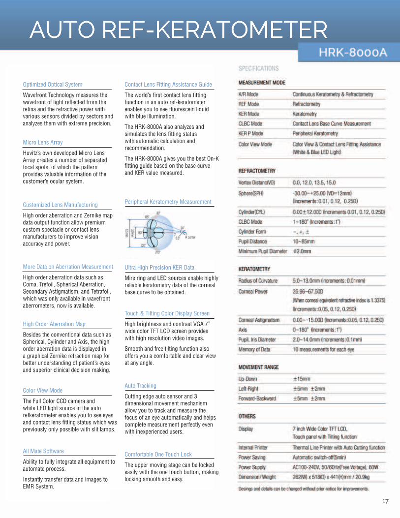
Contact Lens Fitting Assistance guide
The world’s first contact lens fitting function in an auto ref-keratometer enables you to see fluorescein liquid with blue illumination.
The HRK-8000A also analyzes and simulates the lens fitting status with automatic calculation and recommendation.
The HRK-8000A gives you the best On-K fitting guide based on the base curve and KER value measured.
Comfortable One Touch Lock
The upper moving stage can be locked easily with the one touch button, making locking smooth and easy.
Touch & Tilting Color Display Screen
High brightness and contrast VgA 7” wide color TFT LCD screen provides with high resolution video images.
Smooth and free tilting function also offers you a comfortable and clear view at any angle.
Ultra High Precision KER Data
Mire ring and LED sources enable highly reliable keratometry data of the corneal base curve to be obtained.
Peripheral Keratometry Measurement
Auto Tracking
Cutting edge auto sensor and 3 dimensional movement mechanism allow you to track and measure the focus of an eye automatically and helps complete measurement perfectly even with inexperienced users.
Optimized Optical System
Wavefront Technology measures the wavefront of light reflected from the retina and the refractive power with various sensors divided by sectors and analyzes them with extreme precision.
Micro Lens Array
Huvitz’s own developed Micro Lens Array creates a number of separated focal spots, of which the pattern provides valuable information of the customer’s ocular system.
Customized Lens Manufacturing
High order aberration and Zernike map data output function allow premium custom spectacle or contact lens manufacturers to improve vision accuracy and power.
More Data on Aberration Measurement
High order aberration data such as Coma, Trefoil, Spherical Aberration, Secondary Astigmatism, and Tetrafoil, which was only available in wavefront aberrometers, now is available.
High Order Aberration Map
Besides the conventional data such as Spherical, Cylinder and Axis, the high order aberration data is displayed in a graphical Zernike refraction map for better understanding of patient’s eyes and superior clinical decision making.
Color View Mode
The Full Color CCD camera and white LED light source in the auto refkeratometer enables you to see eyes and contact lens fitting status which was previously only possible with slit lamps.
All Mate Software
Ability to fully integrate all equipment to automate process.
Instantly transfer data and images to EMR System.
17

AUTO LENSMETER
18

AUTO LENSMETER
SPECIFICATIONS
MEASUREMENT
Sphere
Cylinder
Axis
Add
Cylinder Mode
Prism
Measurable Lens Diameter
Wavelength
Measurement Method
Contact Lens Measurement
UV Transmittance
Blue Light Transmittance
Display
Printer
Interface
Dimensions
Power Supply
0D~±25D (0.01/0.06/0.12/0.25)
0D~±10D (0.01/0.06/0.12/0.25)
0°~180° (1° step)
0~10D (0.01/0.06/0.12/0.25)
0 to ±10.00D (Mix/-/+)
0~20 (0.01/0.06/0.12/0.25)
ø20 to 120mm (Contact Lens : ø5 mm over)
545nm (green)
Hartmann Sensor
Hard / Soft
0~100%
0~100%
Tiltable 7” Color LCD IPS panel (800x480) /Touch panel
Auto Cutting Printer
RS-232C / USB 2.0 Port / Wi-Fi(802.11b, 2.4gHz)
222(W) X 240(D) X 370(H)mm, 5.4kg
100-240VAC~, 0.5-0.3A, 50/60Hz
Designs and details can be changed without prior notice for the purposes of improvement.
Multi-focal Lens Measurement
Automatic recognition of multi-focal lenses supports easy measurement with measurement guidance on display and even measurement of sunglasses and prism multi-focal lenses is simple.
Improved Accuracy with green Light Beam
green light beam(545nm), which is nearly the same as Fraunhofer e-line(546.1nm) of ISO standards, allows higher accuracy in measurement than general infrared light.
Hartmann Sensor Wavefront Analysis Tech
Implementation of Hartmann Sensor Wavefront Analysis Technology with more measuring spots maximizes accuracy in measurement even for multi-focal and high curved lenses.
Blue Light Hazard Measurement
As usage of smart phones, LCD monitors and many electric devices increases, blue light hazard emitted from LED displays is recognized as one of harmful rays. HLM-9000 measures blue light transmittance of blue light blocking lens.
UV Measurement
Ease of operation and display of UV transmittance allow for understanding of UV transmittance level from single vision lenses and sunglasses.
7” Color LCD Display
Wide display with unlimited viewing angle (178°) minimizes work fatigue and maximizes work efficiency.
Simple gUI
gUI readable at the first glance is user-friendly with easy operation and anyone can easily conduct measurement without expert knowledge.
Wide Tilting Angle
Clear and bright display is readable from any direction with wide tilting angle.
Minimized gap between PD Bar and Nose
Bi- or multi-focal lenses of small sizes are measurable and accurate measurement is possible over entire area of lens.
Intuitive Prism Direction
Moving directions of both actual lens and lens on display are in same direction to avoid any confusion during measurement.
Auto Cutting Printer
Fast and quiet printer with automatic cutting function shows all data to customers quickly. Replacement of paper roll is a one touch action.
Wireless Communication
Wireless communication via Wi-Fi allows perfect data transmission with HRK-9000A and HDR-9000 regardless of working environment. Classic communication via RS-232 cable is available for data transmission with previous models.
Extra Storage
Extra storage on upper section allows small accessories to be stored without any dust penetration by cover, made of rubber material.
Auto Lens Recognition
Single vision, progressive and other lenses are recognized automatically and turned into corresponding measurements.
Contact Lens Measuring Kit
Hard and soft contact lenses are measurable. (Soft Contact Lens Jig : Optional)
All Mate Software
Ability to fully integrate all equipment to automate process.
Instantly transfer data and images to EMR System 19

DIGITAL LENSMETER
20

11
DIGITAL LENSMETER
Newly Designed PD Bar and Measurement Nose
Measure small, progressive, or multi-focal glasses. The operator can still use the measurement nose when measuring the near sight addition.
Compact Lens Table
Smaller sized glasses or children’s glasses can be measured without interfering with the temples of the glasses.
Adjustable Tilting LCD Monitor
The LCD monitor has a tilting capability of -5°to 60°offering unparalleled visual and operational comfort whether sitting or standing.
User-friendly graphical Interface
The graphical User Interface suggests immediate guidance for easy operation. The well recognizable icons assure rapid response to everyone.
TFT LCD, The Best in Image Quality
TFT LCD images provide higher clarity and increased brightness for an even more efficient operation.
Slim & Compact Design
The newly designed HLM-7000 with its compact size (190 x 377 x 237 mm) offers more space and freedom on limited table space.
Built-in Thermal Printer
Print paper can easily be changed with one-touch lever.
Illustration of Axis & PD helps customers to understand the data better.
Pen Type Marking
Pen type marking assembly guarantees clean and precise marking.
Contact Lens Measurement
HLM-7000 offers fast and accurate measurement data of hard / soft contact lenses.
Uniquely designed Soft Contact lens Measurement Jig* improves stability and comfort when measuringsoft contact lenses.
*Contact lens Measurement Jig is optional accessory.
Incomparable UV Measurement Level Assessments
Few lensmeters provide UV assessments with the exact numerical value. Provide patients with the exact UV protection figure.
PD Measurement
The built-in PD sensor enables you measure PD of frames easily. The power of lens can be captured simultaneously.
Dark Sunglasses Mode
you can measure dark sunglasses better by using Dark Sunglasses Mode.
Additional Prism Display Mode
Now with an additional prism mode, you have a choice of Five or Ten Prism Display. For high prism, you may choose the Ten prism mode to get the status of a wide area and for normal and low prism, you may use the Five prism display mode.
Wide Measurement Range
The extensive diopter measurement range of +25D to –25D gives you the ability to measure wide range of lenses.
Progressive Measurement Now More Efficient
The advanced algorithm helps to automatically measure the far and near sight addition with improved accuracy and speed.
All Mate Software
Ability to fully integrate all equipment to automate process.
Instantly transfer data and images to EMR System 21

22

PORTABLE SLIT LAMP
SLIT-PROJECTORLight Source
Slit Section
Slit Width
Spot
Slit Length
Light intensity adjusting
Slit’s illuminating angle
Filter
Continuous lamp operating time
White LED
Turret
0.1mm, 0.2mm, 0.8mm
ø 1mm, ø 5mm, ø 12 mm and shapes of ellipse
12mm
Continuously-variable (Limit is until 20,000 lux)
± 60° with respect to its horizontal outer periphery
Built in blue filter
130 minutes (new Alkaline batteries)
ELECTRICAL RATINgSInput Voltage
Power Consumption
DC 4.8 to 6.4V (4pcs of AAA batteries [Alkaline or Ni-MH])
3.6 to 4.5 VA
MAIN UNITDimensions / Weight
Power Consumption
220 (W)×95 (D)×220 (H) mm 745g (No batteries)
8 years
SPECIFICATIONS
MICROSCOPE
Type of microscope
Angle of convergence
Total Magnification
Objective lens’ working distance
Practical field of view
Reticles
Variable power type
Interpupillary distance adjustment range
Diopter adjustment range
Binocular-stereoscopic-orthoscopic microscope
13°
10, 16X
80 mm (when a magnifying power of 16 is selected)100 mm (when a magnifying power of 10 is selected)
ø 10 mm (when a magnifying power of 16 is selected)ø 15 mm (when a magnifying power of 10 is selected)
Built in both eyepieces
2-magnifying power selectable / moving objective lens type
50 to 72 mm
-8 to +5D
2
1 Instrument description
1Main unit (slit-lamp)
Bottom view
Light intensity control dialTurn the light intensity control dial to the right or left; the light become b r igh te r o r darke r respectively
replace batteries by blinks when battery level becomes low.
Upper cover
Lamp switch
lever with grip in hand. Release the lever to turn it off.
Magnifying power select leverTurn the lever in the directions of to select a magnifying power of 10 or 16.
Coupled arm
Grip
Battery box
Revolving arm
Objective lensAn image 10 t imes as large as a subject can be observed. An image 16 times as large as a subject can be observed by advancing the lens.
Eyecup
Slit-disk
Spot-diskSelect the spot diameter or use of the blue filter. Select a spot diameter of
12mm.
Air vent
Arm swing
angle scale
Point to the swing angle sca le of illumination light.
Projection lens
EyepieceDiopter adjustment ring
Adjusts the diopter.
Prism box
part both ways.
Red dot
20,000 Lux White LED Source
Illumination is thumb wheel controlled and illuminated for easy viewing in a darkened room
Powered by AAA batteries
Kowa Portable Slit Lamps use the following commercially available batteries:
• AAA rechargeable batteries• AAA dry cell batteries(Except manganese dry cell batteries)
Four color options available
Anti-tip design
New circular base increases slit lamp stability, providing a safer place to charge and store the instrument
White Pink Green Aqua Blue
All Mate Software
Ability to fully integrate all equipment to automate process.
Instantly transfer data and images to EMR System
Standard Headrest
23

24

13
SLIT LAMP SERIES
All Mate Software
Ability to fully integrate all equipment to automate process.
Instantly transfer data and images to EMR System
25

Brighter imaging. Sharper pictures.
IMAGING SYSTEMS
26

15
LED CHART PROJECTOR
27

Wavefront technologyUnlike many conventional diagnostic devices, hrK-8000a is based on hartmann-Shack wavefront sensor, which analyzes many focal spots of a light wavefront.
It has the ability to measure not the basic refraction error of a patient, but to obtain a spatially resolved refraction map.
the new hrK-9000a utilizes a unique wavefront analysis algorithm and surpasses conventional and simple refraction offering added values with high order aberration data output for customized lenses and observation of patients before and after refractive surgery. (See more on pg. 14-15)
coburn technologies, Inc. is the world’s leading provider of computer-integrated ophthalmic lens processing systems. We design, manufacture, distribute and service equipment and supplies used in all aspects of surfacing prescriptions for lens blanks, coating lenses and machining lenses to fit into patient frames. For over 60 years, we’ve supplied optical practices and labs with the equipment, application support, field service, and turn-key solutions they need to take their business to new heights.
coburn technologies now provides a complete line of diagnostic systems throughout the United States, manufactured by huvitz co., ltd., a leading innovator, developer and manufacturer specializing in optometric medical equipment.
Your Complete Solution
coburn technologies, Inc.55 gerber road, South Windsor, ct 06074
800-262-8761 - www.coburntechnologies.com
800 262 8761 305 592 4705 514-326 7930 +44-145-420-0780 +86 21 54450505 +65 6253 8577 +91 80 2206 7048Middle east / indiaGreater ChinaCanadalatin aMeriCa europe / afriCa sinGaporenorth aMeriCa
huvitz hrK-9000a
connected to an
external monitor
(optional)


