REVIEW ARTICLE
Opportunistic toenail onychomycosis. The fungalcolonization of an available nail unit space bynon-dermatophytes is produced by the trauma of theclosed shoe by an asymmetric gait or other trauma. Aplausible theoryN. Zaias,* S.X. Escovar, G. Rebell
Dermatology division, Greater Miami Skin and Laser, Mount Sinai Medical Center, Miami Beach, FL, USA
*Correspondence: N. Zaias. E-mail: [email protected]
AbstractOpportunistic onychomycosis is defined, when a non-dermatophyte mould is cultured from an abnormal nail unit in the
absence of a dermatophyte. The presumption is that the mould has caused the abnormal clinical appearance of the nail
unit, yet there are no data available to substantiate this claim. Reports have only identified the mould being recovered
from the nail unit niche. A review of the published dermatologic literature describing toenail opportunistic onychomycosis
by non-dermatophyte fungi has shown toenails with onycholysis, nail bed (NB) keratosis and nail plate surface abnormal-
ities. The appearance of these clinical changes is indistinguishable from the diagnosis of the Asymmetric Gait Nail Unit
Signs (AGNUS). AGNUS is produced by the friction of the closed shoe in patients with an asymmetric gait, resulting pri-
marily from the ubiquitous uneven flat feet. Most commonly, species of Acremonium (Cephalosporium), Aspergillus,
Fusarium, Scopulariopsis and rarely species of many different fungi genera are capable of surviving and reproducing in a
keratinous environment and change the clinical appearance of the involved nail unit. AGNUS toenails predispose to the
colonization by the non-dermatophyte opportunistic fungi but not by dermatophyte fungi.
Received: 2 January 2014; Accepted: 12 February 2014
Conflicts of interestNone declared.
Funding sourcesNone declared.
IntroductionA recent report1 clinically identified very prevalent toenail unit
signs, dermatophyte free, resulting from the pressure to the toes
and foot by the closed shoe, in subjects who had an asymmetric
gait due to the ubiquitous uneven flat feet. Clinically one or
more signs can be seen depending on which location of the toe-
nail unit the pressure is focused by the closed shoe while walking.
Initially, signs are seen unilaterally and when they are bilateral,
one side is always more severe than the other. These signs are:
1 Nail Plate (NP) curved on one side due to pressure of shoe on
the NP matrix while walking, Fig. 1 (lateral arrows inward).
2 Onycholysis and hyperkeratosis of distal toe skin, Fig. 1
(arrow up and down).
3 NB keratosis, similar to distal subungual onychomycosis
(DSO), dermatophyte free, Fig. 2.
4 Changes of the surface of the NP, similar to white superficial
onychomycosis (WSO), dermatophyte free, Fig. 3.
Onychomycosis is a general term that defines a physical rela-
tionship between the nail unit and a member of the order My-
cota. Onychomycosis can exist when a fungus either initiates the
invasion of the nail unit, as we see in the chronic dermatophyto-
sis and scytalidium syndromes, where there is involvement of
not only the nail units but also the skin of the soles and glabrous
skin.
Opportunistic onychomycosis by non-dermatophyte fungi(moulds) with the exception of scytalidium‘The infected’ nail unit is usually a solitary event, not
accompanied by tinea pedis as seen in onychomycosis by
dermatophytes2 and it does not follow an inheritance pattern,
as do dermatophyte onychomycosis.3 The fungi recovered are
all environmental and easily accessible to the human toenail
niche from the shoe. These fungi include many families and
genera, but only those that are capable to survive and repro-
© 2014 European Academy of Dermatology and VenereologyJEADV 2014, 28, 1002–1006
DOI: 10.1111/jdv.12458 JEADV
duce in a keratinous environment can colonize the available
nail unit niche.
Thus, it is a reasonable hypothesis to propose that opportu-
nistic fungi colonize available spaces of the toenail unit. Why are
toenail unit niches available? Mainly because of Asymmetric
Gait Nail Unit Signs (AGNUS). The prevalence of AGNUS is
greater than the clinician suspects. Of the available studies com-
bined, over a thousand patients with a clinical impression of
onychomycosis that were cultured for dermatophyte fungi, only
27–30% had dermatophyte fungi isolated.4–7 That could mean
that AGNUS was responsible for the majority of the remaining
70–73% of abnormal toenails. In these nails a large variety of
moulds were recovered. It is possible that a mould that can
sustain itself in nail unit niche can alter the substrate and make
the involved nail unit look more abnormal. Whenever there is
toenail onycholysis, NB keratosis and NP surface abnormalities,
there will be a possible colonization by environmental fungi.
MethodsThe dermatologic literature relating to ‘Opportunistic onycho-
mycosis’ from 1960 to 2012 was reviewed. The clinical pictures
of the affected toenail units included in the reports were anal-
ysed looking for AGNUS clinical signs such as onycholysis,
NB keratosis, half of an omega-shaped NP, NP surface damage
and hyperkeratosis of the affected skin of the distal toes.
ResultsAGNUS is the most common toenail unit damage. It is dermato-
phyte free but can coexist with any other affliction of toenails for
independent reasons. Typical AGNUS images, Figs 1–3, demon-
strate the toenail unit niches available from AGNUS. The figures
presented in all reviewed articles on opportunistic fungi show
characteristic AGNUS features, see Figs 4–14.
Fungi reports and their confirmation are summarized in
Table 1.8–24 All the clinical images of the halluces are identical to
what is described as AGNUS.
DiscussionWe propose the theory that opportunistic environmental fungi
of many genera can colonize toenail niches that exist because of
an asymmetric gait and the closed shoe (AGNUS), as long as
Figure 1 Asymmetric Gait Nail Unit Signs (AGNUS) – dermato-phyte free, showing the shoe pressure bending the nail plate matrixmedially (lateral arrows) and producing the half omega curvature ofAGNUS. At same time it also produces onycholysis (arrows down)and the hyperkeratosis of the distal toe skin (arrows up).
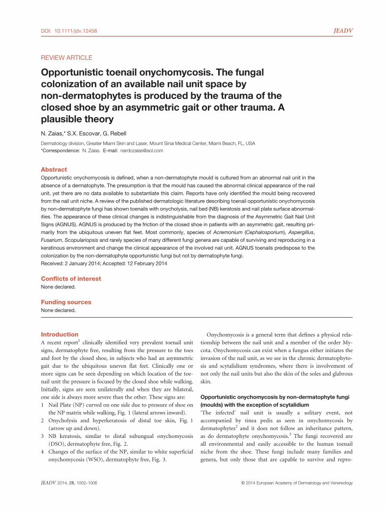
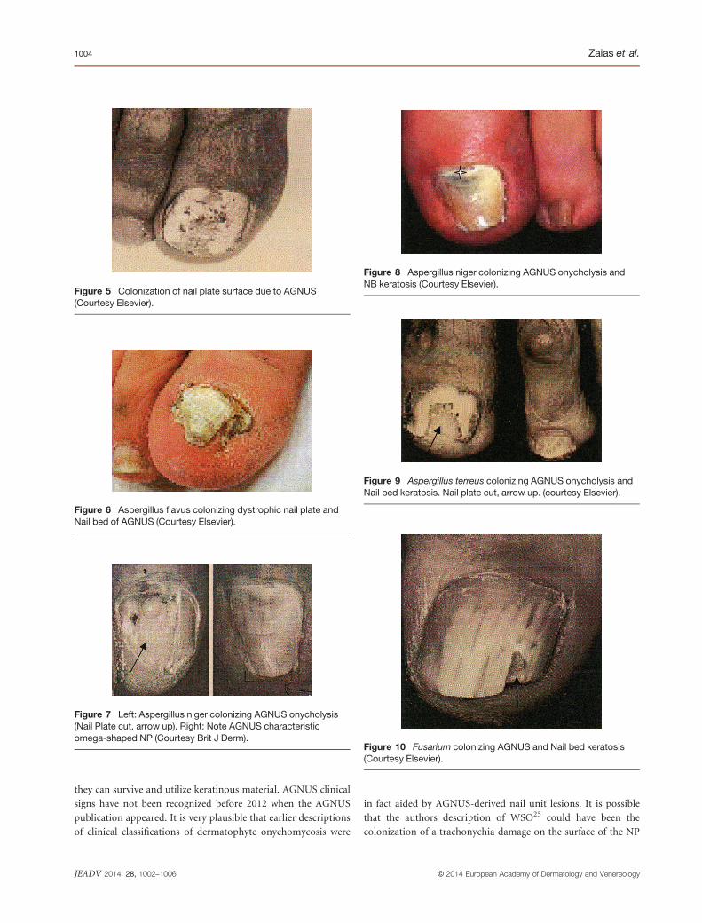
Figure 4 Acremonium species (Cephalosporium) colonizing theonycholysis of AGNUS. Arrow up points at onycholysis (CourtesyElsevier).
Figure 2 Asymmetric Gait Nail Unit Signs subungual hyperkera-tosis, from shoe pressure on nail plate and subsequently on nailbed, dermatophyte free.
Figure 3 Asymmetric Gait Nail Unit Signs – White superficialonychomycosis like clinical but dermatophyte free (arrows down),curved nail plate (lateral arrows) and onycholysis (arrows up).
© 2014 European Academy of Dermatology and VenereologyJEADV 2014, 28, 1002–1006
Opportunistic toenail onychomycosis 1003
they can survive and utilize keratinous material. AGNUS clinical
signs have not been recognized before 2012 when the AGNUS
publication appeared. It is very plausible that earlier descriptions
of clinical classifications of dermatophyte onychomycosis were
in fact aided by AGNUS-derived nail unit lesions. It is possible
that the authors description of WSO25 could have been the
colonization of a trachonychia damage on the surface of the NP
Figure 5 Colonization of nail plate surface due to AGNUS(Courtesy Elsevier).
Figure 6 Aspergillus flavus colonizing dystrophic nail plate andNail bed of AGNUS (Courtesy Elsevier).
Figure 7 Left: Aspergillus niger colonizing AGNUS onycholysis(Nail Plate cut, arrow up). Right: Note AGNUS characteristicomega-shaped NP (Courtesy Brit J Derm).
Figure 8 Aspergillus niger colonizing AGNUS onycholysis andNB keratosis (Courtesy Elsevier).
Figure 9 Aspergillus terreus colonizing AGNUS onycholysis andNail bed keratosis. Nail plate cut, arrow up. (courtesy Elsevier).
Figure 10 Fusarium colonizing AGNUS and Nail bed keratosis(Courtesy Elsevier).
© 2014 European Academy of Dermatology and VenereologyJEADV 2014, 28, 1002–1006
1004 Zaias et al.
commonly seen in AGNUS and that Trichophyton interdigitale
(mentagrophytes) also found in the interdigital spaces, set up
household there to clinically appear as WSO.
In another experiment by a group of Spanish dermatologists26
attempted to prove Koch’s postulates, inoculated cultures of der-
matophyte on the surface of scarified normal toenail plates and
occluded them. Lesions of WSO were seen after 1 month but as
soon as the occlusion was removed all lesions disappeared. No
lesions of DSO were seen. Could it be that the artifactual scarifi-
cation of the surface of the NP needs to be continuous, as seen
in the shoe damage produced by AGNUS?
Other descriptions and new classifications merit discussion
here. Recently described dermatophytoma, Fig. 12,27 is a fungus
ball of Fusarium in an onycholytic area of the NB produced by
AGNUS in a patient who for independent reasons also had T.
rubrum DSO.
(a)
(c)
(b)
(d)
Figure 12 So-called dermatophytoma. (a) AGNUS onycholysis(arrow). (b) Onycholysis and NB keratosis (arrow). (c) Nail plate cutto show onycholysis and fungal colony (up right lines and asterisk).(d) Fungal mass in onycholytic space (Courtesy Elsevier).
Figure 14 AGNUS changes in a patient who also has Paraneo-plastic acral vascular syndrome (courtesy Elsevier).
Figure 11 Scopulariopsis brevicaulis, colonizing AGNUS ony-cholysis and Nail bed keratosis (arrow down) (Courtesy Elsevier).
Figure 13 Pseudomonas colonizing AGNUS onycholysis and NBkeratosis (Courtesy Elsevier).
© 2014 European Academy of Dermatology and VenereologyJEADV 2014, 28, 1002–1006
Opportunistic toenail onychomycosis 1005
Another example of a mixed diagnosis is shown in Fig. 13. A
patient who had dermatophyte DSO coexisting with AGNUS
and finally the onycholytic space inhabited by Pseudomonas,
which tinted the nail space green.
Other diseases have been described to cause toenail abnormal-
ities, as shown in Fig. 14, who clinically had AGNUS and devel-
oped paraneoplastic vascular disease in that toe.
The treatment of opportunistic onychomycosisTreatment of onychomycosis caused by non-dermatophyte
moulds (NDM) is still not well standardized and several authors
underline the fact that NDM onychomycosis frequently does not
respond to systemic antifungals. The use of topicals with the NP
avulsion is commonly described but without reproducible
results.
In an interesting in vitro study, Vander-Straten and col-
leagues8 found that most opportunistic fungi had a very high
minimal inhibitory concentration (MIC) to 5-flurocytosine and
fluconazole. The best results were produced with amphotericin
B and itraconazole was a little better than ketoconazole.
In the reported studies, the treatment time with systemic anti-
fungals appeared to be very short to accomplish complete cure
when compared with the growth rate of the hallux NP and the
length of the infected nail plate.
In summary, we theorize it is impossible for opportunistic
fungi to infect a normal toenail unit without a previous
alteration of the nail unit anatomy, as for example onycholysis,
NB keratosis and superficial NP damage, as seen classically in
the majority of AGNUS cases and trauma.
References1 Zaias N, Rebel G, Casals G, Appel J. The asymmetric gait toenail unit sign
(AGNUS), fungus negative, produced by an asymmetric walking gait that
could be correctable in early life. Skinmed 2012; 10: 213–217.2 Zaias N, Rebell G. Introducing the syndromes of human dermatophyto-
sis. Cutis 2001; 6(Suppl. 5): 9–47.3 Zaias N, Tosti A, Rebell G et al. Autosomal dominant pattern of distal
subungual onychomycosis caused by T. rubrum. J Am Acad Dermatol
1996;34:302–304.4 Za�ıas N, Oertel I, Elliott DF. Fungi in nail. J Invest Dermatol 1969; 53:
140–142.5 Gupta AK, Konnikov N, MacDonald PM et al. Prevalence and epidemi-
ology of toe nail onychomycosis in diabetic subjects: a multicenter sur-
vey. Br J Dermatol 1998; 139: 665–671.6 Haneke E, Roseeeuw D. The scope of onychomycosis: epidemiology and
clinical features. Int J Dermatol 1999; 38: 7–127.7 Scherer WP, McCreary JP, Hayes WW. The diagnosis of onychomycosis
in a geriatric population. J Am Podiatr Med Assoc 2001; 91: 456–464.8 Vander Straten MR, Balkis MM, Ghannoum MA. The role of non derma-
tophyte molds in onychomycosis: diagnosis and treatment. Dermatol
Ther 2002;15: 89–98.9 Negroni P. Una nueva Musedinacea (Cephalosporium) parasitica del
hombre. Rev Soc Argent Biol 1930; 6: 653.
10 Gupta AK, Drummond-Maine C, Cooper EA et al. Systematic review of
non-dermatophyte mold onychomycosis: diagnosis, clinical types, epide-
miology and treatment. J Am Acad Dermatol 2011; 66: 494–502.11 Blomqvist K. Athroderma tuberculatum isolated from finger nail and
beard. Dermatologica 1969; 138: 229.
12 Tosti A, Piraccini BM, Lorenzi S, Iorizzo M. Treatment of non-dermato-
phyte mold and Candida onychomycosis. Dermatol Clin 2003; 21: 491–497.
13 Kaben U. Aspergilus candidum link als erreger einer Konikomycose.
Hauthr Geshlechtskr 1962; 32: 50.
14 Bereston ES, Keil H. Onychomycosis due to Aspergilus flavus. Arch Derm
Syphilol 1941; 44: 420.
15 Bereston ES, Waring WS. Aspergillus infection of the nails. Arch Derm
Syphilol 1946; 54: 552.
16 Tosti A, Piraccini BM. Proximal subungual onychomycosis due to Asper-
gillus niger: report 2 cases. Br J Dermatol 1998; 139: 156–157.17 Moore Weiss R. Onychomycosis caused by Aspergillus terreus. J Invest
Dermatol 1948; 11: 215.
18 Rush-Munro FM, Black Dingley JM. Onychomycoses caused by Fusarium
oxysporum. Aust J Dermatol 1971; 12: 18.
19 Restrepo A, Arango M, Herta H, Uribe L. The isolation of (Lasiodiplodia)
Brotriodiplodia theobromae from a nail lesion. Sabouraudia 1976; 14: 1.
20 Sigler L, Congly H. Toe nail infection caused by Onychocola canadiensis.
J Med Vet Mycol 1990; 28: 405–417.21 Gupta AK, Horgan-Bell CB, Summerbell RC. Onychomycosis associated
with Onychocola canadiensis: ten case report and a review of the litera-
ture. J Am Acad Dermatol 1998; 39: 410–417.22 Gip L, Paldrok H. Onychomycosis caused by Phyllostictina Sydow. Acta
Derm Venereol 1967; 47: 186–189.23 Punithalingam E, English MP. Pyrenochaeta unguis-hominis on human
toenails. Trans Br Mycol Soc 1975; 64: 539.
24 Brumpt E. Precis de Parasitologie. Masson, Paris, 1949.
25 Zaias N. Superficial white onychomycosis. Sabouraudia 1966; 5: 99–103.26 Vilanova X, Cassanovas M, Francino F. Onychomycosis an experimental
study. J Invest Dermatol 1956; 27: 77–101.27 Roberts DT, Evans EG. Dermatophytoma. Br J Dermatol 1998; 138: 188.
Table 1 Reported and confirmed non-dermatophytic fungi produ-cing opportunistic onychomycosis
Organism Confirmedreports
Acremonium (Cephalosporium) species.Figure 4
59,10
Arthroderma trabeculatum Not confirmed11
Aspergillus candidus. Figure 5 412,13
Aspergillus flavus. Figure 6 210–14
Aspergillus glaucus 312
Aspergillus nidulans Not confirmed13
Aspergillus niger. Figures 7–8 515,16
Aspergillus terreus. Figure 9 510–17
Aspergillus ustus 310–15
Aspergillus versicolor 28–10
Fusarium oxysporum and F. solani. Figure 10 44,18
Lasiodiplodia sydowii Not Confirmed 19
Onychocola canadiensis 520,21
Phyllostictina sydowii Not Confirmed22
Pyrenocheta unguis hominis Not Confirmed23
Scopulariopsis brevicaulis. Figure 11 Many reports8,24
© 2014 European Academy of Dermatology and VenereologyJEADV 2014, 28, 1002–1006
1006 Zaias et al.