63
www.semrock.com Optical Filters: Filters for Fluorescence Turan Erdogan, PhD (CTO and Co-founder) Semrock, A Unit of IDEX Corporation May 31, 2011

www.semrock.com
Optical Filters:Filters for Fluorescence
Turan Erdogan, PhD (CTO and Co-founder)Semrock, A Unit of IDEX Corporation
May 31, 2011

2
Basics of fluorescence
• Fluorescence is the property of some atoms and molecules to absorb light over a particular wavelength range and to subsequently emit longer-wavelength light after a brief interval termed the “fluorescence lifetime” The process of phosphorescence occurs in a manner similar to fluorescence, but with a
much longer excited-state lifetime
Diagram courtesy ofMolecular Expressions™
3
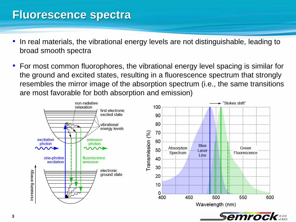
• In real materials, the vibrational energy levels are not distinguishable, leading to broad smooth spectra
• For most common fluorophores, the vibrational energy level spacing is similar for the ground and excited states, resulting in a fluorescence spectrum that strongly resembles the mirror image of the absorption spectrum (i.e., the same transitions are most favorable for both absorption and emission)
Fluorescence spectra
4
Fluorescence filters
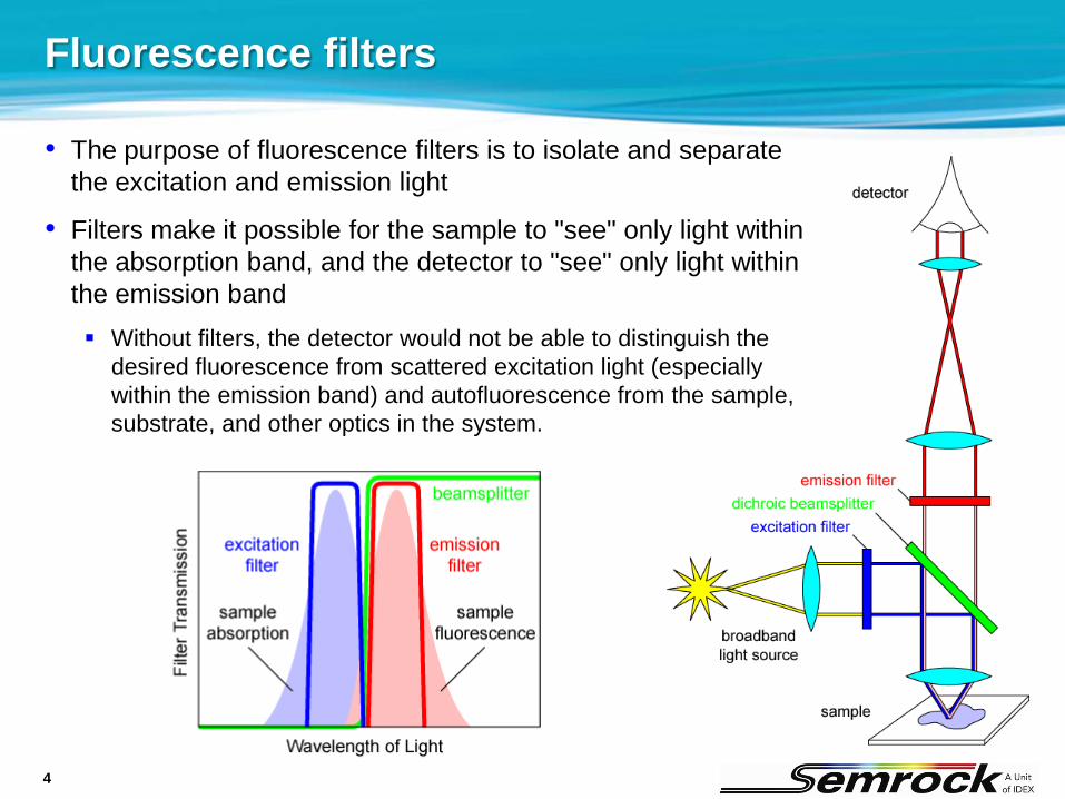
• The purpose of fluorescence filters is to isolate and separate the excitation and emission light
• Filters make it possible for the sample to "see" only light within the absorption band, and the detector to "see" only light within the emission band Without filters, the detector would not be able to distinguish the
desired fluorescence from scattered excitation light (especially within the emission band) and autofluorescence from the sample, substrate, and other optics in the system.

5
Fluorescence microscopy
Source: Olympus Source: Molecular ExpressionsTM

6
BrightLine® filters* – spectacular spectra
• Typical filter set optimized for Green Fluorescent Protein
* US Patents 6,809,859, 7,411,679, and pending
7
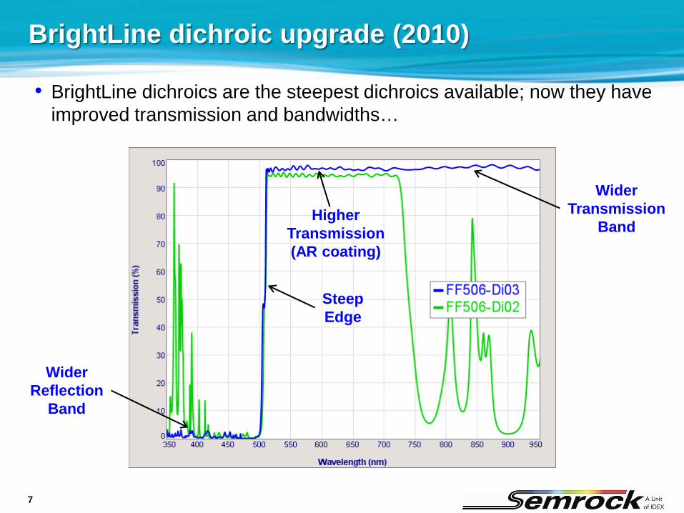
• BrightLine dichroics are the steepest dichroics available; now they have improved transmission and bandwidths…
BrightLine dichroic upgrade (2010)
HigherTransmission(AR coating)
WiderTransmission
Band
WiderReflection
Band
SteepEdge
8
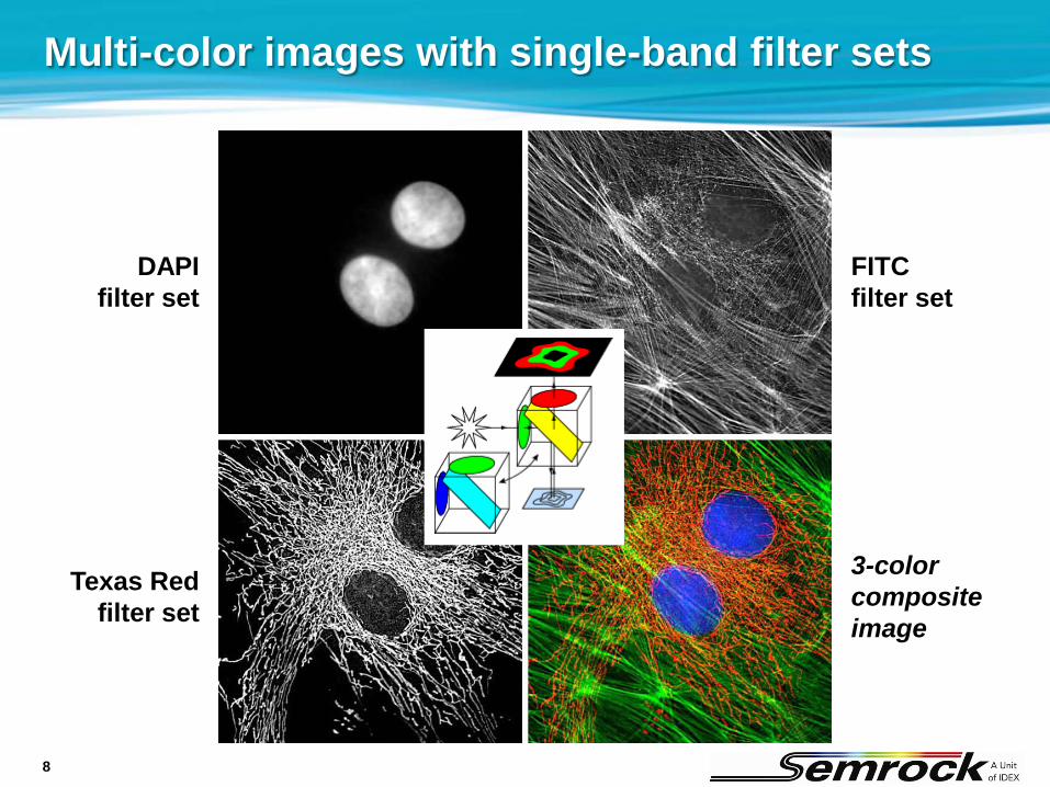
Multi-color images with single-band filter sets
DAPIfilter set
Texas Redfilter set
FITCfilter set
3-colorcompositeimage
9
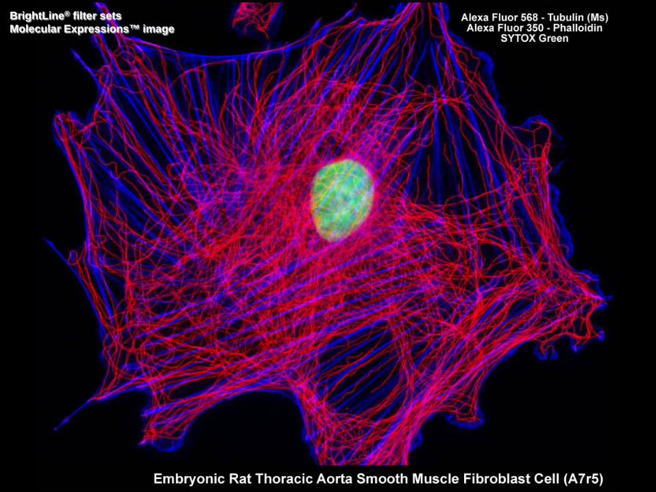
BrightLine® filter setsMolecular Expressions™ image

10
Configurations for fluorescence filters in systems
Standard Epi-fluorescence• Most popular• Widely used in
microscopes and imaging systems
Epifluorescence without a dichroic• Only useful for
small diameter excitation beams
• Laser excitation with moving x-y scanning sample
Side collection• For systems with
a self-contained sample that is accessible from all sides
• Flow cytometry
Forward collection• Rarely used• Difficult to achieve
sufficient blocking of scattered light in real systems
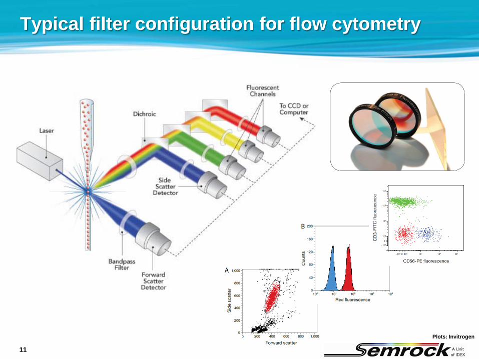
11
Typical filter configuration for flow cytometry
Plots: Invitrogen
12
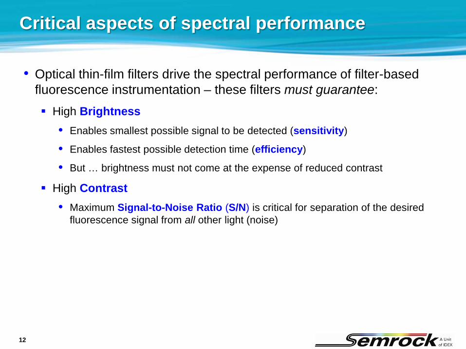
Critical aspects of spectral performance
• Optical thin-film filters drive the spectral performance of filter-based fluorescence instrumentation – these filters must guarantee: High Brightness
• Enables smallest possible signal to be detected (sensitivity)
• Enables fastest possible detection time (efficiency)
• But … brightness must not come at the expense of reduced contrast
High Contrast• Maximum Signal-to-Noise Ratio (S/N) is critical for separation of the desired
fluorescence signal from all other light (noise)
13
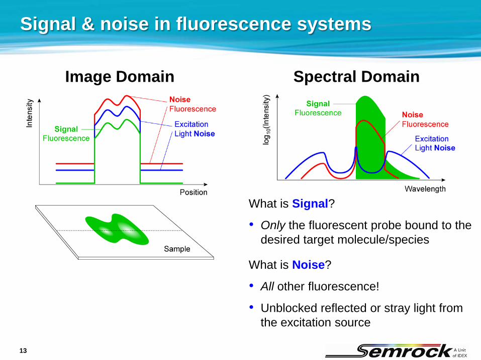
Signal & noise in fluorescence systems
Image Domain Spectral Domain
What is Signal?
• Only the fluorescent probe bound to the desired target molecule/species
What is Noise?
• All other fluorescence!
• Unblocked reflected or stray light from the excitation source

14
More on noise in fluorescence systems
• Noise due to fluorescence “Background” sample fluorescence
• Aromatic amino acids (like Tryptophan, Tyrosine, and Phenylalanine) and proteins
• Enzyme cofactors NAD(P)H, Flavins (FAD), Pyridoxal Phosphate Derivatives
• Level can be substantial (several % of signal)
Bleed-through noise
Non-specific binding and/or unbound (excess) fluorescent probe (minimized by careful specimen preparation)
• Excitation light noise Reflected and/or scattered light that is not blocked by any filter
• Other sources of noise for lowest detection thresholds Detector (thermal and shot) noise and electronic noise
Thermal (blackbody) radiation
15
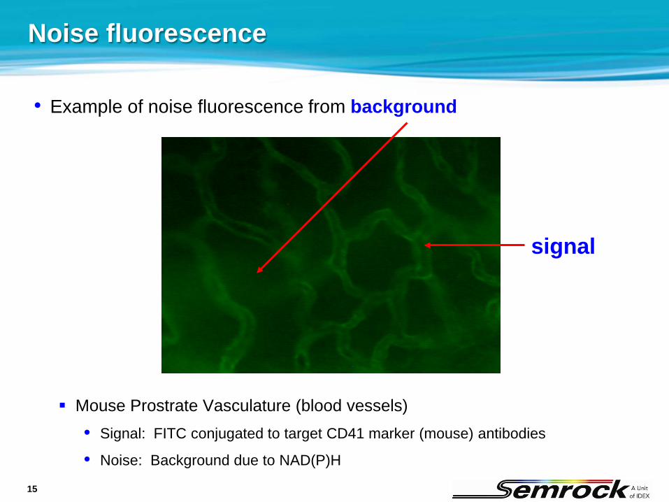
Noise fluorescence
• Example of noise fluorescence from background
Mouse Prostrate Vasculature (blood vessels)• Signal: FITC conjugated to target CD41 marker (mouse) antibodies
• Noise: Background due to NAD(P)H
signal
16
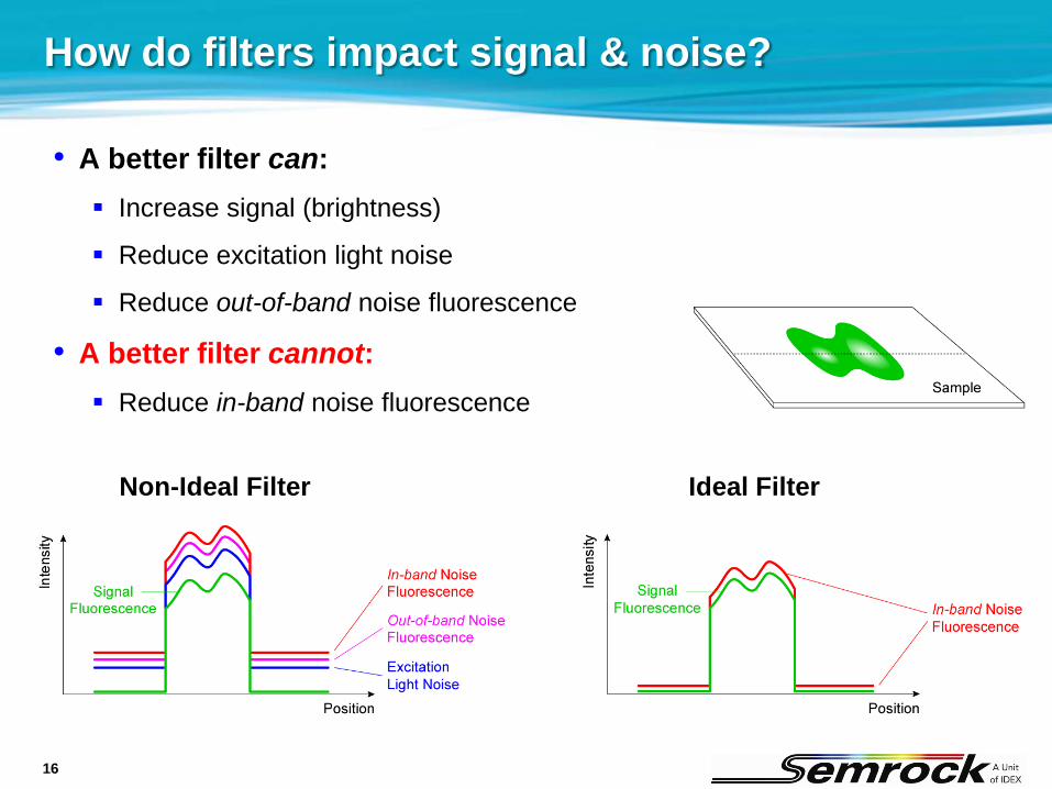
How do filters impact signal & noise?
• A better filter can: Increase signal (brightness)
Reduce excitation light noise
Reduce out-of-band noise fluorescence
• A better filter cannot: Reduce in-band noise fluorescence
Non-Ideal Filter Ideal Filter
17
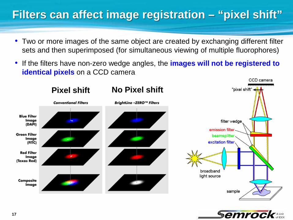
Filters can affect image registration – “pixel shift”
• Two or more images of the same object are created by exchanging different filter sets and then superimposed (for simultaneous viewing of multiple fluorophores)
• If the filters have non-zero wedge angles, the images will not be registered to identical pixels on a CCD camera
Pixel shift No Pixel shift

18
BrightLine filters – “zero pixel shift”
• Impact of filter sets on imaging a particular point on the sample ( )
x (pixels)
y (pixels)
Radius within which image pointsalways occur when using filter sets…
= several pixels for non-ZERO filter sets
< 1 pixel for “-ZERO” filter sets
Location of the imagepoint with NO filter set
Location of the imagepoint WITH filter set
Error due to an emitter
Error due to a dichroic
Offset associatedwith all filter sets
19
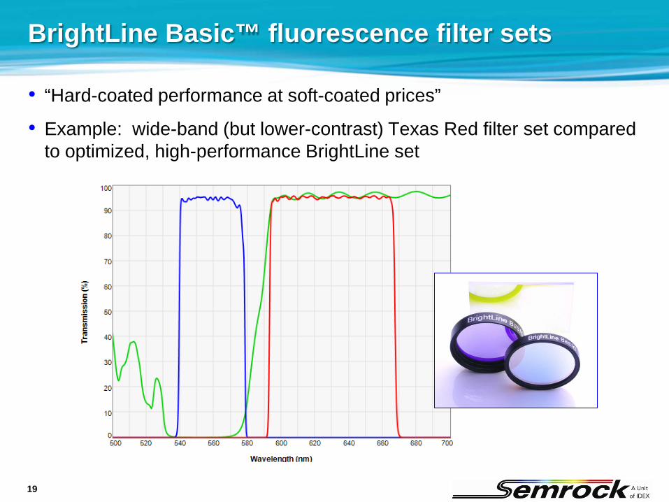
BrightLine Basic™ fluorescence filter sets
• “Hard-coated performance at soft-coated prices”
• Example: wide-band (but lower-contrast) Texas Red filter set compared to optimized, high-performance BrightLine set

20
BrightLine Basic™ – compared to competitors

21
Filter sets for low fluorophore concentration
• For applications like single-molecule imaging with very low fluorophore concentration, long-pass emission filters capture the most possible light
• However, these sets should only be used when sample preparation and system performance yield low background autofluorescence, since strong background will swamp the desired signal at longer wavelengths
22
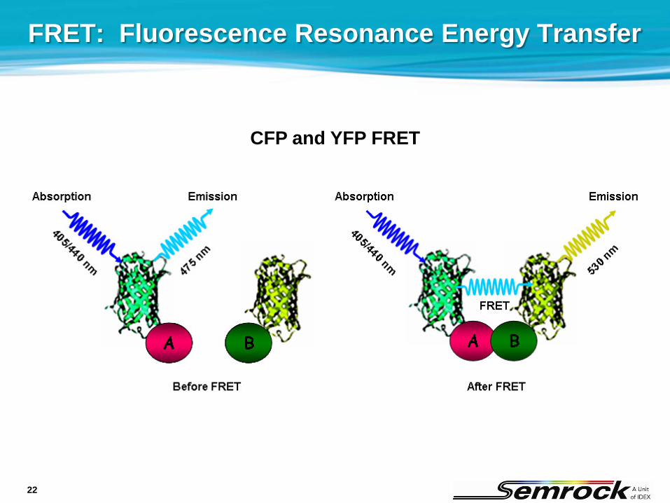
FRET: Fluorescence Resonance Energy Transfer
CFP and YFP FRET
23
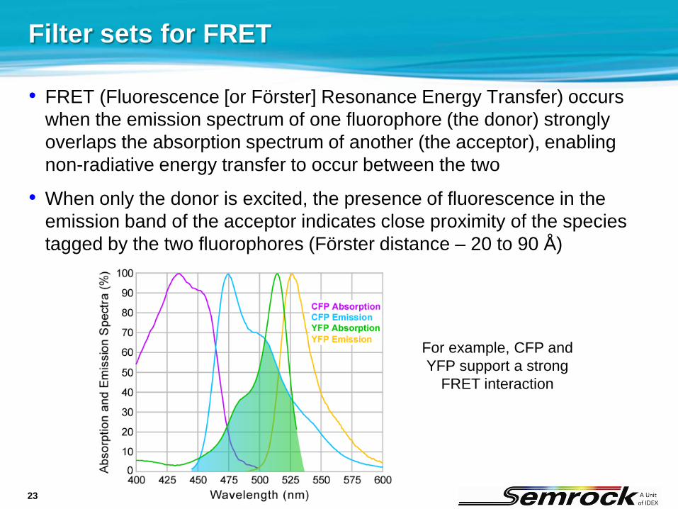
Filter sets for FRET
• FRET (Fluorescence [or Förster] Resonance Energy Transfer) occurs when the emission spectrum of one fluorophore (the donor) strongly overlaps the absorption spectrum of another (the acceptor), enabling non-radiative energy transfer to occur between the two
• When only the donor is excited, the presence of fluorescence in the emission band of the acceptor indicates close proximity of the species tagged by the two fluorophores (Förster distance – 20 to 90 Å)
For example, CFP and YFP support a strong
FRET interaction

24
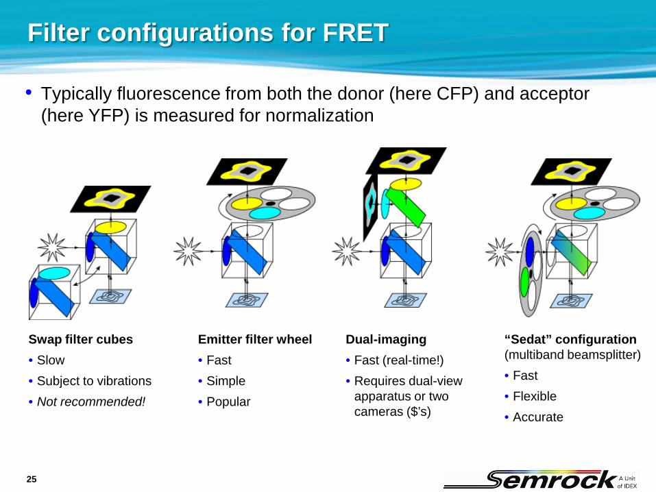
Filter sets for FRET
• Typically fluorescence from both the donor (here CFP) and acceptor (here YFP) is measured for normalization
• This can be done with an emitter filter wheel (requires 4 filters total) or two separate filter cubes (requires 6 filters total)
Filter combination for measuring CFP (donor) fluorescence
Filter combination for measuring YFP (acceptor) FRET fluorescence
25
• Typically fluorescence from both the donor (here CFP) and acceptor (here YFP) is measured for normalization
Filter configurations for FRET
Swap filter cubes• Slow• Subject to vibrations• Not recommended!
Emitter filter wheel• Fast• Simple• Popular
Dual-imaging• Fast (real-time!)• Requires dual-view
apparatus or two cameras ($’s)
“Sedat” configuration(multiband beamsplitter)• Fast• Flexible• Accurate
26
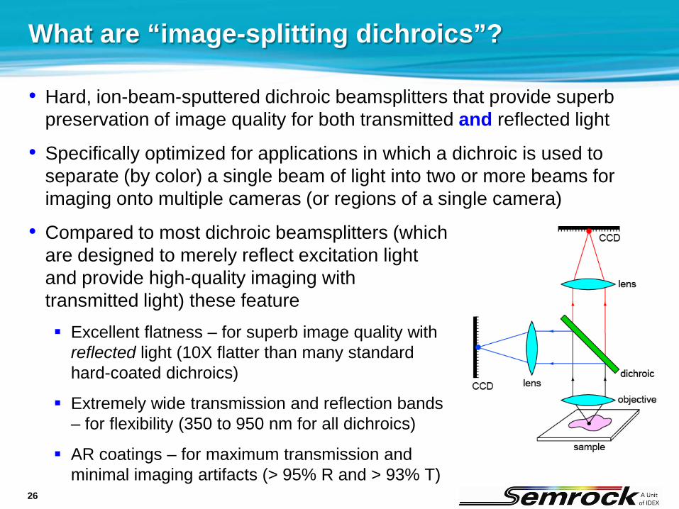
What are “image-splitting dichroics”?
• Hard, ion-beam-sputtered dichroic beamsplitters that provide superb preservation of image quality for both transmitted and reflected light
• Specifically optimized for applications in which a dichroic is used to separate (by color) a single beam of light into two or more beams for imaging onto multiple cameras (or regions of a single camera)
• Compared to most dichroic beamsplitters (which are designed to merely reflect excitation light and provide high-quality imaging with transmitted light) these feature Excellent flatness – for superb image quality with
reflected light (10X flatter than many standard hard-coated dichroics)
Extremely wide transmission and reflection bands – for flexibility (350 to 950 nm for all dichroics)
AR coatings – for maximum transmission and minimal imaging artifacts (> 95% R and > 93% T)
27
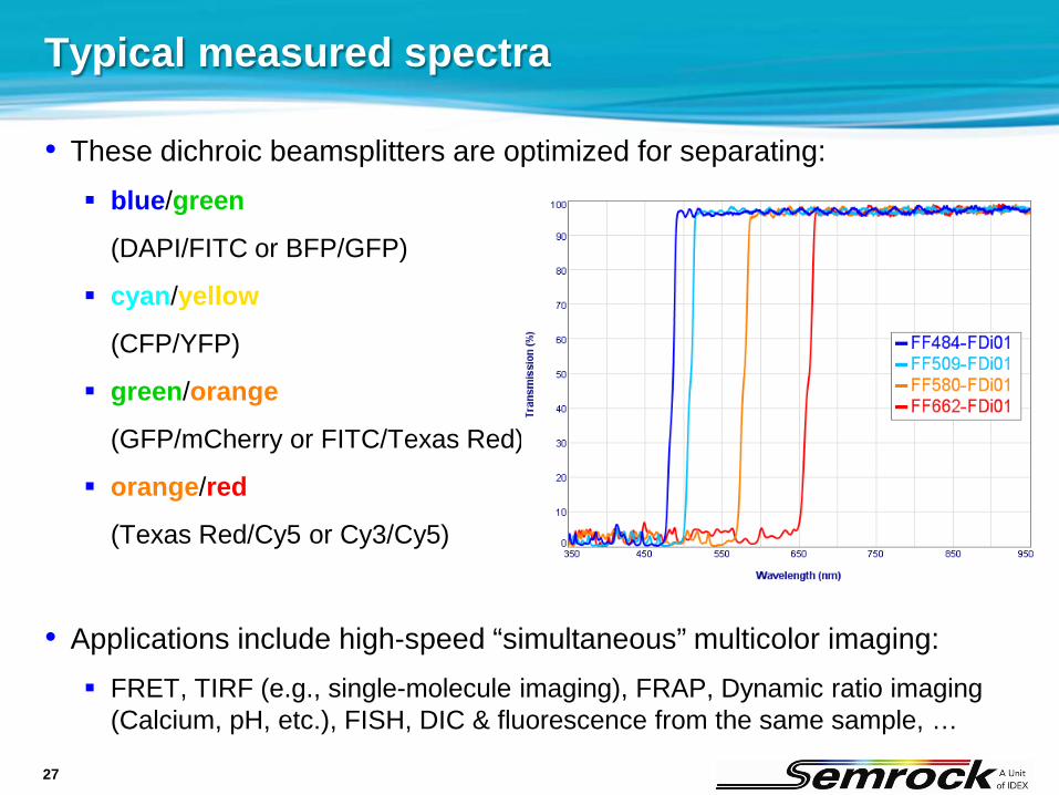
Typical measured spectra
• These dichroic beamsplitters are optimized for separating: blue/green
(DAPI/FITC or BFP/GFP)
cyan/yellow
(CFP/YFP)
green/orange
(GFP/mCherry or FITC/Texas Red)
orange/red
(Texas Red/Cy5 or Cy3/Cy5)
• Applications include high-speed “simultaneous” multicolor imaging: FRET, TIRF (e.g., single-molecule imaging), FRAP, Dynamic ratio imaging
(Calcium, pH, etc.), FISH, DIC & fluorescence from the same sample, …
28
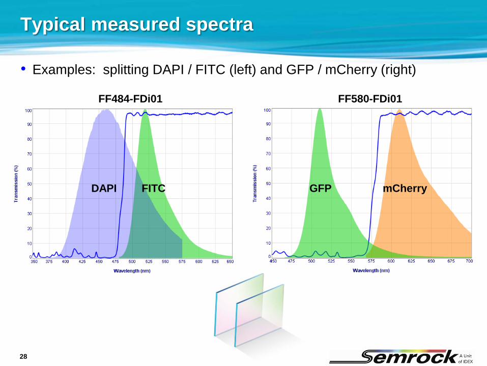
Typical measured spectra
• Examples: splitting DAPI / FITC (left) and GFP / mCherry (right)
FF484-FDi01 FF580-FDi01
DAPI FITC GFP mCherry

29
• Example: Fura-2 is a fluorophore with an absorption spectrum that shifts significantly based on how much calcium (Ca2+) is present near the fluorophore molecule
• By measuring the ratio of digital images taken with two different excitation filters, the (spatial) location of Ca2+ can be tracked
Filter sets for ratiometric imaging

30
Filter spectra for Fura-2 Ca2+ indicator set
High (saturated Ca2+) concentration
Low (Ca2+-free) concentration
BrightLine Fura-2 four-filter set• Fast and accurate ratiometric imaging
• High brightness
• Minimized crosstalk
• Excellent saturated-to-free signal balance

31
Near-IR fluorescence filter sets
• Near-infrared (near-IR) fluorescence imaging is powerful because tissue transmission is higher at these longer wavelengths – ideal for applications like small-animal imaging
• Light levels are typically low, so BrightLine brightness is critical!
Filter set for Cy5.5™ and Alexa Fluor® 680 Filter set for Cy7™ and Alexa Fluor® 750
32
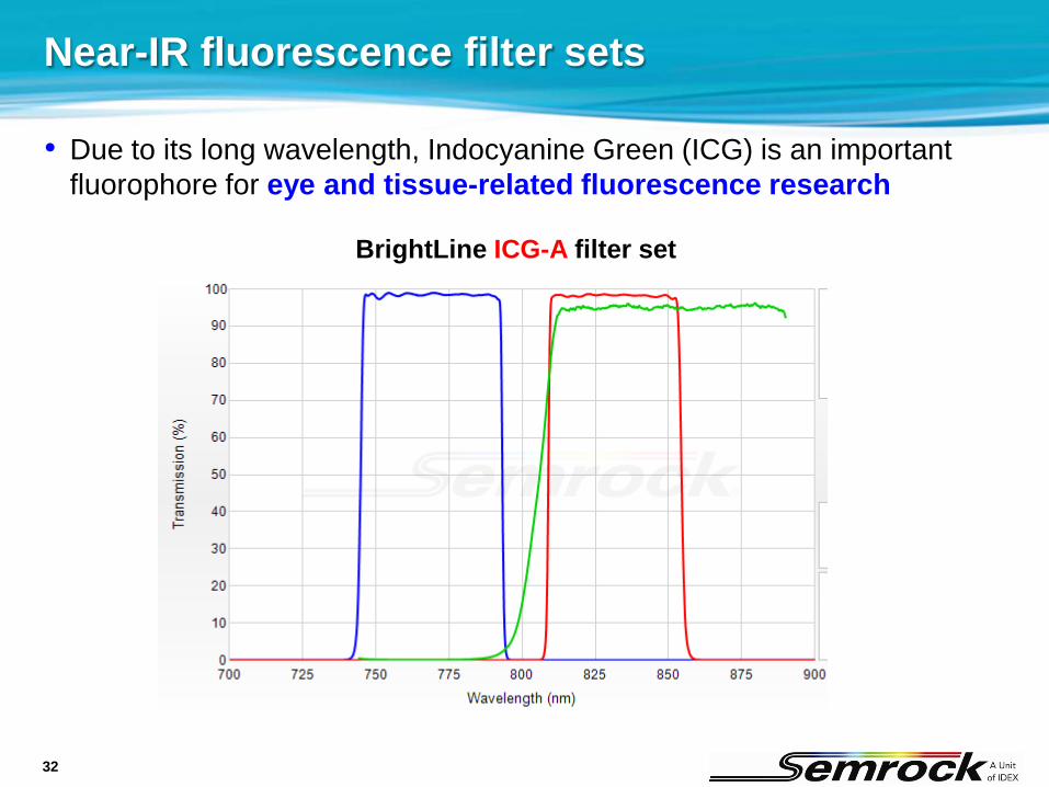
Near-IR fluorescence filter sets
• Due to its long wavelength, Indocyanine Green (ICG) is an important fluorophore for eye and tissue-related fluorescence research
BrightLine ICG-A filter set
33
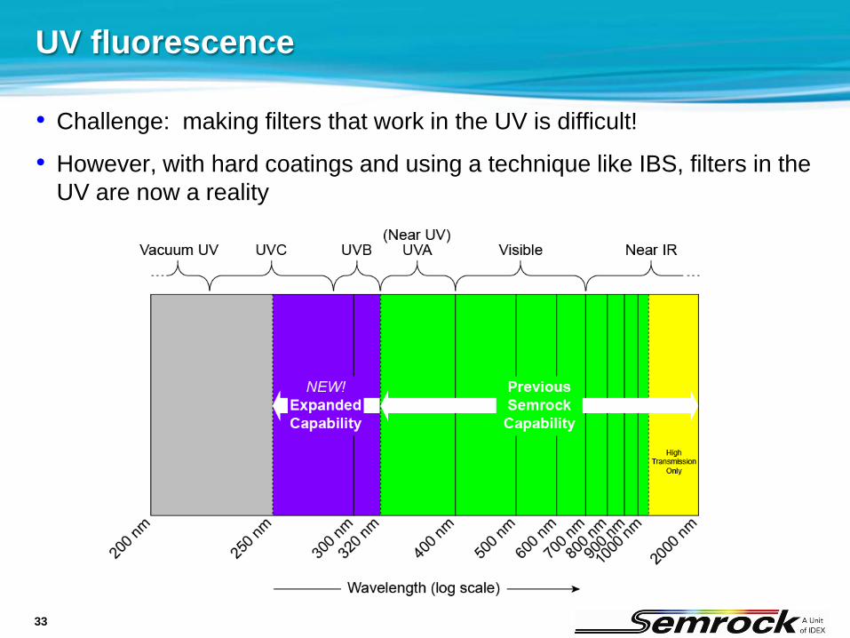
UV fluorescence
• Challenge: making filters that work in the UV is difficult!
• However, with hard coatings and using a technique like IBS, filters in the UV are now a reality
34
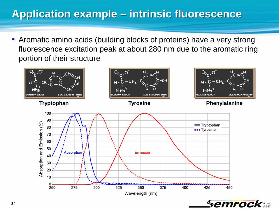
Application example – intrinsic fluorescence
• Aromatic amino acids (building blocks of proteins) have a very strong fluorescence excitation peak at about 280 nm due to the aromatic ring portion of their structure
PhenylalanineTryptophan Tyrosine
35
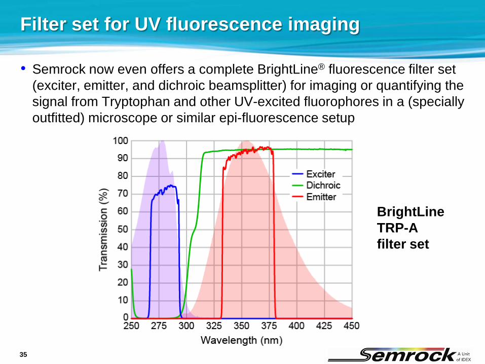
Filter set for UV fluorescence imaging
• Semrock now even offers a complete BrightLine® fluorescence filter set (exciter, emitter, and dichroic beamsplitter) for imaging or quantifying the signal from Tryptophan and other UV-excited fluorophores in a (specially outfitted) microscope or similar epi-fluorescence setup
BrightLineTRP-Afilter set
36
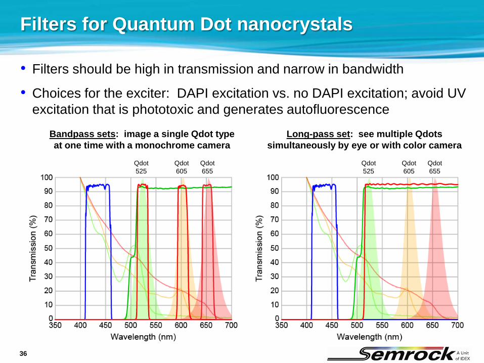
Bandpass sets: image a single Qdot typeat one time with a monochrome camera
Filters for Quantum Dot nanocrystals
• Filters should be high in transmission and narrow in bandwidth
• Choices for the exciter: DAPI excitation vs. no DAPI excitation; avoid UV excitation that is phototoxic and generates autofluorescence
Qdot525
Qdot605
Qdot655
Qdot525
Qdot605
Qdot655
Long-pass set: see multiple Qdotssimultaneously by eye or with color camera

37
BrightLine® multiband filters
• Semrock makes multiband fluorescence filters and sets with spectral performance comparable to single-band filters Highest brightness
Best contrast
Superb color balance
38
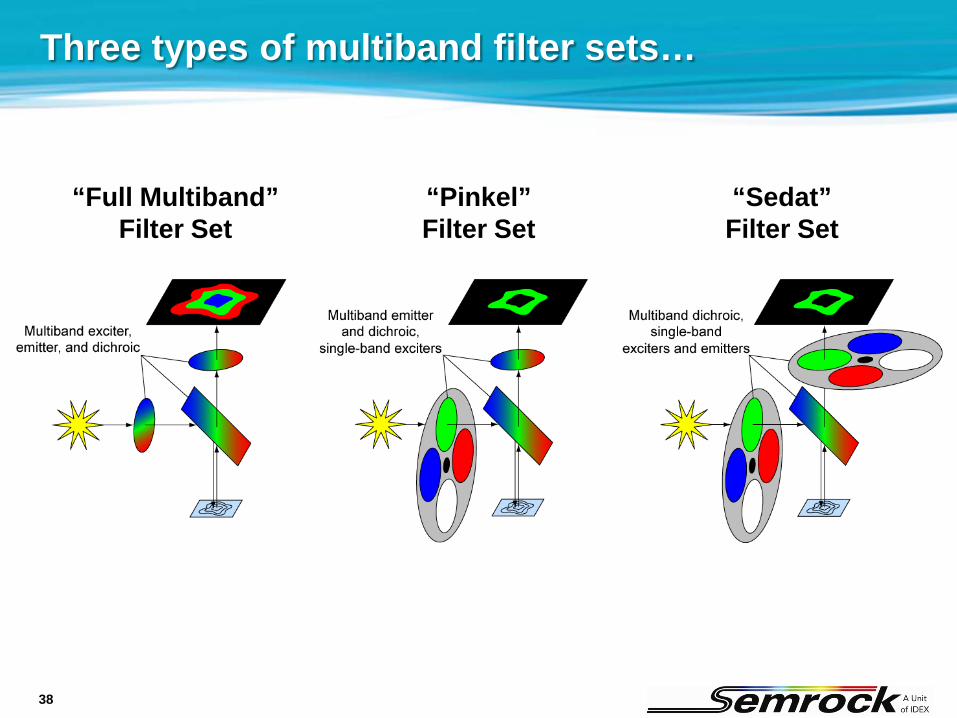
Three types of multiband filter sets…
“Full Multiband”Filter Set
“Pinkel”Filter Set
“Sedat”Filter Set
39
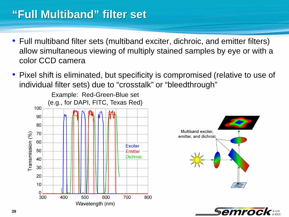
“Full Multiband” filter set
• Full multiband filter sets (multiband exciter, dichroic, and emitter filters) allow simultaneous viewing of multiply stained samples by eye or with a color CCD camera
• Pixel shift is eliminated, but specificity is compromised (relative to use of individual filter sets) due to “crosstalk” or “bleedthrough”
Example: Red-Green-Blue set(e.g., for DAPI, FITC, Texas Red)
40
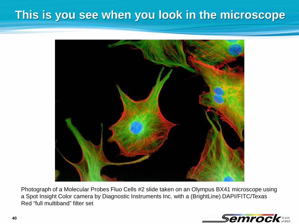
This is you see when you look in the microscope
Photograph of a Molecular Probes Fluo Cells #2 slide taken on an Olympus BX41 microscope using a Spot Insight Color camera by Diagnostic Instruments Inc. with a (BrightLine) DAPI/FITC/Texas Red “full multiband” filter set
41
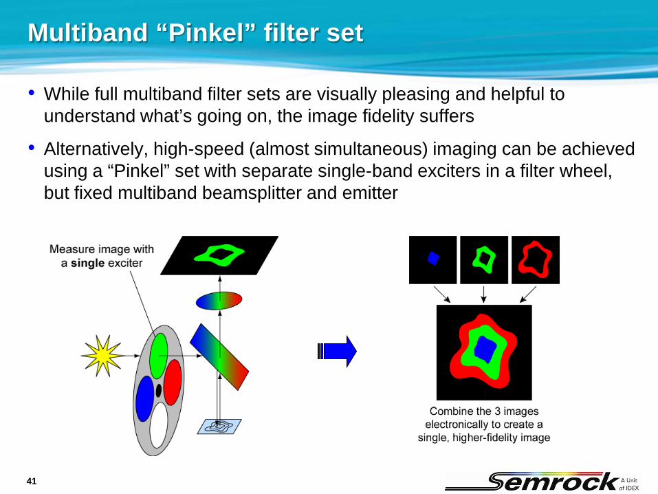
Multiband “Pinkel” filter set
• While full multiband filter sets are visually pleasing and helpful to understand what’s going on, the image fidelity suffers
• Alternatively, high-speed (almost simultaneous) imaging can be achieved using a “Pinkel” set with separate single-band exciters in a filter wheel, but fixed multiband beamsplitter and emitter

42
Multiband “Pinkel” filter set
• Example below shows a dual-band Pinkel set optimized for use with fluorescent proteins (CFP and YFP)
• When used with a monochrome camera, so long as each exciter excites only its respective fluorophore, the fidelity can be almost as good as that achieved with multiple single-band sets (while achieving the advantages of high speed color change and no pixel shift)
43
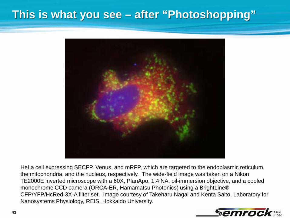
This is what you see – after “Photoshopping”
HeLa cell expressing SECFP, Venus, and mRFP, which are targeted to the endoplasmic reticulum, the mitochondria, and the nucleus, respectively. The wide-field image was taken on a Nikon TE2000E inverted microscope with a 60X, PlanApo, 1.4 NA, oil-immersion objective, and a cooled monochrome CCD camera (ORCA-ER, Hamamatsu Photonics) using a BrightLine® CFP/YFP/HcRed-3X-A filter set. Image courtesy of Takeharu Nagai and Kenta Saito, Laboratory for Nanosystems Physiology, REIS, Hokkaido University.

44
Bleedthrough (or “crosstalk”)
• Bleedthrough occurs when the emission of one fluorophore is detected in the filter passband that is reserved for a different fluorophore
Example: imaging CFP in a sample co-labeled with YFP
• All types of filter sets (including single-band sets and all types of multiband sets) exhibit at least some bleedthrough of the YFP emission within the CFP emitter band
• “Full multiband” and “Pinkel” sets also exhibit bleedthrough of the YFP emission within the YFP emitter band, which can be substantially larger
45
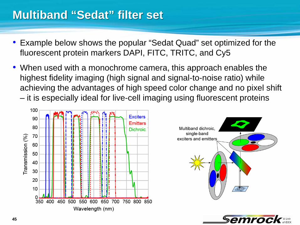
Multiband “Sedat” filter set
• Example below shows the popular “Sedat Quad” set optimized for the fluorescent protein markers DAPI, FITC, TRITC, and Cy5
• When used with a monochrome camera, this approach enables the highest fidelity imaging (high signal and signal-to-noise ratio) while achieving the advantages of high speed color change and no pixel shift – it is especially ideal for live-cell imaging using fluorescent proteins
46
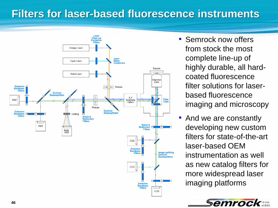
Filters for laser-based fluorescence instruments
• Semrock now offers from stock the most complete line-up of highly durable, all hard-coated fluorescence filter solutions for laser-based fluorescence imaging and microscopy
• And we are constantly developing new custom filters for state-of-the-art laser-based OEM instrumentation as well as new catalog filters for more widespread laser imaging platforms
47
Laser fluorescence instrumentation filters
• Laser-based fluorescence microscopes and instruments put special demands on fluorescence filters
• Laser-scanning confocal
• Spinning-disk confocal
• Total Internal Reflection Fluorescence (TIRF)
• Multi-photon
• DNA/protein microarray scanners
• Flow cytometry
• High-content (confocal) imaging
• Single-molecule gene sequencing ($1K human genome project!)
Fluorescence Microscopy Other Fluorescence Instruments
48
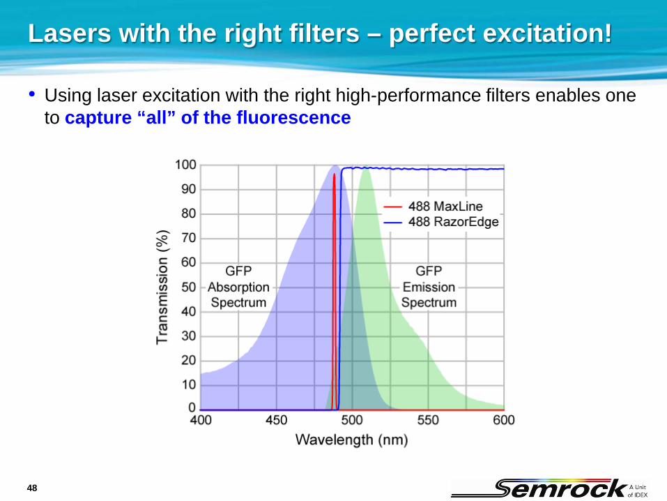
Lasers with the right filters – perfect excitation!
• Using laser excitation with the right high-performance filters enables one to capture “all” of the fluorescence

49
Confocal microscopes – often require special filters
• Example: Yokogawa CSU10/22/X spinning-disk confocal scanner filters
Laser-blockingemission filters
Laser-transmittingdichroic beamsplitters
Photos from Yokogawa

50
Critical characteristics of filters for lasers
• All filters Edge wavelengths should be keyed to the laser wavelengths
Low transmission ripple minimizes intensity fluctuations
High laser damage threshold is necessary
• Dichroics Flatness is often critical
Low autofluorescence glass should be used
Should be anti-reflection (AR) coated to eliminate interference fringes resulting from the coherent laser light

51
• TIRF = Total Internal Reflection Fluorescence
Example: laser TIRF microscopy
Source:OlympusMicroscopyResourceCenter

52
New DPSS lasers – hot for fluorescence & Raman
• Why are DPSS lasers so good? Plenty of power (10’s to 100’s of mW)
Excellent beam quality (M2 < 1.1)
Single-longitudinal-mode (often with 10’s MHz linewidth)
Very low noise (< 1% RMS)
Extremely efficient (the lab does not heat up!)
• Wavelengths that have been around for several years 532 nm (most mature – popular for Raman due to high powers)
491 nm (ideal for GFP and FITC – most popular fluorophores!)
473 nm (for GFP with minimal orange/red fluorophore excitation)
561 nm (for longer-wavelength fluorophores)
• Hot new wavelengths: 515 nm (replaces 514.5 nm Ar-ion line for YFP)
594 nm (replaces 594.1 nm HeNe, and ideal for mCherry RFP and Texas Red)
53
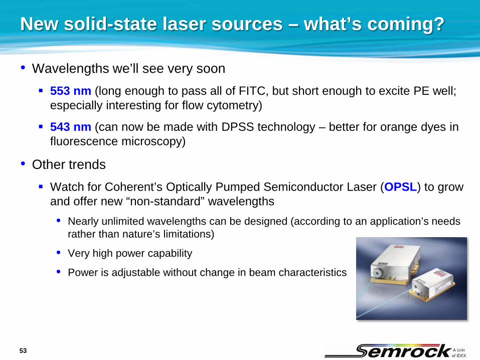
New solid-state laser sources – what’s coming?
• Wavelengths we’ll see very soon 553 nm (long enough to pass all of FITC, but short enough to excite PE well;
especially interesting for flow cytometry)
543 nm (can now be made with DPSS technology – better for orange dyes in fluorescence microscopy)
• Other trends Watch for Coherent’s Optically Pumped Semiconductor Laser (OPSL) to grow
and offer new “non-standard” wavelengths• Nearly unlimited wavelengths can be designed (according to an application’s needs
rather than nature’s limitations)
• Very high power capability
• Power is adjustable without change in beam characteristics
54
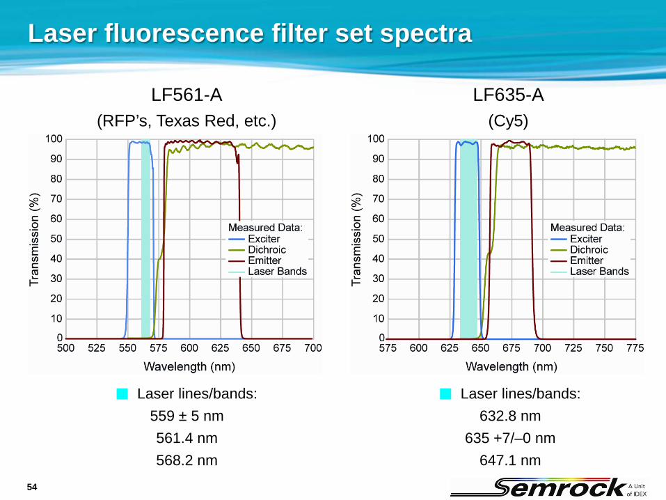
Laser fluorescence filter set spectra
LF561-A(RFP’s, Texas Red, etc.)
LF635-A(Cy5)
Laser lines/bands:559 ± 5 nm561.4 nm568.2 nm
Laser lines/bands:632.8 nm
635 +7/–0 nm647.1 nm
55
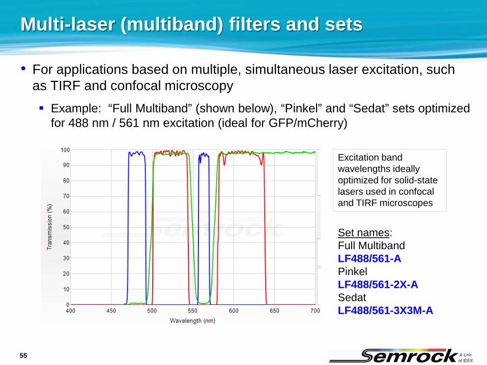
Multi-laser (multiband) filters and sets
• For applications based on multiple, simultaneous laser excitation, such as TIRF and confocal microscopy Example: “Full Multiband” (shown below), “Pinkel” and “Sedat” sets optimized
for 488 nm / 561 nm excitation (ideal for GFP/mCherry)
Excitation band wavelengths ideally optimized for solid-state lasers used in confocal and TIRF microscopes
Set names:Full MultibandLF488/561-APinkelLF488/561-2X-ASedatLF488/561-3X3M-A
56
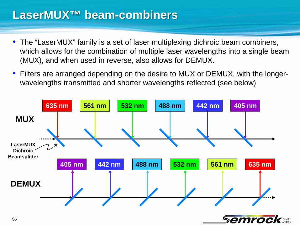
LaserMUX™ beam-combiners
• The “LaserMUX” family is a set of laser multiplexing dichroic beam combiners, which allows for the combination of multiple laser wavelengths into a single beam (MUX), and when used in reverse, also allows for DEMUX.
• Filters are arranged depending on the desire to MUX or DEMUX, with the longer-wavelengths transmitted and shorter wavelengths reflected (see below)
405 nm 442 nm 488 nm 532 nm 561 nm 635 nm
DEMUX
405 nm442 nm488 nm532 nm561 nm635 nm
MUX
LaserMUXDichroic
Beamsplitter
57
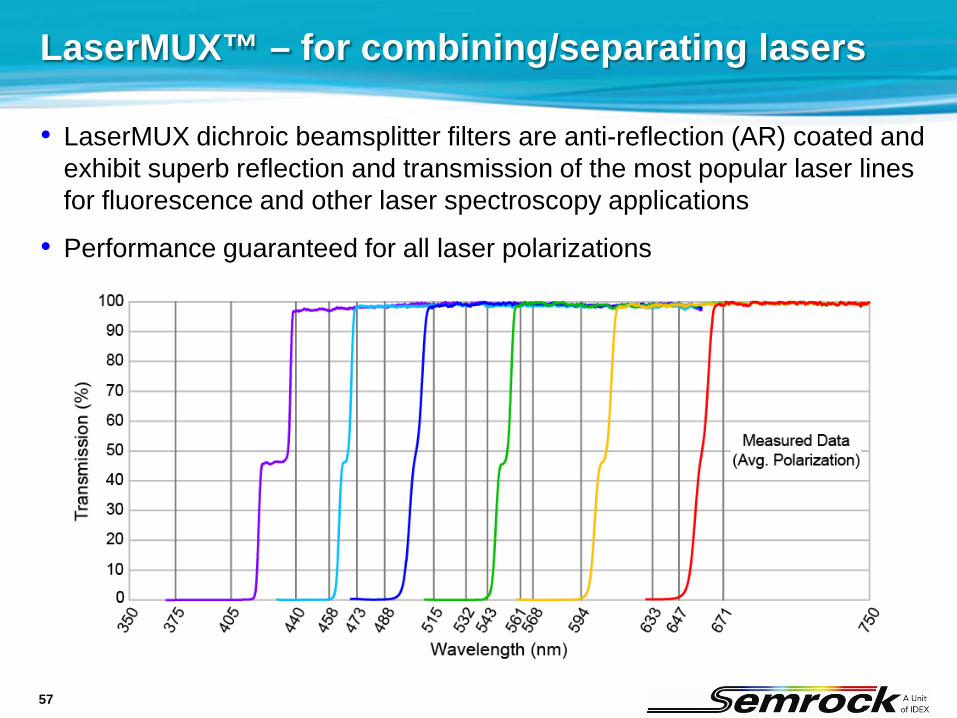
LaserMUX™ – for combining/separating lasers
• LaserMUX dichroic beamsplitter filters are anti-reflection (AR) coated and exhibit superb reflection and transmission of the most popular laser lines for fluorescence and other laser spectroscopy applications
• Performance guaranteed for all laser polarizations
58
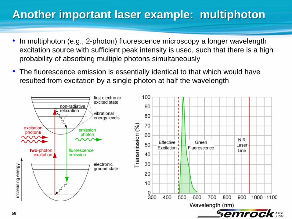
• In multiphoton (e.g., 2-photon) fluorescence microscopy a longer wavelength excitation source with sufficient peak intensity is used, such that there is a high probability of absorbing multiple photons simultaneously
• The fluorescence emission is essentially identical to that which would have resulted from excitation by a single photon at half the wavelength
Another important laser example: multiphoton
59
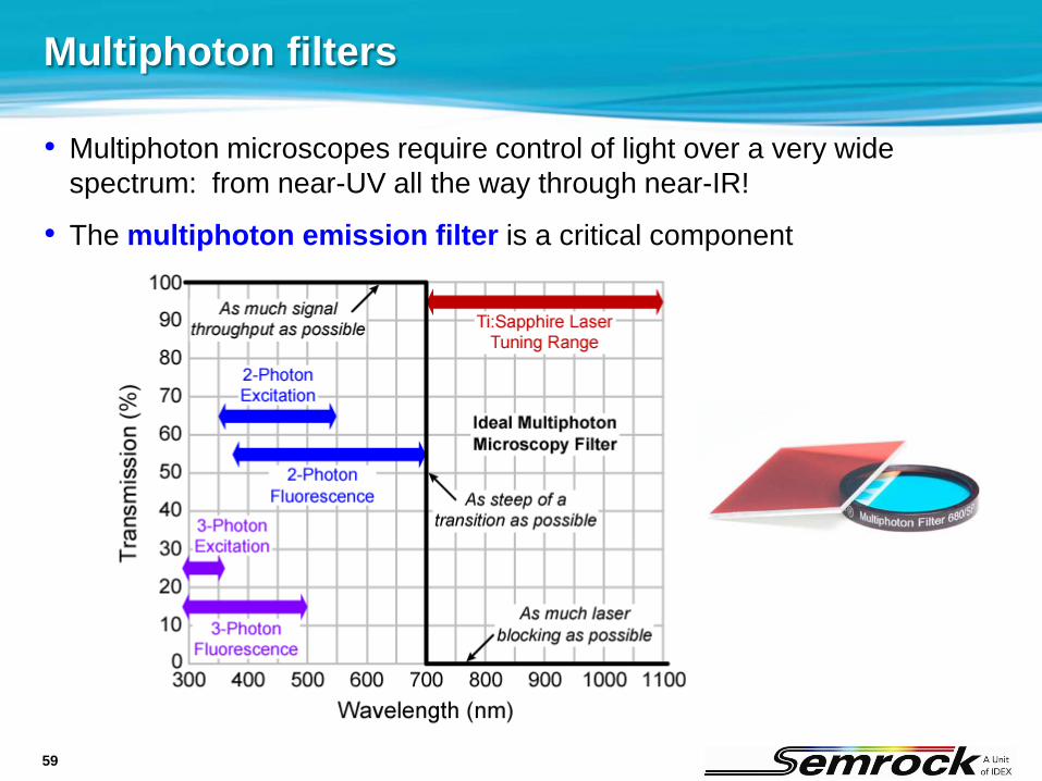
• Multiphoton microscopes require control of light over a very wide spectrum: from near-UV all the way through near-IR!
• The multiphoton emission filter is a critical component
Multiphoton filters
60
Multiphoton emission filters
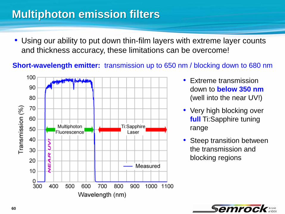
• Extreme transmission down to below 350 nm(well into the near UV!)
• Very high blocking over full Ti:Sapphire tuning range
• Steep transition between the transmission and blocking regions
Short-wavelength emitter: transmission up to 650 nm / blocking down to 680 nm
• Using our ability to put down thin-film layers with extreme layer counts and thickness accuracy, these limitations can be overcome!

61
Multiphoton dichroic beamsplitters
Matching short-wavelength dichroic beamsplitter
• Very high reflection into near-UV
• Very high transmission over Ti:Sapphire tuning range
• Steep transition between the reflection and transmission regions
• Using our ability to put down thin-film layers with extreme layer counts and thickness accuracy, these limitations can be overcome!
62
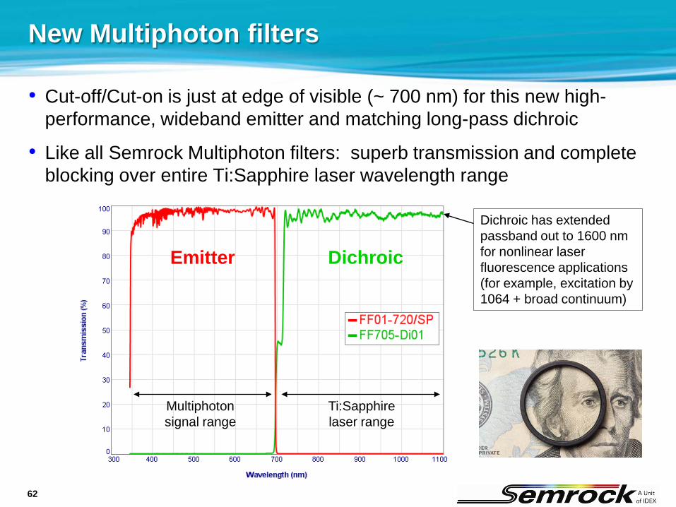
New Multiphoton filters
• Cut-off/Cut-on is just at edge of visible (~ 700 nm) for this new high-performance, wideband emitter and matching long-pass dichroic
• Like all Semrock Multiphoton filters: superb transmission and complete blocking over entire Ti:Sapphire laser wavelength range
Dichroic has extended passband out to 1600 nm for nonlinear laser fluorescence applications (for example, excitation by 1064 + broad continuum)
Emitter Dichroic
Ti:Sapphirelaser range
Multiphotonsignal range

63
Thank you!


