BIOCHEMISTRY Optical Rotatory Dispersion of L-Aspartate ,&Decarboxylase and Its Derivatives* Edith M. Wilson and Alton Meister ABSTRACT: The optical rotatory dispersion of aspartate @-decarboxylase and a number of its derivatives has been determined over the range 225-500 mp. The pyridoxal 5 '-phosphate enzyme (holoenzyme) exhibits anomalous rotation in the region of the absorption of the chromophore (maximum, about 360 mp), while the dispersion of the apoenzyme is plain in this region. The difference disperiion curve (holoenzyme - apoen- zyme) exhibits a Cotton effect with a point of inflection at 360 mp. The magnitude of the trough of rotation at 233-234 mp is substantially the same for the apo- enzyme and holoenzyme. Rotatory dispersion titration of the apoenzyme with pyridoxal 5 '-phosphate indicates that about 15.5 moles of pyridoxal 5'-phosphate are bound/800,000 g of enzyme, a value which is in close agreement with spectrophotometric, chemical, and microbiological data. The pyridoxamine 5 '-phosphate form of the enzyme exhibits a plain dispersion similar to that of the apoenzyme, while the 4'-deoxypyridoxine 5'-phosphate enzyme exhibits a small Cotton effect with a point of inflection at about 320 mp. The 4'- A spartate @-decarboxylase has been isolated in ap- parently homogeneous form from Alcaligenes faecalis (Novogrodsky et al., 1963; Novogrodsky and Meister, 1964a) and from Achromobacter (Wilson, 1963 ; Wilson and Kornberg, 1963). Studies with the Alcaligenes enzyme led to the conclusion that the enzyme acts both as a relatively nonspecific L-amino acid transaminase and as an L-aspartate @-decarboxylase. The slow trans- amination reaction with L-aspartate to yield oxaloace- tate and the pyridoxamine 5'-phosphate form of the enzyme results in inactivation of the enzyme for aspar- tate decarboxylation ; this inactivation may be pre- vented by adding a variety of a-keto acids which re- generate the pyridoxal 5'-phosphate form of the en- zyme by transamination. In the absence of a-keto acids, pyridoxamine 5 '-phosphate dissociates readily, leaving the apoenzyme, which may be reactivated by pyridoxal 5 '-phosphate or by pyridoxamine 5 '-phosphate plus an a-keto acid. In addition to pyridoxal 5'-phosphate and -~ * From the Department of Biochemistry, Tufts University School of Medicine, Boston, Massachusetts. Received December 27,1965. Supported in part by grants from the Nutrition Founda- tion and the National Institutes of Health, U. S. Public Health Service. A preliminary account of this work has appeared (Wilson 1 166 and Meister, 1965). deoxypyridoxine 5 '-phosphate derivative of porcine glutamateaspartate transaminase was also prepared and found to exhibit a Cotton effect similar to that of the corresponding aspartate @-decarboxylase deriva- tive. When aspartate @-decarboxylaseholoenzyme was reduced with sodium borohydride its maximum ab- sorbancy shifted from 360 to 320 mp, and the char- acteristic Cotton effect exhibited by the holoenzyme was replaced by a smaller Cotton effect with a point of inflection at 320 mp. Derivatives of the holoenzyme of aspartate @-decarboxylase obtained by treatment with sodium cyanide, hydroxylamine, and threo-@- hydroxy-DL-aspartate exhibited rotatory dispersion curves that were, within experimental error, the same as that of the apoenzyme. The optical rotatory dispersion curves of aspartate @-decarboxylaseand its derivatives are compared with earlier and present data on the corresponding derivatives of glutamate-aspartate trans- aminase, and the significance of the data is considered in relation to the nature of the cofactor-enzyme link- ages. pyridoxamine 5 '-phosphate the apoenzyme can bind the inhibitory cofactor analog 4'-deoxypyridoxine 5 I- phosphate (Novogrodsky and Meister, 1964b). Other derivatives of the pyridoxal 5 '-phosphate enzyme from Achromobacter formed by reaction with sodium boro- hydride, carbonyl reagents, and the substrate analog P-hydroxyaspartate have also been reported (Wilson and Kornberg, 1963). In an effort to learn more about the binding of the various vitamin Bg cofactors and derivatives to the enzyme, we have carried out determinations of optical rotatory dispersion. The binding of a symmetrical chromophoric molecule to a protein may induce anoma- lous optical rotatory dispersion, a Cotton effect, which is believed to be due to asymmetric orientation on the protein of the otherwise optically inactive chromo- phore. Spectropolarimetry has only recently been ap- plied to the study of chromophoric proteins; Ulmer and Vallee (1965) have reviewed this field and have summarized its present status. This paper describes the rotatory dispersion char- acteristics of aspartate @-decarboxylase and compares these findings with those previously reported for glu- tamate-aspartate transaminase ; the latter enzyme is the only other pyridoxal 5 '-phosphate enzyme which has thus far been so studied (Fasella and Hammes, EDITH M. WILSON AND ALTON hlElSTER
Transcript
B I O C H E M I S T R Y
Optical Rotatory Dispersion of L-Aspartate ,&Decarboxylase and Its Derivatives*
Edith M. Wilson and Alton Meister
ABSTRACT: The optical rotatory dispersion of aspartate @-decarboxylase and a number of its derivatives has been determined over the range 225-500 mp. The pyridoxal 5 '-phosphate enzyme (holoenzyme) exhibits anomalous rotation in the region of the absorption of the chromophore (maximum, about 360 mp), while the dispersion of the apoenzyme is plain in this region. The difference disperiion curve (holoenzyme - apoen- zyme) exhibits a Cotton effect with a point of inflection at 360 mp. The magnitude of the trough of rotation at 233-234 mp is substantially the same for the apo- enzyme and holoenzyme. Rotatory dispersion titration of the apoenzyme with pyridoxal 5 '-phosphate indicates that about 15.5 moles of pyridoxal 5'-phosphate are bound/800,000 g of enzyme, a value which is in close agreement with spectrophotometric, chemical, and microbiological data. The pyridoxamine 5 '-phosphate form of the enzyme exhibits a plain dispersion similar to that of the apoenzyme, while the 4'-deoxypyridoxine 5'-phosphate enzyme exhibits a small Cotton effect with a point of inflection at about 320 mp. The 4'-
A spartate @-decarboxylase has been isolated in ap- parently homogeneous form from Alcaligenes faecalis (Novogrodsky et al., 1963; Novogrodsky and Meister, 1964a) and from Achromobacter (Wilson, 1963 ; Wilson and Kornberg, 1963). Studies with the Alcaligenes enzyme led to the conclusion that the enzyme acts both as a relatively nonspecific L-amino acid transaminase and as an L-aspartate @-decarboxylase. The slow trans- amination reaction with L-aspartate to yield oxaloace- tate and the pyridoxamine 5'-phosphate form of the enzyme results in inactivation of the enzyme for aspar- tate decarboxylation ; this inactivation may be pre- vented by adding a variety of a-keto acids which re- generate the pyridoxal 5'-phosphate form of the en- zyme by transamination. In the absence of a-keto acids, pyridoxamine 5 '-phosphate dissociates readily, leaving the apoenzyme, which may be reactivated by pyridoxal 5 '-phosphate or by pyridoxamine 5 '-phosphate plus an a-keto acid. In addition to pyridoxal 5'-phosphate and
-~
* From the Department of Biochemistry, Tufts University School of Medicine, Boston, Massachusetts. Received December 27,1965. Supported in part by grants from the Nutrition Founda- tion and the National Institutes of Health, U. S. Public Health Service. A preliminary account of this work has appeared (Wilson
1 166 and Meister, 1965).
deoxypyridoxine 5 '-phosphate derivative of porcine glutamateaspartate transaminase was also prepared and found to exhibit a Cotton effect similar to that of the corresponding aspartate @-decarboxylase deriva- tive. When aspartate @-decarboxylase holoenzyme was reduced with sodium borohydride its maximum ab- sorbancy shifted from 360 to 320 mp, and the char- acteristic Cotton effect exhibited by the holoenzyme was replaced by a smaller Cotton effect with a point of inflection at 320 mp. Derivatives of the holoenzyme of aspartate @-decarboxylase obtained by treatment with sodium cyanide, hydroxylamine, and threo-@- hydroxy-DL-aspartate exhibited rotatory dispersion curves that were, within experimental error, the same as that of the apoenzyme. The optical rotatory dispersion curves of aspartate @-decarboxylase and its derivatives are compared with earlier and present data on the corresponding derivatives of glutamate-aspartate trans- aminase, and the significance of the data is considered in relation to the nature of the cofactor-enzyme link- ages.
pyridoxamine 5 '-phosphate the apoenzyme can bind the inhibitory cofactor analog 4'-deoxypyridoxine 5 I- phosphate (Novogrodsky and Meister, 1964b). Other derivatives of the pyridoxal 5 '-phosphate enzyme from Achromobacter formed by reaction with sodium boro- hydride, carbonyl reagents, and the substrate analog P-hydroxyaspartate have also been reported (Wilson and Kornberg, 1963).
In an effort to learn more about the binding of the various vitamin Bg cofactors and derivatives to the enzyme, we have carried out determinations of optical rotatory dispersion. The binding of a symmetrical chromophoric molecule to a protein may induce anoma- lous optical rotatory dispersion, a Cotton effect, which is believed to be due to asymmetric orientation on the protein of the otherwise optically inactive chromo- phore. Spectropolarimetry has only recently been ap- plied to the study of chromophoric proteins; Ulmer and Vallee (1965) have reviewed this field and have summarized its present status.
This paper describes the rotatory dispersion char- acteristics of aspartate @-decarboxylase and compares these findings with those previously reported for glu- tamate-aspartate transaminase ; the latter enzyme is the only other pyridoxal 5 '-phosphate enzyme which has thus far been so studied (Fasella and Hammes,
E D I T H M. W I L S O N A N D A L T O N h l E l S T E R
V O L . 5 , N O . 4, A P R I L 1 9 6 6
1964, 1965; Torchinsky and Koreneva, 1964; Breusov et al., 1964). The large Cotton effect observed with aspartate @-decarboxylase has been used as an indicator in the titration of the apoenzyme with pyridoxal 5'- phosphate. We have also observed smaller Cotton effects with the reduced and 4'-deoxypyridoxine 5 I -
phosphate derivatives of aspartate @-decarboxylase, and with the 4'-deoxypyridoxine 5 '-phosphate deriva- tive of glutamate-aspartate transaminase, a form of the latter enzyme not previously studied in this manner. The significance of the spectropolarimetric data is con- sidered in relation to the nature of the cofactor-enzyme linkage.
Experimental Section
Materials. L-Cysteine suliinic acid, threo-@-hydroxy- DL-aspartic acid, pyridoxal 5 '-phosphate, 4 '-deoxy- pyridoxine 5 '-phosphate, and pyridoxamine 5 '-phos- phate were purchased from California Corp. for Bio- chemical Research. Protamine sulfate, maleic acid, and sodium borohydride were obtained from Nutritional Biochemical Corp., Cleveland, Ohio, Matheson, Cole- man and Bell, East Rutherford, N. J., and Metal Hy- drides Inc., Beverly, Mass., respectively. DEAE-cellu- lose was purchased from Brown Co., Berlin, New Hampshire, and prepared for use according to Peterson and Sober (1962). Glutamateaspartate transaminase (porcine heart), a product of C. F. Boehringer and Soehn (Mannheim, Germany), was purchased from California Corp. for Biochemical Research.
Isolation of Aspartate @- Decarboxylase and Preparation of Its Derivatives
Aspartate &decarboxylase was isolated from A. faecalis by an extensive modification of several pre- viously published procedures (Novogrodsky and Meister, 1964a; Soda et al., 1964; Wilson, 1963). The cells were grown in 580-1. batches on the medium pre- viously described (Novogrodsky and Meister, 196k) which contains 25 m~ sodium succinate as the carbon source and 12.5 m~ N H C l as the nitrogen source.' The cells were harvested several hours after cessation of the logarithmic growth ; such preparations were found to give the highest yields of enzyme. The cells were washed with distilled water and stored as a frozen paste at
Step I . CELL EXTRACT. Cells (800 g of frozen paste) were suspended in water (final volume, 900 ml) and were then disrupted by treatment for 15 min in a 20 kc MSE ultrasonic disintegrator (Measuring and Scientific Equipment Co., Ltd., London) in 300-ml batches. The extract thus obtained was centrifuged at 20,OOOg for 30 min, the supernatant solution was decanted, and the precipitate was resuspended in a final volume of 300 ml of water and again sonicated and centrifuged. To the combined supernatant solutions were added 0.2 M
-70".
'Carried out at the New England Enzyme Center, Tufts University School of Medicine.
maleic acid, pyridoxal 5'-phosphate, EDTA, and 2- mercaptoethanol to achieve final concentrations, re- spectively, of 10 mM, 0.1 m ~ , 1 mM, and 1 mM; the pH was adjusted to 5.0 by addition of 1 N HCI.
Step I I . HEAT TREATMENT. The solution was kept at 50" for 1 hr, after which it was cooled to 15", and mixed with protamine sulfate (1 g/10 g of protein); the mixture was centrifuged at 20,000g for 30 min. The pH of the supernatant solution was adjusted to 7.0 by addition of 4 N KOH.
Step III . DEAE-CELLULOSE CHROMATOGRAPHY. The enzyme was applied to the top of a DE&-cellulose column (5 X 60 cm) prepared from 180 g of DEAE- cellulose in 0.01 M Tris-maleate buffer, pH 7.0, con- taining 1 mM EDTA and 1 mM 2-mercaptoethanol. The column was developed with a linear gradient between 5 1. of this buffer and 5 1. of this buffer containing 0.3 M NaCl. The active enzyme fractions, which were eluted when between 6 and 7 1. of buffer had emerged from the column, were combined (1200 ml), and diluted with 3 volumes of water; this solution was applied to a column of DEAE-cellulose ( 5 x 6 cm) under 1 psi of pressure, and eluted with Tris-maleate buffer containing 0.5 M NaC1. The active fractions (120 ml) were combined and concentrated to 7.3 ml by ultrafiltration through 0.25411. diameter dialysis tubing in vacuo (Peterson and Sober, 1962) with simultaneous dialysis us. 0.1 M potassium phosphate, pH 6.8.
Step IV. SUCROSE GRADIENT CENTRIFUGATION. The concentrated enzyme was applied to the top of six separate 25-ml linear sucrose gradients (8-25 sucrose in 0.1 M potassium phosphate, pH 6.8). Centrifugation was carried out in the swinging bucket rotor (No. 25) of the Spinco Model L ultracentrifuge at 25,000 rpm for 20 hr. The major protein peak (containing almost all of the activity) was found in the lower half of the tube; this fraction was concentrated by ultrafiltration, followed by dialysis us. 0.1 M potassium phosphate (pH 6.8) to remove sucrose. A representative purification is de- scribed in Table I. The enzyme was essentially homo- geneous when examined in the analytical ultracentri- fuge.2 Agar gel electrophoresis showed a single minor contaminant. Further studies on the physicochemical characterization of the enzyme will be reported subse- quently.
Resolution of the Enzyme. Solutions of the purified enzyme (2-5 mg/ml) were treated with L-aspartate to give a final concentration of 0.1 M and then dialyzed cs. 1 1. of 1 M sodium acetate buffer (pH 5.0) con- taining 0.1 M L-aspartate for 2-3 days with a change of buffer each 24 hr. The enzyme was then dialyzed us. several changes of 0.1 M potassium phosphate (pH 6.8) or 0.1 M sodium acetate (pH 6.8).
Reduction of the Enzyme with Sodium Borohydride. One-tenth volume of a freshly prepared aqueous solu-
2 We are indebted to Dr. Rudy H. Haschemeyer for these determinations. The refractive index increment was determined in the Model E ultracentrifuge using the double-sector synthetic boundary ceIl and interference optics (Richards and Schachman, 1959). 1167
A S P A R T A T E p - D E C A R B 0 y y A S E
B I O C H E M I S T R Y
TABLE I : Purification of Aspartate /3-Decarboxylase:
Step Fraction
Protein Activity
Concn Total Specificb Total Volume (ml) (mglrnl) (mg) (units/mg) (units)
a Experimental details are given in the text. tein under the conditions described in the text.
Micromoles of product formed per hour (units) per milligram of pro-
-1000-
-2000-
-3000 - -4000 - -5000 - -6000 -
-7000 -
c4:'
I I I I I I I 1 I 1 I I
220 230 240 2% 260 270 280 320 360 400 440 480 WAVELENGTH IN m g
FIGURE 1: Rotatory dispersion curves of aspartate /3- decarboxylase (0.1 M potassium phosphate, pH 6.8). Measurements were made in a cell with a 1-cm path length at 9.2 mg/ml in the 300-500-mp range and at 0.92 mg/ml below 300 mp: interrupted curve, apo- enzyme; solid curve, apoenzyme plus M pyridoxal 5 '-phosphate.
tion of sodium borohydride (10 mg/ml) was added to a solution of the enzyme (5-10 mg/ml) in 0.1 M potassium phosphate (pH 6.8) in a centrifuge tube; the tube was immediately centrifuged at 4" for 30 min, at low speed to reduce foaming (Hughes et al., 1962). The solution was then dialyzed overnight vs. 0.1 M sodium acetate (pH 6.8).
Resolution of GlutamateAspartate Transaminase. The pyridoxal phosphate form of glutamateaspartate transaminase was resolved (Wada and Snell, 1962) by adjusting a 1 suspension of the holoenzyme in 6 0 z ammonium sulfate to pH 3.0. After standing at 4" over- night the pellet obtained by centrifugation was dis- solved in 0.1 M dipotassium hydrogen phosphate and dialyzed vs. 0.1 M potassium phosphate (pH 6.8). Assay by the method of Sizer and Jenkins (19621, in which the transaminase reaction is coupled with malate dehydro- genase, showed that the enzyme was 90-96x resolved.
Methods. Enzyme activity was determined as de- scribed previously by the colorimetric determination of sulfite liberated from L-cysteine sulfinic acid, which is a 1 168
WAVELENGTH my
FIGURE 2 : Rotatory dispersion titration of aspartate p- decarboxylase apoenzyme with pyridoxal 5 '-phosphate. Pyridoxal 5 '-phosphate was added to a solution of the apoenzyme [9.9 mg/ml (1-111) or 4.8 mg/ml (IV-VIII)] in 0.1 M sodium acetate (pH 7.0) to give the following ratios of pyridoxal 5 '-phosphate to protein (moles of pyridoxal 5'-phosphate added/800,000 g of enzyme): I, 4.15; 11, 8.4; 111, 12.7; IV, 17.1; V, 21.4; VI, 25.8; VII, 30.1 ; VIII, 34.4. Rotatory dispersion curves were recorded after 15 min at 23'; path length, 1 cm; differ- ence dispersion curves were calculated after correction for dilution.
substrate for aspartate &decarboxylase (Soda et al., 1964). The standard assay system consisted of sodium a-ketoglutarate (0.5 pmole), pyridoxal 5 '-phosphate (0.5 pmole), sodium acetate buffer, pH 5.0 (350 pmoles), and enzyme in a final volume of 1.0 ml. After pre- incubation for 10 min at 37", 0.5 ml of sodium L-cysteine sulfinate (50 pmoles) was added to start the reaction; after 10 min, 0.5 ml of 50% trichloroacetic acid was added. Sulfite was determined by the fuchsin method of Grant (1947); the colors were compared at 550 mp. During purification, the protein concentration was de- termined by the procedure of Lowry et al. (1951) using crystalline bovine serum albumin as standard. Specific
EDITH M. W I L S O N A N D A L T O N M E I S T E R
V O L . 5, N O . 4, A P R I L 1 9 6 6
IO0 I I I I I I
MOLES PLP/EOO,OOOg ENZYME
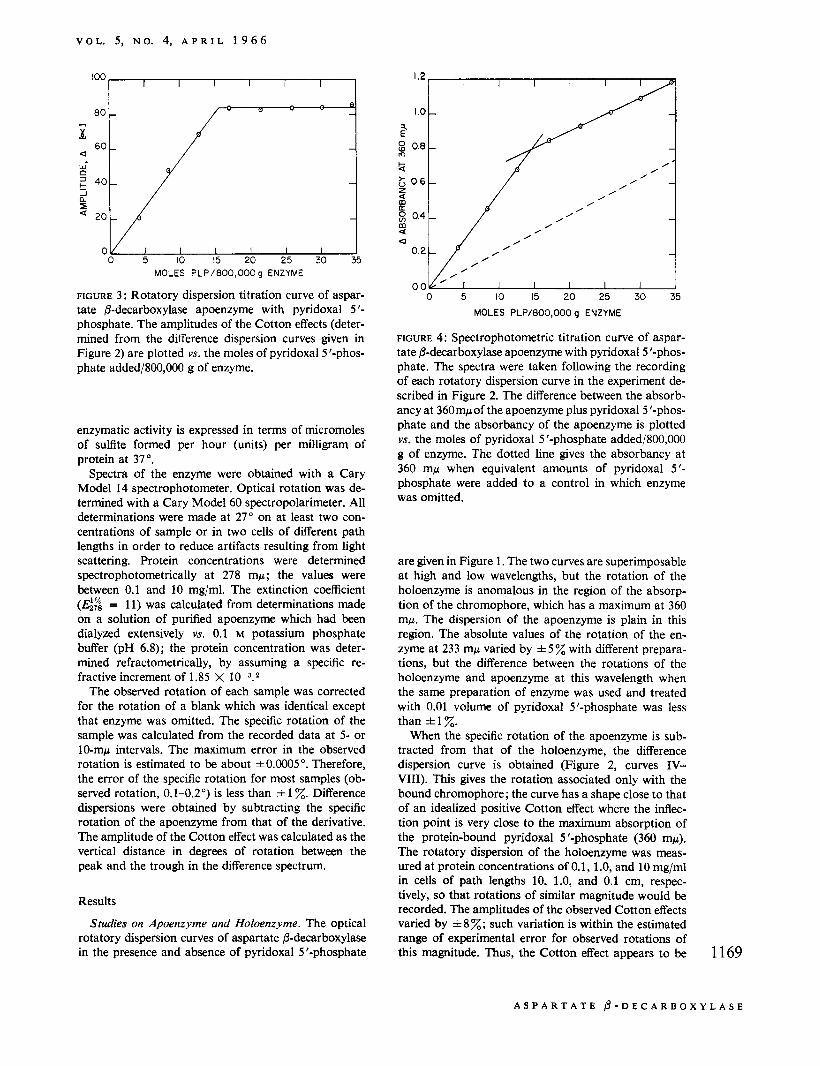
FIGURE 3 : Rotatory dispersion titration curve of aspar- tate &decarboxylase apoenzyme with pyridoxal 5 '- phosphate. The amplitudes of the Cotton effects (deter- mined from the difference dispersion curves given in Figure 2) are plotted vs. the moles of pyridoxal 5'-phos- phate added/800,000 g of enzyme.
enzymatic activity is expressed in terms of micromoles of sulflte formed per hour (units) per milligram of protein at 37".
Spectra of the enzyme were obtained with a Cary Model 14 spectrophotometer. Optical rotation was de- termined with a Cary Model 60 spectropolarimeter. All determinations were made at 27" on at least two con- centrations of sample or in two cells of different path lengths in order to reduce artifacts resulting from light scattering. Protein concentrations were determined spectrophotometrically at 278 mp ; the values were between 0.1 and 10 mg/ml. The extinction coefficient (GZ = 11) was calculated from determinations made on a solution of purified apoenzyme which had been dialyzed extensively YS. 0.1 M potassium phosphate buffer (pH 6.8); the protein concentration was deter- mined refractometrically, by assuming a specific re- fractive increment of 1.85 X 10-3.2
The observed rotation of each sample was corrected for the rotation of a blank which was identical except that enzyme was omitted. The specific rotation of the sample was calculated from the recorded data at 5- or 10-mp intervals. The maximum error in the observed rotation is estimated to be about f0.0005". Therefore, the error of the specific rotation for most samples (ob- served rotation, 0.1-0.2") is less than f 1 %. Difference dispersions were obtained by subtracting the specific rotation of the apoenzyme from that of the derivative. The amplitude of the Cotton effect was calculated as the vertical distance in degrees of rotation between the peak and the trough in the difference spectrum.
Results
Studies on Apoenzyme and Holoenzyme. The optical rotatory dispersion curves of aspartate @-decarboxylase in the presence and absence of pyridoxal 5'-phosphate
1.2 I I I I I
0 5 IO 15 20 25 30 35 MOLES PLP/EOO,OOO g ENZYME
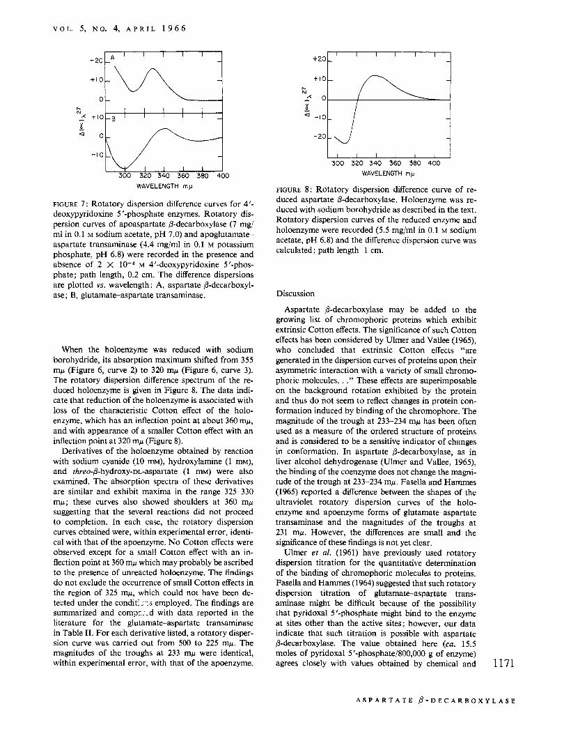
FIGURE 4: Spectrophotometric titration curve of aspar- tate &decarboxylase apoenzyme with pyridoxal 5 '-phos- phate. The spectra were taken following the recording of each rotatory dispersion curve in the experiment de- scribed in Figure 2. The difference between the absorb- ancy at 360mp of the apoenzyme plus pyridoxal 5'-phos- phate and the absorbancy of the apoenzyme is plotted YS. the moles of pyridoxal 5 '-phosphate added/800,000 g of enzyme. The dotted line gives the absorbancy at 360 mp when equivalent amounts of pyridoxal 5 ' - phosphate were added to a control in which enzyme was omitted.
are given in Figure 1. The two curves are superimposable at high and low wavelengths, but the rotation of the holoenzyme is anomalous in the region of the absorp- tion of the chromophore, which has a maximum at 360 mp. The dispersion of the apoenzyme is plain in this region. The absolute values of the rotation of the en- zyme at 233 mp varied by f 5 % with different prepara- tions, but the difference between the rotations of the holoenzyme and apoenzyme at this wavelength when the same preparation of enzyme was used and treated with 0.01 volume of pyridoxal 5'-phosphate was less than * 1 %.
When the specific rotation of the apoenzyme is sub- tracted from that of the holoenzyme, the difference dispersion curve is obtained (Figure 2, curves IV- VIII). This gives the rotation associated only with the bound chromophore; the curve has a shape close to that of an idealized positive Cotton effect where the inflec- tion point is very close to the maximum absorption of the protein-bound pyridoxal 5 '-phosphate (360 mp). The rotatory dispersion of the holoenzyme was meas- ured at protein concentrations of 0.1, 1.0, and 10 mg/ml in cells of path lengths 10, 1.0, and 0.1 cm, respec- tively, so that rotations of similar magnitude would be recorded. The amplitudes of the observed Cotton effects varied by *8z; such variation is within the estimated range of experimental error for observed rotations of this magnitude. Thus, the Cotton effect appears to be 1169
A S P A R T A T E @ - D E C A R B O X Y L A S E
B I O C H E M I S T R Y
0 I I I I I I
- 100 c _____.---- 2 I +&-- I
1170
-4(
u 300 320 340 360 380 400 4M 440
WAVELENGTH IN m y
FIGURE 5 : Effect of pyridoxamine 5'-phosphate and pyridoxamine 5 '-phosphate plus a-ketoglutarate on the rotatory dispersion of aspartate @-decarboxylase. Rotatory dispersion was determined after incubation of a solution of the apoenzyme (12 mgiml) in 0.1 M
potassium phosphate (pH 6.8) containing the indicated additions for 30min at 37"; path length 0.2 cm: curve 1, apoenzyme; curve 2, apoenzyme plus 1 mM pyridox- amine 5'-phosphate; curve 3, apoenzyme plus 1 mM pyridoxamine phosphate and 1 mM a-ketoglutarate.
independent of protein concentration over the range studied. The amplitude of the Cotton effect was also independent of pH between 5.7 and 7.7.
The several curves shown in Figure 2 were obtained in an experiment in which the apoenzyme was treated with increasing amounts of pyridoxal 5 '-phosphate. In Figure 3, the amplitude of the Cotton effect is plotted YS. the moles of pyridoxal 5 '-phosphate added/800,000 g of enzyme. [A molecular weight of about 800,OOO has previously been estimated for this enzyme (Novo- grodsky and Meister, 1964a).] No significant increase in amplitude was observed after the fourth addition of pyridoxal 5 '-phosphate. The amplitude is proportional to the amount of pyridoxal 5'-phosphate added until a saturation point is reached at about 15.5 moles of pyridoxal 5 '-phosphate/800,000 g of protein. The spectrum of the enzyme was recorded after each of the additions of pyridoxal 5 '-phosphate carried out in the experiment described in Figure 2. In Figure 4, the ab- sorbancy at 360 mp minus the absorbancy of the apo- enzyme at this wavelength is plotted vs. the moles of pyridoxal 5 '-phosphate added/800,000 g of enzyme. This curve changes slope sharply after about 15 moles of pyridoxal 5'-phosphate had been added/800,000 g of enzyme. The slope after the sharp break is close to that of a control in which enzyme was omitted. Although free pyridoxal 5 '-phosphate exhibits maximum absorbancy at 388 mp, it exhibits appreciable absorption at 360 mp; thus, the curve shown in Figure 4 reflects the presence of both enzyme-bound and free pyridoxal 5 ' - phosphate. The dotted line represents the absorbancy of free pyridoxal 5 '-phosphate.
Studies on Other Forms of the Enzyme. As shown in Figure 5 (curve 2), the rotatory dispersion curve ob- tained after incubation of the apoenzyme with 1 mM pyridoxamine 5 '-phosphate was very similar to that exhibited by the apoenzyme. The available data indicate that pyridoxamine 5'-phosphate is bound to the en-
WAVELENGTH m y
FIGURE 6: Absorption spectra of aspartate @-decar- boxylase derivatives. Enzyme concentration is 7.2 mg/ml in 0.1 M sodium acetate (pH 6.8): curve 1, apoenzyme; curve 2, holoenzyme; curve 3, sodium borohydride reduced derivative; curve 4, 4 '-deoxypyridoxine 5'- phosphate derivative.
zyme under these conditions. Thus, the K, for pyridox- amine 5'-phosphate is about 0.1 mM, and, in the assay system used here (which contains a-ketoglutarate), pyridoxal 5 '-phosphate and pyridoxamine 5 '-phosphate are equally effective cofactors when studied at a con- centration of 0.3 mM. After incubation of the enzyme with 1 mM pyridoxamine 5'-phosphate followed by dialysis overnight, the spectrum exhibited a small maxi- mum at 325 mp indicating enzyme-bound pyridoxamine 5 '-phosphate; a small peak at 360 mp was also observed, suggesting that some of the pyridoxamine 5 '-phosphate had been converted to pyridoxal 5 '-phosphate during dialysis. When the enzyme was incubated with both 1 mM pyridoxamine 5'-phosphate and 1 mM cr-keto- glutarate (Figure 5, curve 3) an anomalous dispersion curve characteristic of the holoenzyme was observed.
4'-Deoxypyridoxine 5 '-phosphate is an inhibitory analog of pyridoxal 5 '-phosphate for both aspartate 6-decarboxylase (Novogrodsky and Meister, 1964b) and glutamate-aspartate transaminase (Meister et at., 1954). The spectrum of the 4'-deoxypyridoxine 5'- phosphate derivative of aspartate @-decarboxylase (Figure 6, curve 4) exhibits a maximum at 315 mp, while that of the corresponding derivative of glutamate- aspartate transaminase is maximal at 320 mp. The dif- ference dispersion curves for the 4'-deoxypyridoxine 5'-phosphate derivatives of these enzymes are given in Figure 7. Both derivatives exhibited small3 but definite Cotton effects with points of inflection at about 320 mp. Under the experimental conditions employed, the amplitude of the Cotton effects varied between 10 and 20" for aspartate 6-decarboxylase and between 20 and 30" for glutamate-aspartate transaminase. The varia- tion in amplitudes observed may be ascribed to ex- perimental error inherent in the calculation of small differences between large numbers.
a The small Cotton effects observed with the 4'-deoxypyridox- h e 5'-phosphate enzyme and the NaBH~reduced enzyme were not detected under the conditions employed for the studies described in our preliminary report (Wilson and Meister, 1965).
E D I T H M. W I L S O N ANT) A L T O N M E I S T ' E R
V O I . . 5, N O . 4, A P R I L 1 9 6 6
I I I I
IC N I I I I I
B I ,..
WAVELENGTH rnp
FIGURE 7 : Rotatory dispersion difference curves for 4’- deoxypyridoxine 5 ’-phosphate enzymes. Rotatory dis- persion curves of apoaspartate /?-decarboxylase (7 mg/ ml in 0.1 M sodium acetate, pH 7.0) and apoglutamate- aspartate transaminase (4.4 mg/ml in 0.1 M potassium phosphate, pH 6.8) were recorded in the presence and absence of 2 X M 4’-deoxypyridoxine 5’-phos- phate; path length, 0.2 cm. The difference dispersions are plotted vs. wavelength : A, aspartate P-decarboxyl- ase; B, glutamate-aspartate transaminase.
When the holoenzyme was reduced with sodium borohydride, its absorption maximum shifted from 355 mp (Figure 6, curve 2) to 320 mp (Figure 6, curve 3). The rotatory dispersion difference spectrum of the re- duced holoenzyme is given in Figure 8. The data indi- cate that reduction of the holoenzyme is associated with loss of the characteristic Cotton effect of the holo- enzyme, which has an inflection point at about 360 mp, and with appearance of a smaller Cotton effect with an inflection point at 320 mp (Figure 8).
Derivatives of the holoenzyme obtained by reaction with sodium cyanide (10 m ~ ) , hydroxylamine (1 mM), and threo-P-hydroxy-DL-aspartate (1 m ~ ) were also examined. The absorption spectra of these derivatives are similar and exhibit maxima in the range 325-330 mp; these curves also showed shoulders at 360 mp suggesting that the several reactions did not proceed to completion. In each case, the rotatory dispersion curves obtained were, within experimental error, identi- cal with that of the apoenzyme. No Cotton effects were observed except for a small Cotton effect with an in- flection point at 360 mp which may probably be ascribed to the presence of unreacted holoenzyme. The findings do not exclude the occurrence of small Cotton effects in the region of 325 mp, which could not have been de- tected under the condit: :-:s employed. The findings are summarized and corn$:;-d with data reported in the literature for the glutamate-aspartate transaminase in Table 11. For each derivative listed, a rotatory disper- sion curve was carried out from 500 to 225 mp. The magnitudes of the troughs at 233 mp were identical, within experimental error, with that of the apoenzyme.
I I I I I I I I 300 320 340 360 300 400
WAVELENGTH my
FIGURE 8: Rotatory dispersion difference curve of re- duced aspartate @-decarboxylase. Holoenzyme was re- duced with sodium borohydride as described in the text. Rotatory dispersion curves of the reduced enzyme and holoenzyme were recorded (5.5 mg/ml in 0.1 M sodium acetate, pH 6.8) and the difference dispersion curve was calculated; path length 1 cm.
Discussion
Aspartate 6-decarboxylase may be added to the growing list of chromophoric proteins which exhibit extrinsic Cotton effects. The significance of such Cotton effects has been considered by Ulmer and Vallee (1965), who concluded that extrinsic Cotton effects “are generated in the dispersion curves of proteins upon their asymmetric interaction with a variety of small chromo- phoric molecules. . .” These effects are superimposable on the background rotation exhibited by the protein and thus do not seem to reflect changes in protein con- formation induced by binding of the chromophore. The magnitude of the trough at 233-234 mp has been often used as a measure of the ordered structure of proteins and is considered to be a sensitive indicator of changes in conformation. In aspartate P-decarboxylase, as in liver alcohol dehydrogenase (Ulmer and Vallee, 1965), the binding of the coenzyme does not change the magni- tude of the trough at 233-234 mp. Fasella and Hammes (1965) reported a difference between the shapes of the ultraviolet rotatory dispersion curves of the holo- enzyme and apoenzyme forms of glutamateaspartate transaminase and the magnitudes of the troughs at 231 mp, However, the differences are small and the significance of these findings is not yet clear.
Ulmer er al. (1961) have previously used rotatory dispersion titration for the quantitative determination of the binding of chromophoric molecules to proteins. Fasella and Hammes (1964) suggested that such rotatory dispersion titration of glutamateaspartate trans- aminase might be difficult because of the possibility that pyridoxal 5’-phosphate might bind to the enzyme at sites other than the active sites; however, our data indicate that such titration is possible with aspartate 8-decarboxylase. The value obtained here (ca. 15.5 moles of pyridoxal 5 ’-phosphate/800,000 g of enzyme) agrees closely with values obtained by chemical and 1171
A S P A R T A T E P - D E C A R B O X Y L A S E
B I O C H E M I S T R Y
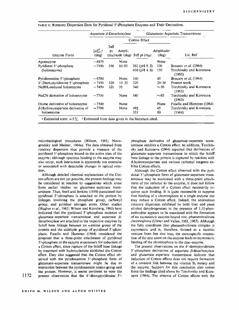
TABLE 11 : Rotatory Dispersion Data for Pyridoxal 5 '-Phosphate Enzymes and Their Derivatives.
Oxime derivative of holoenzyme P-Hydroxyaspartate derivative of
holoenzyme
-6870 -7190
-6780 - 7450 - 7450
- 7700
- 7500 - 7700
None 358 60-85
None 320 10-20 320 35
None
None None
362 (pH 8.2) 430 (pH 4.8)
340 320 340
340
492 333
None 100 150
45 20-30 -50
-65
None 45 80
Breusov et af. (1964) Torchinsky and Koreneva
Breusov et a[. (1964) Present work Torchinsky and Koreneva
Torchinsky and Koreneva
Fasella and Hammes (1964) Torchinsky and Koreneva
(1963)
(1 963)
(1963)
(1 964)
a Estimated error f 5 %. b Estimated from data given in the literature cited.
1172
microbiological procedures (Wilson, 1963 ; Novo- grodsky and Meister, 1964a). The data obtained from rotatory dispersion thus provide a measure of the pyridoxal 5 '-phosphate bound to the active sites of the enzyme; although spurious binding to the enzyme may also occur, such interaction is apparently not extensive or associated with detectable changes in optical rota- tion.
Although detailed chemical explanations of the Cot- ton effects are not yet possible, the present findings may be considered in relation to suggestions which arose from earlier studies on glutamate-aspartate trans- aminase. Thus, Snell and Jenkins (1959) postulated that pyridoxal 5 '-phosphate is attached to the protein by linkages involving the phosphate group, carbonyl group, and pyridine nitrogen atom. Other studies (Hughes et al., 1962; Wilson and Kornberg, 1963) have indicated that the pyridoxal 5 '-phosphate moieties of glutamate-aspartate transaminase and aspartate 8- decarboxylase are attached to the respective enzymes by Schiff base linkage between an e-amino group of the protein and the aldehyde group of pyridoxal 5'-phos- phate. Fasella and Hammes (1964) considered the proposal that a three-point attachment of pyridoxal 5'-phosphate to the enzyme is necessary for induction of a Cotton effect, since rupture of the Schiff base linkage by treatment with hydroxylamine abolished the Cotton effect. They also suggested that the Cotton effect ob- served with the pyridoxamine 5 '-phosphate form of glutamateaspartate transaminase might be due to interaction between the pyridoxamine amino group and the protein. However, it Seems pertinent to note the present observation that the 4'-deoxypyridoxine 5 '-
phosphate derivative of glutamate-aspartate trans- aminase exhibits a Cotton effect. In addition, Torchin- sky and Koreneva (1964) reported that derivatives of glutamateaspartate transaminase in which the Schiff base linkage to the protein is ruptured by reaction with P-hydroxyaspartate and various carbonyl reagents ex- hibit Cotton effects.
Although the Cotton effect observed with the pyri- doxal 5 '-phosphate form of glutamateaspartate trans- aminase may be associated with a three-point attach- ment of the cofactor to the enzyme, it does not follow that the induction of a Cotton effect necessarily re- quires such binding. It is quite reasonable to suppose that binding of a chromophore at a single enzyme site may induce a Cotton effect. Indeed, the anomalous rotatory dispersion exhibited by both liver and yeast alcohol dehydrogenases in the presence of 1,lO-phen- anthroline appears to be associated with the formation of an asymmetric enzyme-bound zinc-phenanthrolinate chromophore (Ulmer and Vallee, 1961,1965). Although the fully coordinate zinc-phenanthrolinate complex is asymmetric and is, therefore, formed as a racemic mixture from free zinc ions, the stereospecific orienta- tion of the zinc atom on the enzyme leads to asymmetric binding of the chromophore to the zinc-enzyme.
The present observations on the 4'-deoxypyridoxine 5 '-phosphate derivatives of aspartate P-decarboxylase and glutamateaspartate transaminase indicate that induction of Cotton effects does not require formation of a covalent link between the vitamin B6 moiety and the enzyme. Support for this conclusion also comes from the findings cited above by Torchinsky and Kore- neva (1964). The absence of Cotton effects with the
E D I T H M. W I L S O N A N D A L T O N M E I S T E R
V O L . 5, N O . 4, A P R I L 1 9 6 6
cyanohydrin, oxime, and P-hydroxyaspartate deriva- tives of aspartate @-decarboxylase may indicate that these forms of the enzyme do not possess sufficient asymmetry to induce Cotton effects that can be de- tected under the experimental conditions employed. It is technically difficult to detect small Cotton effects which exhibit points of inflection appreciably lower than 360 mp; the sensitivity of the procedure cannot be greatly increased by using higher concentrations of protein because of the high absorbancy of the chromophoric protein. Circular dichroism (Breusov et al., 1964; John- son and Graves, 1965) may be a more effective tool for further studies in this area.
A striking difference between aspartate P-decar- boxylase and glutamate-aspartate transaminase lies in the markedly different optical rotatory dispersions observed with the pyridoxamine 5 '-phosphate forms of these enzymes. Thus, no Cotton effect was observed with the pyridoxamine 5 '-phosphate form of aspartate @-decarboxylase, while a substantial effect has been reported for the pyridoxamine 5'-phosphate of glu- tamate-aspartate transaminase. Previous studies have shown that the affinity of glutamate-aspartate trans- aminase for pyridoxamine 5'-phosphate is about the same as that for pyridoxal 5'-phosphate ( K , 4.4 X 10⁻⁶ M) (Meister et al., 1954). In contrast, experiments on aspartate @-decarboxylase indicate that the K,, value for pyridoxamine 5 '-phosphate is approximately M or about one thousand times greater than that for pyridoxal 5'-phosphate. Under the conditions of the present studies, a concentration of pyridoxamine 5 '- phosphate was used that was sufficient to saturate the apoenzyme. However, the marked differences in the values of K , for pyridoxal 5'-phosphate and pyridox- amine S'-phosphate, and the finding that (in contrast to glutamate-aspartate transaminase) the pyridoxamine 5 '-phosphate of aspartate @-decarboxylase dissociates readily from the enzyme (Novogrodsky and Meister, 1964a), suggest that there is a significant difference be- tween these enzymes in the manner in which pyridox- amine 5 '-phosphate is bound. Presumably, the pyridox- amine 5 '-phosphate of the transaminase is bound more tightly and with greater asymmetry than that of the decarboxylase. It seems to be of significance that aspartate @-decarboxylase and glutamateaspartate transaminase exhibit approximately equal affinity for 4'-deoxypyridoxine 5 '-phosphate and that extrinsic Cotton effects of about the same magnitude were ob- served with both enzymes.
In conclusion, the present work shows that determina- tions of optical rotatory dispersion can serve usefully in quantitative studies on the binding of pyridoxal 5 ' - phosphate. Optical rotatory dispersion titration offers a distinct advantage as compared to spectrophotometric titration in that there is no background due to unbound cofactor. Such an advantage would be of greater sig- nificance in studies on pyridoxal 5 '-phosphate enzymes that exhibit maximum absorbancy at wavelengths rela- tively close to that of free pyridoxal 5 '-phosphate. The present studies also indicate that optical rotatory disper- sion is a useful tool for the investigation of the binding
of cofactors and cofactor analogs to aspartate @- decarboxylase, and also for the comparison of binding phenomena of various pyridoxal 5 '-phosphate enzymes. Optical rotatory dispersion may prove useful in examin- ing the effects of inhibitors that affect coenzyme bind- ing. Thus, dispersion measurements used together with information derived from kinetic, spectrophotometric, and chemical approaches may provide farther insight into the structure and function of the active site of the pyridoxal 5 '-phosphate enzymes.
Acknowledgment
The authors are indebted to Dr. Bert L. Vallee and to Dr. David Ulmer for their valuable criticisms of this work.
References
Breusov, Yu. N., Ivanov, V. I. , Karpeisky, M. Ya., and Morozov, Yu. V. (1964), Biochim. Biophys. Acta 92, 388.
Fasella, P., and Hammes, G. G. (1964), Biochemistry 3, 530.
Fasella, P., and Hammes, G. G. (1965), Biochemistry 4, 801.
Grant, W. M. (1947), Anal. Chem. 19, 345. Hughes, R. C., Jenkins, W. T., and Fischer, E. H.
(1962), Proc. Natl. Acad. Sei. U. S . 48, 1615. Johnson, G. F., and Graves, D. J. (1965), Abstracts,
150th National Meeting of the American Chemical Society, Atlantic City, N. J., Sept 1965, p 97C.
Lowry, 0. H., Rosebrough, N. J., Farr, A. L., and Randall, R. J. (1951), J . Biol. Chem. 193, 265.
Meister, A., Sober, H. A., and Peterson, E. A. (1954), J . Biol. Chem. 206, 89.
Novogrodsky, A., and Meister, A. (1964a), J . Biol. Chem. 239, 879.
Novogrodsky, A., and Meister, A. (1964b), Biochim. Biophys. Acta 85, 170.
Novogrodsky, A., Nishimura, J. S., and Meister, A. (1963), J . Biol. Chem. 238, PC1903.
Peterson, E. A., and Sober, H. A. (1962), Methods Enzymol. 5 , 3.
Richards, E. G., and Schachman, H. K. (1959), J . Phys. Chem. 63, 1578.
Sizer, I., and Jenkins, W. T. (1962), Methods Enzymol. 5,677.
Snell, E. E., and Jenkins, W. T. (1959), J . Cellular Comp. Physiol. 54 (Suppl. l), 161.
Soda, K., Novogrodsky, A., and Meister, A. (1964), Biochemistry 3, 1450.
Torchinsky, Yu. M., and Koreneva, L. G. (1963), Biokhimiya 28, 1087.
Torchinsky, Yu. M., and Koreneva, L. G. (1964), Biochim. Biophys. Acta 79, 426.
Ulmer, D. D., Li, T. K., and Vallee, B. L. (1961), Proc. Natl. Acad. Sci. U. S . 47, 1155.
Ulmer, D. D., and VaUee, B. L. (1961), J . Biol. Chem. 236, 730.
Ulmer, D. D., and Vallee, B. L. (1965), Advan. Enzymol. 27,37. 1173
A S P A R T A T E 0 - D E C A R B O X Y L A S E
B I O C H E M I S T R Y
Wada, H., and Snell, E. E. (1962), J . B i d . Chem. 237,
Wilson, E. M. (1963), Biochim. Biophys. Acta 67, 345. Wilson, E. M., and Kornberg, H. L. (1963), Biochem. J .
88,578. 127. Wilson, E. M., and Meister, A. (1965), Abstracts,
150th National Meeting of the American Chemical Society, Atlantic City, N. J., Sept 1965, p 36C.
Protein Synthesis Systems from Rat Brain*
Mary K. Campbell, Henry R. Mahler,: Walter J. Moore, and Sujata Tewari
ABSTRACT: Two distinct cell-free protein synthetic systems have been isolated from brain tissue of 18-21 day old white rats, one based on purified ribosomes and the other on mitochondria. The ribosomal system was typical in its requirements for pH 5 enzymes and an exogenous source of adenosine triphosphate (ATP), and its almost complete inhibition by ribonuclease (RNA- ase).
The mitochondrial system was one of the most active yet reported. It had no requirement for pH 5 enzymes or exogenous ATP, was not inhibited by RNAase, but was moderately inhibited by 0.5 pmole of dinitrophenol, 1.6 mpmoles of rotenone, or 2 pg of ant’- mycin-A per ml. The activity was sensitive to the concen- trations of inorganic phosphate, adenosine diphosphate
T wo distinct cell-free protein synthetic sysiems have been isolated from immature rat brain, one localized in purified ribosomes and one in mitochondria. These systems, both highly active, differ in various biochemical properties. The purpose of this paper is to describe and contrast the properties of these two systems, as a con- tribution to an eventual understanding of functional aspects of protein synthesis in brain.
Ribosomal System
There have been several recent studies of ribosomal protein synthetic systems from brain, including those of Zomzely et al. (1964) on rat, of Rubin and Stenzel (1965) on rabbit, and of Murthy and Rappoport (1965a) on rat. A cell-free microsomal system from guinea pig brain has been characterized by Satake et al. (1964).
Experimental Procedures We used immature rats ir view of reports (Murthy
_ _ ~ -
* From the Chemical Laboratory, Indiana University, Bloom- ington, Indiana. Received November 4, 1965. This work was supported by the National Science Foundation and the U. S. Office of Naval Research; presented at the International Neuro- chemical Conference, Oxford, July 26, 1965.
1 174 $ Research Career Award (PHSGM-05060) of the USPHS.
(ADP), and ATP in the medium. Various neurochemi- cals had specific effects, notably stimulation by 7- aminobutyric acid in both systems. Proteins from the ribosomal system were fractionated on DEAE-cellulose columns, yielding about 3.5% of the label in a soluble acidic protein fraction. Most of the label, however, was in a protein fraction associated with ribosomal ribo- nucleic acid (RNA). In presence of an artificial messen- ger, polyuridylic acid, the system incorporated [ 14C]- phenylalanine at several times the standard rate, pro- vided the ribosomes were first incubated in a KCI medium low in ATP. Relations of these protein syn- thetic systems to functional activity of neurons are considered, including a possible role of mitochondria in synthesis of synaptic vesicle proteins.
and Rappoport, 1965b) that immature rat brains pro- vided more active enzyme extracts. The rats were male, Sprague-Dawley strain (Simonsen Laboratories, White Bear Lake, Minn.), 18-21 days old, approxi- mately 45 g. The preparation of ribosomes followed that devised by Munro et al. (1964) for the liver system, with a fractionation procedure as outlined in Figure 1, Elec- tron micrographs of the ribosomal preparations, for example, Figure 2, show a fairly high proportion of double ribosomes as well as some larger aggregates. Ultracentrifuge data on this point will be discussed later.
The standard ribosomal system is summarized in Table I. Ribosomes (0.6-0.7 mg of protein) and pH 5 enzymes (1.0-1.5 mg of protein) were separately sus- pended in medium M. The ATP1 and GTP, pH 5 en- zymes, and ribosomes were added in that order to medium M in 13 mm X 10 cm tubes held in a constant temperature block, and the run was started by addition of labeled [lF]leucine. The reaction was stopped after