The human eye is a paradigmatic example of a rela-tively simple optical instrument providing excep-tional functionality. But even with the simplicity ofocular optics, there was a long process in reachingthe nearly complete understanding we have today.The optics of the eye resemble an aplanatic design[1] with a partial correction of both spherical aberra-tion and coma. This is due to a natural balance be-tween the aberrations of the cornea and the lensin the young eye [2], which is progressively lost dur-ing normal aging [3]. Although interest in the eye isintrinsic to human nature, it was around Galileo’stime when scientific exploration of the eye began.From then, it continuously advanced, in part dueto the great evolution of artificial optical instru-ments, paradoxically developed to actually bypassthe eye’s limitations of resolution.
At the time of Galileo’s first use of the telescope forastronomical observations, there was already a basicunderstanding of some of the refractive errors of thehuman eye [4,5]. It was evident that a normal emme-tropic eye was most comfortable viewing light origi-nating from a far distance. This observation was one
of the reasons why the Galilean telescope workedadequately when a secondary negative lens (Galileaneyepiece) was placed in combination with a largefocal length positive lens (objective lens).
In the years to come, three factors were identifiedto limit the optical performance of the Galilean tele-scope [6–8]: a limited field of view—intrinsic to a tele-scope with a negative eyepiece–and chromatic andspherical aberrations (also, some historical studiessuggest that coma might have been an issue forGalileo’s observations). Concerning these three de-fects, only the field of view in the eye is superiorto the Galilean telescope. However, the human eyeis affected by chromatic and spherical aberrations,coma, and other higher order aberrations.
In the case of the telescope, the impact of Galileo’sdiscoveries generated a “telescope race,” quickly im-proving the optical quality of the instrument: in-creasing the field of view with Kepler’s eyepiecedesign, suppressing chromatic effects with Newton’sreflective designs, and minimizing aberrations withthe adequate surface shape calculated after Snell’slaw. But in the case of the human eye, understandingand correction of ocular aberrations followed a slowerevolution. Previous to Galileo’s time, spectacles forthe correction of presbyopia and myopia were al-ready used and sold by Italian glassmakers in Flor-ence [4,5] based on empirical testing, until Kepler
correctly described how spherical lenses compensatemyopic and hyperopic refractive errors. However, ittook a long time to characterize and correct astigma-tism. At the beginning of the nineteenth century,Thomas Young correctly described the astigmatismof his left eye [9], and it took almost 30 years to findthe appropriate astigmatic correction. It was Airy, in1827, who suggested the use of cylindrical-shapedsurfaces to compensate for refractive errors alongcertain meridians, a spectacle design that is still verycommon today [5].
The ocular spherical and chromatic aberrationswere both well known, due to the limitation they im-posed on telescopes and microscopes. The first obser-vation of spherical aberration in the human eyeseems to be made by Thomas Young in his famous1801 publication “On the mechanism of the eye”[9,10] and later by Helmholtz in his Treatise on Phys-iological Optics in 1855. The chromatic effects in theeye were initially mentioned by Newton [11] andlater by Young [12]. However, the correction of chro-matic aberration by using achromatizing lenses wasonly tried in the middle of the twentieth century [13],and some variations of corrector providing wide-angle performance were proposed even recently [14].
In this article, we provide a brief historical per-spective of how we gained knowledge of the opticalcomponents of the eye, the cornea, and the crystal-line lens, describing the on-axis monochromatic aber-rations of each component, and the step-by-stepprocedure of buildingmore andmore accurate opticalmodels of the eye, until the current understanding ofthe eye’s optics.
2. Brief History of Main Advances on Optics of theEye
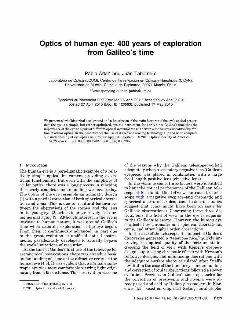
In the eye, only two lenses are used to focus lightfrom a distance source on the retina. If comparedto an artificial optical system designed for the samepurpose (for instance, a teleobjective lens), this num-ber may appear as too small (see a schematic repre-sentation in Fig. 1). The number of elementsmounted in the artificial optical system is due to amore or less demanding correction of aberrations.Whether this type of optimization strategy was alsopresented in the human eye could not be elucidateduntil a characterization of each separate componentwas given, and that took 400 years from Galileo’stime.
In a simplified historical revision [15], Galileo’scontemporaries, Kepler, Scheiner, and Descartes,contributed to the initial understanding of the eyeas an optical instrument, realizing that the imageon the retina was inverted and providing the first de-scription of the optical components. Later, Huygensbuilt a physical eye model made of two hemispheresfilled with water and a diaphragm. But, it was only atthe beginning of the nineteenth century when Tho-mas Young produced the first geometric optics de-scription of the cornea and the lens. The radii ofcurvature of corneal and lens surfaces, as well as
the anterior chamber depth and the refractive indexvalues, were strikingly well estimated for thosetimes. Later, Moser (1844) and Listing (1851) builtschematic eyes using spherical surfaces to describethe cornea and lens. The Listing eye model was im-proved by Helmholtz modifying the positions of thelens surfaces. Those schematic eyes were three sur-face models, one for the cornea and two for the crys-talline lens. The radius of curvature of the posteriorcorneal surface was measured for the first time byTscherning, and he described the first four-surfaceschematic eye model in 1900. After that, based onthe improvement of the techniques for measuringthe eye curvatures and axial distances, several eyemodels were proposed in the past century, and someof them became very popular, such as the Gullstrandmodel discussed in Ref. [16], the Le Grand [17] andthe Emsley [18] eye models. The use of these spheri-cal models should be restricted to the paraxial geo-metric optics. They were used to estimate thecardinal planes and points of the eye, to designnew spectacles and, after the first modern cataractsurgery {in 1949 an intraocular lens (IOL) was im-planted in an eye for the first time [19]}, to calculatethe power of the IOL.
However, these simple models could not be used tofully understand the aberrations of each component.Because the surfaces used were rotationally symme-trical spheres, ray tracing through the schematiceyes would immediately show that the predicted val-ues did not agree with the measurements. The accu-racy of the models was restricted to the paraxialoptics. After nearly 350 years, the actual aberrationsof the cornea and the lens were largely unknown.
The first attempt to localize where astigmatismoriginates in the eye was performed by Young in1801 [9]. His scientific curiosity led him to questionif his own astigmatism was originated either by the
Fig. 1. (Color online) Schematic example of a human eye modelcompared with an artificial optical objective.
cornea or by the crystalline lens. He noticed thatwhen immersed in water (with a similar refractiveindex as the cornea), he would cancel the cornealcontribution, and since the astigmatism persisted,he assumed it was induced by the crystalline lens.
The sources of aberrations in the eye were not stu-died until the decade of 1970 (previously, in 1961Smirnov had measured the wave aberration of thehuman eye for the first time [20] with a vernier-typesubjective method). At this time, the first versions ofcorneal topographers (based on the deformations ob-served in the reflected image of a series of concentricrings at the cornea) began to be used as ophthalmictools. This permitted the reconstruction of the firstcorneal surface shape, and therefore the calculationof corneal (first surface) aberrations. In 1973, ElHage and Berny [21], using one of these instruments,estimated the spherical aberration of the cornea, ob-taining positive values, i.e., the peripheral rays crossthe optical axis in front of the paraxial rays. Becausethere was no direct access to the crystalline lensin vivo, they measured spherical aberration of the to-tal eye using a Foucault test (also a well-known tech-nique from telescope optics). When they comparedboth sets of data, they realized that the sphericalaberration of the cornea was much higher than thespherical aberration of the complete eye. They in-ferred that the spherical aberration of the crystallinelens should have negative values, providing a bal-ance of the positive spherical aberration of the cor-nea. An experiment performed later by Millodot andSivak [22], using goggles filled with water to cancelthe corneal contribution (similar to what Young didover 200 years before), obtained more variability inthe sign of the lens spherical aberration. In 1993,Tomlinson et al. [23] confirmed the results from ElHage and Berny [21] using a mixed objective/psycho-physic technique.
Some of the advances in the instruments to assessobjectively the optical quality of the eye allowed us torevisit this issue in the 1990s. By using the double-pass technique [24,25], we suggested [26] that notonly the spherical aberration, but also other higherorder aberrations, could be balanced between the cor-nea and the lens. The introduction of the Hartmann–Shack wavefront sensor to measure the aberrationsof the eye [27,28] permitted the design of additionalexperiments, including revisiting the measurementsof the aberrations of the eye immersed in water[29,30].
In addition, the use of improved Hartmann–Shackwavefront sensors allowed us to perform real-timeanalysis of the aberrations [31] and to build adaptiveoptics systems for the human eye [32–34].
The advances on the understanding of the opticalcomponents led to a higher degree of complexity inthe elaboration of the schematic eye models. Tryingto improve the prediction of the spherical aberrationmeasurements, several authors incorporated conicsurfaces to describe the cornea and the lens [35–37].The corneal asphericity values were taken from cor-
neal topography measurements, but the lens surfaceasphericity values were somehow difficult to mea-sure, and, in many cases, they were used as variablesto adjust the model to certain values or they weremeasured from ex vivo lenses. Today, the exact valuesof asphericity of the lens surfaces are still a matter ofdiscussion, with only available data from opticallycorrected Scheimpflug images [38] that show largestandard deviation errors in repeatability. Addi-tional advances in the schematic eyes added a refrac-tive gradient index to the lens [39–41]. However, themeasurements of this parameter are also difficultand scarce. It is usually incorporated into the opticalmodels via optimization to fit a more easily measur-able variable, such as the peripheral refractive er-rors. The new models also benefited from advancesin the understanding of the chromatic properties ofthe eye, either the longitudinal chromatic aberrationor the lateral chromatic aberration [42–44]. Anotheraspect traditionally modeled is accommodation, fromthe purely paraxial models (see, for instance, theBennett and Rabbets eye model [45] that consideronly changes in lens curvature) to more sophisticatedeye models that include the changes in thickness, as-phericity, and refractive index as a function of accom-modation [37,46].
In general, the latest schematic eyemodels provideresults that are close to an average population. How-ever, due to the individual variability in the aberra-tions, they might not be individually accurate. Newcustomized eye models incorporating individual datawill be discussed in the next section.
3. Current Understanding of Eye Optics:Aplanatic Design
The magnitude of the eye’s higher order aberrations(beyond defocus and astigmatism) only representsapproximately around 10% of the total aberrationsof the normal eye [47]. Although a large variabilitybetween subjects is typically presented, the effectof these aberrations is crucial to degrade the retinalimage quality of the human eye. In terms of statisticsin a relatively large population [47–49], only spheri-cal aberration has a slightly positive mean value dif-ferent from zero. However, of interest is not only theglobal magnitude of aberrations in the eye, but alsothe contribution to those aberrations of the ocularcomponents.
In this respect, two aberration terms show a signif-icant level of compensation between cornea and lens:spherical aberration and horizontal coma. In thesetwo cases, the cornea has higher values than the com-plete eye. To understand the underlying mechanismfor this compensation, we must know more about thesources of these aberrations. The crystalline lensmay induce negative spherical aberration mainlydue to three factors: curvature, asphericity, and gra-dient index. Simulations with a lens model with onlyspherical surfaces show that curvature alone cannotbe responsible for inducing negative spherical aber-ration. Therefore, the lens contribution must be
determined by asphericity or gradient index. To whatextent the lens contribution is from either one oranother factor, or a combination of both, is not experi-mentally determined yet.
The improvement of the in vivo imaging tech-niques of the anterior chamber depth might allowus to acquire more precise data of the lens surfaceprofiles that, in combination with other optical pa-rameters, could be used to infer the gradient indexand its actual contribution to the negative sphericalaberration. The understanding of the compensationof coma required more experiments. It was initiallyhypothesized that a particular location of the lens,with respect to the cornea or the gradient refractiveindex of the lens, might induce this effect [29]. How-ever, new experiments indicated that the angularmisalignment between the line of sight (the lineconnecting the center of the entrance pupil andthe fixation stimulus) and the pupillary axis (the lineperpendicular to the cornea passing through the cen-ter of the entrance pupil) was linearly related to thegeneration of coma in both the cornea and the lens[30,50]. This angular misalignment is called the an-gle kappa, or lambda, of the eye, and the averagemagnitude in normal eyes is around 5° [17]. Thoseeyes with a small angle kappa showed nearly nocoma compensation, while those with a large anglekappa had large values of both corneal and lenticularcoma, but with opposite signs.
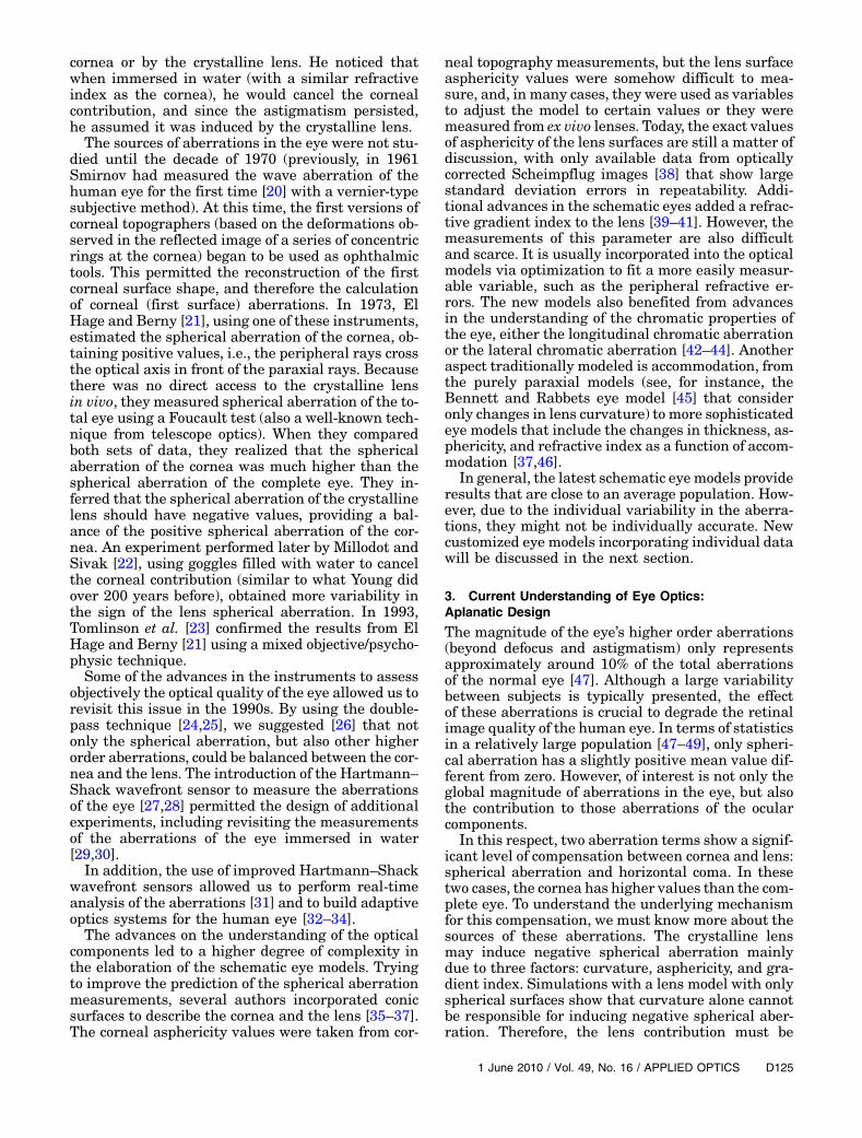
By using a method based on the recording of Pur-kinje images, it was possible to measure the anglekappa together with the lens tilt and decentration[51]. Figure 2 presents some alignment results ina group of normal young eyes from a recent experi-ment [2]. The left panel shows the decentration of
the lens with respect to the center of the pupil.The values are small—around zero (not larger than0:3mm). The values of angle kappa are shown on theright panel. The horizontal component clearly domi-nated over the vertical component and the magni-tudes were around 5°. Individually ray tracingcustomized eye models that introduce the completeactual cornea topography and a crystalline lens withthe corresponding adjusted values of spherical aber-ration for each subject showed that the real values oflens decentration had a very small effect in the com-pensation of coma. However, neglecting the values ofangle kappa in any of the personalized eye modelshad very significant impact on the values of coma.This suggested that coma was “somehow” closelyrelated to the values of angle kappa.
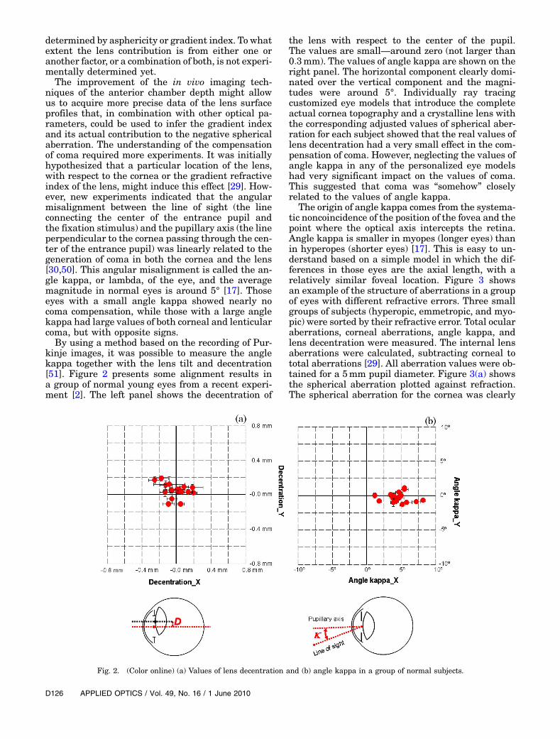
The origin of angle kappa comes from the systema-tic noncoincidence of the position of the fovea and thepoint where the optical axis intercepts the retina.Angle kappa is smaller in myopes (longer eyes) thanin hyperopes (shorter eyes) [17]. This is easy to un-derstand based on a simple model in which the dif-ferences in those eyes are the axial length, with arelatively similar foveal location. Figure 3 showsan example of the structure of aberrations in a groupof eyes with different refractive errors. Three smallgroups of subjects (hyperopic, emmetropic, and myo-pic) were sorted by their refractive error. Total ocularaberrations, corneal aberrations, angle kappa, andlens decentration were measured. The internal lensaberrations were calculated, subtracting corneal tototal aberrations [29]. All aberration values were ob-tained for a 5mm pupil diameter. Figure 3(a) showsthe spherical aberration plotted against refraction.The spherical aberration for the cornea was clearly
Fig. 2. (Color online) (a) Values of lens decentration and (b) angle kappa in a group of normal subjects.
positive, as opposed to the negative values of thelens. The compensation was not affected by the re-fractive error, which is also a consequence of the axialnature of the refractive errors (i.e., the myopic eyesare longer and hyperopic eyes are shorter than thenormal eye). Concerning coma, compensation is ob-served in the horizontal direction [Fig. 3(b)]but mainly for the hyperopic eyes (those with largerangle kappa). Along the vertical direction, Fig. 3(c)data are scattered and the compensation effectwas not statistically significant. The compensationof horizontal coma was related to the angle kappaof the eye [Fig. 3(d)]. The larger the angle, the largerwill be the corneal and lenticular coma, but both withopposite signs. The next step in the understanding ofthe compensation of the aberrations compensationmechanism was to realize that the opposite signsin coma for the cornea and the lens originated fromthe different shape factors of each component [1,2]. Asimplified model using Seidel aberration theory forlenses immersed in nonsymmetrical refractive indexmediums [52] shows that coma depends on the shapeof the components, the linear angular dependence,the position of the object plane, and the refractive in-dices. These results were corroborated with exact raytracing calculations.
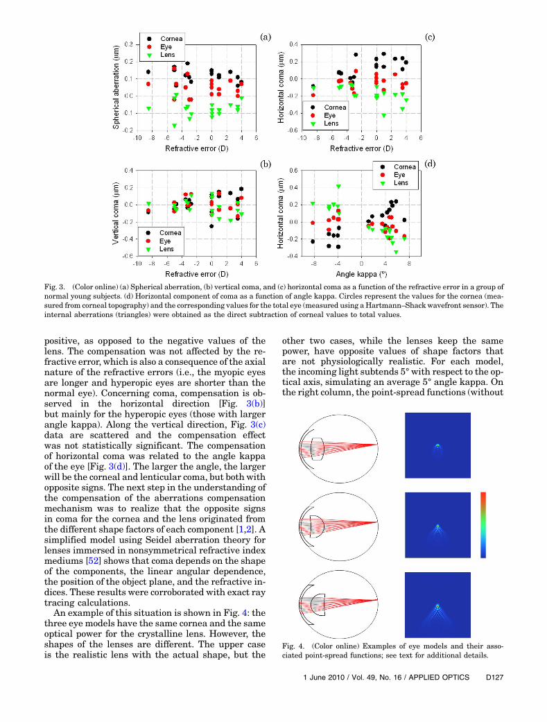
An example of this situation is shown in Fig. 4: thethree eye models have the same cornea and the sameoptical power for the crystalline lens. However, theshapes of the lenses are different. The upper caseis the realistic lens with the actual shape, but the
other two cases, while the lenses keep the samepower, have opposite values of shape factors thatare not physiologically realistic. For each model,the incoming light subtends 5° with respect to the op-tical axis, simulating an average 5° angle kappa. Onthe right column, the point-spread functions (without
Fig. 3. (Color online) (a) Spherical aberration, (b) vertical coma, and (c) horizontal coma as a function of the refractive error in a group ofnormal young subjects. (d) Horizontal component of coma as a function of angle kappa. Circles represent the values for the cornea (mea-sured from corneal topography) and the corresponding values for the total eye (measured using a Hartmann–Shack wavefront sensor). Theinternal aberrations (triangles) were obtained as the direct subtraction of corneal values to total values.
Fig. 4. (Color online) Examples of eye models and their asso-ciated point-spread functions; see text for additional details.
the rotationally symmetrical terms) of each case areshown. The situation yielding the lower amount ofcoma corresponds precisely to the physiologically ac-curate model, while the rest of the lens shapes,although optically possible, produce higher valuesof off-axis coma.
These results support the idea that the eye’s opticsis an aplanatic design, a configuration where spheri-cal aberration and coma are approximately cor-rected. Because the compensation is not perfect, ascan exist in an artificial system, the eye is stillaffected by aberrations.
4. From Eye’s Design to Ophthalmic Corrections:Current and Future Advances
The fact described in the previous section, that cor-neal and crystalline lens aberrations are partiallybalanced, has generated research on the hypotheticalsituations in which this fine tuning might be dis-rupted. Certainly, some modification of either thecornea or the lens can occur in ophthalmic surgerysituations, such as refractive or cataract surgery.Standard refractive surgery procedures can disruptthe aberration compensation, especially in hyperopiccorrections [53], leading to the idea that improvedablation profiles, inducing less aberration, may be re-quired. Cataracts are opacifications of the crystallinelens that typically occur with age. The common cur-rent solution is to replace the opaque lens by an ar-tificial IOL. The first experimental works on thein vivo optical quality of eyes implanted with IOLsshowed an apparent paradox [54,55]. The retinal im-age quality appeared to be similar to that of normalpatients of the same age. IOLs are manufacturedwith high optical quality standards [56], better thanisolated older human lenses. However, when im-planted into the eye, the resulting optical qualitywas not significantly improved. The compensationof aberrations in the normal eye provided an expla-nation to this paradox and, more interestingly, a po-tential solution. The best IOL is not diffractionlimited, but a lens with opposite aberrations to thatof the cornea, mimicking the situation in the youngcrystalline lens. This actually opened a new era forthe optical design of IOLs. Some designs with asphe-rical optics had been proposed before [57], but it waswithin the context of the compensation of aberration[58], with a better understanding of the optics of theeye, when this technology was ready to enter the fieldof ophthalmology [59]. Along these lines, more re-cently we proposed another design of IOL to correctalso the coma generated by the typical kappa anglesin the eye [60]. The idea for this design was to main-tain the appropriate shape factors of the IOL to com-pensate corneal coma for each refractive powermodel. This model also incorporated an aspheric sur-face to compensate for corneal spherical aberration.
The current, better understanding of the eye’s op-tics reviewed in this paper will surely contribute tonew potential practical applications. Virtually everyfuture idea for visual correction will need to consider
carefully the optical properties of the eye. Combiningnew materials and surgical technologies with opticshas the potential for improvements in everyapproach. Adaptive optics applications [32,33] forvisual simulation and testing [61,62] and high-resolution retinal imaging [63,64], still mostlyrestricted to the research laboratories, may soon be-come routine clinical procedures. Completing the un-derstanding of the optical properties of the lens, andthe changes related to aging and accommodation, arestill challenging open topics. Of course this revisiondoes not cover all the issues on eye optics, since weare restricted to monochromatic and on-axis fovealsituations. However, peripheral aberrations [65],with possible potential in relation with experimentsof myopia progression and control, and chromaticaberrations are other interesting aspects still subjectof extensive research.
In the study of the human eye, there is a conver-gence of optical physics and photonics together withmaterials, instrumentation, and surgery. Ideally, acombination of all these disciplines will make it pos-sible to provide patients with a better assessment,evaluation, and correction in the future. The giantsof the field, from Galileo to Young or Helmholtz,would surely be amazed to see the current under-standing of most of the main problems.
This research was supported, in part, by the Min-isterio de Educación y Ciencia, Spain (grantFIS2007-64765) and Fundación Séneca, Región deMurcia, Spain (grant 04524/GERM/06).
References1. P. Artal and J. Tabernero, “The eye’s aplanatic answer,” Nat.
Photon. 2, 586–589 (2008).2. J. Tabernero, A. Benito, E. Alcón, and P. Artal, “Mechanism of
compensation of aberrations in the human eye,” J. Opt. Soc.Am. A 24, 3274–3283 (2007).
3. P. Artal, E. Berrio, A. Guirao, and P. Piers, “Contribution of thecornea and internal surfaces to the change of ocular aberra-tions with age,” J. Opt. Soc. Am. A 19, 137–143 (2002).
4. N. J. Wade and S. Finger, “The eye as an optical instrument:from camera obscura to Helmholtz’s perspective,” Perception30, 1157–1177 (2001).
5. N. J. Wade, “Image, eye, and retina,” J. Opt. Soc. Am. A 24,1229–1249 (2007).
6. Y. Zik, “Galileo and optical aberrations,” Nuncius J. Hist.Science 17, 455–465 (2002).
7. V. Greco, G. Molesini, and F. Quercioli, “Telescopes of Galileo,”Appl. Opt. 32, 6219–6226 (1993).
8. A. Van Helden, “Introduction,” in Sidereus Nuncius, GalileoGalilei, translated by A. Van Helden (U. Chicago Press,1989), pp. 13–14.
9. T. Young, “On themechanism of the eye,” Philos. Trans. R. Soc.London 91, 23–88 (1801).
10. M. Koomen, R. Tousey, and R. Scolnik, “The spherical aberra-tion of the eye,” J. Opt. Soc. Am. 39, 370–376 (1949).
11. I. Newton, Opticks (1730), 4th ed., Book 1, Part ∂2, Prop. VIII.(reprinted by Bell, 1931).
12. T. Young, “An account of some cases of the productions of col-ors, not hitherto described,” Philos. Trans. R. Soc. London 92,387–397 (1802).
13. A. C. S. Van Heel, “Correcting the spherical and chromaticaberrations of the eye,” J. Opt. Soc. Am. 36, 237–239 (1946).
14. Y. Benny, S. Manzanera, P. M. Prieto, E. N. Ribak, and P. Artal,“Wide-angle chromatic aberration corrector for the humaneye,” J. Opt. Soc. Am. A 24, 1538–1544 (2007).
15. D. Atchison and G. Smith, Optics of the Human Eye (Butter-worth-Heinemann, 2000).
16. J. P. C. Southall, Helmholtz’s Treatise on Physiological Optics(Optical Society of America, 1924), Vol. 1.
17. Y. Le Grand and S. G. El Hage, Physiological Optics(Springer, 1980).
18. H. H. Emsley, Visual Optics (Butterworth, 1952).19. N. H. L. Ridley, “Intraocular acrylic lenses after cataract
extraction,” Lancet 259, 118–129 (1952).20. M. S. Smirnov, “Measurement of the wave aberration of the
human eye,” Biofizika 6, 776–795 (1961).21. S. G. El Hage and F. Berny, “Contribution of the crystalline
lens to the spherical aberration of the eye,” J. Opt. Soc. Am.63, 205–211 (1973).
22. M. Millodot and J. Sivak, “Contribution of the cornea and lensto the spherical aberration of the eye,”Vision Res. 19, 685–687(1979).
23. A. Tomlinson, R. P. Hememger, and R. Garriott, “Methodfor estimating the spherical aberration of the human crystal-line lens in vivo,” Invest. Ophthalmol. Visual Sci. 34, 621–629(1993).
24. J. Santamaría, P. Artal, and J. Bescós, “Determination of thepoint-spread function of the human eye using a hybrid opti-cal–digital method,” J. Opt. Soc. Am. A 4, 1109–1114 (1987).
25. I. Iglesias, E. Berrio, and P. Artal, “Estimates of the ocularwave aberration from pairs of double-pass retinal images,”J. Opt. Soc. Am. A 15, 2466–2476 (1998).
26. P. Artal and A. Guirao, “Contribution of corneal and lens to theaberrations of the human eye,” Opt. Lett. 23, 1713–1715(1998).
27. J. Liang, B. Grimm, S. Goelz, and J. F. Bille, “Objective mea-surement of the WA’s aberration of the human eye with theuse of a Hartmann–Shack sensor,” J. Opt. Soc. Am. A 11,1949–1957 (1994).
28. P.M. Prieto, F. Vargas-Martín, S. Goelz, and P. Artal, “Analysisof the performance of the Hartmann–Shack sensor in the hu-man eye,” J. Opt. Soc. Am. A 17, 1388–1398 (2000).
29. P. Artal, P. A. Guirao, E. Berrio, and D. R. Williams, “Compen-sation of corneal aberrations by the internal optics in thehuman eye,” J. Vision 1, 1–8 (2001).
30. P. Artal, A. Benito, and J. Tabernero, “The human eye is anexample of robust optical design,” J. Vision 6, 1–7 (2006).
31. H. Hofer, P. Artal, B. Singer, J. L. Aragón, and D. R. Williams,“Dynamics of the eye’s wave aberration,” J. Opt. Soc. Am. A 18,497–506 (2001).
32. J. Liang, D. R. Williams, and D. T. Miller, “Supernormal visionand high-resolution retinal imaging through adaptive optics,”J. Opt. Soc. Am. A 14, 2884–2892 (1997).
33. E. J. Fernández, I. Iglesias, and P. Artal, “Closed-loop adaptiveoptics in the human eye,” Opt. Lett. 26, 746–748 (2001).
34. H. Hofer, L. Chen, G. Y. Yoon, B. Singer, Y. Yamauchi, and D. R.Williams, “Improvement in retinal image quality with dy-namic correction of the eye’s aberrations,” Opt. Express 8,631–643 (2001).
35. W. Lotmar, “Theoretical eyemodel with aspherics,” J. Opt. Soc.Am. 61, 1522–1529 (1971).
36. A. C. Kooijman, “Light distribution on the retina of a wide an-gle theoretical eye,” J. Opt. Soc. Am. 73, 1544–1550 (1983).
37. R. Navarro, J. Santamaría, and J. Bescós, “Accommodation-de-pendent model of the human eye with aspherics,” J. Opt. Soc.Am. A 2, 1273–1281 (1985).
38. M. Dubbelman, G. L. van der Heijde, and H. A. Weeber,“Change in shape of the aging human crystalline lens withaccommodation,” Vision Res. 45, 117–132 (2005).
39. H. L. Liou and N. A. Brennan, “Anatomically accurate, finitemodel eye for optical modelling,” J. Opt. Soc. Am. A 14, 1684–1695 (1997).
40. D. Siedlecki, H. Kasprzak, and B. Pierscionek, “Schematic eyewith a gradient index lens and aspheric surfaces,” Opt. Lett.29, 1197–1199 (2004).
41. A. Goncharov and C. Dainty, “Wide-field schematic eye modelswith gradient index lens,” J. Opt. Soc. Am. A 24, 2157–2174(2007).
42. L. N. Thibos, M. Ye, X. Zhang, and A. Bradley, “The chromaticeye: a new reduced-eye model of ocular chromatic aberrationin humans,” Appl. Opt. 31, 3594–3660 (1992).
43. M. Rynders, B. Lidkea, W. Chisholm, and L. N. Thibos, “Sta-tistical distribution of foveal transverse chromatic aberration,pupil centration, and angle Ψ in a population of young adulteyes,” J. Opt. Soc. Am. A 12, 2348–2357 (1995).
44. S. Marcos, S. A. Burns, E. Moreno-Barriuso, and R. Navarro,“A new approach to the study of ocular chromatic aberrations,”Vision Res. 39, 4309–4323 (1999).
45. R. B. Rabbetts, Bennett and Rabbetts’ Clinical Visual Optics(Butterworth-Heinemann, 1998).
46. S. Norrby, “The Dubbelman eye model analysed by ray tracingthrough aspheric surfaces,”Ophthalmic Physiol. Opt. 25, 153–61 (2005).
47. J. F. Castejón-Mochón, N. López-Gil, A. Benito, and P. Artal,“Ocular wave-front aberration statistics in a normal young po-pulation,” Vision Res. 42, 1611–1617 (2002).
48. J. Porter, A. Guirao, I. G. Cox, and D. R. Williams, “Monochro-matic aberrations of the human eye in a large population,”J. Opt. Soc. Am. A 18, 1793–1803 (2001).
49. L. N. Thibos, X. Hong, A. Bradley, and X. Cheng, “Statisticalvariation of aberration structure and image quality in a nor-mal population of healthy eyes,” J. Opt. Soc. Am. A 19, 2329–2348 (2002).
50. J. E. Kelly, T. Mihashi, and H. C. Howland, “Compensation ofcorneal horizontal/vertical astigmatism, lateral coma, andspherical aberration by internal optics of the eye,” J. Vision4, 262–271 (2004).
51. J. Tabernero, A. Benito, V. Nourrit, and P. Artal, “Instrumentfor measuring the misalignments of ocular surfaces,” Opt. Ex-press 14, 10945–10956 (2006).
52. L. N. Hazra and C. A. Delisle, “Primary aberrations of a thinlens with different object and image space media,” J. Opt. Soc.Am. A 15, 945–953 (1998).
53. A. Benito, M. Redondo, and P. Artal, “Laser in situ keratomi-leusis disrupts the aberration compensationmechanism of thehuman eye,” Am. J. Ophthalmol. 147, 424–431 (2009).
54. R. Navarro, M. Ferro, P. Artal, and I. Miranda, “Modulationtransfer functions of eyes implanted with intraocular lenses,”Appl. Opt. 32, 6359–6367 (1993).
55. P. Artal, S. Marcos, R. Navarro, I. Miranda, and M. Ferro,“Through focus image quality of eyes implanted with monofo-cal and multifocal intraocular lenses,” Opt. Eng. 34, 772–779(1995).
56. N. E. S. Norrby, “Standardized methods for assessing the ima-ging quality of intraocular lenses,” Appl. Opt. 34, 7327–7333 (1995).
57. D. A. Atchison, “Design of aspheric intraocular lenses,”Ophthalmic Physiol. Opt. 11, 137–146 (1991).
58. A. Guirao, M. Redondo, E. Geraghty, P. Piers, S. Norrby, and P.Artal, “Corneal optical aberrations and retinal image qualityin patients in whom monofocal intraocular lenses were im-planted,” Arch. Ophthalmol. 120, 1143–1151 (2002).
59. J. T. Holladay, P. A. Piers, G. Koranyi, M. van der Mooren, andN. E. Norrby, “A new intraocular lens design to reduce sphe-rical aberration of pseudophakic eyes,” J. Refract. Surg. 18,683–691 (2002).
60. J. Tabernero, P. Piers, and P. Artal, “Intraocular lens to correctcorneal coma,” Opt. Lett. 32, 406–408 (2007).
61. E. J. Fernández, S.Manzanera, P. Piers, andP.Artal, “Adaptiveopticsvisual simulator,”J.Refract.Surg.18, S634–S638 (2002).
62. E. J. Fernández, P. M. Prieto, and P. Artal, “Binocular adaptiveoptics visual simulator,” Opt. Lett. 34, 2628–2630 (2009).
63. A.RoordaandD.R.Williams,“Thearrangementofthethreeconeclasses in the living human eye,”Nature 397, 520–522 (1999).
64. B. Hermann, E. J. Fernández, A. Unterhuber, H. Sattmann, A.F. Fercher, W. Drexler, P. M. Prieto, and P. Artal, “Adaptive-optics ultrahigh-resolution optical coherence tomography,”Opt. Lett. 29, 2142–2144 (2004).
65. L. Lundstrom, A. Mira-Agudelo, and P. Artal, “Peripheraloptical errors and their change with accommodation differbetween emmetropic and myopic eyes,” J. Vision 9, 1–11(2009).