79 RPCV (2015) 110 (593-594) 79-85 Optimization of a real time PCR for the detection of rabbit haemorrhagic disease virus in infected rabbits from Azores Otimização de um PCR em tempo real para deteção do vírus da doença hemorrágica do coelho nos Açores Sandra E.A.A. Benevides 1* , Sandra de F.G. Toste 1 , Susana M.A.A. Bernardo 1 , Lídia M.G. Flor 1 1 Laboratório Regional de Veterinária (LRV) do Governo dos Açores Secretaria Regional da Agricultura e Ambiente (SRAA) Direção Regional da Agricultura (DRAg) Direção de Serviços de Veterinária (DSV) Summary: Some cases of Rabbit Haemorragic Disease (RHD) have been reported in the Azores and do the correct detection of agent pathogenic to this disease is important. Consequently, it was necessary to develop an in-house assay, which needed an adequate optimization and validation before be introduced in routine diagnostics. The aim of this study was to optimize the identification of the virus of haemorrhagic disease by RT-qPCR in order to use this technique in routine of Veterinary Regional Laboratory (LRV) of Azorean Government. The optimization of a RT-qPCR method for the rapid detection of the rabbit haem- orrhagic disease virus (RHDV) was described using repeatabil- ity, reproducibility, specificity, sensitivity or detection limit. A primer set that targets 3’part of VP60 gene and a Taq Man probe specific for the conserved region in RHDV genome was used in the method. The assay is able to detect genetic material in rab- bits infected with classical RHDV and RHDVa. The specificity was 100% and the lowest limit of detection for series of dilution of a RHDV RNA-positive sample was found in 10 -5 dilution, to Ct=32. The hallmarks for an optimized RT-qPCR assay were reached, the reaction efficiency found was 94.5%, the R 2 value was 0.998 and consistency across samples (reproducibility) and replicates reactions (repeatability) was obtained. In conclusion, the studied method detects the RHDV and will be used in the routine of LRV. The replication of experiment by others veteri- nary laboratories is possible. Furthermore is a sensitive and reli- able technique to RHD diagnosis. Keywords: RHDV, rabbit, RT-qPCR, optimization. Resumo: Alguns casos de doença hemorrágica do coelho (RHD) foram relatados nos Açores e fazer a deteção correta do agente patogénico causador desta doença torna-se importante. Por isso, foi necessário o desenvolvimento de um ensaio in-house, que precisava de uma otimização e validação adequadas antes de ser introduzido no diagnóstico de rotina. O objetivo deste estudo foi otimizar a identificação do vírus da doença hemorrágica por RT- qPCR, a fim de se utilizar esta técnica na rotina do Laboratório Regional de Veterinária (LRV) do Governo dos Açores. A otimi- zação de um método RT-qPCR para a deteção rápida do vírus da doença hemorrágica dos coelhos (RHDV) foi descrita utilizando a repetibilidade, reprodutibilidade, a especificidade, a sensi- bilidade ou o limite de deteção. Foram utlizados um conjunto de primers que tem como alvo a parte 3´do gene VP60 e uma sonda específica de TaqMan da região conservada do genoma de RHDV. O ensaio utilizado é capaz de detetar material genético em coelhos infetados com a estirpe clássica de RHDV e RHDVa. A especificidade encontrada foi de 100% e o limite de deteção, para uma série de diluições de uma amostra de RNA positiva ao RHDV, foi encontrado na diluição 10 -5 com um Ct = 32. Foram alcançados todos os critérios necessários para a otimização do método RT-qPCR, designadamente a eficiência da reação foi de 94,5%, o valor de R 2 foi 0,998, obteve-se uma boa uniformidade entre as amostras ensaiadas (reprodutibilidade) e entre réplicas das amostras (repetibilidade). Em conclusão, o método estudado permitiu a deteção de RHDV e vai ser utilizado na rotina do LRV. A replicação da experiência por outros laboratórios veterinários é possível. Além disso é uma técnica sensível e confiável para diagnóstico da RHD. Palavras-chave: RHDV, coelho, RT-qPCR, otimização. Introduction Rabbit haemorrhagic disease (RHD) is caused by a non-enveloped ribonucleic acid (RNA) virus (OIE, 2010) that belongs to the genus Lagovirus and Caliciviridae family (Parra and Prieto, 1990; OIE, 2010). It is a virus constituted by a single stranded positive-sense genome of 7.5 Kb (Fitzner et al., 2011), with a capsid composed mostly of a single structural protein of 60 kDa in size (VP60) (Asgari et al., 1998) that causes a high contagious and lethal infection of wild and domestic rabbits of the species Oryctolagus cuniculus (Gall et al., 2007, Abrantes et al., 2012; Esteves et al., 2014; Duarte et al., 2014). Transmission of RHDV may occurs through direct contact with an infected animal, since infected rabbits may shed viral particles in their secretions and excre- tion, or indirectly by means of fomites-contaminated food, bedding, water, clothing, cages and equipment or vector-borne transmission by scavenging mammals, birds and insects. The possible routes for transmis- sion of the disease are the oral, nasal, conjunctival and *Correspondence: Sandra Elisabete Azevedo Alves Benevides Phone number - + 351 295 40 42 45; Fax number- + 351 295 216492
Transcript
79
RPCV (2015) 110 (593-594) 79-85
Optimization of a real time PCR for the detection of rabbit haemorrhagic disease virus in infected rabbits from Azores
Otimização de um PCR em tempo real para deteção do vírus da doença hemorrágica do coelho nos Açores
1Laboratório Regional de Veterinária (LRV) do Governo dos Açores Secretaria Regional da Agricultura e Ambiente (SRAA)
Direção Regional da Agricultura (DRAg)Direção de Serviços de Veterinária (DSV)
Summary: Some cases of Rabbit Haemorragic Disease (RHD) have been reported in the Azores and do the correct detection of agent pathogenic to this disease is important. Consequently, it was necessary to develop an in-house assay, which needed an adequate optimization and validation before be introduced in routine diagnostics. The aim of this study was to optimize the identification of the virus of haemorrhagic disease by RT-qPCR in order to use this technique in routine of Veterinary Regional Laboratory (LRV) of Azorean Government. The optimization of a RT-qPCR method for the rapid detection of the rabbit haem-orrhagic disease virus (RHDV) was described using repeatabil-ity, reproducibility, specificity, sensitivity or detection limit. A primer set that targets 3’part of VP60 gene and a Taq Man probe specific for the conserved region in RHDV genome was used in the method. The assay is able to detect genetic material in rab-bits infected with classical RHDV and RHDVa. The specificity was 100% and the lowest limit of detection for series of dilution of a RHDV RNA-positive sample was found in 10-5 dilution, to Ct=32. The hallmarks for an optimized RT-qPCR assay were reached, the reaction efficiency found was 94.5%, the R2 value was 0.998 and consistency across samples (reproducibility) and replicates reactions (repeatability) was obtained. In conclusion, the studied method detects the RHDV and will be used in the routine of LRV. The replication of experiment by others veteri-nary laboratories is possible. Furthermore is a sensitive and reli-able technique to RHD diagnosis.
Keywords: RHDV, rabbit, RT-qPCR, optimization.
Resumo: Alguns casos de doença hemorrágica do coelho (RHD) foram relatados nos Açores e fazer a deteção correta do agente patogénico causador desta doença torna-se importante. Por isso, foi necessário o desenvolvimento de um ensaio in-house, que precisava de uma otimização e validação adequadas antes de ser introduzido no diagnóstico de rotina. O objetivo deste estudo foi otimizar a identificação do vírus da doença hemorrágica por RT-qPCR, a fim de se utilizar esta técnica na rotina do Laboratório Regional de Veterinária (LRV) do Governo dos Açores. A otimi-zação de um método RT-qPCR para a deteção rápida do vírus da doença hemorrágica dos coelhos (RHDV) foi descrita utilizando a repetibilidade, reprodutibilidade, a especificidade, a sensi-bilidade ou o limite de deteção. Foram utlizados um conjunto
de primers que tem como alvo a parte 3´do gene VP60 e uma sonda específica de TaqMan da região conservada do genoma de RHDV. O ensaio utilizado é capaz de detetar material genético em coelhos infetados com a estirpe clássica de RHDV e RHDVa. A especificidade encontrada foi de 100% e o limite de deteção, para uma série de diluições de uma amostra de RNA positiva ao RHDV, foi encontrado na diluição 10-5 com um Ct = 32. Foram alcançados todos os critérios necessários para a otimização do método RT-qPCR, designadamente a eficiência da reação foi de 94,5%, o valor de R2 foi 0,998, obteve-se uma boa uniformidade entre as amostras ensaiadas (reprodutibilidade) e entre réplicas das amostras (repetibilidade). Em conclusão, o método estudado permitiu a deteção de RHDV e vai ser utilizado na rotina do LRV. A replicação da experiência por outros laboratórios veterinários é possível. Além disso é uma técnica sensível e confiável para diagnóstico da RHD.
Rabbit haemorrhagic disease (RHD) is caused by a non-enveloped ribonucleic acid (RNA) virus (OIE, 2010) that belongs to the genus Lagovirus and Caliciviridae family (Parra and Prieto, 1990; OIE, 2010). It is a virus constituted by a single stranded positive-sense genome of 7.5 Kb (Fitzner et al., 2011), with a capsid composed mostly of a single structural protein of 60 kDa in size (VP60) (Asgari et al., 1998) that causes a high contagious and lethal infection of wild and domestic rabbits of the species Oryctolagus cuniculus (Gall et al., 2007, Abrantes et al., 2012; Esteves et al., 2014; Duarte et al., 2014).
Transmission of RHDV may occurs through direct contact with an infected animal, since infected rabbits may shed viral particles in their secretions and excre-tion, or indirectly by means of fomites-contaminated food, bedding, water, clothing, cages and equipment or vector-borne transmission by scavenging mammals, birds and insects. The possible routes for transmis-sion of the disease are the oral, nasal, conjunctival and
Benevides S. et al. RPCV (2015) 110 (593-594) 79-85
80
parenteral, blood-feeding insects have also been shown to be efficient mechanical vectors (Abrantes et al., 2012). Flies and others insects are the example of very efficient mechanical vectors (Asgari et al., 1998). In natural infections, the fecal-oral route is considered the preferential mode of transmission. In the field, carcasses of RHDV- infected rabbits may be the major source for viral spreading since the virus seems to be highly re-sistant and stable when exposed to harsh environmental conditions. Indeed, carcasses of RHDV-infected rabbits exposed at environmental conditions have been found to contain viable viral particles for up to three months (Abrantes et al., 2012). It was recently shown if dogs consume contaminated corpses can spread the virus through their feces and contaminate further through casual contact with other rabbits. The virus is not de-stroyed by freezing and contaminated meat can be transported one country to another implanting of virus in the importing countries (Boucher e Nouaille, 1996). Major histopathological lesions can be observed on liver (Abrantes et al., 2012) and it contains the highest vi-ral concentration. There is primary liver necrosis and a massive disseminated intravascular coagulopathy in all organs and tissues (OIE, 2010). Haemorrhagic conges-tions can be seen in several organs, particularly in lungs, heart and kidneys. However trachea, spleen, digestive tract, chest and abdominal cavity, muscles and central nervous system are also affected (Abrantes et al., 2012).
All known strains of RHDV belong to one serotype, and this virus does not reproduce in cell culture. Small, non-enveloped viral particles are resistant to ether and chloroform (Fitzner. et al., 2011).
The RHDV does not affect whit the same intensity in all age groups, in adults rabbits mortality is around 90%, and 60% in rabbits whit ages between 4 and 6 months (Carvalho et al., 1993). The maternal antibod-ies have been suggested as one possible explanation for the survival in young rabbits (Abrantes et al., 2012). The incubation period of the disease in rabbits is three days (Carvalho and Almeida, 1990), and death usually occurs 12-36 hours after the onset of fever (OIE, 2010).
The origin and evolution of RHDV are not well un-derstood. Although first reported in Chine in Angora rabbits imported from Germany, is was not clear if rabbits were already infected whit RHDV when they arrived in China, since the disease might have been previously observed in Germany, or if they became infected later in China. The idea of RHDV being of Chinese origin has been challenged by several studies. Indeed, these studies have shown that the pathogenic form of RHDV originated before 1984 and the Chinese strain isolated in 1984 had its origin in European iso-lates (Abrantes et al., 2012). In Europe, an outbreak (named “Mallatia X”) was reported in 1986 in Italy (Fitzne et al., 2011). Cases of RHD in the wild were reported in Spain in June of 1988. Soon afterwards the disease was reported in France, in Scandinavia by January 1990 and in Great Britain by August 1998, but had also been reported among domestic rabbits in the
Russian Federation, in Middle East and parts of Africa, and in Cuba, Mexico and India (Cooke, 2002).
In Australia and New Zealand, where the rabbits are considered an important agricultural pest, as well as major threat to the endemic wild flora and fauna, rabbit haemorrhagic disease (RHDV) was soon considered as an agent for rabbit control (Abrantes et al., 2012). In Portugal, the disease was for the first time described in 1987 in Madeira Island. In the following years, the disease was reported in different islands of the Azores. In 1988, it was reported in Faial Island (Duarte et al., 2014) followed by a report in São Jorge Island in January of 1989 where this disease caused a mortality above 80%. In 1990, was reported the RHD in Santa Maria Island (Carvalho and Almeida, 1990) and in the same year RHD wiped out rabbit population in most of Azores Archipelago islands (Duarte et al., 2014).
In Portugal mainland, the first cases date back to 1989 (Duarte et al., 2014). Recently, rabbit Azorean samples have been used for genotyping studies, in or-der to integrate de RHDV in the existing genogroups. All the tested strains of RHDV obtained from Azores were identified as beloging to G5 genogroup, these G5 strains, seem to be the dominant group in these Atlantic islands (Duarte et al., 2014).
Hardly any commercial assays are yet available for detection many important pathogenic agents. Consequently, it is often necessary to develop in-house assays, which need adequate optimization and valida-tion before they are introduced in routine diagnostics. The fluorescent-based quantitative real time polymer-ase chain reaction (qPCR) is the technique that com-bines the advantage of conventional PCR whit a real-time detection of the amplification products. Such qPCR assays have been adapted and developed for various types of studies. Assay validation criteria are the characterizing traits of an assay that represent deci-sive factors, measures or standards upon which a judg-ment or decision may be based. Validation is a process that determines the fitness of an assay, which has been properly developed, optimized and standardized, for an intended purpose (OIE, 2013). In order to obtain spe-cific results to detect RHDV by reverse transcription (RT) qPCR, the availability of an accurate and reliable detection method is essential to limit the risk of false positive and false negative results.
The aim of this study was to optimize the identifi-cation of the RHDV by RT-qPCR in order to use this technique in our laboratory routine.
Material and Methods
Samples
The rabbit liver samples (n=8) used for this study were admitted to our laboratory for routine analy-sis, and their diagnosis was confirmed by national reference laboratory in Lisbon, National Institute of Agrarian and Veterinarian Research (INIAV). We also
Benevides S. et al. RPCV (2015) 110 (593-594) 79-85
81
used RHDV positive liver samples (n=2) from Office International des Epizooties (OIE) reference laboratory.
Firstly the dead rabbits were submit to necropsy and samples of lung, liver, kidney, trachea and spleen were collected for histopathology and virology exam. Tissue samples were fixed in 10% buffered formalin and embedded in paraffin. Sections of 3 µm thick were stained with haematoxylin and eosin (H&E) for routine microscopical examination. The rabbit liver samples admitted to routine virology analysis were from domestic (n=2) and wild (n=6) rabbits origi-nated from Terceira (n=6), Pico (n=1) and Graciosa (n=1) islands.
Nucleic acid extraction
A total of 0.02-0.04 g of liver tissue was homog-enized whit 1 ml of ribonucleases (RNases) free wa-ter, and centrifuged at 3000 rpm for 2 minutes. The volume of 100 µl from supernatant was used in the extraction of RNA whit “Mag Vet Universal Isolation Kit” from Laboratoire Service International (LSI), ac-cording to manufacturer’s instruction. The extraction was performed automatically on Thermo Scientific KingFisher ML equipment.
Extracted undiluted RNA, and serial dilutions were stored at -20˚C and used for the RT-qPCR.
Real-time reverse transcription
To realize the RT-qPCR, we used: a probe (6986-7010) FAM-5’- CCAARAGCACRCTCGT GTTCAACCT-3’-TAMRA and a pair of oli-gonucleotide primers P7vp60 (6941-6961) 5’-ACYTGACTGAACTYATTGACG-3’ and P8vp60 (7044-7022) 5’- TCAGACATAAGAAAAGCCATTGG -3’ as describe by Fitzner et al., 2011, Gall et al., 2007, OIE Terrestrial Manual, 2010 (GenBank Accession Number DQ205345). The assay was performed in 7500 Real Time PCR System (Applied Biosystems) as one-step reaction using “Verso 1-step qRT –PCR Low Rox Kit” (Thermo Scientific). The reaction mix-ture has a final volume of 25 µl containing: 0.25 µl of Verso Enzyme Mix, 12.5 µl of 2x 1-Step qPCR Low Rox Mix, 1.25 µl of RT Enhancer, 1.0 µl of Forward Primer (10 µM), 1.0 µl of Reverse Primer (10 µM), 0.4 µl of Probe (10 µM), 2.5 µl of extracted RNA and 7.25 µl of free RNases Water. All reagents were kept cold (4˚C) while being used.
The cycling conditions were as follows: cDNA Synthesis 50˚C/15 min (1 cycle); Thermo-start activa-tion 95˚C/15 min (1 cycle); Denaturation 95˚C/15 s (40 cycles); Annealing/Extension 60˚C/1 min (40 cycles).
After finished the PCR run, crossing values of threshold cycle (C
q) were established automatically on
the program “7500 System SDS Software” with the 7500 Real Time PCR System (Applied Biosystems) equipment.
Internal control
The use of an internal control (IC) is an important as-pect of quality control. An IC is necessary for ensuring adequate efficiency of RNA extraction and confirming the absence of PCR inhibitors in each sample, in or-der to avoid false negative results. In practice, differ-ent IC systems can be used (Hoffmkann et al., 2009). We used an endogenous gene that occurs naturally in the test specimen like liver, the Eucaryotic 18S ribos-omal RNA. All samples were run in two wells, one to detect the 18S ribosomal RNA and another to detect RHDV. The reaction was made in 7500 Real-Time PCR System (Applied Biosystems) whit one-Step reaction using Verso 1-Step qRT-PCR Low Rox kit (Thermo Scientific). The final mixture has the volume of 25 µl, containing: 0.25 µl of Verso Enzyme Mix, 12.5 µl of 2x 1-Step qPCR Low Rox Mix, 1.25 µl of RT Enhancer, 1.25 of Eucaryotic 18S rRNA endogenous control (VIC/TAMRA Probe, Primer Limited 20X TaqMan Life Technology), 2.5 µl of extracted RNA and 7.25 µl of DNases and RNases free water. The cycling were the same used on RT-qPCR, already described on this paper in 2.3.
Positive and negative control
Analysis of a negative control, simultaneously with the specimen, enables detection of possible contami-nation during the extraction or the amplification (Raymaekers et al., 2009). The positive controls are analyzed to verify that the method is capable of ad-equately recover and amplify the target (Cunha and Inácio, 2014). In this study were used a RHDV RNA-positive control, and a RHDV RNA-negative control. The RHDV RNA-positive control corresponding to a reference liver sample sent from OIE laboratory and the RHDV RNA-negative control corresponding to a liver sample admitted to our laboratory for routine analysis and confirmed as RHDV RNA-negative in INIAV. The RHDV RNA-positive and RHDV RNA-negative controls were extracted the same way that liver samples as described in 2.2.
Equipment
All the equipment used in the validation process were properly maintained and calibrated. To ampli-fy the extracted RNA on RT-qPCR were used clear plates sealed with “Optically Clear Adhesive Seal Sheets” (Thermo Scientific). All reagents used are in within the expiring date.
Validation
Repeatability and reproducibility of RT- qPCR assaysThe repeatability (intra-assay precision) is the
closeness of agreement between successive and in-dependent results, obtained under identical con-
Benevides S. et al. RPCV (2015) 110 (593-594) 79-85
82
ditions and gives important information about the assay before further validation tests (OIE, 2013). It refers to the precision and robustness of the assay with the same samples repeatedly analyzed in the same assay (Bustin et al., 2009). Repeatability was assessed by the same operator during the same work session, using three individual samples. Five indi-vidual aliquots obtained from starting samples were extracted and amplified as an independent sample. The intra-assay variability was calculated by assess-ing the homogeneity among replicates by the per-centage of total variance obtained with replicas of different samples. The repeatability was expressed as SD (standard deviation) and %CV (percent coef-ficient of variation) for the C
t variance obtained to
each sample.Reproducibility (inter-assay precision) is the close-
ness of agreement between successive and independ-ent results (OIE, 2013), obtained under variations of measurement conditions. It refers to the variation in results between runs or between different laboratories (Bustin et al., 2009). Reproducibility was assessed by two operators during two different work sessions, using three individual samples. Three individual al-iquots obtained from the starting samples were ex-tracted and amplified as an independent sample. The reproducibility was expressed as the SD and %CV for the C
t variance obtained to each sample.
Specificity
To prove the specificity, that is the capacity of the assay to detect an analysis or genomic sequence that is unique to a targeted organism, and excludes all other known organisms that are potentially cross re-active (OIE, 2013), were tested a panel of pathogens. The pathogens were Neospora Caninum, Bovine Viral Diarrhea Virus (BVDV), Mycoplasma bovis, Bovine Herpes Virus 1 (BHV 1), Chlamydophila spp., classical RHDV, RHDVa, RHDV2 and RHDV RNA-negative liver samples.
Limit of detection and PCR efficiency
To determine the limit of detection (LOD) was used a reference RHDV RNA-positive liver sample from OIE laboratory. This positive liver sample was ex-tracted and the product of the extraction was diluted in a RHDV RNA-negative liver sample according to a range of 10, until signal extinction. The undiluted sam-ple and the serial dilutions were amplified in triplicate. The results were plotted as a standard curve construct-ed by plotting the log of the dilution factor against the C
t value obtained during the amplification of each dilu-
tion. The equation of the linear regression line, along with Pearson’s correlation coefficient (r) and the coef-ficient of determination (R2) was used to evaluated how well the experimental data fit standard curve and how linear the data are.
The PCR efficiency is dependent on the assay, the master mix performance, and sample quality. Generally, efficiency between 90 and 110% is considered accept-able which corresponds to a slope of between -3.58 and -3.10 (Cunha and Inácio, 2014).
The efficiency (% Efficiency) was estimated through a linear regression of the dilution curve. According the formula: %Efficiency = (10(-1/slope) -1) x 100 and the amplification efficiency (E) according the formula: E = 10(-1/slope).
Results
Repeatability and reproducibility
Mean Ct values, SD and %CV obtained with RT-
qPCR carried out on total RNA extracted from RHDV RNA-positive samples and RHDV RNA-negative samples are shown in Table 1. Very low variation was obtained in the intra-assays to detect RHDV RNA (0.6291-0.6595%), and to detect 18S rRNA (0.4591-0.7962%) indicating that the method is highly repeat-able. The 18S rRNA was detected in all samples what ensure adequate efficiency of RNA extraction and con-firms the absence of PCR-inhibitors in each sample
Table 1 - Repeatability and reproducibility of RHDV RT-qPCR
Sample 1: RHVD RNA positive; Sample 2: RHDV RNA positive; Sample 3: RHDV RNA negative. N= number of replicates.
Benevides S. et al. RPCV (2015) 110 (593-594) 79-85
83
and validate the negative results. Furthermore the high reproducibility of RT-qPCR determinations is reflected in the standard deviation values obtained.
Limit of detection
The lowest limit of detection for series of RNA dilu-tion of RHDV RNA-positive sample was found in 10-5 dilution (Fig. 1), to C
t=31.67. A mean C
t values from
14.34 at undiluted sample to 31.67 at 10-5 dilution were obtained. At the highest dilution (10-5), C
t values fluc-
tuated around 31.47 and 31.67 (Table 2). The optimal dilution of cDNA (C
t 18-25) was obtained with first 3
dilutions. First five sets of replicates are good, but then variability is high when cDNA is too diluted.
The 18S RNA was detect in all samples. A mean Ct
values from 26.29 at undiluted sample to 13.43 to di-luted samples were obtained (Table 3; Fig. 2).
Fig. 1 - Amplification plots of undiluted and diluted RNA sam-ples (log10 dilution) of RHDV RNA-positive sample by RT-qPCR. From the left to right: RNA undiluted, 10-1, 10-2, 10-3, 10-4, 10-5.
Table 2 - Ct values, Means and limit of detection obtained from
dilutions of a pure RHDV RNA-positive extracted sample to de-tect RHDV.
RNA Dilution Ct
Ct
Ct
Mean SD %CV
Ct Pure 14.38 14.27 14.37 14.34 0.06 0.42
10-1 17.96 18.09 18.20 18.08 0.12 0.66
10-2 21.52 21.49 21.53 21.51 0.02 0.10
10-3 24.84 24.88 25.02 24.91 0.09 0.38
10-4 28.25 28.38 28.40 28.34 0.08 0.29
10-5 31.47 31.79 31.74 31.67 0.17 0.54
Table 3 - Ct values, Means and limit of detection obtained from
dilutions of a pure RHDV RNA-positive extracted sample to de-tect 18S rRNA.
RNA Dilution Ct
Ct
Ct
Mean SD %CV
Pure 26.38 26.21 26.29 26.29 0.09 0.32
-1 13.47 13.75 13.76 13.66 0.16 1.21
-2 13.42 13.26 13.23 13.30 0.10 0.77
-3 13.46 13.44 13.20 13.37 0.14 1.08
-4 13.26 13.57 13.43 13.42 0.16 1.16
-5 13.34 13.36 13.58 13.43 0.13 0.99
Fig. 2 - Amplification plots of undiluted and diluted RNA sam-ples (log10 dilution) of 18S RNA by RT-qPCR. On de left: dilu-ted samples (10-1, 10-2, 10-3, 10-4, 10-5). On the right: Undiluted sample.
PCR efficiency
At the end of the each cycle, the amplicon copy number increased 1.954-fold (E=1.954), or 95.4% of the template was amplified, indicating that 95.4% of the product was doubled with each cycle. The R2 = 0.998 reflects a good linearity of the standard curve (Fig. 3).
Fig. 3 - Standard curve showing amplification of successive 10-fold dilution of RHDV RNA-positive sample.
Specificity
The specificity was confirmed in vitro since no am-plification was achieved with the pathogens Neospora Caninun, BVDV, Mycoplasma bovis, BHV 1, Chlamydophila spp, RHDV 2 and with RHDV nega-tive samples (Table 4). The C
t values to RHDV RNA-
positive samples ranged from 15.42 to 30.18, with a mean value 22.62 (SD=6.56). The 18S RNA was de-tect in all samples, a C
t mean value 21.89 (SD=5.12)
was obtained to RHDV RNA-positive samples and a C
t mean value 15.47 (SD=0.25) was obtained to
RHDV RNA-negative samples.
Benevides S. et al. RPCV (2015) 110 (593-594) 79-85
84
Table 4 - Specificity of the RT-qPCR
Pathogens N Positive fluorescence
Neospora Caninum 1 No
BVDV 1 No
Mycoplasma bovis 1 No
BHV 1 1 No
Chlamydophila spp 1 No
RHDV (classical) 3 Yes
RHDVa 1 Yes
RHDV 2 1 No
RHDV negative samples 5 No
N: Number of samples
Necropsy and histopathological examination
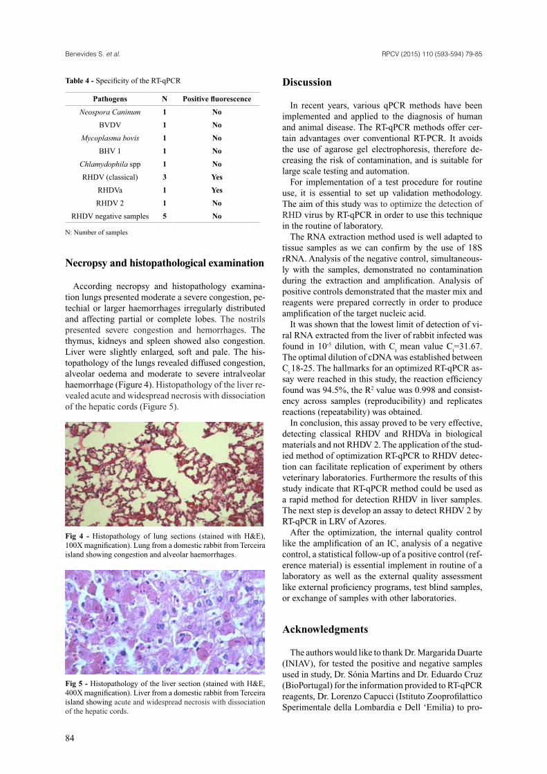
According necropsy and histopathology examina-tion lungs presented moderate a severe congestion, pe-techial or larger haemorrhages irregularly distributed and affecting partial or complete lobes. The nostrils presented severe congestion and hemorrhages. The thymus, kidneys and spleen showed also congestion. Liver were slightly enlarged, soft and pale. The his-topathology of the lungs revealed diffused congestion, alveolar oedema and moderate to severe intralveolar haemorrhage (Figure 4). Histopathology of the liver re-vealed acute and widespread necrosis with dissociation of the hepatic cords (Figure 5).
Fig 4 - Histopathology of lung sections (stained with H&E), 100X magnification). Lung from a domestic rabbit from Terceira island showing congestion and alveolar haemorrhages.
Fig 5 - Histopathology of the liver section (stained with H&E, 400X magnification). Liver from a domestic rabbit from Terceira island showing acute and widespread necrosis with dissociation of the hepatic cords.
Discussion
In recent years, various qPCR methods have been implemented and applied to the diagnosis of human and animal disease. The RT-qPCR methods offer cer-tain advantages over conventional RT-PCR. It avoids the use of agarose gel electrophoresis, therefore de-creasing the risk of contamination, and is suitable for large scale testing and automation.
For implementation of a test procedure for routine use, it is essential to set up validation methodology. The aim of this study was to optimize the detection of RHD virus by RT-qPCR in order to use this technique in the routine of laboratory.
The RNA extraction method used is well adapted to tissue samples as we can confirm by the use of 18S rRNA. Analysis of the negative control, simultaneous-ly with the samples, demonstrated no contamination during the extraction and amplification. Analysis of positive controls demonstrated that the master mix and reagents were prepared correctly in order to produce amplification of the target nucleic acid.
It was shown that the lowest limit of detection of vi-ral RNA extracted from the liver of rabbit infected was found in 10-5 dilution, with C
t mean value C
t=31.67.
The optimal dilution of cDNA was established between C
t 18-25. The hallmarks for an optimized RT-qPCR as-
say were reached in this study, the reaction efficiency found was 94.5%, the R2 value was 0.998 and consist-ency across samples (reproducibility) and replicates reactions (repeatability) was obtained.
In conclusion, this assay proved to be very effective, detecting classical RHDV and RHDVa in biological materials and not RHDV 2. The application of the stud-ied method of optimization RT-qPCR to RHDV detec-tion can facilitate replication of experiment by others veterinary laboratories. Furthermore the results of this study indicate that RT-qPCR method could be used as a rapid method for detection RHDV in liver samples. The next step is develop an assay to detect RHDV 2 by RT-qPCR in LRV of Azores.
After the optimization, the internal quality control like the amplification of an IC, analysis of a negative control, a statistical follow-up of a positive control (ref-erence material) is essential implement in routine of a laboratory as well as the external quality assessment like external proficiency programs, test blind samples, or exchange of samples with other laboratories.
Acknowledgments
The authors would like to thank Dr. Margarida Duarte (INIAV), for tested the positive and negative samples used in study, Dr. Sónia Martins and Dr. Eduardo Cruz (BioPortugal) for the information provided to RT-qPCR reagents, Dr. Lorenzo Capucci (Istituto Zooprofilattico Sperimentale della Lombardia e Dell ‘Emilia) to pro-
Benevides S. et al. RPCV (2015) 110 (593-594) 79-85
85
vided reference material to classical RHDV, RHDVa and RHDV 2, to Laboratório Regional de Veterinária (LRV) do Governo dos Açores - Secretaria Regional da Agricultura e Ambiente (SRAA) – Direção Regional da Agricultura (DRAg) – Direção de Serviços de Veterinária (DSV) to approve all technical support and to the Virology and Pathology departments for techni-cal assistance.
Bibliography
Abrantes J, Loo W, Pendu JL, Esteves P (2012). Rabbit hae-morrhagic disease (RHD) and rabbit haemorrhagic disease virus (RHDV): a review. Veterinary Research, 43(12).
Asgari S, Hardy JRE, Sinclair RG, Cooke BC (1998). Field evidence for mechanical transmission of rabbit haemorrha-gic disease virus (RHDV) by flies (Diptera: Calliphoridae) among wild rabbits in Australia. Virus Research. 54, 123-132.
Bustin SA, Benes V, Garson JA, Hellemans J, Huggett J, Kubista M, Mueller R, Nolan T, Pfaff MW, Shipley GL, Vandesompele J, Witttwer CT (2009). The MIQE Guidelines: Minimum Information for Publication of Quantitative Real Time PCR Experiments. Clinical Chemistry, 55, 4.
Boucher S and Nouaille L (1996). Maladie des lapins.1re Édtion, 95-96.
Carvalho GDF and Almeida LMM (1990). Contribuição para o estudo de uma população de coelhos selvagens (Oryctolagus cuniculus, L.) na Ilha de Santa Maria e o impacto do R.V.H.D na população local. Relatório e Comunicações do Departamento de Biologia, Universidade dos Açores, 61-67.
Carvalho G, Ferrand N, Fonseca A, Branco M, Azevedo M, Mendes R, Batista P, Mantua P (1993). Estudo de uma po-pulação de coelhos selvagens (Oryctolagus cuniculus, L.) na ilha de São Jorge – Açores, Relatório e Comunicações do Departamento de Biologia, Universidade dos Açores, 92, 8-20.
Cooke BD (2002). Rabbit haemorrhagic disease: field epi-demiology and the management of wild rabbit population, Rev. Sci. Tech. Off. Int. Epiz, 21 (2), 347-358.
Cunha MV and Inácio J (2014). Abordagens moleculares em veterinária. LIDEL – Edições Técnicas, Lda. 1, 16-17; 3, 55-99.
Duarte MD, Henriques AM, Barros S, Luís T, Fagulha T, Ramos F, Fevereiro M (2014), New insight into the epi-demiology of rabbit haemorrhagic disease viroses in Portugal: Retrospective study reveals the circulation of genogroup 5 (G5) in Azores and discloses the circulation of G1 and G6 and strains in mainland until 2008. Infection Genetics and Evolution, 27, 149-155.
Esteves PJ, Lopes AM, Magalhães MJ, Pinheiro A, Gonçalves D, Abrantes J (2014). Rabbit Haemorrhagic Disease Virus Detected in Pico, Azores, Portugal, Revealed a Unique Endemic Strain whit More Than 17 Years if Independent Evolution. Viruses 6, 2698-2707.
Fitzner A, Niedbalski WW, Kesy A, Paprocka G (2011). Detection of RHD Virus by Real-time reverse transcription PCR. Bull Vet Inst Pulawy, 55, 581-586.
Gall A, Hoffmann B, Teifke JP, Lange B, Schirrmeier H (2007). Persistence of viral RNA in rabbits wich overco-me an experimental RHDV infection detected by a highly sensitive multiplex real-time RT-PCR. Vet Microbiol, 120, 17-32.
Hoffmann B, Beer M, Reid SM, Mertens P, Oura CAL, Rijn PA, Slomka MJ, Banks J, Brown IH, Alexander DJ, King DK (2009). A review of RT-PCR Technologies used in veterinary virology and disease control: Sensitive and specific diagnosis of five livestock diseases notifiable to the World Organization for Animal Health. Veterinary Microbiology. 139, 1-23.
Parra F and Prieto M (1990). Purification and characteri-zation of a calicivirus as the causative agent of a lethal Haemorrhagic disease in rabbits. J. Virol. 64, 4013-4015.
Principles and methods of validation of diagnostic assays for infectious disease, Chapter 1.1.5 OIE Terrestrial Manual 2013, 1-17.
http://www.oie.int/f ileadmin/Home/fr/Health_standards/tahm/1.01.05_VALIDATION.pdf . Accessed 2 October 2014.
Raymaekers M., Smets R., Maes B., Cartuyvels R., 2009, Checklist for Optimization and Validation of Real-Time PCR Assays. Journal of Clinical Laboratory Analysis 23, 145-151.
Real-Time PCR Handbook, Life Technologies, 2012. http://www.gene-quantification.de/real-time-pcr-handbook-life-technologies-update-flr.pdf. Accessed 2 October 2014.
Validation and Quality control of polymerase chain reac-tion methods used for the diagnosis of infectious disease, Chapter 1.1.5 OIE Terrestrial Manual 2008, 46-55. http://web.oie.int/eng/normes/MMANUAL/2008/pdf/1.1.05._VALID_PCR.pdf. Accessed 8 October 2014.