HBB. 1(2): 25-38 25 Copyright © 2017, Health Biotechnology and Biopharma. All rights reserved.
Health Biotechnology and Biopharma (2017), 1(2): 25-38
Original Research Article
Optimization of cyclotide like peptide extraction methods and
characterization of these peptides from Viola tricolor
Mehrnaz Hosseini Jafari 1, Mahboubeh Zarrabi
1, Parinaz Ghadam
1*, Maryam
Keshavarzi 2
1Department of Biotechnology, Faculty of Biological Sciences, Alzahra University, Tehran, Iran
2Department of Botany, Faculty of Biological Sciences, Alzahra University, Tehran, Iran
*Corresponding author: Parinaz Ghadam, Department of Biotechnology, Faculty of Biological Sciences, Alzahra
University, Tehran, Iran. Email: [email protected]
Received: June 17, 2017; Accepted: August 15, 2017
ABSTRACT
Cyclotides are antimicrobial peptides and play significant role as bio preservative in the food
industry. Since foodborne diseases are a universal issue, in the present study the different methods
for partial purification of cyclotide like peptides from Viola tricolor were compared and the
antimicrobial activities of these peptides were investigated on Staphylococcus aureus Escherichia
coli, which are important in foodborne diseases. Cyclotide like peptides from the aerial parts of the
plant were extracted and it was partially purified with three methods including reverse phase
chromatography, two phase system with reverse phase chromatography and ammonium sulfate
precipitation. The samples were analyzed by SDS-PAGE, Tricine–PAGE, and reverse phase HPLC.
It was found that the first method has lower MIC (Minimum Inhibitory Concentration) and is the
best method.
Keywords: Cyclotides, Peptides, Viola tricolor
Hosseini et al.
26 HBB. 1(2): 25-38
INTRODUCTION
Plant antimicrobial compounds are
investigated for their functional bio preservative
properties and the plant antimicrobial peptides
pAMPs) are one of them. The pAMPs consist of
cyclotides, snakins, hevein, and knottin-like
peptides, lipid transfer proteins (LTPs), peptides
from hydrolysates, plant defensins, myrosinase-
binding proteins (MBPs), and thionin [1].
Cyclotides are the largest family of plant
peptides and are typically comprised of 28 to 37
amino acids, which are ribosomally synthesized.
The N-terminal of the original chain of cyclotide
peptides is attached to the C-terminal and makes
a structural motif called CCK (Cyclic Cistine
Knot). These features have the unique structure
and remarkable stability of cyclopeptides
against heat, chemicals, and enzymes. They
have a wide variety of biological activities such
as anti HIV, antimicrobial, anti pests, anti-
fungal, and anti-tumor. Cyclotides are
appropriate for drug design and food
preservatives [2-6].
In the 1970s, the first cyclotide of Oldenlandia
affinis (Rubiaceae), African plant leaves was
found and called kalata B1 [7], and then the
other cyclotides were isolated from other plants
with different methods [8]. Nowadays, more
than 150 cyclotides of approximately 30 plants
of Violaceae and Rubiaceae families have been
isolated [9, 10]. Violat ricolor is a member of
the Violaceae plant family and is utilized in
traditional medicine in order to soothe coughs
and relieve fever, besides its antitoxic nature [6].
Three cyclotides containing cytotoxic activity
have been reported based on a study on the
cytotoxicity of the plant extraction [11]. In the
present research, we compared the Viola tricolor
(Fig. 1) cyclotides partial purification methods.
Ultimately, the antimicrobial effects of cyclotide
solution were measured on gram-positive (S.
aureus) and gram-negative (E. coli) bacteria.
Fig. 1. Viola tricolor
MATERIALS AND METHODS
Cyclotides extraction
Viola tricolor was collected from Damavand
in Iran and dried at 50 o
C. About 25 g of aerial
parts of the plant were powdered. The extraction
was done by dichloromethane/methanol (1:1)
overnight at 21 o
C in a shaker incubator. The
solution was passed through Whatman paper
filter. Then the water was added to this solution
(1/2 whole volume). Two phases were separated
Viola tricolor peptides extraction
HBB. 1(2): 25-38 27
and then methanol was evaporated and the
obtained solution was freeze-dried. The powder
was dissolved in water; acetic acid was added to
the extract (2% whole volumes), passed through
the polyamide filter and dried. Further
purification was performed by three different
methods.
At the first method (reverse phase
chromatography) the powder was dissolved in
the ammonium bicarbonate buffer (pH 8.05) and
immediately passed through SPE-C18 column,
and the column was washed with ethanol 20%,
50% and 80%. Finally separated fractions were
collected, dried and analyzed.
At the second method (two phase system and
reverse phase chromatography), butanol and
water (1:1) were added to the powder. The
solution was shaken, and the butanol phase and
aqueous phase separated. This step was repeated
three times and in each time butanol phases
were collected and the solvent evaporated. As
above, the powder was dissolved in the
ammonium bicarbonate buffer (pH 8.05) and
immediately passed through SPE-C18 column.
Then the column was washed with ethanol 20%,
50% and 80%. Finally the resulting fractions
were collected, dried and analyzed. The third
method (ammonium sulfate precipitation) the
resulting powder was dissolved in water and
precipitated by ammonium sulfate (saturation
concentration 90%). The suspension was
prepared and centrifuged for 20 min at 4 o
C.
The obtained precipitation was isolated and
resolved. Then it was separated using dialysis
bag (2000 MWCO).
Bradford assay
Bradford assay is a colorimetric method for
determination of protein and peptide
concentration.
Bradford solution preparation
10 mg of Coomassie Briliant Blue G250 was
dissolved in 5 ml of ethanol 95%. Then 10 ml of
phosphoric acid 85% was added and mixed
thoroughly. Final volume was reached to 100 ml
with water. The solution was filtered using filter
paper.
Standard protein solution
To determine the protein concentration, the
standard graph was drawn. 1 mg of BSA
(Bovine Serum Albumin) was dissolved in
water slowly. Then different concentrations of
BSA were prepared in 6 tubes. 1 ml of Bradford
solution was added to 20 μl of them and samples
then mixed. Absorption was repoted at 595 nm
[12].
SDS-PAGE
The discontinuous polyacrylamide gel
electrophoresis (15%) containing SDS was used
Hosseini et al.
28 HBB. 1(2): 25-38
to investigate extracted proteins in according to
Laemmeli method (120V) and stained with
Coomassie Brilant Blue R250 [13].
Tricine-PAGE
Tricine-PAGE was performed in order to
determine the exact weight of the peptides. It
contains running buffer, cathode buffer and
anode buffer. It has three different parts consists
of condensing gel (in this part proteins are
condensed), spacing gel and separating gel, in
this part proteins are separated based on their
molecular weights. Separating gel: 3% C (the
percentage of bisacrylamide), 16.5% T (the
percentage of acrylamide and bisacrylamide),
spacer gel: 3% C, 10% T, stacking gel: 3% C,
4% T. Electerophoresis was done at constant
voltage 30V for about 1h. Voltage was raised up
to 100V.
At the first, the gel was fixed in solution
containing 50 % methanol and 10 % acetic acid
for 1 h. Then it was stained with solution
containing 0.025 % Serva blue G in 10 % acetic
acid for 2 h, finally it was destained with
solution containing 10 % acetic acid for 2 h
[14].
Reverse Phase- HPLC (RP- HPLC)
Reverse phase high performance liquid
chromatography (RP-HPLC) was used to
separate organic molecules based on their
partitioning between stationary phase and the
mobile phase [21]. However, proteins and
peptides as organic molecules behave
differently. They were absorbed to the stationary
phase through hydrophobic forces and eluted by
organic solvent. The elution of peptides from
reverse phase supports was done by reducing the
polarity of washing solution. HPLC analysis
was performed using KNAUER (Germany)
system and C18 column (Length × ID: 250 × 4.6
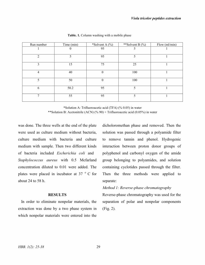
mm with precolumn) (Table. 1).
Two different phases were used to wash
column. Mobile phase A consists of 0.05% TFA
and mobile phase B consists of acetonitrile
(ACN) (% 90) and trifluoroacetic acid (0.05 %).
At the first, the column was washed with the 95
% gradient of solvent A and 5 % gradient of
solvent B. Then the process of washing was
continued by different percent of solvent A and
solvent B (according to Table 1). Due to the
hydrophobic surface, cyclotides was eluted in a
late retention time.
Determination of antibacterial activity
Antibacterial activity was determined by
Micro-broth dilution method according to CLSI
[15]. At first, 20 µl of fluid nutrient broth
culture was added to all the wells except the first
well. Then 10 μl of each extraction was added to
the first well. 5 µl of sample from the first well
was transferred to the next well and the dilution
Viola tricolor peptides extraction
HBB. 1(2): 25-38 29
Table. 1. Column washing with a mobile phase
Run number Time (min) *Solvent A (%) **Solvent B (%) Flow (ml/min)
1 0 95 5 1
2 5 95 5 1
3 15 75 25 1
4 40 0 100 1
5 50 0 100 1
6 50.2 95 5 1
7 55 95 5 1
*Solution A: Trifluoroacetic acid (TFA) (% 0.05) in water
**Solution B: Acetonitrile (ACN) (% 90) + Trifluoroacetic acid (0.05%) in water
was done. The three wells at the end of the plate
were used as culture medium without bacteria,
culture medium with bacteria and culture
medium with sample. Then two different kinds
of bacteria included Escherichia coli and
Staphylococcus aureus with 0.5 Mcfarland
concentration diluted to 0.01 were added. The
plates were placed in incubator at 37 o
C for
about 24 to 58 h.
RESULTS
In order to eliminate nonpolar materials, the
extraction was done by a two phase system in
which nonpolar materials were entered into the
dicholoromethan phase and removed. Then the
solution was passed through a polyamide filter
to remove tannin and phenol. Hydrogenic
interaction between proton donor groups of
polyphenol and carbonyl oxygen of the amide
group belonging to polyamides, and solution
containing cyclotides passed through the filter.
Then the three methods were applied to
separate:
Method 1: Reverse-phase chromatography
Reverse-phase chromatography was used for the
separation of polar and nonpolar components
(Fig. 2).
Hosseini et al.
30 HBB. 1(2): 25-38
Fig. 2. Method1. M: molecular weight marker, 1: SPE-
C18 column output (washing by ethanol 20%), 2 and 3:
SPE-C18 column Output (washing by ethanol 50%), 4:
SPE-C18 column Output (washing by ethanol 80%).
polyacrylamide gel (15%), stained by coomassie blue,
constant voltage 120 V.
Method 2: Two-phase system and reverse phase
chromatography
The two-phase system method was adopted for
Viola odorata cyclotides extraction by Zarrabi
et al. and finally, the separation was performed
by SPE-C18 column (Fig. 3). In this protocol we
combined the methods used by Zarrabi and
Hashempour [17, 20].
Fig. 3. Method 2. M: molecular weight marker, 1: The
SPE-C18 column Output (washing by ethanol 20%), 2:
SPE-C18 column Output (washing by ethanol 50%), 3:
SPE-C18 column Output (washing by ethanol 80%).
Method 3. 4: peptides precipitated by 90% of ammonium
sulfate saturation (dialysis), 5: peptides precipitated by
ammonium sulfate saturation of 90% (not dialysis).
Polyacrylamide gel (15%), stained by Coomassie blue,
constant voltage 120 V.
Method 3: Precipitation by ammonium sulfate
The extraction containing a lot of components
such as protein. To separate protein moiety from
the other materials; proteins were precipitated
using 90% of ammonium sulfate saturation (Fig.
3). Finally, all the three methods were analyzed
by SDS-PAGE (Fig. 2 and 3), Tricine-PAGE
(Fig. 4), and RP-HPLC.
Fig. 4. Tricine-PAGE. M: molecular weight marker.
Method 2. 1: The SPE-C18 column Output (washing by
ethanol 50%), 3: SPE-C18 column Output (washing by
ethanol 80%). Method1. 2: SPE-C18 column Output
(washing by ethanol 80%). Method3. 5: peptides
precipitated by 90% of ammonium sulfate saturation (not
dialysis), 6: peptides precipitated by 90% of ammonium
sulfate saturation (dialysis).
Bradford assay was applied to these methods.
SDS-PAGE and Tricine-PAGE were performed
in order to determine the weight of the peptides.
Viola tricolor peptides extraction
HBB. 1(2): 25-38 31
Due to the hydrophobic surface on the
cyclotides, the RP-HPLC with C18 column was
used. Since the C18 column is nonpolar; the
nonpolar compounds interact with the column
through hydrophobic interactions. Different
compounds, based on their polarity, were
separated by decreasing the solvent polarity. In
the end, the strongest nonpolar compounds were
eluted. According to the standard proteins which
were extracted by Farhadpour et al [21], the
cyclotides were eluted at 23 to 28 min (Fig. 5).
Fig. 5. RP-HPLC chromatogram. Standard. A: Cyclotides. According to the standard proteins, cyclotides were eluted at
the time of 23-28 min on the charts.
At first, the column was washed by a polar
solvent. During this time, the nonpolar
compounds attached to the column, and polar
compounds came out of the column. The
nonpolar compounds with different affinity
attached to the column, were then washed and
separated by the reduction of washing solvent
polarity. Therefore, the nonpolar compounds
which strongly attached to the column were
eluted sooner. RP-HPLC was performed on both
samples and standard; by comparison, between
all three methods, the elution time of the
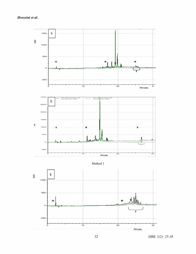
peptides was determined. In all three methods
according to the chromatogram, cyclotides like
peptides were eluted at 23 to 28 min, and these
compounds were considered as a solution
containing cyclotides like peptides (Fig. 6).
Hosseini et al.
32 HBB. 1(2): 25-38
Method 1
Viola tricolor peptides extraction
HBB. 1(2): 25-38 33
Method 2
Method 3
Fig. 6. RP-HPLC chromatogram. Method 1 and Method 2, SPE column washing with ethanol (a) 50% and (b) 80%.
Method3, (a) without dialysis and (b) with dialysis. Peak intensity of polar compounds (A), which was eluted initially.
The peaks are decreasing, and the peak of non-polar compounds (B) was appeared. Non-polar compound retention time
is more. Non-polar compounds (B) were appeared at the time of 23-28 minutes on the charts were considered as a
solution containing Cyclotides.
Hosseini et al.
34 HBB. 1(2): 25-38
Tricine-PAGE was applied for the analysis of
proteins in the range of 1 to 100 KDa [14].
Antimicrobial activities of the same amount of
cyclotide like peptides were extracted by three
methods. The antimicrobial activities of the
samples on gram-positive and gram-negative
bacteria, including S. aureus and E. coli and the
related MIC were determined (Table2).
Table. 2. The amount of MIC (µg/ml)
Bacteria
sample
resulting from
SPE column
washing by
ethanol 50 %
(Method 1)
sample
resulting from
SPE column
washing by
ethanol 80 %
(Method 1)
sample
resulting from
SPE column
washing by
ethanol 50 %
(Method2)
sample
resulting from
SPE column
washing by
ethanol 80 %
(Method2)
sample
resulting from
Amonium
sulfate
precipitation
(method 3)
E. coli
ATTC25922 6.71 6.28 10.83 16.9 15.6
S. aureus
PTCC1431 0.41 6.28 0.338 16.9 7.8
Bacteriostatic capability of samples was
measured by MIC determination. It was
determined that the Solid Phase Extraction
(SPE) column samples washed with 80 %
ethanol showed the same MIC for E.coli and
S.aureus in both the first and second method.
The SPE column samples washed with 50 %
and 80 % ethanol had the lower MIC in the first
method in comparison with the other methods. It
means that a lower concentration of samples had
inhibiting effects on bacteria, and they have
more bacteriostatic capability. The MIC results
showed that S. aureus was more sensitive to
these peptides than E. coli. Because the SPE
column samples washed with 20 % ethanol had
the lowest protein concentration according to
the Bradford results and showing the colorless
band in SDS-PAGE, these samples have not
been studied by RP-HPLC. According to the
SDS-PAGE and RP-HPLC results, cyclotides
like peptides in terms of hydrophobicity and
molecular weight properties, were more
extracted in the second method, but according to
the MIC results, antimicrobial effects were
extracted more in the first method.
DISCUSSION
Viola tricolor is extensively employed as an
herbal plant in traditional medicine [6]. As a
result, a great number of researchers have been
attracted to the analysis of cyclic peptides,
called cyclotides [10, 16].
Cyclotides are resistant to extreme
environmental conditions [6, 17]. Several
Viola tricolor peptides extraction
HBB. 1(2): 25-38 35
cyclotides were extracted from the plants, and
their cytotoxic effects were studied on cancer
cells [16]. The antimicrobial activity of
cycloviolacin O2 cyclotide of Viola odorata was
investigated 0n gram-negative bacteria [18].
Plant antimicrobial peptides have been separated
from different parts of a plant, and they were
active against plants and humans pathogenesis.
Cyclotides have exceptional structural
properties and biological activities [18, 19].
They have been extracted by various methods
from numerous plants like the violet, legume,
coffee, and cucurbit families [17]. Cyclotide
extraction from violaceae for instance, V.
odorata, V. ignobilis, V. arvensis, V. philippica,
V. hederaceae, and V.tricolor had been
performed [16-25]. Extraction in all of these
methods was performed by dichloromethane or
ethanol, and then the tannin was removed. In
2004, butanol extraction was passed through a
SPE column by Goransson et al., and then
cyclotide expression profiles were obtained by
liquid chromatography –mass spectrometry
(LC-MS) [22]. Extraction method which was
used in 2008, extraction was done by
dichloromethane/methanol/water (1: 1: 1) or
acetonitrile/water (1: 1) and then was passed
through solid phase columns and SPE-C18, in
the next step, physical and chemical properties
of the cyclotides such as hydrophobicity
properties were evaluated [26]. In 2013,
Hashempur et al. conducted cyclotides
extraction from Viola ignobilis. They used a
dichloromethane/methanol (1: 1) extraction
method. Finally the extraction was passed
through SPE-C18. It contained fewer steps for
extraction [17]. In this study, we applied the
first and third methods in order to extract the
cyclotides like peptide on Viola tricolorBecause
the method used by Zarrabi was time-
consuming, the second method was applied,
which also optimizes the cyclotides like peptide
extraction processes. The second method was
done through a combination of Hashempour and
Zarrabi methods.
CONCLUSION
In all three methods, lower MIC amounts were
obtained for the inhibition of S. aureus than
E.coli. Method 1 was more appropriate because
it was performed through fewer steps.
Consequently, a fraction of the bacteriostatic
ability that was eluted with 50 % ethanol was
stronger than S. aureus.
The conflict of interest
Authors declare no conflict of interests.
ACKNOWLEDGMENT
The results described in this paper were part of
student thesis and this survey has been
Hosseini et al.
36 HBB. 1(2): 25-38
supported by Vice Chancellor of Alzahra
University, Tehran, Iran.
REFERENCES
[1]. Lee NK, Paik HD. Status, antimicrobial
mechanism, and regulation of natural
preservatives in livestock food systems. Korean
J Food Sci Anim Resour, 2016; 36(4): 547-57.
[2]. Hintz T, Matthews KK, Di R. The use of
plant antimicrobial compounds for food
preservation. BioMed Res Int, 2015; 246264.
[3]. Chen B, Colgrave ML, Daly NL, Rosengren
KJ, Gustafson KR, Craik DJ. Isolation and
characterization of novel cyclotides from Viola
hederaceae: solution structure and anti-HIV
activity of vhl-1, a leaf-specific expressed
cyclotide. J Biol Chem, 2005; 280(23): 22395-
405.
[4]. Craik DJ, Daly NL, Bond T, Waine C. Plant
cyclotides: A unique family of cyclic and
knotted proteins that defines the cyclic cystine
knot structural motif. J Mol Biol, 1999; 294(5):
1327-36.
[5]. Craik DJ, Daly NL. The cyclotides: novel
macrocyclic peptides as scaffolds in drug
design. Curr Opin Drug Discov Devil, 2002;
5(2): 251-60.
[6]. Saether O, Craik DJ, Campbell ID, Sletten
K, Juul J, Norman DG. Elucidation of the
primary and three- dimensional structure of the
uterotonic polypeptide kalata B1. Biochemistry,
1995; 34(13): 4147-58.
[7]. Gran L. Oxytocic principles of oldenlandia
affinis. Lloydia, 1973; 36(2): 174-78.
[8]. Poth AG, Colgrave ML, Philip R, Kerenga
B, Daly NL, Anderson MA, Craik DJ.
Discovery of cyclotides in the Fabaceae plant
family provides new insights into the
cyclization, evolution and distribution of
circular proteins. ACS Chem Boil. 2011; 6(4):
345-55.
[9]. Herrmann A, Burmann R, Mylne, JS,
Karlsson G, Gullbo J, Craik DJ, Clark RJ,
Göransson U. The alpine violet, Viola biflora, is
a rich source of cyclotides with potent
cytotoxicity. Phytochemmistry, 2008; 69(4):
939-52.
[10]. Wang CK, Kaas Q, Chiche L, Craik DJ.
CyBase: a database of cyclic protein sequences
and structures, with applications in protein
discovery and engineering. Nucleic Acids Res,
2008; 36: 206-10.
[11]. Svangard E, Goransson UHocaoglo Z,
Gullbo J, Larsson R, Claeson P, Bohlin L.
Cytotoxic cyclotides from Viola tricolor. J Nat.
Prod, 2004; 67(2): 144-47.
[12]. Bradford MM. A rapid and sensitive
method for the quantitation of microgram
quantities of protein utilizing the principle of
protein-dye binding. Anal Biochem, 1976; 72:
248-54.
Viola tricolor peptides extraction
HBB. 1(2): 25-38 37
[13]. Laemmeli U. Cleavage of structural
proteins during the assemble of the head of
bacteriophage T4. Nature, 1970; 227(5259):
680-85.
[14]. Schagger H. Tricine-SDS-PAGE. Nat
Protoc, 2006; 1(1): 16-22.
[15]. Paredes D, Ortiz C, Torres R. Synthesis,
characterization, and evaluation of antibacterial
effect of Ag nanoparticles against Escherichia
coli O157:H7 and methicillin resistant
Staphylococcus aureus (MRSA). Int J
Nanomedicine, 2014; 9(1): 1717-29.
[16]. Tang J, Wang CK, Pan X, Yan H, Zeng G,
Xu W, He W, Daly NL, Craik DJ, Tan N.
Isolation and characterization of cytotoxic
cyclotides from Viola tricolor. Peptides, 2010;
31(8): 1434-40.
[17]. Hashempour H, Koehbach J, Daly NL,
Ghassempour A, Gruber CW. Characterizing
circular peptides in mixtures: sequence fragment
assembly of cyclotides from a violet plant by
MALDI-TOF/TOF mass spectrometry. Amino
Acids, 2013; 44(2): 581-95.
[18]. Pranting M, Loov C, Burman R,
Goransson U, Andersson DI. The cyclotide
cycloviolacin O2 from Viola odorata has potent
bactericidal activity against Gram-negative
bacteria. J Antimicrob Chemother, 2010; 65(9):
1964-71.
[19]. Nawrot R, Barylski J, Nowicki G,
Broniarczyk J, Buchwald W. Plant antimicrobial
peptides. Folia microbial, 2014; 59(3): 181-96.
[20]. Zarrabi M, Dalirfardouei R, Sepehrizade
Z, Kermanshahi RK. Comparison of the
antimicrobial effects of semi purified cyclotides
from Iranian Viola odorata against some of
plant and human pathogenic bacteria. J Appl
Microbial, 2013; 115(2): 367-75.
[21]. Farhadpour M, Hashempour H, Talebpour
Z, A-Bagheri N, Shushtarian MS, Gruber CW,
Ghassempour A. Microwave-assisted extraction
of cyclotides from Viola ignobilis. Anal
Biochem, 2016; 497: 83-89.
[22]. Goransson U, Svangard E, Claeson P,
Bohlin L. Novel strategies for isolation and
characterization of cyclotides: The discovery of
bioactive macrocyclic plant polypeptides in the
violaceae. Curr Protein Pept Sci, 2004; 5(5):
317-29.
[23]. He W, Chan LY, Zeng G, Daly NL, Craik
DJ, Tan N. Isolation and characterization of
cytotoxic cyclotides from Viola philippica. J
Peptides, 2011; 32(8): 1719-23.
[24]. Ireland DC, Colgrave ML, Craik DJ. A
novel suite of cyclotides from Viola odorata:
sequence variation and the implications for
structur, function and stability. Biochem J, 2006;
400(1): 1-12.
[25]. Trabi M, Craik DJ. Tissue-specific
expression of head-to-tail cyclized miniproteins
Hosseini et al.
38 HBB. 1(2): 25-38
in violaceae and structure determination of the
root cyclotide Viola hederaceae root cyclotide1.
Plant Cell, 2004; 16(8): 2204-16.
[26]. Gruber CW, Elliobtt AG, Ireland DC,
Delprete PG, Dessein S, Göransson U, Trabi M,
Wang CK, Kinghorn AB, Robbrecht E, Craik
DJ. Distribution and evolution of circular
miniproteins in flowering plants. plant cell,
2008; 20(9): 2471-83.