Optimization of Encoded Hydrogel Particles for Nucleic Acid Quantification Daniel C. Pregibon and Patrick S. Doyle* Department of Chemical Engineering, Massachusetts Institute of Technology, 77 Massachusetts Avenue, Cambridge, Massachusetts 02139 The accurate quantification of nucleic acids is of utmost importance for clinical diagnostics, drug discovery, and basic science research. These applications require the concurrent measurement of multiple targets while de- manding high-throughput analysis, high sensitivity, speci- ficity between closely related targets, and a wide dynamic range. In attempt to create a technology that can simul- taneously meet these demands, we recently developed a method of multiplexed analysis using encoded hydrogel particles. Here, we demonstrate tuning of hydrogel poros- ity with semi-interpenetrating networks of poly(ethylene glycol), develop a quantitative model to understand hy- bridization kinetics, and use the findings from these studies to enhance particle design for nucleic acid detec- tion. With an optimized particle design and efficient fluorescent labeling scheme, we demonstrate subattomole sensitivity and single-nucleotide specificity for small RNA targets. Molecular screening lies at the foundation of biological tests in the clinic and at the bench, with specific examples including expression profiling of mRNA to connect drug responses to disease (i.e., the “connectivity map”) 1 and microRNA profiling for cancer diagnostics. 2-5 These and most other screening applica- tions require the quantification of tens to thousands of biomarker targets in a single sample. In comparison to serial testing, multiplexed assays require smaller sample volumes, leading to reductions in assay cost and increases in speed. Two broad classes of technologies are used for multiplexing: planar microarrays and particle-based arrays. Although microarrays typically provide superior screening density, particles provide faster kinetics via mixing (although planar kinetics are diffusion-limited), increased capacity afforded by increased surface area, and higher versatility for adapting target sets. 6 Although particles are preferred over planar substrates for high-throughput screening, current approaches for particle-based multiplexed analysis involve complicated or expensive processes for encoding, functionalizing, or decoding active substrates and also yield a very limited number of analyte-specific codes. 7,8 Few commercial platforms exist and are limited by their coding scheme to scan up to ∼100 targets at a time. 9 This limit in “density” is restrictive for several applications in biomarker discovery, drug discovery, and diagnostics that require the quantification of hundreds or thousands of targets. 1,10 It can also be viewed as a limitation in throughput, when considering sample pooling (si- multaneously scanning several samples for few targets). A technology that could accommodate higher density without loss of performance or increase in cost would be an enabling tool. Beyond limitations in throughput or density, the materials used in most commercial arrays are not ideal for biological interactions. The environment in which ligands are immobilized has tremen- dous impact on the quality of target capture, dictating how many molecules can bind to a surface, how specific that interaction is for a given molecule, and how strong that binding event is. 11,12 All of these attributes are extremely important for biomolecule quantification as they determine the sensitivity, specificity, and dynamic range of detection. Ideally, substrates used for biomol- ecule quantification would be based on nonfouling materials, have a high target capacity, and provide solution-like hybridization thermodynamics. With these considerations in mind, hydrogels are proving to be excellent substrates for biomolecule capture and quantification. Hydrogels are a class of biofriendly materials that characteristi- cally retain water, allowing biological interactions to occur in three- * To whom correspondence should be addressed. Phone: 617-253-4534. Fax: 617-258-5042. E-mail: [email protected]. (1) Lamb, J.; Crawford, E. D.; Peck, D.; Modell, J. W.; Blat, I. C.; Wrobel, M. J.; Lerner, J.; Brunet, J.-P.; Subramanian, A.; Ross, K. N.; Reich, M.; Hiero- nymus, H.; Wei, G.; Armstrong, S. A.; Haggarty, S. J.; Clemons, P. A.; Wei, R.; Carr, S. A.; Lander, E. S.; Golub, T. R. Science 2006, 313, 1929–1935. (2) Calin, G. A.; Ferracin, M.; Cimmino, A.; Di Leva, G.; Shimizu, M.; Wojcik, S. E.; Iorio, M. V.; Visone, R.; Sever, N. I.; Fabbri, M.; Iuliano, R.; Palumbo, T.; Pichiorri, F.; Roldo, C.; Garzon, R.; Sevignani, C.; Rassenti, L.; Alder, H.; Volinia, S.; Liu, C.-g.; Kipps, T. J.; Negrini, M.; Croce, C. M. N. Engl. J. Med. 2005, 353, 1793–1801. (3) Cummins, J. M.; Yiping He, Y.; Leary, R. J.; Pagliarini, R.; Diaz, L. A., Jr.; Sjoblom, T.; Barad, O.; Bentwich, Z.; Szafranska, A. E.; Labourier, E.; Raymond, C. K.; Roberts, B. S.; Juhl, H.; Kinzler, K. W.; Vogelstein, B.; Velculescu, V. E. Proc. Natl. Acad. Sci. U.S.A. 2006, 103, 3687–3692. (4) Jay, C.; Nemunaitis, J.; Chen, P.; Fulgham, P.; Tong, A. W. DNA Cell Biol. 2007, 26, 293–300. (5) Volinia, S.; Calin, G. A.; Liu, C.-G.; Ambs, S.; Cimmino, A.; Petrocca, F.; Visone, R.; Iorio, M.; Roldo, C.; Ferracin, M.; Prueitt, R. L.; Yanaihara, N.; Lanza, G.; Scarpa, A.; Vecchione, A.; Negrini, M.; Harris, C. C.; Croce, C. M. Proc. Natl. Acad. Sci. U.S.A. 2006, 103, 2257–2261. (6) Nolan, J. P.; Sklar, L. A. Trends Biotechnol. 2002, 20, 9–12. (7) Finkel, N. H.; Lou, X.; Wang, C.; He, L. Anal. Chem. 2004, 76, 352A– 359A. (8) Braeckmans, K.; De Smedt, S. C.; Leblans, M.; Pauwels, R.; Demeester, J. Nat. Rev. Drug Discovery 2002, 1, 447–456. (9) Fulton, R. J.; McDade, R. L.; Smith, P. L.; Kienker, L. J.; Kettman, J. J. R. Clin. Chem. 1997, 43, 1749–1756. (10) Ramaswamy, S.; Tamayo, P.; Rifkin, R.; Mukherjee, S.; Yeang, C. H.; Angelo, M.; Ladd, C.; Reich, M.; Latulippe, E.; Mesirov, J. P.; Poggio, T.; Gerald, W.; Loda, M.; Lander, E. S.; Golub, T. R. Proc. Natl. Acad. Sci. U.S.A. 2001, 98, 15149–15154. (11) Halperin, A.; Buhot, A.; Zhulina, E. B. Biophys. J. 2005, 89, 796–811. (12) Vainrub, A.; Pettitt, B. M. Biopolymers 2003, 68, 265–270. Anal. Chem. 2009, 81, 4873–4881 10.1021/ac9005292 CCC: $40.75 2009 American Chemical Society 4873 Analytical Chemistry, Vol. 81, No. 12, June 15, 2009 Published on Web 05/12/2009 Downloaded by MIT on September 7, 2009 | http://pubs.acs.org Publication Date (Web): May 12, 2009 | doi: 10.1021/ac9005292
Transcript
Optimization of Encoded Hydrogel Particles forNucleic Acid Quantification
Daniel C. Pregibon and Patrick S. Doyle*
Department of Chemical Engineering, Massachusetts Institute of Technology, 77 Massachusetts Avenue,Cambridge, Massachusetts 02139
The accurate quantification of nucleic acids is of utmostimportance for clinical diagnostics, drug discovery, andbasic science research. These applications require theconcurrent measurement of multiple targets while de-manding high-throughput analysis, high sensitivity, speci-ficity between closely related targets, and a wide dynamicrange. In attempt to create a technology that can simul-taneously meet these demands, we recently developed amethod of multiplexed analysis using encoded hydrogelparticles. Here, we demonstrate tuning of hydrogel poros-ity with semi-interpenetrating networks of poly(ethyleneglycol), develop a quantitative model to understand hy-bridization kinetics, and use the findings from thesestudies to enhance particle design for nucleic acid detec-tion. With an optimized particle design and efficientfluorescent labeling scheme, we demonstrate subattomolesensitivity and single-nucleotide specificity for small RNAtargets.
Molecular screening lies at the foundation of biological testsin the clinic and at the bench, with specific examples includingexpression profiling of mRNA to connect drug responses todisease (i.e., the “connectivity map”)1 and microRNA profiling forcancer diagnostics.2-5 These and most other screening applica-tions require the quantification of tens to thousands of biomarkertargets in a single sample. In comparison to serial testing,multiplexed assays require smaller sample volumes, leading toreductions in assay cost and increases in speed. Two broad classesof technologies are used for multiplexing: planar microarrays and
particle-based arrays. Although microarrays typically providesuperior screening density, particles provide faster kinetics viamixing (although planar kinetics are diffusion-limited), increasedcapacity afforded by increased surface area, and higher versatilityfor adapting target sets.6
Although particles are preferred over planar substrates forhigh-throughput screening, current approaches for particle-basedmultiplexed analysis involve complicated or expensive processesfor encoding, functionalizing, or decoding active substrates andalso yield a very limited number of analyte-specific codes.7,8 Fewcommercial platforms exist and are limited by their coding schemeto scan up to ∼100 targets at a time.9 This limit in “density” isrestrictive for several applications in biomarker discovery, drugdiscovery, and diagnostics that require the quantification ofhundreds or thousands of targets.1,10 It can also be viewed as alimitation in throughput, when considering sample pooling (si-multaneously scanning several samples for few targets). Atechnology that could accommodate higher density without lossof performance or increase in cost would be an enabling tool.
Beyond limitations in throughput or density, the materials usedin most commercial arrays are not ideal for biological interactions.The environment in which ligands are immobilized has tremen-dous impact on the quality of target capture, dictating how manymolecules can bind to a surface, how specific that interaction isfor a given molecule, and how strong that binding event is.11,12
All of these attributes are extremely important for biomoleculequantification as they determine the sensitivity, specificity, anddynamic range of detection. Ideally, substrates used for biomol-ecule quantification would be based on nonfouling materials, havea high target capacity, and provide solution-like hybridizationthermodynamics. With these considerations in mind, hydrogelsare proving to be excellent substrates for biomolecule capture andquantification.
Hydrogels are a class of biofriendly materials that characteristi-cally retain water, allowing biological interactions to occur in three-
* To whom correspondence should be addressed. Phone: 617-253-4534. Fax:617-258-5042. E-mail: [email protected].
(1) Lamb, J.; Crawford, E. D.; Peck, D.; Modell, J. W.; Blat, I. C.; Wrobel, M. J.;Lerner, J.; Brunet, J.-P.; Subramanian, A.; Ross, K. N.; Reich, M.; Hiero-nymus, H.; Wei, G.; Armstrong, S. A.; Haggarty, S. J.; Clemons, P. A.; Wei,R.; Carr, S. A.; Lander, E. S.; Golub, T. R. Science 2006, 313, 1929–1935.
(2) Calin, G. A.; Ferracin, M.; Cimmino, A.; Di Leva, G.; Shimizu, M.; Wojcik,S. E.; Iorio, M. V.; Visone, R.; Sever, N. I.; Fabbri, M.; Iuliano, R.; Palumbo,T.; Pichiorri, F.; Roldo, C.; Garzon, R.; Sevignani, C.; Rassenti, L.; Alder,H.; Volinia, S.; Liu, C.-g.; Kipps, T. J.; Negrini, M.; Croce, C. M. N. Engl.J. Med. 2005, 353, 1793–1801.
(3) Cummins, J. M.; Yiping He, Y.; Leary, R. J.; Pagliarini, R.; Diaz, L. A., Jr.;Sjoblom, T.; Barad, O.; Bentwich, Z.; Szafranska, A. E.; Labourier, E.;Raymond, C. K.; Roberts, B. S.; Juhl, H.; Kinzler, K. W.; Vogelstein, B.;Velculescu, V. E. Proc. Natl. Acad. Sci. U.S.A. 2006, 103, 3687–3692.
(4) Jay, C.; Nemunaitis, J.; Chen, P.; Fulgham, P.; Tong, A. W. DNA Cell Biol.2007, 26, 293–300.
(5) Volinia, S.; Calin, G. A.; Liu, C.-G.; Ambs, S.; Cimmino, A.; Petrocca, F.;Visone, R.; Iorio, M.; Roldo, C.; Ferracin, M.; Prueitt, R. L.; Yanaihara, N.;Lanza, G.; Scarpa, A.; Vecchione, A.; Negrini, M.; Harris, C. C.; Croce, C. M.Proc. Natl. Acad. Sci. U.S.A. 2006, 103, 2257–2261.
(6) Nolan, J. P.; Sklar, L. A. Trends Biotechnol. 2002, 20, 9–12.(7) Finkel, N. H.; Lou, X.; Wang, C.; He, L. Anal. Chem. 2004, 76, 352A–
359A.(8) Braeckmans, K.; De Smedt, S. C.; Leblans, M.; Pauwels, R.; Demeester, J.
Nat. Rev. Drug Discovery 2002, 1, 447–456.(9) Fulton, R. J.; McDade, R. L.; Smith, P. L.; Kienker, L. J.; Kettman, J. J. R.
M.; Ladd, C.; Reich, M.; Latulippe, E.; Mesirov, J. P.; Poggio, T.; Gerald,W.; Loda, M.; Lander, E. S.; Golub, T. R. Proc. Natl. Acad. Sci. U.S.A. 2001,98, 15149–15154.
(11) Halperin, A.; Buhot, A.; Zhulina, E. B. Biophys. J. 2005, 89, 796–811.(12) Vainrub, A.; Pettitt, B. M. Biopolymers 2003, 68, 265–270.
Anal. Chem. 2009, 81, 4873–4881
10.1021/ac9005292 CCC: $40.75 2009 American Chemical Society 4873Analytical Chemistry, Vol. 81, No. 12, June 15, 2009Published on Web 05/12/2009
Dow
nloa
ded
by M
IT o
n Se
ptem
ber
7, 2
009
| http
://pu
bs.a
cs.o
rg
Pub
licat
ion
Dat
e (W
eb):
May
12,
200
9 | d
oi: 1
0.10
21/a
c900
5292
dimensional space. In comparison to glass substrates, hydrogelmaterials (e.g., poly(ethylene glycol), PEG) are nonfouling, thuslimiting nonspecific interactions, and can be derived from a broadlist of precursors. Moreover, although hybridization is dramaticallyinhibited on solid surfaces,13 Mirzabekov’s group has shown thatnucleic acid hybridization in gels closely resembles that insolution.14 Furthermore, by collecting fluorescence from a three-dimensional volume as opposed to a two-dimensional plane, agreater number of fluorophores can be captured to provideenhanced sensitivity. It was demonstrated that gel substratesexhibit better sensitivity and a higher capacity than their glasscounterparts for both nucleic acids15 and proteins.16 Although gelshave been used successfully for planar arrays, the application ofhydrogel for particle arrays is very limited.
In planar or particle arrays, solid or gel-based, an understand-ing of the kinetics for target capture can be used to design andoptimize assays. The kinetics of biomolecule capture involves masstransport (convection and/or diffusion) of target molecules to anactive substrate and subsequent chemical reaction with animmobilized probe. Hybridization kinetics have been studied andmodeled extensively over a variety of substrates and conditions.Analysesincludecaptureonsolidsubstrateswithforcedconvection17-19
or instagnantfluids20-22 andalsocapture inhydrogelsubstrates.19,23
Typically, analytical solutions can only be found for specificregimes where the nonlinear, coupled equations governingtransport can be simplified. Furthermore, although modeling hasbeen accomplished for particle arrays24 and planar hydrogelarrays,18,19,23 modeling has not been done for hydrogel particlearrays.
Recently, we developed a method for molecular screeningbased on multifunctional encoded hydrogel particles.25 Theseparticles are generated using flow lithographysa process thatallows for the rapid generation of morphologically complex,monodisperse (coefficients of variation <2%) particles from a broadrange of precursor materials.26,27 For bioassays, we use PEGprecursors that cross-link upon UV exposure to form encodedparticles composed of a porous hydrogel network. Encoded
particles are incubated with a sample containing unknown speciesand subsequently scanned for fluorescence in a flow-throughdevice where barcodes are read and the corresponding targetsquantified. Our multiplexing technology provides a virtuallyunlimited number of codes, single-color, rapid flow-throughscanning, and the ability to detect several targets on singleparticles.25 Although we demonstrated proof-of-concept multi-plexed nucleic acid detection, the performance of our system wasnot previously assessed. Here, we elucidate the parametersdictating assay sensitivity and subsequently optimize particle andassay design to demonstrate high-performance nucleic acidquantification.
MATERIALS AND METHODSFabrication and Assembly of Microfluidic Devices. Mi-
crofluidic channels were molded on 4 in. silicon wafers usingstandard soft lithography. Briefly, SU-8 photoresist (MicroChem)was spin-coated on a clean silicon wafer for 30 s at a speed selectedto obtain the desired layer thickness. After a brief 65 °C prebakeon a hot plate, the wafer was exposed to UV irradiation througha transparency mask. The photoresist was then postbaked at 95°C, and subsequently, unexposed photoresist was removed usinga developer. Poly(dimethylsiloxane) (PDMS) (Sylgard 184, DowCorning) was mixed at a base to curing agent ratio of 10:1. Theelastomer was degassed for 30 min and poured over the siliconwafer mold. The PDMS was then cured overnight at 65 °C. Holesfor external connection were punched out using a blunt-endedsyringe needle. Glass slides were coated with PDMS and partiallycured (for 20 min at 65 °C). Cleaned channels were then placedon the slides and contact-sealed. The assembled devices were thenbaked for an additional 45 min at 65 °C.
Microscope Setup. All experiments were performed usingan Axiovert 200 (Zeiss) inverted microscope with a VS25 shuttersystem (UniBlitz) in place to precisely control UV exposure dose.A 100 W HBO mercury lamp in conjunction with wide-rangeexcitation UV filter (11000v2:UV, Chroma) provided irradiationof the desired wavelength. Transparency masks designed usingAutocad were printed by CAD/Art Services, Inc. (Bandon, OR)at 10 000 dpi resolution. Each mask was designed to be circular,2.5 cm in diameter, with features typically printed no more then0.5 cm radially from the mask center. During an experiment, amask was sandwiched between two 25 mm circular glass cover-slips (VWR), placed in the first slot of the filter slider bar, andsecured with an O-ring. The filter slider was then positioned inthe field-stop position of the microscope. Images were processedusing NIH Image.
Particle Synthesis Using Stop-Flow Lithography. Precursorsolutions consisted of blends of poly(ethylene glycol) diacrylate(PEG-DA, Mn ) 700, ∼70 cP at 25 °C, Aldrich) and PEG (Mw
) 200, ∼50 cP at 25 °C, Aldrich) in 35% 3× Tris-EDTA buffer(pH ) 8.0, EMD) with 5% Darocur 1173 photoinitiator (Alrich).When applicable, DNA probe modified with an acrydite group(IDT) was included at concentrations ranging from 10 to 100µM. The sequences of the oligomers used are given in Table1. These precursor samples were loaded into channels using pipettips (200 µL, Molecular BioProducts), connected with rubbertubing (Tygon) to a common pressure source (regulated by apressure valve, Controlair Inc.). The tips were filled with ∼100µL of polymer and inserted into the channel inlet ports. A three-
(13) Levicky, R.; Horgan, A. Trends Biotechnol. 2005, 23, 143–149.(14) Fotin, A. V.; Drobyshev, A. L.; Proudnikov, D. Y.; Perov, A. N.; Mirzabekov,
A. D. Nucleic Acids Res. 1998, 26, 1515–1521.(15) Sorokin, N. V.; Chechetkin, V. R.; Pan’kov, S. V.; Somova, O. G.; Livshits,
M. A.; Donnikov, M. Y.; Turygin, A. Y.; Barsky, V. E.; Zasedatelev, A. S.J. Biomol. Struct. Dyn. 2006, 24, 57–66.
(16) Zubtsov, D. A.; Savvateeva, E. N.; Rubina, A. Y.; Pan’kov, S. V.; Konovalova,E. V.; Moiseeva, O. V.; Chechetkin, V. R.; Zasedatelev, A. S. Anal. Biochem.2007, 368, 205–213.
(17) Squires, T. M.; Messinger, R. J.; Manalis, S. R. Nat. Biotechnol. 2008, 26,417–426.
(18) Zubtsov, D. A.; Ivanov, S. M.; Rubina, A. Y.; Dementieva, E. I.; Chechetkin,V. R.; Zasedatelev, A. S. J. Biotechnol. 2006, 122, 16–27.
(19) Sorokin, N. V.; Yurasov, D. Y.; Cherepanov, A. I.; Kozhekbaeva, J. M.;Chechetkin, V. R.; Gra, O. A.; Livshits, M. A.; Nasedkina, T. V.; Zasedatelev,A. S. J. Biomol. Struct. Dyn. 2007, 24, 571–578.
(20) Gadgil, C.; Yeckel, A.; Derby, J. J.; Hu, W. S. J. Biotechnol. 2004, 114,31–45.
(21) Erickson, D.; Li, D. Q.; Krull, U. J. Anal. Biochem. 2003, 317, 186–200.(22) Bishop, J.; Chagovetz, A. M.; Blair, S. Biophys. J. 2008, 94, 1726–1734.(23) Livshits, M. A.; Mirzabekov, A. D. Biophys. J. 1996, 71, 2795–2801.(24) Henry, M. R.; Wilkins Stevens, P.; Sun, J.; Kelso, D. M. Anal. Biochem.
1999, 276, 204–214.(25) Pregibon, D. C.; Toner, M.; Doyle, P. S. Science 2007, 315, 1393–1396.(26) Dendukuri, D.; Pregibon, D. C.; Collins, J.; Hatton, T. A.; Doyle, P. S. Nat.
Mater. 2006, 5, 365–369.(27) Dendukuri, D.; Gu, S. S.; Pregibon, D. C.; Hatton, T. A.; Doyle, P. S. Lab
Chip 2007, 7, 818–828.
4874 Analytical Chemistry, Vol. 81, No. 12, June 15, 2009
Dow
nloa
ded
by M
IT o
n Se
ptem
ber
7, 2
009
| http
://pu
bs.a
cs.o
rg
Pub
licat
ion
Dat
e (W
eb):
May
12,
200
9 | d
oi: 1
0.10
21/a
c900
5292
way solenoid valve (Burkert) allowed for the oscillation betweenpressurized (typically ∼3 psi, high velocity) and ambient-pressure(no flow) states as shown in Figure 1. A valving system withresistive elements (filter-top pipet tips, Molecular BioProducts)and needle valves (Swagelok) provided independent control ofthe stream widths. Visual alignment for polymerization wasachieved using a CCD camera (KPM1A, Hitachi) with NIH Imagesoftware. Control of flow (via solenoid valve) and UV exposuredoses was accomplished using a custom-written script in LabViewto allow continuous synthesis of particles. Typical times for flow,hold, UV exposure, and hold were 500, 300, 75, and 125 ms,respectively.
Hybridization Assay. All assays were carried out using ahybridization buffer containing either 0.2 M (composition study)or 0.5 M NaCl (sensitivity/specificity studies) in 1× Tris-EDTA(pH ) 8) with 0.05% Tween-20. The samples were incubated in0.65 mL Eppendorf tubes at the desired temperature and durationusing a thermomixer (Quantifoil Rio) with a mixing speed of 1800rpm.
Labeling Biotinylated Targets. After hybridization, particleswere rinsed 2× with PBS containing 0.05% Tween-20 (PBST).Then, streptavidin-r-phycoerythrin reporter (SAPE) was diluted1:50 in PBST and added to obtain a final dilution of 1:500. Thesamples were then incubated at 37 °C for 30 min with mixing at1800 rpm. Before imaging, particles were rinsed 2× with PBSTand then 1× in PTET (5× Tris-EDTA buffer, pH ) 8, with 25%PEG (n ) 200) and 0.05% Tween-20).
Imaging for Quantitative Analysis. Rinsed particle sampleswere pipetted into glass slides and sealed with a coverslip, whichwas then mounted on a Zeiss Axiovert 200 microscope. We usedNIH Image to visualize images captured from an EB-CCD camera(C7190-20, Hamamatsu) mounted to the sideport of the micro-scope with camera settings of 10, 1.6, and 9.7 for gain, offset, andsensitivity, respectively. We used a Zeiss A-Plan 10× objective (NA) 0.25) and an Exfo X-Cite illumination source (series 120) at
the highest setting. Movies taken in NIH Image at 20 frames/sover 10 frames were averaged and saved as a single image. Theseimages were analyzed using ImageJ software.
RESULTS AND DISCUSSIONModeling Hybridization. In order to optimize hybridization
assays, it is essential to determine how system parameters dictateassay kinetics and sensitivity. As stated earlier, kinetic modelinghas not been done for assays involving porous particles, particu-larly hydrogels. To better understand hybridization kinetics usingencoded hydrogel particles, we develop a quantitative model usingdimensional analysis to simplify the problem and identify impor-tant parameters.
The encoded particles used in our system typically bear abarcode region, an inert region, and one or multiple probe regions.For this exercise, we consider particles bearing a single proberegion flanked by two inert regions (Figure 1). Such particles aresynthesized using stop-flow lithography27 with a three-inlet mi-crofluidic device. In order to have precise control over the probe-region width, we utilize a custom-built valving system to indepen-dently adjust the pressure of each inletsthis allows us to set therelative width of each stream flowing along the channel (Figure1a). In an assay, particles are hybridized in a sample containingtagged or fluorescently labeled targets at unknown concentrations,with continuous mixing to facilitate mass transfer. The porousnature of our particles allows targets to diffuse and react deepwithin the particle interior, as shown by the thick, bright edgeson the probe region of the particle in Figure 1b. After hybridiza-tion, particles are scanned for fluorescence along their length withtheir broad face down (Figure 1b). In our system, fluorescenceis captured through the entire depth of the particle for maximumsensitivity. The “signal” obtained for a scan represents thefluorescent intensity at the center of the probe region minus theaverage background fluorescence measured in the inert regionsof the particle as shown in Figure 1c. We are interested indeveloping a model that will allow us to understand and predictsuch signals with a given incubation time and initial targetconcentration.
For this analysis, we consider rectangular particles (2L × 2W)of extruded thickness 2l with a single probe-region stripe (ofthickness 2d) flanked by two inert gel regions serving as negativecontrols, as shown in Figure 1d. We locate the origin of thecoordinate system at the center of the particle. Due to thesymmetry of the problem, we designate our region of interest tobe the volume contained by (x, y, z) g 0 (Figure 1d).
During hybridization, target oligonucleotides (denoted Ts insolution and T within the particles) diffuse into the particlesurface and bind with incorporated probes P to form complexesTP (Figure 1e). The particles are assumed to be homogeneouswith a target diffusivity in the gel denoted Dgel. We consider asample of volume Vs where there are Np particles each with aprobe-region volume of Vp. The dissociation constant for atarget-probe complex is given by Kd ) kd/ka where kd and ka
are the first-order dissociation and second-order associationrate constants for a given target-probe pair, respectively.
We assume that the solution is well-mixed such that theconcentration of target is homogeneous throughout the solution.Thus, while species in the particle can vary by location, r, and time,t (i.e., [T], [P], [TP] ) f(r, t)), the target in solution only varies with
Table 1. List of Nucleic Acid Probes and Targets Usedin This Worka
a For composition studies, the DNA probe Pcomp was used with DNAtargets T20-T200. For sensitivity and specificity studies, the probesP7a-P7d were used with RNA target T7a.
4875Analytical Chemistry, Vol. 81, No. 12, June 15, 2009
Dow
nloa
ded
by M
IT o
n Se
ptem
ber
7, 2
009
| http
://pu
bs.a
cs.o
rg
Pub
licat
ion
Dat
e (W
eb):
May
12,
200
9 | d
oi: 1
0.10
21/a
c900
5292
time ([Ts] ) [Ts](t)). It is also important to note that the samplevolume is several orders of magnitude larger than the total particlevolume (the ratio of sample/particle volume is typically ∼103),so it can be assumed that the concentration of target in solution(Ts) is unaffected by the presence of target within the particles(T). The equations governing the conservation of species in thisproblem are given by
∂[T]∂t
) Dgel∇2[T] - ka[P][T] + kd[TP] (1)
∂[P]∂t
) -ka[P][T] + kd[TP] (2)
[TP] ) [P]o - [P] (3)
Vs
d[Ts]dt
) -Np∫S[(Dgel∇[T])·n] dS (4)
where n is a unit vector normal to the particle surface, S. Initially(t ) 0), all target is in solution and unbound probe is evenlydistributed throughout the particle probe region (with no probe
in the inert regions). The boundary conditions for target comefrom symmetry, giving zero net flux through the center of theparticle and concentration matching at the particle/solutioninterface (i.e., a partition coefficient of one as has been used forother gel systems23).
Unfortunately, these equations are nonlinear and coupled. Asset up, it is not possible to obtain a general analytical solution.However, we will show that in a specific regime, which is relevantto most assays, the system can be simplified to a one-dimensionalproblem and solved analytically.
Scaling arguments can be made to reduce the complexity ofthis problem. Specifically, we are interested in dimensionlessgroups that describe (1) the ratio of target to probe molecules (γ) [Ts]oVs/([P]oNpVp)), (2) the rate of association versusdiffusion which is given by the Damkohler number (Da )ka[P]o/(Dgel/l2)), and (3) the relative strength of hybridization(κ ) Kd/[Ts]o).
We consider the reaction of short oligonucleotides (∼20 bp)at moderate to low levels (<500 × 10-18 mol). In this scenario(with a typical particle design), probe is in great excess (γ ,1), the rate of association is much greater than diffusion (Da
Figure 1. (a) Schematic of particle synthesis. Three streams with controllable widths are flowed along a microfluidic channel, stopped, and polymericparticles are formed when bursts of UV light cross-link the monomer precursors. (b) Fluorescence image of a particle after hybridization with fluorescenttarget. The particle, with a probe region flanked by two inert regions, is outlined with a dotted white line. (c) Scan of fluorescence along the length ofthe particle shown in panel b with the measured signal taken as the probe-region fluorescence minus background fluorescence. (d) Particle design formodeling. The origin of the coordinate system is designated at the center of the particle. (e) Target oligonucleotides (Ts in solution, T within particles)in solution diffuse to the particle and through the porous interior, binding with incorporated probes P to form complexes TP. Scale bar is 50 µm.
4876 Analytical Chemistry, Vol. 81, No. 12, June 15, 2009
Dow
nloa
ded
by M
IT o
n Se
ptem
ber
7, 2
009
| http
://pu
bs.a
cs.o
rg
Pub
licat
ion
Dat
e (W
eb):
May
12,
200
9 | d
oi: 1
0.10
21/a
c900
5292
. 1), and hybridization is very strong at the initial targetconcentration (κ , 1), specifically γ ∼ 2 × 10⁻², Da ∼ 4 × 10²,and κ ∼ 2 × 10⁻³. These values were found using typical assayparameters23 of ka ∼ 5 × 10⁶ M s-1, kd ∼ 10-7 s-1, [P]o ∼ 5 ×10⁻⁶ M, [Ts]o ∼ 10-11 M, Vs ∼ 5 × 10⁻⁵L, Np ∼ 30, Vp ∼ 15 ×10-11L, 2l ∼ 25 × 10-6 m, and Dgel ∼ 10-11 m2 s-1.
With Da . 1, the penetration distance for target moleculesinto the probe region can be approximated as l/Da1/2 ∼ 0.5 µm,which is orders of magnitude smaller than the other lengthscales in the problem (W, L, l, and d). This implies that in theprobe region, mass transport is occurring close to the particlesurface, creating a core-shell profile for bound target. Thelarge Da also implies that diffusion of targets through the inertgel regions toward the interior interfaces of the probe regionwill be much slower than binding at those interfaces. Theseconsiderations justify simplification to one-dimension (1D),where we can model the system as a semi-infinite slab, ignoringthe inert regions of the particles and any edge effects. We willfind a solution to this 1D problem and apply it across theexposed probe-region surface, which has an area Ap, keepingthe z-dimension as our single coordinate.
Although eqs 2 and 3 remain unchanged with 1D simplification,eqs 1 and 4 become
∂[T]∂t
) Dgel∂
2[T]∂z2 - ka[P][T] + kd[TP] (5)
Vs
d[Ts]dt
) -NpApDgel∂[T]∂z |
z)l(6)
The initial conditions are given by
[Ts](t ) 0) ) [Ts]o (7)
[T](z, 0) ) [TP](z, 0) ) 0 (8)
[P](z, 0) ) [P]o (9)
and boundary conditions, remembering that we are modeling thesystem as a semi-infinite slab, by
[T](z ) 0, t) ) 0 (10)
[T](z ) l, t) ) Ts(t) (11)
To find the time scale of the problem, we consider the scenariothat leads to maximum signal, which in the case of excess probehappens when all of the target is captured in the particles. Thistime is associated with eq 6. We can scale the length andconcentrations using η ) Da1/2(l - z)/l, Ts ) [Ts]/[Ts]o, and T) [T]/[Ts]o. We can then group all terms on the left-hand sideand choose a time scale that makes all terms in the equationof order one. The resulting dimensionless time governing targetdepletion from solution is
τ ) t/( Vs
NpAp(Dgelka[P]o)12) (12)
It is important to notice that this time scale governing Ts is oforder 103 s, which is much longer than the time scalegoverning species evolution in eqs 1-3 (ka[P]o ∼ 1 s).Therefore, a pseudo-steady-state approximation can be madefor T, P, and TP.
Target-probe complex can be scaled using TP ) [TP]/([Ts]Vs/Vp), which represents the concentration relative tothat if all target from solution was hybridized homogeneouslythroughout the total probe-region volume, whereas probeis scaled naturally as P ) [P]/[P]o. We now scale the fourgoverning equations using the time scale in eq 12 and thescalings for length and concentrations given above. In eachequation, we can group parameters and use dimensionalanalysis to neglect terms that are not significant. In particular,we can solve for [P] in eq 3, substitute it into eq 5, and applythe appropriate scaling. Realizing that the lumped parametersof the resulting equation, NpApl/(Da1/2), Vs[Ts]o/(NpVp[P]o),and Kd/[P]o are all , 1, we can neglect several terms fromthe equation to find
0 ) ∂2T
∂η2 - T (13)
which has a general solution of T ) C1e-η + C2eη, where C1
and C2 are parameters to be found using the boundaryconditions. In this regime where Da f ∞ and T f 0 at η f∞, our scaled boundary conditions for this 1D case become
T(η f ∞, τ) ) 0 (14)
T(η ) 0, τ) ) Ts(τ) (15)
Applying these boundary conditions to the general solution, wefind that the concentration of target within the particle is givenby
T ) Tse-η (16)
This solution can be applied to eq 6, which in dimensionless formbecomes
dTs
dτ) ∂T
∂η |η)0
) -Ts (17)
The general solution to eq 17 is Ts ) C1e-τ where C1 is a constantto be determined. Using the scaled initial condition of Ts(τ )0) ) 1 from eq 8 with this general solution, we find that thedepletion of target from solution is governed by
Ts ) e-τ (18)
This result suggests the exponential decay of target fromsolution, governed by a time scale dependent on probeconcentration, sample volume, particle surface area, and dif-fusivity of target in the gel particles. The rate at which targetdepletes from solution is inversely related to the rate oftarget-probe complex formation in the particles. Assuming aneven distribution of captured target molecules across the probe-
4877Analytical Chemistry, Vol. 81, No. 12, June 15, 2009
Dow
nloa
ded
by M
IT o
n Se
ptem
ber
7, 2
009
| http
://pu
bs.a
cs.o
rg
Pub
licat
ion
Dat
e (W
eb):
May
12,
200
9 | d
oi: 1
0.10
21/a
c900
5292
region surface for all particles in the assay, we can estimatethe fluorescent signal intensity (I) seen on the particles using
I )FeVs[Ts]o(1 - e-τ)
NpAp(19)
where Fe is a signal efficiency factor (with units AU m2/mol)that takes into account fluorophore and detector efficienciesas well as the number of hybridization surfaces through whichsignal is measured (which is two in the case of our particles).This result indicates that for maximum sensitivity, longhybridization times should be used with minimum particlenumbers and probe-region surface while for the fastest kinetics,assays should be in small volumes with maximum probeconcentration, diffusivity, and association kinetics.
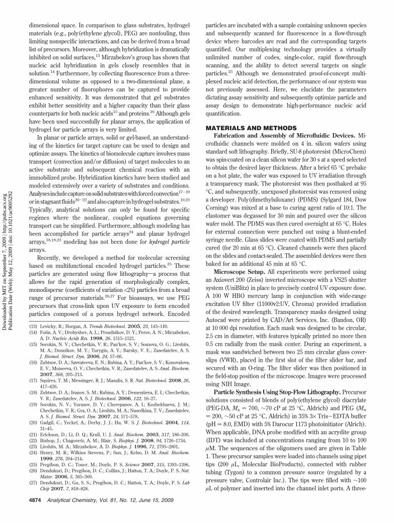
Investigation of Particle Composition. It is clear from thetime scale shown in eq 12 that the diffusivity of targets in the gelmatrix, Dgel, and also the concentration of probe incorporated,[P]o, will play a major role in determining the system kinetics.Diffusivity is directly related to the porosity of a gel matrix,which can be varied by altering the composition of prepolymersolutions. Pore size can be tuned efficiently using blends ofreactive and inert species, forming a semi-interpenetratingnetwork28 (semi-IPN). Although larger pores will allow fastertransport, it is also expected that they will lead to a decreasein probe incorporation efficiency and particle rigidity. In order
to investigate the effects of prepolymer composition, we useda semi-IPN consisting of both reactive PEG-DA (Mn ) 700)and inert PEG (Mw ) 200) mixed at different ratios.
To efficiently study the effects of particle composition on probeincorporation and hybridization signal, we synthesized pentafunc-tional “ladder” particles using stop-flow lithography.27 As shownin Figure 2a, each rung of the ladder had a unique composition.All prepolymer solutions contained a total of 60% PEG (PEG-DA+ PEG), with the amount of PEG-DA ranging from 15% to 35%.In each monomer solution, we used 5% Darocur 1173 and 35% of3× TE. The monomer solutions were mixed at 9:1 with a 50 bpDNA probe, which was modified with a fluorescein group to assessincorporation efficiency. The final DNA concentration in themonomer blends was 5 µM.
In addition to probe incorporation efficiency, particles werealso assessed for hybridization signal obtained after incubationwith targets varying in length from 20 to 200 bp. For thesehybridization studies, we used two different target labelingschemes based on SAPE or PicoGreen as fluorescent reporters.These two reporters were chosen due to their dramatic differencein size. Whereas PicoGreen is a small, DNA-binding cyanine dyeon the order of 1 nm, streptavidin and r-phycoerythrin are bothproteinswithdiametersontheorderof4and10nm,respectively.29,30
Although SAPE is a very efficient reporter for fluorescent detec-tion, it is also one of the largest, making it a good test of the upperlimit for target labeling. Alternatively, fluorescently labeled targets
(28) Witte, R. P.; Blake, A. J.; Palmer, C.; Kao, W. J. J. Biomed. Mater. Res., PartA 2004, 71, 508–518.
(29) Green, N. M. Methods Enzymol. 1990, 184, 51–67.(30) MacColl, R.; Eisele, L. E.; Williams, E. C.; Bowser, S. S. J. Biol. Chem. 1996,
271, 17157–17160.
Figure 2. Particle composition study. (a) Pentafunctional particles made from monomer solutions containing 15-35% PEG-DA were investigatedfor probe incorporation (b) and hybridization signal using two methods of fluorescent detection (c and d). The error bars shown in panel brepresent the standard deviation over three measurements. In panels c and d, fluorescent scans along the particles were normalized to theintensity measured for the 15% PEG-DA regions in order to show penetration trends for the two labeling schemes. Particles have dimensionsof 400 × 100 × 30 µm3.
4878 Analytical Chemistry, Vol. 81, No. 12, June 15, 2009
Dow
nloa
ded
by M
IT o
n Se
ptem
ber
7, 2
009
| http
://pu
bs.a
cs.o
rg
Pub
licat
ion
Dat
e (W
eb):
May
12,
200
9 | d
oi: 1
0.10
21/a
c900
5292
can be directly captured and detected without the use of a reporter,as shown in previous work25 and also in the model validationexperiments discussed later.
Probe Incorporation Efficiency. The incorporation of fluo-rescent probe at various precursor compositions is shown inFigure 2b. To find values of incorporated probe relative to that inprecursor, the fluorescence at each particle composition wasnormalized using the fluorescence obtained from particles madeusing 60% PEG-DA, which were assessed immediately aftersynthesis. At this high concentration of PEG-DA, it can beassumed that nearly all of the 50 bp (rg ∼ 4 nm) probe isincorporated within the particles, either by covalent linkageor physical entrapment (as fully cross-linked PEG-DA (Mn )700) is known to have a pore size of ∼1 nm31,32).
The results of this analysis show that the amount of reactivespecies in precursor solutions affect probe incorporation in a linearfashion over the compositions studied, with incorporations rangingfrom ∼5% to 25%. This trend is expected as the propagation rateis linear with respect to double-bond concentration for multifunc-tional, reactive monomers.33 Although it is by no means alimitation of our system, it is possible that the incorporationefficiency may be increased by matching the reaction rates of themonomer and probe species, which in this experiment wereacrylates and methacrylates, respectively. It is known that acrylatesreact faster than methacrylates, so it is possible that if methacry-lated monomers or acrylated probes were used, the probeincorporation would be higher.
Target Hybridization Signal. We expected that changing theparticle composition would alter the resulting pore size. To studythis in the context of DNA hybridization, we performed assaysusing biotinylated DNA targets with varying sizes of 20, 50, 100,and 200 bp. Using the Kratky-Porod equation,34 we can estimatethat oligonucleotides of these lengths have radii of gyration (rg)on the order of 2, 4, 7, and 10 nm with the ionic strength used(0.1 M). It is important to realize that the use of polymer targets(such as DNA) will not provide a direct measurement of thehydrogel pore size as these semiflexible polymer chains cantraverse the gel via reptation.
Particles were hybridized with each target present at greatexcess (1 µM) for 90 min and assessed for fluorescence usingboth labeling methods (Figure 2, parts c and d). The absolutevalues of fluorescent signal were dependent on target length,which is expected as both length and secondary structure areknown to alter association rates.35 For this reason, signals fromeach data set were normalized using a scale factor to match theintensities of the 15% PEG-DA regions over all target lengths. Thisis done to emphasize trends with respect to particle composition.
In both labeling schemes, the reporter entities (SAPE orPicoGreen) are added after hybridization. As such, the reportersmay be size-excluded from regions of the particle where their sizeis larger than the pore size. This is the case for the bulky SAPE
reporter, as shown in Figure 2c. Above a composition of 25% PEG-DA, SAPE is excluded from the particle interior; this is shown bya dramatic decrease in hybridization signal with all target sizes.This suggests that particle compositions of less than 25% PEG-DA must be used for SAPE-based labeling schemes.
In order to get a better understanding of DNA hybridizationthroughout the particles, we used PicoGreensa DNA dye thathas ∼100× fluorescent enhancement when bound to dsDNA (orDNA/RNA) versus ssDNA. The small size of this dye allows it topenetrate all regions of the particle. As shown in Figure 2d, the20 bp target signal has an intensity profile mimicking that of probeincorporation in Figure 2b. This suggests that the small, 20 bptarget can completely penetrate all regions of the particle,hybridizing throughout in the 90 min incubation period. The largertargets show this trend for lower PEG-DA concentrations, but itstarts to deviate with smaller pore sizes. For instance, the 50 and100 bp target signals start diminishing at 30% PEG-DA, whereasthe 200 bp target shows decrease beginning at 25%. These resultsshow that, as expected, the particle composition can be tuned toselectively inhibit penetration of larger oligonucleotide targets.
In selecting an “optimized” particle composition for generalassay use, we chose the composition of 20% PEG-DA. Thiscomposition allows use of both fluorescent labeling schemes andensures that particles are mechanically robust (instances ofmorphological deformation were observed for some of the 15%PEG-DA particle regions). As shown in Figure 2b, particles madefrom this composition retain ∼11% of the probe included in theprecursor solution.
Experimental Validation of Model. We investigated thevalidity of our model by performing kinetic studies using particlessimilar to that shown schematically in Figure 1 with varying probeconcentrations (from 1 to 5 µM), probe-region surface areas (2dfrom 45 to 80 µm), and particle numbers (from 20 to 40). In eachcase, we incubated distinct particle samples with 500 amol offluorescein-labeled target at room temperature. At various timepoints, particles from a sample were measured for fluorescence.We plotted the raw data as cumulative fluorescent intensity overall particles, as our model predicts the cumulative target loss fromsolution. As can be seen in Figure 3a, the data covers a widespreadof fluorescent intensity over incubation time.
To evaluate our model, we scaled time using the relationshipin eq 12 and the signal from eq 19 using
INp )IAp
FeVs[Ts]oNp ) 1 - e-τ (20)
We plotted the scaled data (Figure 3b) to find that it collapsesnicely on a similar trend. We fit the parameters kaDgel and Fe tofind a curve that best fit the data. Using these parameters, wecompared the experimentally observed fluorescent signals withsignals predicted from our model as shown in Figure 3b onthe right. The model agreed with experimental data over the entirerange of fluorescent intensities studied, thus validating our modelfor this specific hybridization regime.
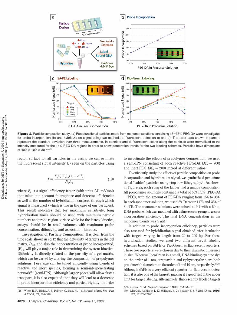
Investigation of Sensitivity. With a quantitative model inplace to understand hybridization in a rare-target regime, weassessed the sensitivity of our system over a range of hybridizationtimes. As an optimized design, we chose particles with 20% PEG-DA composition (which gives large pores with suitable particle
(31) Mellott, M. B.; Searcy, K.; Pishko, M. V. Biomaterials 2001, 22, 929–941.(32) Cruise, G. M.; Scharp, D. S.; Hubbell, J. A. Biomaterials 1998, 19, 1287–
1294.(33) Andrzejewska, E. Prog. Polym. Sci. 2001, 26, 605–665.(34) Meagher, R. J.; Won, J. I.; McCormick, L. C.; Nedelcu, S.; Bertrand, M. M.;
Bertram, J. L.; Drouin, G.; Barron, A. E.; Slater, G. W. Electrophoresis 2005,26, 331–350.
(35) Gao, Y.; Wolf, L. K.; Georgiadis, R. M. Nucleic Acids Res. 2006, 34, 3370–3377.
4879Analytical Chemistry, Vol. 81, No. 12, June 15, 2009
Dow
nloa
ded
by M
IT o
n Se
ptem
ber
7, 2
009
| http
://pu
bs.a
cs.o
rg
Pub
licat
ion
Dat
e (W
eb):
May
12,
200
9 | d
oi: 1
0.10
21/a
c900
5292
mechanics), a probe concentration of 50 µM in precursor solution(for 5.5 µM in particles after 11% incorporation efficiency), andthin, 30 µm probe regions. We investigated hybridization timesup to 3.3 h, at which point the reaction is expected to be ∼75%complete (i.e., τ ∼ 1.3 so Ts ) 0.25 using eq 18). It is importantto note that this is much shorter than typical assay times forcommercially available multiplexing systems, which frequentlyrecommend incubations up to 20 h or more.36,37
For sensitivity assays, we chose to use phycoerythrin (i.e.,SAPE) as the reporting fluorophore, as it is much more efficientthan fluorescein used in our model validation, but requires anextra processing step to report fluorescence. In a typical assay,particles are incubated with biotinylated targets and subsequentlylabeled using SAPE in a 30 min reaction. We found the fluorescentefficiency for SAPE assays, using our detection system, to beFe,SAPE ) 2.5 × 10¹⁴ AU m2/mol.
To measure the detection limits experimentally, we incubatedparticles with target at varying levels near the expected limits.For each concentration, the fluorescent signal was measured and
divided by the pooled standard deviation of the background signal(over all measurements) to obtain a signal-to-noise ratio (S/N).A line was then fit to the S/N data for each time point, specificallyfor the three data points above the limit of detection (LOD) (datanot shown), to obtain and estimate of the sensitivity, which wedesignated as the point where S/N ) 3. With the use of ourdetection settings, the observed noise is typically ∼0.5 AU, suchthat at the LOD the signal, I, would be ∼1.5 AU.
As shown in Figure 4, our system is extremely sensitive,providing subattomole LODs even with short, 1 h hybridizations.Furthermore, the dynamic nature of this sensitivity is predictableusing our model, as indicated by in the figure. The sensitivityand kinetics of our system are very favorable compared tocommercially available systems and will likely improve with theimplementation of photomultiplier-based detection.
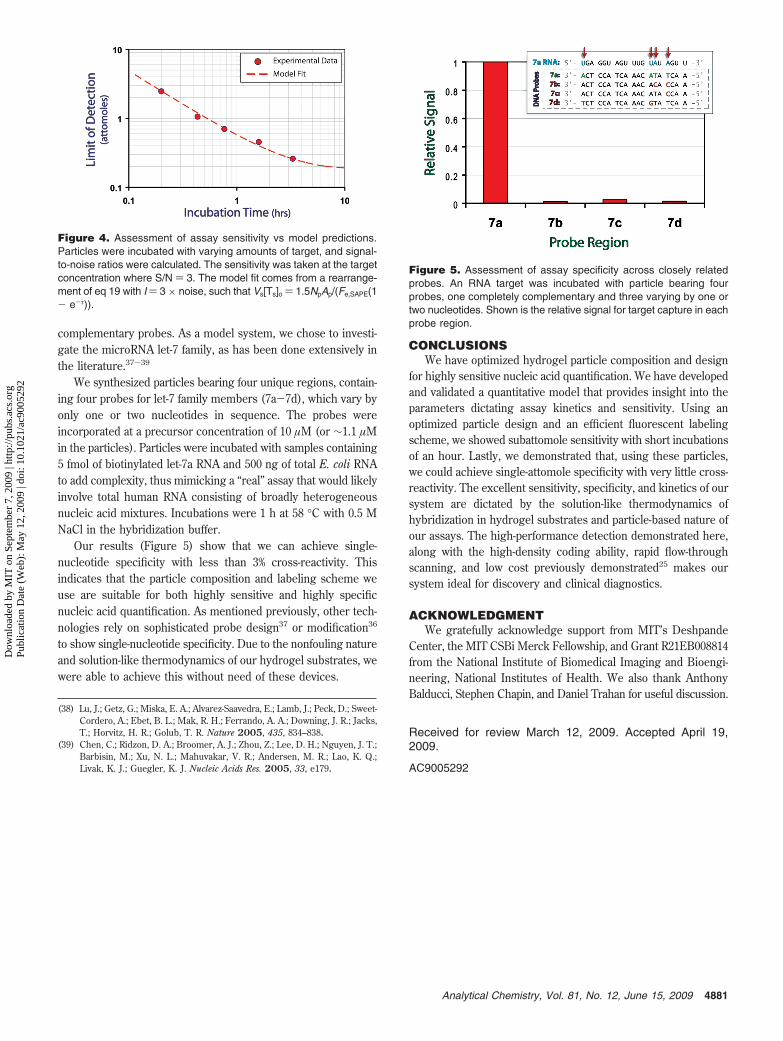
Investigation of Specificity. We demonstrated the optimiza-tion of particle design for highly sensitive detection. Anotherimportant metric for nucleic acid detection is specificityshow wellthe assay can distinguish between closely related targets. In orderto show that the optimized particle design and labeling schemedo not negatively affect the specificity of our system, we investi-gated the cross-reactivity of an RNA target with a closely related
(36) Castoldi, M.; Schmidt, S.; Benes, V.; Noerholm, M.; Kulozik, A. E.; Hentze,M. W.; Muckenthaler, M. U. RNA 2006, 12, 913–920.
(37) Wang, H.; Ach, R. A.; Curry, B. RNA 2007, 13, 151–159.
Figure 3. Validation of model predictions. Particles with varying probe concentrations ([P]o), stripe width (d), and numbers (Np) were incubatedwith 500 amol of complementary, fluorescein-labeled target, and their fluorescence was measured over time. (a) Raw data showing the averageparticle signal multiplied by the number of particles vs incubation time. (b, left) Dimensionless signal vs dimensionless time showing the collapseof the data from panel a onto a universal curve. The dashed line represents a fit of eq 20 to the data with kdDgel ) 5.5 × 10⁻⁵ m2 s-2 M-1 andFe,FITC ) 2.54 × 10¹². (b, right) The observed fluorescence plotted against the model fit. The dashed line (x ) y) represents a perfect fit and isshown to guide the eye.
4880 Analytical Chemistry, Vol. 81, No. 12, June 15, 2009
Dow
nloa
ded
by M
IT o
n Se
ptem
ber
7, 2
009
| http
://pu
bs.a
cs.o
rg
Pub
licat
ion
Dat
e (W
eb):
May
12,
200
9 | d
oi: 1
0.10
21/a
c900
5292
complementary probes. As a model system, we chose to investi-gate the microRNA let-7 family, as has been done extensively inthe literature.37-39
We synthesized particles bearing four unique regions, contain-ing four probes for let-7 family members (7a-7d), which vary byonly one or two nucleotides in sequence. The probes wereincorporated at a precursor concentration of 10 µM (or ∼1.1 µMin the particles). Particles were incubated with samples containing5 fmol of biotinylated let-7a RNA and 500 ng of total E. coli RNAto add complexity, thus mimicking a “real” assay that would likelyinvolve total human RNA consisting of broadly heterogeneousnucleic acid mixtures. Incubations were 1 h at 58 °C with 0.5 MNaCl in the hybridization buffer.
Our results (Figure 5) show that we can achieve single-nucleotide specificity with less than 3% cross-reactivity. Thisindicates that the particle composition and labeling scheme weuse are suitable for both highly sensitive and highly specificnucleic acid quantification. As mentioned previously, other tech-nologies rely on sophisticated probe design37 or modification36
to show single-nucleotide specificity. Due to the nonfouling natureand solution-like thermodynamics of our hydrogel substrates, wewere able to achieve this without need of these devices.
CONCLUSIONSWe have optimized hydrogel particle composition and design
for highly sensitive nucleic acid quantification. We have developedand validated a quantitative model that provides insight into theparameters dictating assay kinetics and sensitivity. Using anoptimized particle design and an efficient fluorescent labelingscheme, we showed subattomole sensitivity with short incubationsof an hour. Lastly, we demonstrated that, using these particles,we could achieve single-attomole specificity with very little cross-reactivity. The excellent sensitivity, specificity, and kinetics of oursystem are dictated by the solution-like thermodynamics ofhybridization in hydrogel substrates and particle-based nature ofour assays. The high-performance detection demonstrated here,along with the high-density coding ability, rapid flow-throughscanning, and low cost previously demonstrated25 makes oursystem ideal for discovery and clinical diagnostics.
ACKNOWLEDGMENTWe gratefully acknowledge support from MIT’s Deshpande
Center, the MIT CSBi Merck Fellowship, and Grant R21EB008814from the National Institute of Biomedical Imaging and Bioengi-neering, National Institutes of Health. We also thank AnthonyBalducci, Stephen Chapin, and Daniel Trahan for useful discussion.
Received for review March 12, 2009. Accepted April 19,2009.
AC9005292
(38) Lu, J.; Getz, G.; Miska, E. A.; Alvarez-Saavedra, E.; Lamb, J.; Peck, D.; Sweet-Cordero, A.; Ebet, B. L.; Mak, R. H.; Ferrando, A. A.; Downing, J. R.; Jacks,T.; Horvitz, H. R.; Golub, T. R. Nature 2005, 435, 834–838.
(39) Chen, C.; Ridzon, D. A.; Broomer, A. J.; Zhou, Z.; Lee, D. H.; Nguyen, J. T.;Barbisin, M.; Xu, N. L.; Mahuvakar, V. R.; Andersen, M. R.; Lao, K. Q.;Livak, K. J.; Guegler, K. J. Nucleic Acids Res. 2005, 33, e179.
Figure 4. Assessment of assay sensitivity vs model predictions.Particles were incubated with varying amounts of target, and signal-to-noise ratios were calculated. The sensitivity was taken at the targetconcentration where S/N ) 3. The model fit comes from a rearrange-ment of eq 19 with I ) 3 × noise, such that Vs[Ts]o ) 1.5NpAp/(Fe,SAPE(1- e-τ)).
Figure 5. Assessment of assay specificity across closely relatedprobes. An RNA target was incubated with particle bearing fourprobes, one completely complementary and three varying by one ortwo nucleotides. Shown is the relative signal for target capture in eachprobe region.
4881Analytical Chemistry, Vol. 81, No. 12, June 15, 2009