Optimization of methodology for the simultaneous speciation of inorganic As, Sb and Se in fluid samples by sector-field ICP- MS coupled to HPLC Dissertation zur Erlangung des Doktorgrades der Naturwissenschaften (Dr. rer. nat.) am Fachbereich Geowissenschaften der Universität Bremen vorgelegt von Debo Wu Bremen, April 2015
Transcript
Optimization of methodology for the simultaneous speciation of inorganic As, Sb and Se in fluid samples by sector-field ICP-
MS coupled to HPLC
Dissertation
zur Erlangung des Doktorgrades der Naturwissenschaften (Dr. rer. nat.)
am Fachbereich Geowissenschaften
der Universität Bremen
vorgelegt von
Debo Wu
Bremen, April 2015
Reviewer:
Prof. Dr. Thomas Pichler
Prof. Dr. Andrea Koschinsky-Fritsche
I
E r k l ä r u n g
Hiermit versichere ich, dass ich
i. die Arbeit ohne unerlaubte fremde Hilfe angefertigt habe, ii. keine anderen als die von mir angegebenen Quellen und
Hilfsmittel benutzt haben und iii. die den benutzten Werken wörtlich oder inhaltlich entnom
2.3.3 Se speciation ................................................................................................................. 41
3. Scopes and objectives ...................................................................................................... 44
4. Simultaneous speciation analysis of As, Sb and Se redox couples by SF-ICP-MS coupled to HPLC .................................................................................................................................... 47
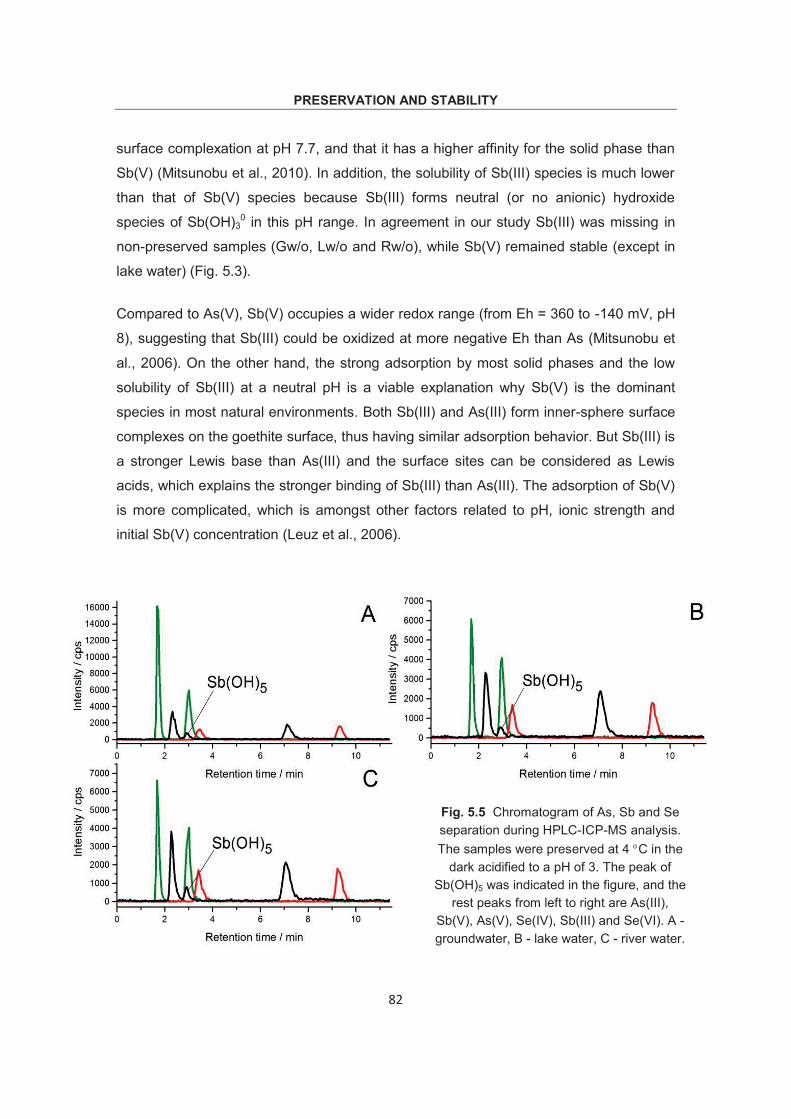
Metal speciation provides information useful in the study of toxicity, bioavailability,
adsorption, and redox behavior of element species. Based on inductively coupled
plasma mass spectrometry (ICP-MS) coupled to high performance liquid
chromatography (HPLC), in this project, a systematic investigation was made regarding
chromatographic methods for the simultaneous speciation of arsenic (As), antimony (Sb)
and selenium (Se) redox couples, and preservation strategies of these species. Finally,
the developed method was applied to the analysis of hydrothermal water samples, with
the purpose of studying As and Sb inorganic species distribution in hydrothermal
systems.
In the first study, a new method was developed for the simultaneous speciation analysis
of inorganic As(III, V), Sb (III, V) and Se(IV, VI) in fluid samples by sector field-ICP-MS
coupled with HPLC. Hamilton PRX-X100 anion-exchange column with EDTA (pH of 4.7)
and 3% methanol as mobile phase was used for the separation of these species. The
overall analysis time was within 11minutes for all six desired species. A thorough
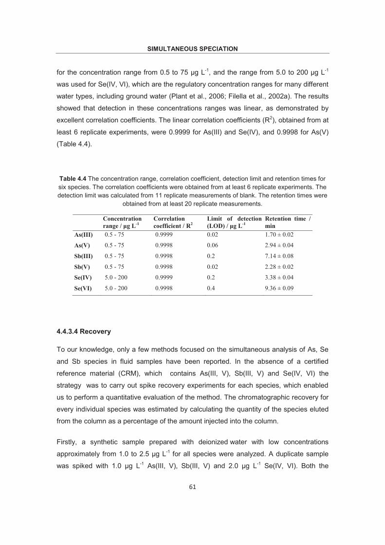
validation concerning stability of retention time, linearity and spike recovery was carried
out. Low detection limits of these species, 0.02 μg L-1 for As(III), 0.06 μg L-1 for As(V),
0.2 μg L-1 for Sb(III), 0.02 μg L-1 for Sb(V), 0.2 μg L-1 for Se(VI) and 0.4 μg L-1 for Se(IV),
make it possible for simultaneous study of competitive adsorption, redox behavior of
these species.
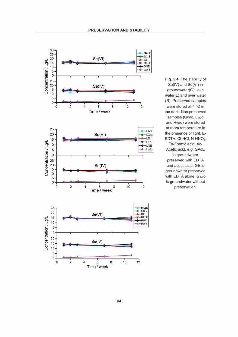
In the second study, preservation method and stability of As, Sb and Se redox couples
were investigated in Fe- and Mn- rich water samples (groundwater, river water and lake
water). As potential preservation strategies EDTA alone and EDTA combined with either
HCl, HNO3, formic acid or acetic acid were studied and compared to unpreserved
samples. The results showed that addition of EDTA combined with acidification to a pH
of 3 successfully preserved all three redox couples for at least 11 weeks stored at 4 C
in the dark. EDTA alone (pH = 6) failed to preserve As and Sb species, especially for
Sb(III) which was eventually completely oxidized in all samples. On the other hand, in
the unpreserved samples, As, Sb and Se redox species showed different adsorption
behaviors. As(III), Sb(III), Se(IV)) and As(V) were strongly adsorbed by Fe-
ABSTRACT
2
(oxy)hydroxide and possibly Mn-(oxy)hydroxide. While Sb(V) and Se(VI) were not
adsorbed in most cases.
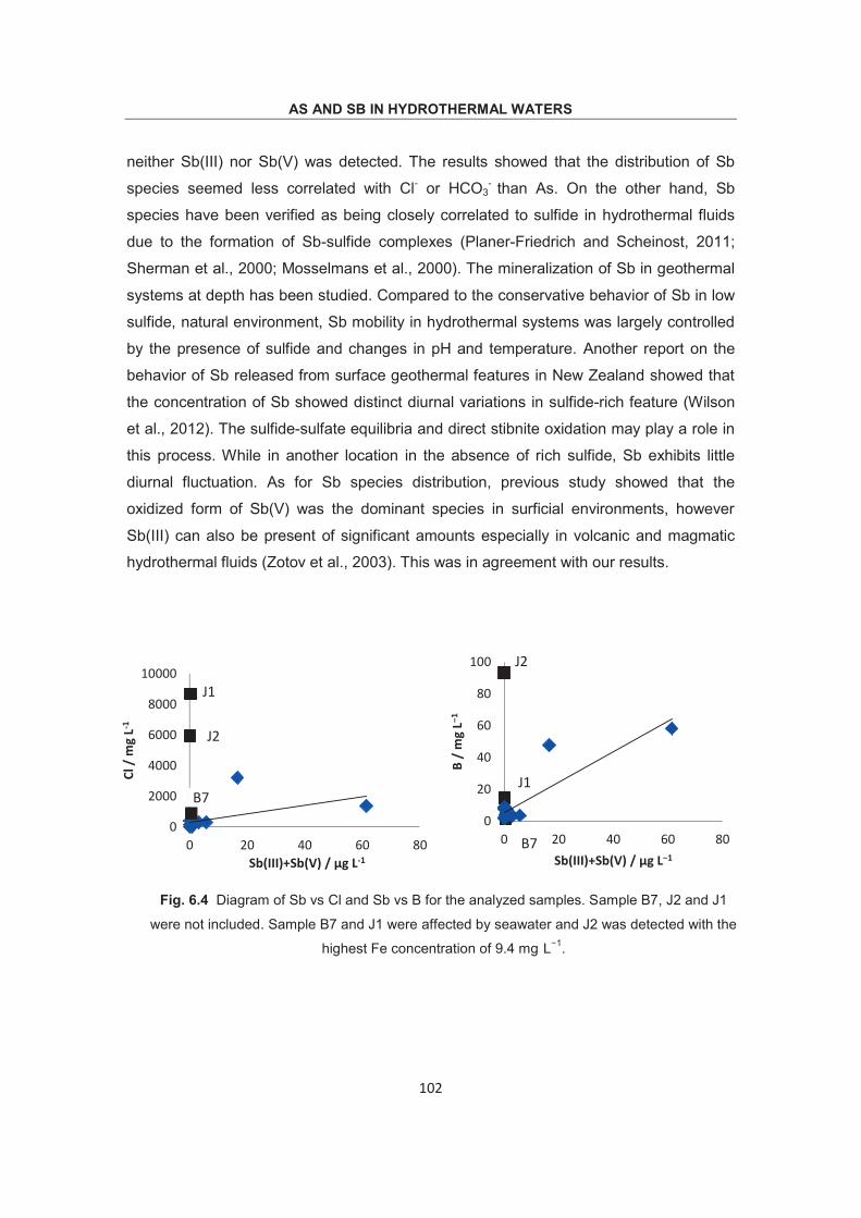
In the third study, the developed speciation method was used for the analysis of
hydrothermal waters from Bali and Java, Indonesia. The results showed that the
distribution of As and Sb species were closely correlated to Cl-, HCO3- and SO4
2-.
Generally, in HCO3-type hydrothermal waters As(V) seemed the dominant species. In
Cl-type samples, it is more complicated. Since extremely high concentration of Cl might
be originated from either magma degassing (HCl gas) or seawater feeding, thus other
oxidation processes may be involved in As species distribution. Our primary speciation
results indicated that when the hydrothermal waters were affected by seawater feeding,
As(V) was the main existing form, probably due to microbial activity. In SO4-type
hydrothermal waters, As distribution is variable, either As(III) or As(V) could be the
dominant species. In addition, an unknown As species was detected in 5 of the 18
samples, particularly in 2 samples this unknown species was even the main existing
form for As, indicating that microbial activity was involved. For Sb species, Sb(V) was
generally the main species in the analyzed samples.
KURZFASSUNG
3
Kurzfassung
Metallspeziation liefert Informationen, die sehr wichtig für die Untersuchung der Toxizität,
der Bioverfügbarkeit, der Adsorption und des Redoxverhaltens von Elementspezies sind.
Basierend auf der Methode der Massenspektrometrie mittels induktiv gekoppleten
Plasma, das mit einer Hochleistungsflüssigchromatographie (HPLC)-Apparatur
verbundenen war, wurden im Rahmen dieses Projektes systematische Untersuchungen
zu chromatographischen Methoden für die simultane Speziation von Arsen (As)-,
Antimon (Sb)- und Selen (Se)-Redoxpaaren sowie von Konservierungsstrategien dieser
Spezies durchgeführt. Anschließlich wurde die entwickelte Methode für die Analyse von
hydrothermalen Wasserproben angewandt.
Im Rahmen der ersten Studie wurde eine neue Methode für die simultane
Speziationsanalyse von anorganischem As(III, V), Sb(III, V) und Se(IV, VI) in
Fluidproben mittels einer mit einer HPLC gekoppelt an ein Sektorfeld-ICP-MS entwickelt.
Für die Trennung dieser Spezies wurde dabei eine Hamilton PRX-X100
Anionenaustauschersäule mit EDTA (pH 4.7) und Methanol (3%) als mobile Phase
verwendet. Die Gesamtanalysenzeit für alle sechs gewünschten Spezies lag innerhalb
von 11 Minuten. Darüber hinaus wurde eine gründliche Validation hinsichtlich der
Stabilität der Retentionszeit, der Linearität und der Spike-Wiederfindung durchgeführt.
Die niedrigen Nachweisgrenzen dieser Spezies (0.02 μg L-1 für As(III), 0.06 μg L-1 für
As(V), 0.2 μg L-1 für Sb(III), 0.02 μg L-1 für Sb(V), 0.2 μg L-1 für Se(VI) und 0.4 μg L-1 für
Se(IV)) ermöglichten die simultane Untersuchung konkurrierender Adsorptions- und
Redoxverhalten dieser Spezies.
In der zweiten Studie wurden einerseits Konservierungsmethoden und andererseits die
Stabilität von As-, Sb- und Se-Redoxpaaren in Fe- und Mn-reichen Wasserproben
(Grund-, Fluss- und Seewasser) untersucht. Als potentielle Konservierungsstrategien
wurden sowohl EDTA, als auch EDTA in Kombination mit entweder HCl, HNO3,
Ameisensäure oder Essigsäure untersucht und die Ergebnisse mit denen nicht-
konservierter Proben verglichen. Es zeigte sich, dass sich alle drei Redoxpaare durch
die Zugabe von EDTA und die Ansäuerung auf pH 3 erfolgreich für mindestens 11
Wochen dunkel gelagert bei 4°C konservieren ließen. EDTA alleine (pH 6) war nicht in
KURZFASSUNG
4
der Lage, As- und Sb-Spezies zu konservieren. Dies gilt insbesondere für Sb(III), das in
allen Proben letztendlich vollständig oxidiert wurde. In den nicht-konservierten Proben
zeigten die As-, Sb- und Se-Redoxspezies dagegen unterschiedliche
Adsorptionsverhalten. As(III), Sb(III), Se(IV) und As(V) adsorbierten stark an Fe- und
möglicherweise auch Mn-(oxi)hydroxiden, während Sb(V) und Se(VI) in den meisten
Fällen nicht adsorbierte.
In der dritten Studie wurde die entwickelte Speziationsmethode für die Analyse von
hydrothermalen Wässern aus Bali und Java (Indonesien) verwendet. Die Ergebnisse
zeigten, dass die Verteilung von As- und Sb-Spezies sehr eng mit den Gehalten an Cl-,
HCO3- und SO4
2- korreliert. Im Allgemeinen schien As(V) die dominierende Spezies in
hydrothermalen Wässern des HCO3-Typs zu sein. In Proben des Cl-Typs ist es
komplizierter. Da extrem hohe Cloridkonzentrationen entweder von HCl ausgasendem
Magma oder Kontakt mit Meerwasser herrühren können, mögen andere
Oxidationsprozesse bei der Verteilung von As-Spezies beteiligt sein. Unsere primären
Speziationsergebnisse zeigten, dass As(V) die vorherrschende Spezies darstellte, wenn
hydrothermale Wässer durch Meerwasserspeisung beeinflusst sind, was möglicherweise
auf mikrobielle Aktivität zurpückzuführen ist. In hydrothermalen Wässern des SO4-Typs
ist die Verteilung des As variable, sowohl As(III), als auch As(V) können die
dominierende Spezies sein. Zusätzlich wurde eine unbekannte As-Spezies in fünf der 18
Proben gefunden, wobei diese in zwei Proben sogar die wichtigste Form darstellte, was
auf eine Beteiligung mikrobieller Aktivität hindeutet. Im Falle des Sb war Sb(V) in der
Regel die Hauptspezies in den analysierten Proben.
INTRODUCTION
5
1. Introduction 1.1 As, Sb and Se in aqueous environment
1.1.1 As
Arsenic (As), a metalloid occurs naturally, being the 20th most abundant element in the
terrestrial crust (Gulledge and O’Connor, 1973). Arsenic and its compounds are mobile
in natural environment. Rock-weathering converts As sulfides to As trioxides, which
subsequently enter into the aquatic environment by dissolving in rain, rivers, or
groundwater. Arsenic has only one stable isotope, 75As. It can exist in the -III, -I, 0, III,
and V oxidation states. Arsenic is highly toxic and leads to a wide range of health
problems in humans. If entering the food chain, As accumulates in animal bodies in the
form of organic species. Arsenic has become increasingly important because of natural
water contamination as well as human activities, e.g. industrial waste and drainage
problem. Numerous studies have shown that excessive intake of As from drinking water
can lead to chronic poisoning and various types of cancers, e.g. skin, lungs, bladder and
kidney (Smedley and Kinniburgh, 1993). Arsenic has been classified as a group I human
carcinogen by the International Agency for Research on Cancer due to the increased
cancer risk. The maximum permissible levels of As in drinking water have been reduced
in many countries. The United States Environmental Protection Agency (USEPA, 2006)
set the maximum contaminant level in drinking water at 10.0 μg L-1, the same as the
guidelines of the World Health Organization (WHO). Australia has a drinking water limit
for arsenic of 7.0 μg L-1 (NHMRC, 2004). The American Natural Resources Defense
Council (NRDC) even recommended an As level of 3.0 μg L-1 (NRDC-report, 2000).
Arsenic concentrations in natural environment can range from less than 0.5 μg L-1 to
more than 5000 μg L-1. Previous study showed that the concentration of As in unpolluted
fresh water typically ranges from 1.0-10.0 μg L-1, rising to 100-5000 μg L-1 in sulfide
mineralization and mining area (Smedley et al., 1996). Some reviews concerning the
occurrence and distribution of As species have been made to enable researchers better
understanding the behavior of As in environment (Mandal, 2002; Wilson et al., 2010;
Plant et al., 2006). Seawater generally contains 1.0-8.0 μg L-1 As, and As(V) was
assumed being the dominant species (As(V) : As(III) = 1026 : 1) from thermodynamic
calculations (oxygenated seawater at pH of 8.1). However, in reality the ratios of As(V):
INTRODUCTION
6
As(III) ranged from 0.1:1 to 10:1 (Johnson, 1972). Biological reduction may play an
important role in affecting the distribution of As species. Arsenic is also an important
constituent in geothermal fluids, ranging from 0.1 to 50 mg L-1, e.g. up to 8.5 mg L-1 in
New Zealand (Ritchie, 1960), 6.4 mg L-1 in Japan (Nakahara et al., 1978), and up to 9.2
mg L-1 (chapter 5) in Java. Speciation analysis of As in geothermal systems indicated
that As occurred in two oxidation states, As(III) and As(V), and As(III) seemed to be the
main aqueous species in hydrothermal fluids (Ballantyne and Moore, 1988; Breuer and
Pichler, 2013). Organic As species such as MMA, DMA and AB were also identified in
marine environment but only minor fractions were detected due to the adsorption on to
suspended particles.
1.1.2 Sb
Antimony (Sb) is a trace element and the 63rd most abundant occurring element in the
Earth’s crust, but its crustal abundance is about one order of magnitude lower than As
(Reimann et al., 2010). Sb in the aquatic environment can be originated from rock-
weathering, soil runoff and anthropogenic activities. Generally, the concentrations of Sb
in unpolluted water are very low, ranging from a few ng L-1 to a few μg L-1 depending on
different chemical and physical conditions (Onishi, 1969; Schutz and Turekian, 1965).
Sb was not well documented and often overlooked, due to its lower abundance and
relative insolubility of most of its compounds. However, anthropogenic related sources,
may lead to up to 100 times higher values. The U.S. Environmental Protection Agency
(EPA) considers it a priority pollutant and the Council of the European Union (1998)
established the maximum admissible level of Sb in drinking waters at 5.0 ug L–1. Sb has
two isotopes; 121Sb and 123Sb with the abundances of 57.21% and 42.76% respectively.
It occurs in four oxidation states (-III, III, IV and V), with two oxidation states +III and +V
being the predominant species in environment. Sb is thought to be chemically similar to
As, as they are both metalloids and have the same oxidation states. However, previous
studies have found that Sb may have quite different behavior regarding oxidation,
adsorption and bioavailability (Wilson et al., 2010).
The existing forms of Sb species are different depending on pH and oxidation states
(section 1.2). Compared to As species, Sb(III) in solution has a complexing properties,
and can form complexation with organic ligands under acidic conditions, such as EDTA,
DTPA. Distribution and speciation of Sb in freshwater and ocean water have not been
INTRODUCTION
7
studied extensively, probably due to the lack of samples preservation methods. Sb
concentration in surface marine waters was 184 ± 45 ng L-1 (Filella et al., 2002b), higher
by a factor of 3 to 4 times higher than in fresh water. Previous studies (Mok and Wai,
1987; Shieh, 1993; Ulrich, 1998; Mok and Wai, 1990) reported that Sb(V) was the
dominant species under oxic conditions. However, significant concentration of Sb(III)
was also detected. Similarly, the Sb(V) was reported under anoxic conditions. This is
contradicting thermodynamic equilibrium predictions. Biological activity or kinetic effects
may partially explain the discrepancy but have not yet been verified (Filella et al., 2002b).
Besides, methylated antimony species were monitored in a few studies but only at trace
level. Sb is present in geothermal systems at substantial concentrations, ranging from
500 mg L-1 up to 10 wt.% (Ritchie, 1960; Stauffer and Thompson, 1984; Weissberg et al.,
1979).
1.1.3 Se
Selenium (Se) has six natural stable isotopes (74Se, 76Se, 77Se, 78Se, 80Se, and 82Se); the
most important are 78Se and 80Se, with natural abundances close to 50 and 24%. Se can
exist in the -II, 0, IV, and VI oxidation states. Se occurs in natural waters principally in
two oxidation states, Se(IV) and Se(VI). Se was introduced into aquatic environment by
both natural processes (weathering or run-off from rocks) and human activity (leachate
from agricultural activity, combustion) (B’Hymer and Caruso, 2006).
In contrast to arsenic, trace concentrations of selenium are essential to human and
animal health. Selenoproteins, incorporated in enzymes, are essential components for
cellular functions in most mammals. However, there is a fine line between low intake
leading to selenium deficiency (< 40 μg d-1) and copious intake leading to toxicity (> 400
μg d-1) in humans (Boyd, 2011). The WHO guideline value for Se in drinking water is
10.0 μg L-1. Though the Se concentration in most natural waters is less than 1.0 μg L-1,
occasionally much higher concentrations were found in groundwater, e.g. extremely high
concentration of up to 1300 μg L-1 were detected in Colorado River catchment, USA
(Engberg, 1999). Groundwaters generally contain higher Se concentrations than surface
waters due to water–rock interactions (Frankenberger and Benson, 1994).
Similar to As and Sb, the existing form and distribution of Se(IV) and Se(VI) are
determined principally by pH and Eh conditions, however, competitive solubility,
INTRODUCTION
8
complexation and biological interaction may also play a part. Previous studies on Se
speciation showed some difference in the Se(IV) to Se(VI) ratio. It did not follow the ratio
of other redox couples (e.g. Fe2+/Fe3+) (White and Dubrovsky, 1994). This reflected the
slow reaction kinetics (Measures and Burton, 1978; Plant et al., 2006). In contrast to As,
the reduced form of Se(IV), is very strongly adsorbed by oxides and clays. This explains
the very low concentration of Se in reducing environment and the remarkable difference
in behavior of As and Se in natural environment. Se in seawater was estimated at 0.17
μg L-1 (Thomson et al., 2001). Detailed study on Se distribution and speciation in
seawater (Cutter and Cutter, 2001) showed that Se(VI) was generally higher than Se(IV)
in marine waters and the concentration of Se with depth showed surface water depletion
and deep water enrichment (due to deposition and mineralization). However, a
substantial fractionation of Se(IV) can also be detected if microbiological processes
(converting Se(VI) to Se(IV)) are involved (Measures and Burton, 1978). Besides,
organic selenide was also found in surface ocean waters but was not detected in mid- or
deep waters.
1.2 Existing forms of As, Sb and Se in aqueous environment
1.2.1 As
Since the solubility, mobility, bioavailability and toxicity of As, Sb and Se are related to
their oxidation states, studies concerning distribution and transformation are necessary
in order to understand their behavior in the environment. Redox potential (Eh) and pH,
as the most important factors controlling inorganic As, Sb and Se species in natural
waters, are used widely to analyze and predict their distributions under different
conditions (Wilson et al., 2010).
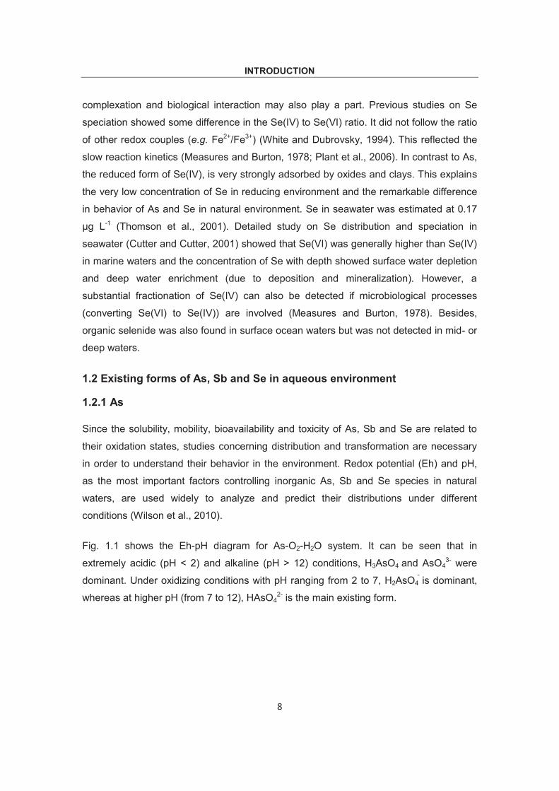
Fig. 1.1 shows the Eh-pH diagram for As-O2-H2O system. It can be seen that in
extremely acidic (pH < 2) and alkaline (pH > 12) conditions, H3AsO4 and AsO43- were
dominant. Under oxidizing conditions with pH ranging from 2 to 7, H2AsO4 is dominant,
whereas at higher pH (from 7 to 12), HAsO42- is the main existing form.
INTRODUCTION
9
Fig. 1.1 Eh-pH stability diagram for As-O2-H2O system at 25 C, 1bar. Dashed lines indicate environmental limits imposed by the dissociation of water to H2(g) and O2(g). (Brookins, 1988)
On the other hand, under reducing conditions with a wide pH range of 0 to 9, As(III)
exists exclusively as non-charged H3AsO3. The lack of charge on the As(III) species
compared to the successive deprotonation of As(V) species implies less charge
dependence associations with solid phases, such as clay minerals and (oxy)hydroxides
in soils. Thus it can be concluded that As(III) species are more mobile than As(V) in a
wide pH range (Bhattacharya et al., 2002). While under alkaline conditions, As(V) exists
as negatively charged oxyanions, such as H2AsO3 at pH of 9 - 10, HAsO3
2- at pH of 11 -
13 and AsO33- at pH higher than 13. In addition, numerous studies have shown that As
and Sb inorganic species predominate over organic species in most environmental
systems (Andreae et al., 1981; Ellwood and Maher, 2002; Sun et al., 1993). It is worth
noting that Fig. 1.1 is a simplified illustration of species distribution, without other
elements involved. In fact other variables could also influence the behavior of As species
in a more complex system. With addition of Fe, As would co-precipitate with Fe-
(oxy)hydroxides, e.g. as the hydrated iron arsenate mineral scorodite (FeAsO4•2H2O)
(Mok and Wai, 1990). While at the presence of extremely high concentration of reduced
S, the formation of dissolved As-sulphide species can be significant, e.g.
(co)precipitation as orpiment (As2S3), realgar (AsS) or other sulphide minerals under
reducing acidic conditions (Bowen, 1979). Therefore, high concentrations of dissolved
INTRODUCTION
10
As were not expected in waters with a high concentrations of free sulphide (Moore et al.,
1988).
1.2.2 Sb
Fig. 1.2 Eh-pH stability diagram for Sb-S-H2O system at 25 C, 1bar with a dissolved Sb of 10-8 mol L-1 and S of 10-3 mol L-1. Dashed lines indicate environmental limits imposed by the dissociation of water to H2(g)
and O2(g). (Filella et al., 2002b)
For Sb (Fig. 1.2) the Eh-pH diagram shows that Sb(V) exclusively exists as negatively
charged Sb(OH)6 (the coordination of Sb(V) with oxygen is octahedral) in a wide pH
range from acid to alkaline, which is different from As(V). As has been mentioned As(V)
was deprotonated in successive steps in a similar pH range. Under extremely acidic
conditions (pH < 1), Sb(V) exists as non-charged Sb(OH)5. As for Sb(III), non-charged
Sb(OH)3 exists in a wide pH range from 2 to 11 with pKa = 11.9 (Table 1.1). Similar to
As, the mobility of Sb (III) is higher than Sb(V). Besides, the exclusive existing form of
Sb(V) ( as Sb(OH)6 ) but successive protonation of As(V) in a wide pH range from acidic
to alkaline indicated that the binding of As(V) to particulate matter in oxygenated
systems is more complicated than that of Sb(V). Previous studies have shown that Sb(V)
formed mainly outer sphere complexes with Fe-(oxy)hydroxides, while As(V) formed
INTRODUCTION
11
inner sphere complexes (Goldberg and Johnston, 2001; Ona-Nguema et al., 2005; Leuz,
2006).
On the other hand, Sb(III) exists as positively charged Sb(OH)2+ under extreme acidic
conditions (pH < 2) and negatively charged Sb(OH)4 under alkaline conditions (pH > 11).
In the wide pH range from 2 to 11, Sb(III) exists as dissolved Sb(OH)3. This diagram was
obtained based on environmentally relevant concentrations: Sb of 10-8 mol L-1 and
dissolved S of 10-3 mol L-1. According to this result, under reducing conditions at
presence of S, stibnite Sb2S3(s) is formed at low to neutral pH range. At higher pHs,
Sb2S42- was formed instead of Sb2S3. However, when the concentration of Sb in the
environment exceeds 10-6 mol L-1, Sb(III) would be present as solid species, e.g. in the
form of polymorphs senarmontite and valentinite (Sb4O6), instead of Sb(OH)3(s) under
acidic to alkaline and moderately reducing to moderately oxidizing conditions (Vink,
1996). As for Sb(V), the ionic species SbO3 (Sb(OH)6
) occupies a large range under
oxidizing conditions from acidic to alkaline conditions, indicating a relatively high mobility.
Noteworthy, Sb(V) was previously thought to be immobile under oxidizing conditions and
existed in the form of Sb2O5 (Brookins, 1986, 1988).
1.2.3 Se
Fig. 1.3 Eh-pH stability diagram for As-O2-H2O system at 25 C, 1bar. Dashed lines indicate environmental limits imposed by the dissociation of water to H2(g) and O2(g). (Brookins, 1988)
INTRODUCTION
12
Similar to As and Sb, Se also is a redox sensitive element. Sulfur and iron compounds
play an important part in the transportation of Se. Se occurs in water solutions principally
in two oxidation states, Se(IV) and Se(VI). For Se(VI), SeO42- mainly exists under
oxidizing condition in a pH range of around 2 to extremely basic conditions. HSeO42-
exists at a pH less than 2. It can be seen in table 1.1 that H2SeO4 is an acid with a pKa
of 2.0. For Se(IV) HSeO3 and SeO3
2- were the main existing forms under reducing
conditions in a wide pH range from 2 to 14. H2SeO3 is formed under very acidic
conditions (pH < 2). In soils and sediments, elemental Se dominates under strong
reducing conditions. Considering the main existing form of Se(VI) and the successive
protonation of Se(IV), Se(IV) is generally more available and more mobile than Se(VI).
Previous study of Se distribution and speciation for seawater showed that the
concentration of Se(VI) was generally higher than Se(IV) (Cutter and Cutter, 2001).
Table 1.1. Equations and pKa values for inorganic As, Sb and Se species.
As(V) pKa H3AsO4 + H2O = H2AsO4- + H3O+ 2.20
H2AsO4- + H2O = HAsO4
2- + H3O+ 6.97
HAsO42- + H2O = AsO4
3- + H3O+ 11.53
Sb(V)
Sb(OH)5 + 2H2O = Sb(OH)6- + H3O+ 2.72
Se(VI)
H2SeO4 + H2O = HSeO4- + H3O+ 2.0
As(III)
H3AsO3 + H2O = H2AsO3- + H3O+ 9.22
H2AsO3- + H2O = HAsO3
2- + H3O+ 12.13
HAsO32- + H2O = AsO3
3- + H3O+ 13.4
Sb(III)
Sb(OH)3 + 2H2O = Sb(OH)4- + H3O+ 11.9
Se(IV)
H2SeO3 + H2O = HSeO3- + H3O+ 2.6
HSeO3- + H2O = SeO3
- + H3O+ 8.3
INTRODUCTION
13
1.3 Interferences in plasma
When analyzing As and Se using ICP-MS, the main difficulties are interferences. There
are many spectral and non-spectral interferences for As and Se determinations. Spectral
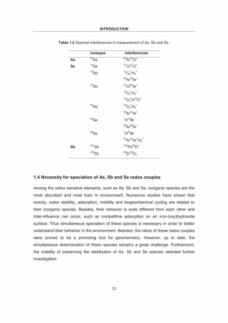
interferences mainly occur as poly atomic species, such as 35Cl40Ar on 75As, 40Ar40Ar on 80Se and 81Br1H+ on 82Se (table 1.2). These interferences could be caused by plasma
gas ions (e.g. 40Ar40Ar), interaction of plasma gas with other species (from reagents or
sample) (e.g. 35Cl40Ar) and sample matrix (e.g. 81Br1H+). Generally four strategies were
used to handle these interferences.
1) Selection of interference-free isotopes for analysis and high resolution mode of
detection (if possible for instrument). e.g. Thermo element 2/XR sector field ICP- MS
provides three resolution modes: low resolution mode (> 300), medium (> 4000) and
high (> 10000). It can analyze almost all kinds of samples and their matrices (seawater,
hydrothermal solution, leachates etc.) free of interferences. Thus, for As measurements,
as it is a mono-isotopic element and a resolution of at least 7775 was needed to
separate 35Cl40Ar and 75As (Jakubowski et al., 2011). Obviously, the high resolution
mode of element 2/XR was sufficient. As for Se measurements, the isotope of 78Se was
monitored in high resolution mode to avoid interferences. However, the using of high-
resolution mode implies a loss of signal intensity, which elevates the detection limit
accordingly, thus retards the measurement with more accuracy and precision, especially
for those elements of low abundance (e.g. 78Se, with an abundance of 23.6%).
2) Using mathematical equation to correct interferences. For the ICP-MS, many
correction equations are built to facilitate automatic corrections of certain isobaric or
polyatomic interferences. For As the most common equation is:
Corrected 75As signal = total signal in mass 75 – (3.127 x (signal in mass 77 – (0.815 x signal in mass 82)))
e.g. 75As (corrected) = 75As – (3.127 x (77Se – (0.815 x 82Se)))
However, this equation was based on two assumptions: a) all signals in mass 82 are
from Se and b) after subtraction of 77Se contribution on mass 77, the remaining signals
on mass 77 are due to 37Cl40Ar. The problem is that if the samples contain high bromine,
INTRODUCTION
14
the signals in mass 82 are a combination of 82Se and 81Br1H. As a result, the correction
equation would produce large bias. As for 82Se, the common used correction equation is:
Corrected 82Se signal = total signal in mass 82 – (0.007833 x signal in mass 83) – (0.00187 x signal in mass 79)
e.g. 82Se (corrected) = 82Se – (0.007833 x 83Kr) – (0.00187 x 79Br)
Obviously, this equation was also matrix dependent, as 79Br was monitored. There is no
universal method for dealing with interferences in ICP-MS. It seems wise to always
monitor more than one isotope (if possible), even if the other isotopes are less abundant.
3) Using chromatography to remove Cl-interferences. Since in aquatic environments Cl
and As species exist as anions, it is possible to use anion exchange chromatography to
remove Cl-related interferences, e.g. 40Ar35Cl+ on 75As. In our previous work (Wu and
Pichler, 2014) the potential interference of 40Ar35Cl+ was solved using a Hamilton PRX-
X100 anion exchange column, as Cl¯ eluted out at a different retention time from As(III)
and As(V).
4) Using other techniques such as “collision / reaction cell”. The collision / reaction cell
technique known as Elan DRC (I, II, e) was introduced by Perkin-Elmer, which is a
chamber placed between the single lens optics chamber and the mass analyzer
chamber of ICP-MS for eliminating isobaric interferences. The chamber has a
quadrupole and can be filled with reaction (or collision) gases (HN3, CH4, He O2 or H2).
The gas reacts with the introduced sample, and eliminates some of the interferences.
The mechanism is based on neutralization of exchange reaction between interfering ions
and reaction gas, producing different m/z+, e.g. methane was used for As and Se
analysis (Komorowicz and Barałkiewicz, 2011).
However, the application of high-resolution mode and collision / reaction cell can both
lead to drop of signal intensity. There is no universal method for dealing with
interferences in ICP-MS. A successful strategy requires a full understanding of the
technique and detailed knowledge of sample matrices.
INTRODUCTION
15
Table 1.2 Spectral interferences in measurement of As, Sb and Se.
isotopes interferences
As 75As 40Ar35Cl+
Se 74Se 37Cl37Cl+ 76Se 12C6
1H4+
36Ar40Ar+ 77Se 37Cl40Ar+
12C61H5
+ 12C5
1H16O+ 78Se 12C6
1H6+
38Ar40Ar+ 80Se 1H79Br
40Ar40Ar+ 82Se 1H81Br
40Ar40Ar1H2+
Sb 121Sb 105Pd16O+ 123Sb 94Zr16O2
1.4 Necessity for speciation of As, Sb and Se redox couples
Among the redox sensitive elements, such as As, Sb and Se, inorganic species are the
most abundant and most toxic in environment. Numerous studies have shown that
toxicity, redox stability, adsorption, mobility and biogeochemical cycling are related to
their inorganic species. Besides, their behavior is quite different from each other and
inter-influence can occur, such as competitive adsorption on an iron-(oxy)hydroxide
surface. Thus simultaneous speciation of these species is necessary in order to better
understand their behavior in the environment. Besides, the ratios of these redox couples
were proved to be a promising tool for geochemistry. However, up to date, the
simultaneous determination of these species remains a great challenge. Furthermore,
the inability of preserving the distribution of As, Sb and Se species retarded further
investigation.
INTRODUCTION
16
1.5 Detector
Various detection systems have been widely used for As, Sb or Se determination, such
as ultra violet (UV) detection (Jaafar et al., 2009; Koshcheeva et al., 2009),
potentiometry and conductometry such as polarography, cathodic stripping voltammetry
(CSV) and anodic stripping voltammetry (ASV) (Smichowski et al., 1998; Domínguez-
Renedo et al., 2009), AFS (Gregori et al., 2005; Price and Pichler, 2005), ICP-AES
(Chausseau et al., 2000) and ICP-MS. However, each type of detection system has its
advantages and limitations, e.g. UV and potentiometry and conductometry systems are
low-cost and easy to operate but their limitations are not low enough to meet the trace or
ultra-trace level determination. ICP-AES has the advantages of high flexibility and
satisfactory accuracy and precision over a broad range of concentrations. Meanwhile,
dissolution of solids may bring about problems, and the detection limits are usually not
low enough for trace elements, like As, Sb and Se. AFS coupled to HG, however, is a
well-established technique, with great sensitivity for As and Sb, even comparable to ICP-
MS. In addition, the purchase and operating costs are low. However, HG technique is
only suitable for those elements which form volatile covalent hydrides, e.g. HG-AFS is
not applicable for simultaneous speciation of Se species due to its inability of forming
Se(VI)-hydride. Thus, the basic speciation includes two replicate measurements, one for
total concentration and the other for one of the inorganic species. The concentration of
the other species was obtained by subtraction of the two. However, the drawback is this
procedure overlooked the presence of other species, such as various organic species.
For ICP-MS, the strong points are: low detection limits for trace element analysis;
excellent possibilities for correcting spectral interferences; high resolution detection
mode for almost all elements free of interferences (sector field ICP-MS). But, the weak
points are also obvious: accuracy and precision are less than ICP-AES for some
particular elements; the costs are much higher than for ICP-MS and special operation
skills may be necessary (Rommers and Boumans, 1996).
Generally, there is no universal detector, which is ideal for all elements determination in
a wide concentration range free of interferences. They may supplement and complement
each other under different conditions. The choice of detectors must be based on various
analytes and analytical requirements. As for the elements of our interest (As, Sb and Se),
the sector field-ICP-MS seems the best choice, because it allowed simultaneous and
interference-free (e.g. complete separation of 75As from 35Cl40Ar and 80Se from 40Ar40Ar)
INTRODUCTION
17
determination at trace level (Wu and Pichler, 2014). Regardless which detection system
was used, the detector itself was not capable of separating different species of a given
element (e.g. As(III) and As(V), Sb(III) and Sb(V) and Se(IV) and Se(VI)) in plasma,
though ICP-MS provides “pseudo” simultaneous detection of different masses. Thus for
speciation analysis, a separation technique (e.g. selective extraction or chromatography
based separation) is needed before introduction in detection system. In addition, the
combination of HPLC to ICP-MS provides another possibility of dealing with isotopic
mass interference. E.g. the common interference of 35Cl40Ar on 75As in direct
determination by ICP-MS can be solved by chromatography, as the species of 35Cl40Ar
and 75As can elute out at different retention times from chromatographic column and
thus are subsequently introduced in plasma separately.
ANALYTICAL METHODS
18
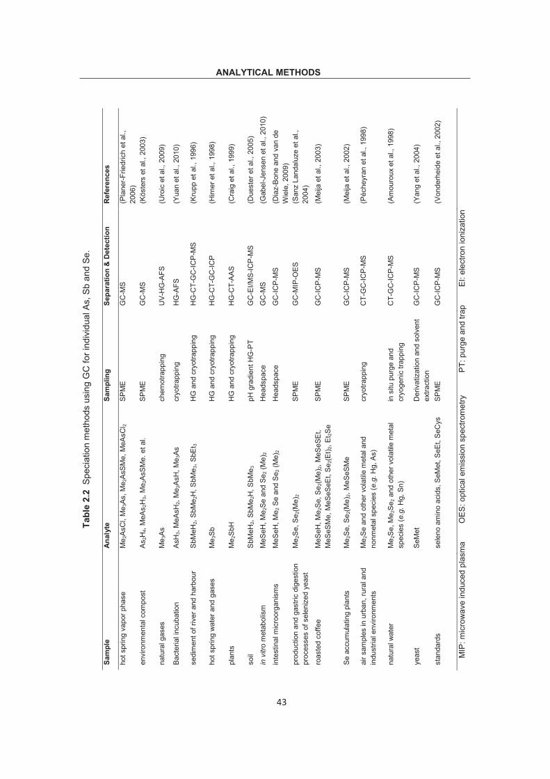
2. Speciation methods for As, Sb and Se species (a review)
Speciation is defined as analytical identification and quantitative determination of
different chemical forms of the elements present in a sample (Templeton et al., 2000).
However, selective determination of each species in the presence of other chemical
forms of the same element is usually impracticable. Thus, the separation and detection
of various analytes of a certain element or even various species of more than two
elements is necessary. Basic separation includes non-chromatographic methods, i.e.
electrokinetic separation methods (Capillary electrophoresis (CE) (Koellensperger et al.,
2002; Sun et al., 2002), supercritical fluid chromatography (SFC) and solid phase
extraction (SPE) (Wu et al., 2009, 2011; Planer-Friedrich et al., 2006), and
chromatographic methods, i.e. gas chromatography (GC), high-performance liquid
extraction (SPE), capillary electrophoresis (CE) and hydride generation (HG). These
techniques provide quantitative information on specific chemical forms of some elements
in many types of samples at reduced cost and time.
2.1.1 Liquid-liquid extraction (LLE)
Liquid–liquid extraction (LLE), also known as solvent extraction and partitioning, is a
method to separate various species based on their relative solubility in two different
immiscible liquids, usually water, and an organic solvent. This technology is extremely
simple at low cost. Great improvement has been achieved based on LLE. Recently, a
micro-extraction technique-dispersive liquid-liquid micro-extraction (DLLME), based on a
ternary solvent system was developed. An appropriate mixture of extraction solvent and
disperser solvent is rapidly injected into an aqueous sample, thus a cloudy solution is
formed. Then the analyte in the sample is transferred to the fine droplets of the
ANALYTICAL METHODS
19
extraction solvent. Phase separation is performed by centrifugation. In an As speciation
analysis study (Escudero et al., 2013), selective separation of As(III) was achieved by
chelation with sodium diethyldithiocarbamate (DDTC) followed by dispersion with 1-
octyl-3-methylimidazolium hexafluorophosphate. As(III) was then extracted with a
packed micro-column and subsequently measured with electrothermal atomic absorption
spectrometry (ETAAS). The concentration of As(V) was deduced by the difference
between total inorganic As and As(III). In another report of As and Sb speciation in
waters (Rivas et al., 2009), As(III) and Sb(III) were complexed with ammonium
pyrrolidine dithiocarbamate at first and then mixed with carbon tetrachloride (extraction
solvent) and methanol (disperser solvent). After centrifugation As(III) and Sb(III) were
extracted in the organic phase and measured with ETAAS, while As(V) and Sb(V)
remained in the aqueous layer.
2.1.2 Liquid-phase microextraction (LPME)
LPME is a simple, and highly sensitive technique for sample pretreatment before trace
analysis of analytes from complex matrices. It is a miniaturized implementation of
conventional liquid-liquid extraction in which only a few μLs of solvents are used. Some
LPME-based methods for As, Sb or Se speciation have been developed. e.g. single-
droplet micro-extraction (SDME) and hollow fiber liquid-phase microextraction.
a) Single-droplet microextraction (SDME)
The basic procedure of SDME is: 1) a precious micro-syringe was used to draw up
extraction solvent (less than 3 μL, typically organic); 2) the micro-syringe was slightly
expelled to make sure that a drop (1-3μL) of extraction solvent suspended at the tip; 3)
expose the droplet to sample under optimized conditions (e.g. temperature and
extraction time); 4) the droplet is retracted and transferred for further determination.
Although originally developed for organic analytes extraction, SDME has been proved to
be also highly effective for pre-concentration and speciation of trace metals. Fan (2007)
developed a speciation method for Sb inorganic species in water samples using SDME
followed by analysis using ETAAS. In the method N-Benzoyl-N-phenylhydroxylamine
(BPHA)-chloroform single drop was used, where BPHA worked as complexing agent.
Total concentration of Sb was determined after pre-reduction (Sb(V) to Sb(III)) by L-
ANALYTICAL METHODS
20
cysteine. Sb(V) was calculated by subtraction. The detection limits were 8.0 ng L−1 for
Sb(III) and 9.2 ng L−1 for total Sb, respectively.
Another type of improved SDME is head-space single-droplet microextraction (HS-
SDME). The biggest difference is in step 3: in HS-SDME the drop was not directly
exposed to the sample but in the sample head-space. The volatile species would be
volatilized under certain temperature into headspace and extracted to the drop. After the
species between head space and the drop achieve equilibrium, the micro-drop was
retracted for determination. Chamsaz et al. (2003) successfully used this method for As
analysis. An organic solvent (a mixture of pyridine and benzyl alcohol, 1:3 v/v) with
dissolved silver diethyldithiocarbamate (AgDDC) was used for extracting As species. As
species in aqueous samples were converted to As-hydrides using sodium
tetrahydroborate (NaBH4). During 7 min extraction at 35 C, the As-hydrides reacted with
AgDDC and were extracted by a 4 μL micro-drop suspended in the tip of micro-syringe.
The determination was carried out on a GFAAS and the detection limit for As (total) was
45 pg mL-1.
b) Hollow fiber liquid-phase microextraction (HFLPME)
HFLPME is a membrane-based separation technique, which was also referred to micro-
porous membrane extraction (Fig. 2.4). The basic extraction process includes: 1)
conditioning of the hollow fiber (make the hydrophobic porous membrane impregnated
with organic solvent); 2) injection of a specific volume of the solvent into the conditioned
hollow fiber using micro-syringe; 3) immersing the hollow fiber into sample (the analytes
would partition from the aqueous sample into the organic solvent); 4) retracting of the
extracted sample for analysis. This method is suitable for extraction of species with large
partitioning coefficients in the organic solvent. It has been used for speciation of
inorganic Se species in natural water samples (Xia et al., 2006). Chloroform was used
as organic solvent and ammonium pyrrolidine dithiocarbamate (APDC) was used as
chelating agent. During extraction Se(IV) was extracted by the organic solvent due to the
formation of a Se(IV)-PDC complex, while Se(VI) remained in the solution as free
species. The reported detection limits are: 0.50 pg mL-1 for Se(IV) and 2.7 pg mL-1 for
Se(VI).
ANALYTICAL METHODS
21
2.1.3. Cloud-point extraction (CPE)
Another separation strategy similar to LLE is cloud-point extraction (CPE), based on the
selective extraction of analytes by non-ionic surfactant. When heated to a certain
temperature (known as could point) the non-ionic surfactant would become turbid. Above
this temperature, the isotropic micelle solution separates into two phases: the surfactant-
rich phase with small volume, and the diluted aqueous phase where the surfactant
concentration is very low (close to the critical micelle concentration). The analytes (or
analyte-chelates, generated by addition of chelation agents) would be extracted
preferentially by the surfactant-rich phase (Stalikas, 2002; Paleologos et al., 2000).
Complete phase separation can be obtained after centrifugation. A method for
simultaneous speciation analysis of inorganic Sb and Se in water samples was
developed (Li et al., 2008) based on the fact that Sb(III) and Se(IV) could form
complexes with diethyldithiocarbamate (DDTC) at a pH of 6. The complexes were
extracted into the surfactant-phase of octylphenoxypolyethoxyethanol (Triton X-114)
when heated in thermostated water bath of 30 C, whereas Sb(V) and Se(VI) remained
in the aqueous solution. The extracted Sb(III) and Se(IV) were subsequently determined
by ETV-ICP-MS. Total concentration of Sb and Se was determined by the same protocol
after pre-reduction by L-cysteine and the concentration of Sb(V) and Se(VI) was
obtained by subtraction. The limits of detection (LODs) were 0.05 μg L-1 for Se(IV) and
0.03 μg L-1 for Sb(III).
Fig. 2.4 Scheme for hollow fiber liquid phase micro-extraction (HFLPME).
ANALYTICAL METHODS
22
2.1.4 Solid-phase extraction (SPE)
Solid-phase extraction can be used to isolate analytes of interest from a wide variety of
matrices. SPE has been frequently used as a technique for speciation analysis. This is
because SPE avoids usage of large amounts of organic solvents and provides larger
pre-concentration factors and lower detection limit. The basic principle is: when sample
passes through stationary phase, the analytes in the sample interact and retain on the
sorbent of stationary phase. Other species would pass through the solid phase and are
then discarded. The desired analytes are eluted with a kind of solvent and then detected.
Some novel speciation methods based on SPE have been developed. Ben Issa et al.
(2010, 2011) combined a strong base anion exchange resin (SBAE) and two hybrid (HY)
resin: HY-Fe (based on behavior of hydrated iron oxide particles on As species) and HY-
AgCl (adsorbent for inorganic As(III) and As(V)) for inorganic As species (As(III) and
As(V)) and organic As species (MMA and DMA). Separation of these species was
achieved based on the following: 1) at pH < 8, SBAE resin separated As(V) from As(III)
by retaining As(V) and allowing As(III) to pass through. So As(III) can be measured in
the effluent. 2) within a wide pH range from 5 to 11, HY-Fe resin retained both As(III)
and As(V), except for DMA. Thus, DMA could be measured. 3) HY-AgCl resin at pH near
9 retained both inorganic As(III) and As(V), but allowed organic As species of MMA and
DMA to pass through, which made detection of organic As species possible (Fig. 2.5).
Fig. 2.5 Scheme for selective separation of As species in water samples using
SBAE, HY-Fe and HY-AgCl resins (from (Ben Issa et al., 2011)).
ANALYTICAL METHODS
23
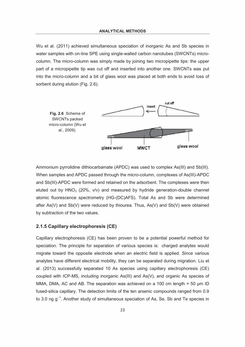
Wu et al. (2011) achieved simultaneous speciation of inorganic As and Sb species in
water samples with on-line SPE using single-walled carbon nanotubes (SWCNTs) micro-
column. The micro-column was simply made by joining two micropipette tips: the upper
part of a micropipette tip was cut off and inserted into another one. SWCNTs was put
into the micro-column and a bit of glass wool was placed at both ends to avoid loss of
sorbent during elution (Fig. 2.6).
Ammonium pyrrolidine dithiocarbamate (APDC) was used to complex As(III) and Sb(III).
When samples and APDC passed through the micro-column, complexes of As(III)-APDC
and Sb(III)-APDC were formed and retained on the adsorbent. The complexes were then
eluted out by HNO3 (20%, v/v) and measured by hydride generation-double channel
atomic fluorescence spectrometry (HG-(DC)AFS). Total As and Sb were determined
after As(V) and Sb(V) were reduced by thiourea. Thus, As(V) and Sb(V) were obtained
by subtraction of the two values.
2.1.5 Capillary electrophoresis (CE)
Capillary electrophoresis (CE) has been proven to be a potential powerful method for
speciation. The principle for separation of various species is: charged analytes would
migrate toward the opposite electrode when an electric field is applied. Since various
analytes have different electrical mobility, they can be separated during migration. Liu et
al. (2013) successfully separated 10 As species using capillary electrophoresis (CE)
coupled with ICP-MS, including inorganic As(III) and As(V), and organic As species of
MMA, DMA, AC and AB. The separation was achieved on a 100 cm length × 50 μm ID
fused-silica capillary. The detection limits of the ten arsenic compounds ranged from 0.9
to 3.0 ng g−1. Another study of simultaneous speciation of As, Se, Sb and Te species in
Fig. 2.6 Scheme of SWCNTs packed
micro-column (Wu et al., 2009).
ANALYTICAL METHODS
24
waters and soil extracts using CE and UV detector was made by Casiot et al. (1998).
The separation was achieved within 5 min at electrolyte pH of 11.2. However, relatively
high detection limits were obtained, from 13 μg L-1 for Se(VI) to 509 μg L-1 for Te(IV),
due to using a low-sensitivity UV detector. Generally, it can be seen that pH plays an
important part in species speciation using CE. The pH of the electrolyte can directly
influence the electrophoretic mobility of the analytes, because the dissociation
(dissociation constant of As, Sb and Se species were listed in Table 1.1) and ionization
capability of the desired species are various under different pH values. e.g. As(V) and
Se(VI) would migrate faster than As(III) and Se(IV), due to their low pKa and two
negative charges in a wide pH range.
However, a special interface for coupling CE with ICP-MS is needed. The first reason is
that CE has a low flow rate of less than 1μL min-1. This requires the use of a very low
uptake rate nebulizer for ICP-MS to ensure high-transport efficiency and relatively high
concentration of analyte brought into plasma. The second problem is the electrical
connection. As is known, for a regular CE both ends of the fused silica capillary were
submerged or in contact with two buffer reservoirs. Thus when CE was coupled with
ICP-MS, the capillary must be connected electrically, and meanwhile still introduce
buffer and analytes into nebulizer to produce a uniform aerosol for detector. Great effort
has been made to improve the designs of CE interfaces, including: usage of sheath
electrolyte (with constant sheath liquid flow rate) to close the electric circuit and addition
of a “make-up” buffer (Majidi and Miller-Ihli, 1998a; Prange and Pröfrock, 2005; Lu et al.,
1995; Taylor et al., 1998). However, due to the inherent complexity, many errors may
still arise when using CE coupled with ICP-MS (Majidi and Miller-Ihli, 1998b).
2.1.6 Hydride generation (HG)
Hydride generation, as one of the most commonly used non-chromatographic speciation
techniques for elements at trace level, was often coupled with AAS or AFS, and further
coupled with HPLC for multi-species speciation, such as As, Sb and Se. This method
was based on the fact that the analytes would form covalent hydrides after introduction
into the atomization systems. Then, after liquid-gas separation, analytes could be
detected in gas phase. The formation of covalent hydrides significantly improves the
sensitivity and lowers the detection limits by several orders of magnitude in comparison
to conventional nebulization. HG has a lot of advantages, such as: 1) easily being
ANALYTICAL METHODS
25
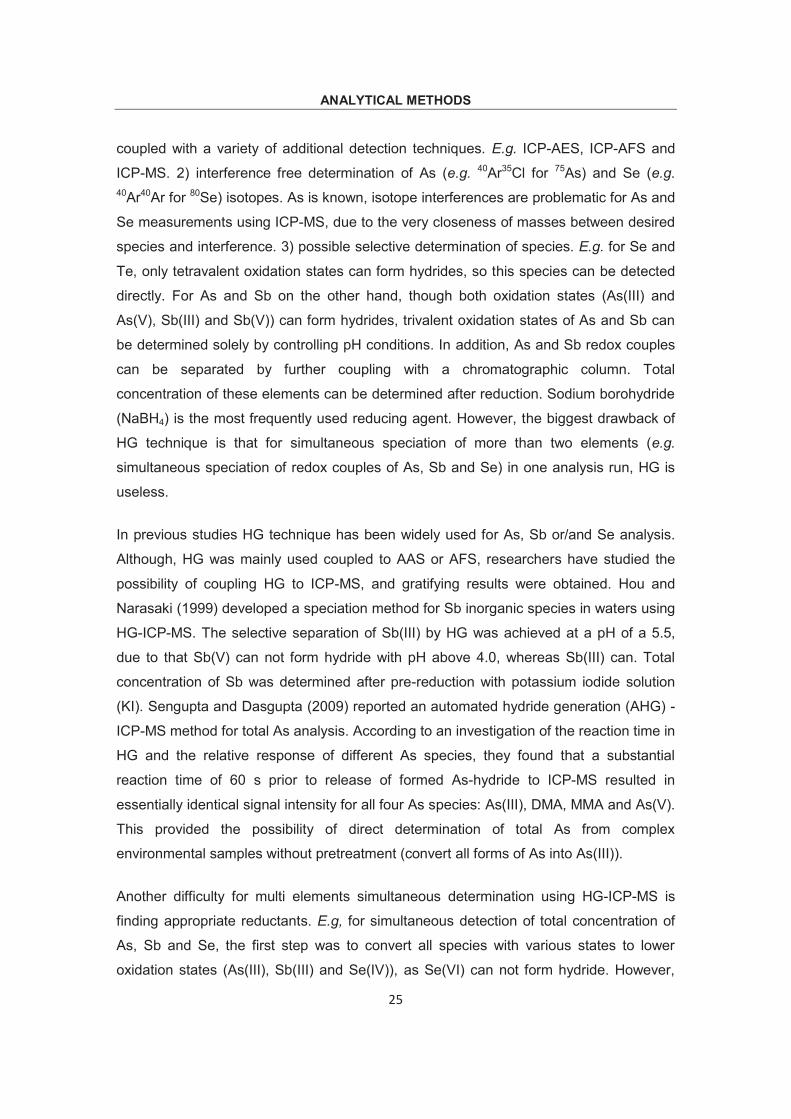
coupled with a variety of additional detection techniques. E.g. ICP-AES, ICP-AFS and
ICP-MS. 2) interference free determination of As (e.g. 40Ar35Cl for 75As) and Se (e.g. 40Ar40Ar for 80Se) isotopes. As is known, isotope interferences are problematic for As and
Se measurements using ICP-MS, due to the very closeness of masses between desired
species and interference. 3) possible selective determination of species. E.g. for Se and
Te, only tetravalent oxidation states can form hydrides, so this species can be detected
directly. For As and Sb on the other hand, though both oxidation states (As(III) and
As(V), Sb(III) and Sb(V)) can form hydrides, trivalent oxidation states of As and Sb can
be determined solely by controlling pH conditions. In addition, As and Sb redox couples
can be separated by further coupling with a chromatographic column. Total
concentration of these elements can be determined after reduction. Sodium borohydride
(NaBH4) is the most frequently used reducing agent. However, the biggest drawback of
HG technique is that for simultaneous speciation of more than two elements (e.g.
simultaneous speciation of redox couples of As, Sb and Se) in one analysis run, HG is
useless.
In previous studies HG technique has been widely used for As, Sb or/and Se analysis.
Although, HG was mainly used coupled to AAS or AFS, researchers have studied the
possibility of coupling HG to ICP-MS, and gratifying results were obtained. Hou and
Narasaki (1999) developed a speciation method for Sb inorganic species in waters using
HG-ICP-MS. The selective separation of Sb(III) by HG was achieved at a pH of a 5.5,
due to that Sb(V) can not form hydride with pH above 4.0, whereas Sb(III) can. Total
concentration of Sb was determined after pre-reduction with potassium iodide solution
(KI). Sengupta and Dasgupta (2009) reported an automated hydride generation (AHG) -
ICP-MS method for total As analysis. According to an investigation of the reaction time in
HG and the relative response of different As species, they found that a substantial
reaction time of 60 s prior to release of formed As-hydride to ICP-MS resulted in
essentially identical signal intensity for all four As species: As(III), DMA, MMA and As(V).
This provided the possibility of direct determination of total As from complex
environmental samples without pretreatment (convert all forms of As into As(III)).
Another difficulty for multi elements simultaneous determination using HG-ICP-MS is
finding appropriate reductants. E.g, for simultaneous detection of total concentration of
As, Sb and Se, the first step was to convert all species with various states to lower
oxidation states (As(III), Sb(III) and Se(IV)), as Se(VI) can not form hydride. However,
ANALYTICAL METHODS
26
the commonly used reducing agents, such as iodide or bromide, L-cysteine and thiourea,
can all reduce Se(IV) to elemental Se which is not able to form hydride as well. Bowman
et al. (1997) developed a procedure for simultaneous detection of As, Sb and Se using
HG-ICP-MS. The method involved an off-line pre-reduction for converting Se(VI) into
Se(IV), combined with an on-line reduction of As(V) and Sb(V) to trivalent state with
thiourea. Although thiourea could also slightly reduce Se(IV) to Se, the conversion was
The principle of separating species with liquid chromatography was demonstrated in Fig.
2.7. Various analytes pass through the stationary phase of column and generate
different velocity due to different adsorption abilities, solubilities or other properties
between mobile and stationary phases. Finally various analytes are separated in column
and eluted out at different retention times. Liquid chromatography, like anion exchange
(AEX), cation exchange (CEX), ion exclusion (IEC), and ion pair chromatography (IPC),
coupled to a sensitive detector (e.g. AFS, ICP-OES and ICP-MS) have been used for As,
Sb or Se speciations. HPLC is more qualified for separation of naturally non-volatile As,
Sb and Se species. These species are not stable if heated to the required temperature
to keep them in gas phase. However, gas chromatography (GC) was qualified for these
volatile organic species.
Fig. 2.7 Scheme of principle of liquid chromatography.
ANALYTICAL METHODS
27
2.2.1 As speciation
As speciation using HPLC, has been well reviewed recently by Komorowicz and
Barałkiewicz (2011) and Ammann (2011). ICP-MS was the most widely used detector for
As species determination due to its high sensitivity, wide linear dynamic range and it can
easily be combined to many separation techniques. The coupling of ICP-MS with liquid
chromatography allows separation, identification and quantification of As species in just
one analysis run. As for separation of various As species, the key factors are pH, mobile
phase, and the type of chromatography. Because, As species vary under different pH
and Eh conditions (section 1.2). Thus the choice of chromatography and mobile phase
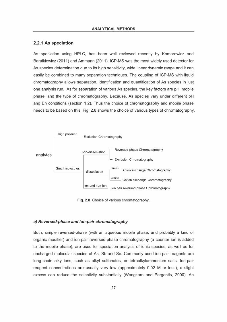
needs to be based on this. Fig. 2.8 shows the choice of various types of chromatography.
Fig. 2.8 Choice of various chromatography.
a) Reversed-phase and ion-pair chromatography
Both, simple reversed-phase (with an aqueous mobile phase, and probably a kind of
organic modifier) and ion-pair reversed-phase chromatography (a counter ion is added
to the mobile phase), are used for speciation analysis of ionic species, as well as for
uncharged molecular species of As, Sb and Se. Commonly used ion-pair reagents are
long-chain alky ions, such as alkyl sulfonates, or tetraalkylammonium salts. Ion-pair
reagent concentrations are usually very low (approximately 0.02 M or less), a slight
excess can reduce the selectivity substantially (Wangkarn and Pergantis, 2000). An
ANALYTICAL METHODS
28
aqueous solution with an organic modifier is often used for elution and separation, e.g.
methanol is usually used as the organic modifier in ICP-MS detectors to improve signal
intensity and maintain plasma stability (for As and Se). Table 1.3 shows that both anion-
pairing and cation-pairing chromatography were used for the separation of As species.
Tetrabutylammonium (TBA, both hydroxide and phosphate) is the commonly used
pairing cation for As species (As(III), As(V), MMA and DMA) (Martín et al., 1995; Pan et
al., 2007). While hexanesulfonic acid (HSA) is often used in cation-pairing
chromatography. The elution order of these species was consistently As(III), DMA, MMA
and AS(V), independent of the various reverse-phase columns. In a wide pH range from
2 to 9, As(III) (pKa = 9.2) (Table 1.1) is a neutral species which eluted out in the void
phase. Generally, the resolutions of these species are dependent of the concentration of
ion-pair reagent, flow rate, ionic strength, and pH of eluent. H2O was one of the most
commonly used mobile phases for ion-pairing chromatography. Martín et al. (1995)
developed a method for simultaneous speciation of As(III), AB, AC, DMA, MMA and
As(V) using anion-pairing chromatography. TBAPO4 was used as ion-pairing reagent,
and H2O as eluent. However, the result showed that AB and AC co-eluted. B’Hymer and
Caruso (2007) speciated the same species using a cation-pairing chromatography with
HSA as ion-pairing reagent. The mobile phase was prepared using citric acid (with a pH
of 2.3) with methanol as modifier.
b) Ion-exchange chromatography
With ion-exchange chromatography, ions or easily ionized analytes of As, Sb and Se
were separated, e.g. anion-exchange columns were used for separation of As(III), As(V),
MMA, DMA, whereas cation exchange columns were used for separation of AB, AC,
TMAO and Me4As+. Commonly, R4N+, SO3, RCOO were used as ion-exchanging groups
(Weis and Weiss, 2004). Charge density and polarizability of the analytes depends on
the molecule size and the charge (controlled by proton association-dissociation
equilibrium). The pKa of As species occupy a large range, many of them being higher
than 8 (Table 1.1). Hence, their negative charges are pH dependent. In addition, the
protonation-deprotonation equilibrium of exchange sites, is also controlled by pH. Ion-
exchange chromatography has been widely used for As inorganic species speciation, as
the eluent pH can be better realized by AEX compared to other chromatography. CEX
did not retain the two most toxic and most common species, As(III) and As(V), thus
eluting them together in the front. Ponthieu et al. (2007) developed a method for As
ANALYTICAL METHODS
29
inorganic and organic species speciation in landfill leachate using CEX on a PRP-X200
column. The results showed that As(III), MMA, As(V) and Cl- eluted out in the front within
3 min, however, Arsenocholine (AC) and Trimethylarsineoxide (TMAO) were co-eluted at
15 min. Generally, anion-exchange chromatography can be used in a wide pH range
(Table 2.1) and different eluents need to be chosen based on the existing form of As
species and pH. HNO3 was often used as mobile phase at low pH. Mattusch and
Wennrich (1998) and Kohlmeyer et al. (2002) used an anion-exchange column with
HNO3 as mobile phase to analyze inorganic and organic As species. Based on this
method, up to 17 As species were identified. For high pH above 9 (As existed as
negatively charged H2AsO4¯), Hydroxide and carbonate containing eluents (NaOH,
NH4HCO3 or (NH4)2CO3) have widely been used on a variety of polymeric anion-
exchange columns (Table 2.1). One of the advantages of this type of AEX is that high
pH eluents substantially increase the dissociation of protonated As species and increase
their affinity for anion exchanger.
However, at oxic/basic conditions the oxidation of As(III) to As(V) may occur fast
(Jackson and Bertsch, 2001; Raab et al., 2004). Besides, separations at high pH can
suffer from metals (Mg, Ca, Al, Mn, Fe, Cu, etc) precipitation as hydroxides inside
columns and adsorb As species. Thus this method was suitable for NaOH extracted soil
samples. An anion-exchange column (e.g. polymeric Hamilton PRP-X100 column) with
medium pH seemed the optimum separation condition for As species. Phosphate-based
mobile phases were widely used (Day et al., 2002; Pizarro et al., 2003). Similar to anion-
pairing chromatography, co-elution of AB and As(III) may occur at neutral pH conditions.
However, As(III) could be separated from AB when pH was higher than 9 or using
tartaric acid as mobile phase, due to the formation of anionic As (Ackley et al., 1999).
Despite the advantages of phosphate as eluent, e.g. playing an indispensable part in
displacing As(V) from strong adsorbent sites, shortcomings are obvious: loading of
phosphorous and sulfur can produce polymeric depositions on the cones and inside of
ICP instrument, thus leading to drop in sensitivity due to clogging (Milstein et al., 2002).
Organic mobile phases such as potassium hydrogenphthalate and
tris(hydroxymethyl)aminomethane (TRIS) were also used as eluents (Woller et al., 1998;
Milstein et al., 2002), though excessive loading of organic carbon can vary As intensities.
In addition, NH4NO3 was also investigated as potential eluent due to its pH-flexibility
(ranging from 2 to 9) and plasma compatibility.
ANALYTICAL METHODS
30
c) Ion-exclusion chromatography
Ion-exclusion chromatography was also used to speciate weakly ionized or neutral As
species. Strong anion- or cation-exchange resins were often used. In contrast to ion-
exchange chromatography, charges on ion-exchange resin are the same as of weakly
ionized species (Haddad and Jackson, 1990). That is, negatively charged analytes are
separated on a cation-exchange resin, e.g. negatively charged As species are separated
using resin containing anionic sulfonate functional groups, whereas positively charged
analytes are separated via anion-exchange chromatography. The basic separation
principle is, strong anions (e.g. inorganic As species) cannot penetrate into the occluded
liquid phase due to the repelling by anionic functional groups on the resin, thus are not
retained by the column. Weakly ionized analytes or neutral molecules of As (e.g. AB)
penetrate the resin zone and move into the occluded liquid phase, thus result in different
retention times. Up to 8 As species (As(III), As(V), MMA, DMA, AB, TMAsO, AC and
TMAs) were determined using an ion-exclusion column packed with a carboxylated
methacrylate resin and Na2SO4 as mobile phase (pH of 3.8), though an overall analysis
time of over 60 min was used (Nakazato et al., 2000).
d) Other techniques
In order to increase the sensitivity of analytes, various nebulizers (ultrasonic nebulizer,
thermospray nebulizer and so on) and hydride generation techniques were investigated.
Among these, HG was favored, because it resulted in the highest sensitivity for As
species, and eliminated clogging of samples and polyatomic ion spectral interferences of 40Ar35Cl on 75As, as only gaseous species were introduced in plasma (Taniguchi et al.,
1999).
Though for As speciation, AEX seemed the primary choice, a combination of an AEX (for
separation of As(III), As(V), MMA and DMA) and a CEX column (for separation of AB,
TMAO, AC and Me4As+ ) sometimes provides more information. This can be achieved
using two columns in two procedures, or two columns in one procedure, e.g. dual
column system (anion-exchange connected with cation-exchange) or dual mode system
(a combination of ion-exclusion and cation-exchange) (Sakai et al., 2001).
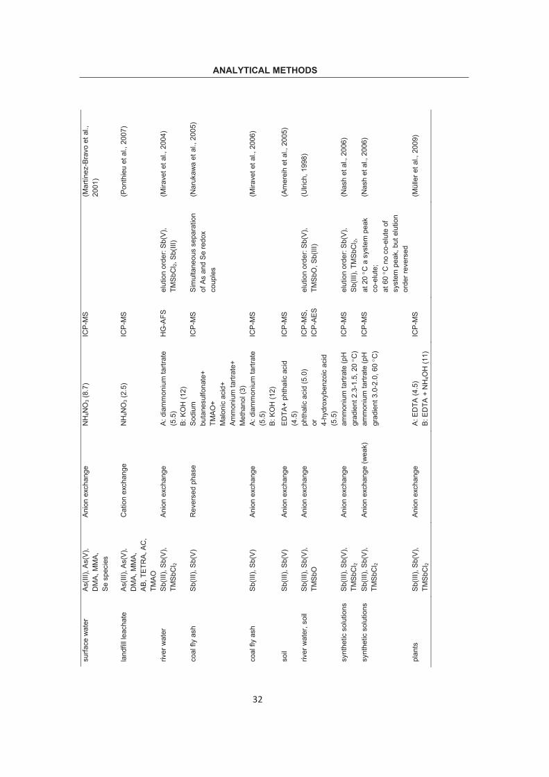
ANALYTICAL METHODS Ta
ble
2.1
Spe
ciat
ion
met
hods
usi
ng H
PLC
for i
ndiv
idua
l As,
Sb
and
Se.
sam
ple
anal
yte
colu
mn
elue
nt (p
H)
dete
ctor
co
mm
ent
Ref
.
stan
dard
As
(III),
AB
, AC
, D
MA,
M
MA,
As(
V)
Ion
pair
(ani
on p
airin
g)
H2O
(5.2
) H
G-A
AS
AB, A
C c
oelu
te;
TBAP
O4 a
s IP
reag
ent
(Mar
tín e
t al.,
199
5)
urin
e AB
, As(
III),
DM
A,
MM
A, A
s(V)
Io
n pa
ir (a
nion
pai
ring)
H
2O (5
.8)
ICP-
MS
TBAO
H a
s IP
reag
ent
(Pan
et a
l., 2
007)
appl
e ex
tract
ion
As(V
), As
(III),
M
MA,
DM
A,
AB, A
C
Ion
pair
(cat
ion
pairi
ng)
citri
c ac
id (2
.3)
ICP-
MS
HSA
as
IP re
agen
t; M
eOH
as
mod
ifier
(B
’Hym
er a
nd C
arus
o,
2007
)
stan
dard
As
(III),
As(
V),
DM
A, A
B,
AC
Anio
n ex
chan
ge
HN
O3
Low
pH
IC
P-M
S BD
SA a
s m
odifi
er
(Mat
tusc
h an
d W
ennr
ich,
19
98)
fish,
mus
sel,
oyst
er a
nd m
arin
e al
gae
As(
III),
As(
V)…
17
As
spec
ies
Anio
n ex
chan
ge
HN
O3
Low
pH
IC
P-M
S BD
SA a
s m
odifi
er;
AB
and
Cl -
coel
ute
(Koh
lmey
er e
t al.,
200
2)
grou
nd w
ater
As
(III),
As(
V),
DM
A, M
MA,
AB
Anio
n ex
chan
ge
CO
32- (1
0.3)
H
igh
pH
ICP-
MS
(L
arse
n, 1
998)
poul
try w
aste
As
(III),
As(
V),
DM
A, M
MA,
p-
ASA,
Rox
Anio
n ex
chan
ge
NaO
H (1
2.7)
H
igh
pH
ICP-
MS
MeO
H a
s m
odifi
er
(Jac
kson
and
Ber
tsch
, 20
01)
urin
e, fi
sh
As(II
I), A
s(V)
, D
MA,
MM
A,
AB
Anio
n ex
chan
ge
(NH
4) 2C
O3 (
9)
Hig
h pH
D
RC
- IC
P-M
S M
eOH
as
mod
ifier
(W
ang
et a
l., 2
007)
wat
er
As(II
I), A
s(V)
, D
MA,
MM
A An
ion
exch
ange
N
a 3PO
4 (6)
M
ediu
m p
H
ICP-
MS
EDTA
as
mod
ifier
(D
ay e
t al.,
200
2)
food
, sed
imen
t As
(III),
As(
V),
DM
A, M
MA,
AB
Anio
n ex
chan
ge
(NH
4) 3PO
4 (6)
M
ediu
m p
H
ICP-
MS
As(II
I) an
d AB
co-
elut
e (P
izar
ro e
t al.,
200
3)
31
ANALYTICAL METHODS su
rface
wat
er
As(II
I), A
s(V)
, D
MA,
MM
A,
Se s
peci
es
Anio
n ex
chan
ge
NH
4NO
3 (8.
7)
ICP-
MS
(M
artın
ez-B
ravo
et a
l.,
2001
)
land
fill l
each
ate
As(II
I), A
s(V)
, D
MA,
MM
A,
AB, T
ETR
A, A
C,
TMAO
Cat
ion
exch
ange
N
H4N
O3 (
2.5)
IC
P-M
S
(Pon
thie
u et
al.,
200
7)
river
wat
er
Sb(II
I), S
b(V)
, TM
SbC
l 2
Anio
n ex
chan
ge
A: d
iam
mon
ium
tartr
ate
(5.5
) B:
KO
H (1
2)
HG
-AFS
el
utio
n or
der:
Sb(V
), TM
SbC
l 2, S
b(III
) (M
irave
t et a
l., 2
004)
coal
fly
ash
Sb(II
I), S
b(V)
R
ever
sed
phas
e
Sodi
um
buta
nesu
lfona
te+
TMAO
+ M
alon
ic a
cid+
Am
mon
ium
tartr
ate+
M
etha
nol (
3)
ICP-
MS
Sim
ulta
neou
s se
para
tion
of A
s an
d Se
redo
x co
uple
s
(Nar
ukaw
a et
al.,
200
5)
coal
fly
ash
Sb(II
I), S
b(V)
An
ion
exch
ange
A:
dia
mm
oniu
m ta
rtrat
e (5
.5)
B: K
OH
(12)
ICP-
MS
(M
irave
t et a
l., 2
006)
soil
Sb(II
I), S
b(V)
An
ion
exch
ange
ED
TA+
phth
alic
aci
d (4
.5)
ICP-
MS
(A
mer
eih
et a
l., 2
005)
river
wat
er, s
oil
Sb(II
I), S
b(V)
, TM
SbO
An
ion
exch
ange
ph
thal
ic a
cid
(5.0
) or
4-
hydr
oxyb
enzo
ic a
cid
(5.5
)
ICP-
MS,
IC
P-AE
S el
utio
n or
der:
Sb(V
), TM
SbO
, Sb(
III)
(Ulri
ch, 1
998)
synt
hetic
sol
utio
ns
Sb(II
I), S
b(V)
, TM
SbC
l 2 An
ion
exch
ange
am
mon
ium
tartr
ate
(pH
gr
adie
nt 2
.3-1
.5, 2
0 C
) IC
P-M
S el
utio
n or
der:
Sb(V
),
Sb(II
I), T
MSb
Cl 2,
(N
ash
et a
l., 2
006)
synt
hetic
sol
utio
ns
Sb(II
I), S
b(V)
, TM
SbC
l 2 An
ion
exch
ange
(wea
k)
amm
oniu
m ta
rtrat
e (p
H
grad
ient
3.0
-2.0
, 60
C)
ICP-
MS
at 2
0 C
a s
yste
m p
eak
co-e
lute
; at
60
C n
o co
-elu
te o
f sy
stem
pea
k, b
ut e
lutio
n or
der r
ever
sed
(Nas
h et
al.,
200
6)
plan
ts
Sb(II
I), S
b(V)
, TM
SbC
l 2 An
ion
exch
ange
A:
ED
TA (4
.5)
B: E
DTA
+ N
H4O
H (1
1)
ICP-
MS
(M
ülle
r et a
l., 2
009)
32
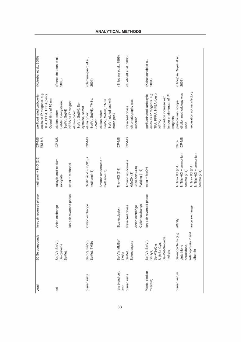
ANALYTICAL METHODS ye
ast
20 S
e co
mpo
unds
Io
n-pa
ir re
vers
ed p
hase
m
etha
nol
+ H
2O (2
.5)
ICP-
MS
ESI-M
S pe
rfluo
rinat
ed c
arbo
xylic
ac
ids
as IP
reag
ents
. e.g
. TF
A, P
FPA,
HFB
A(be
st).
Ove
rall
time
of 7
0 m
in
(Kot
reba
i et a
l., 2
000)
soil
Se(IV
), Se
(VI),
Se
-cys
tein
e Se
Met
Anio
n ex
chan
ge
salic
ylic
aci
d-so
dium
sa
licyl
ate
ICP-
MS
elut
ion
orde
r: Se
Met
, Se-
cyst
eine
, Se
(IV),
Se(V
I)
(Pon
ce d
e Le
ón e
t al.,
20
03)
Ion-
pair
reve
rsed
pha
se
wat
er +
met
hano
l H
FBA
as IP
reag
ent
elut
ion
orde
r: Se
(VI),
Se(
IV),
Se-
cyst
eine
, SeM
et
hum
an u
rine
Se(IV
), Se
(VI),
Se
Met
, TM
Se
Cat
ion
exch
ange
O
xalic
aci
d +
K 2SO
4 +
met
hano
l (3)
IC
P-M
S el
utin
ord
er:
Se(IV
), Se
(VI),
TM
Se,
SeM
et
(Gam
mel
gaar
d et
al.,
20
01)
Am
mon
ium
form
ate
+ m
etha
nol (
3)
elut
ion
orde
r: Se
(IV),
SeM
et, T
MSe
. Se
(VI)
elut
ed la
st w
ith
broa
d pe
ak
rats
blo
od c
ell,
liver
, Se
(VI),
MM
Se*
TMSe
Si
ze e
xclu
sion
Tr
is–H
Cl (
7.4)
IC
P-M
S
(Shi
obar
a et
al.,
199
9)
hum
an u
rine
SeM
et,
Sele
nosu
gars
R
ever
sed
phas
e Am
mon
ium
form
ate
+MeO
H (3
) IC
P-M
S R
ever
sed
phas
e ch
rom
atog
raph
y w
as
supe
rior
(Kue
hnel
t et a
l., 2
005)
Anio
n ex
chan
ge
Citr
ic a
cid
(4.8
) C
atio
n ex
chan
ge
Pyrid
ine
(1.6
)
Plan
ts, (
Indi
an
mus
tard
) Se
(IV),
Se(V
I),
SeC
ys,
Se-M
SeC
ys,
S-(M
Se)C
ys,
Se-M
et-S
e-ox
ide
hydr
ate
Ion-
pair
reve
rsed
pha
se
wat
er +
MeO
H
pe
rfluo
rinat
ed c
arbo
xylic
ac
ids
as IP
reag
ents
. e.g
. TF
A, P
FPA,
HFB
A (b
est),
N
FPA;
re
solu
tion
incr
ease
with
lo
nger
cha
in-le
ngth
of I
P re
agen
ts
(Kah
akac
hchi
et a
l.,
2004
)
hum
an s
erum
Se
leno
prot
eins
(e.g
. gl
utat
hion
e pe
roxi
dase
, se
leno
prot
ein
P an
d al
bum
in
affin
ity
A: T
ris-H
Cl (
7.4)
B:
Tris
-HC
l + a
mm
oniu
m
acet
ate
(7.4
)
OR
S-
ICP-
MS
post
-col
umn
isot
ope
dilu
tion
met
hodo
logy
was
us
ed
(Hin
ojos
a R
eyes
et a
l.,
2003
)
anio
n ex
chan
ge
A: T
ris-H
Cl (
7.4)
B:
Tris
-HC
l + a
mm
oniu
m
acet
ate
(7.4
)
sepa
ratio
n no
t sat
isfa
ctor
y
33
ANALYTICAL METHODS bi
olog
ical
tiss
ues
Se(IV
), Se
(VI),
Se
Met
Se
Cys
TM
Se
catio
n ex
chan
ge
pyrid
ine
solu
tion
(2.8
an
d 4.
7)
ICP-
MS
elut
ion
orde
r dep
end
on
pH
(Mor
eno
et a
l., 2
004)
size
exc
lusi
on
phos
phat
e bu
ffer i
n N
aCl (
7.2)
wat
er s
ampl
e Se
(IV),
Se(V
I),
SeM
et
SeC
ys
reve
rsed
pha
se
phos
phat
e (6
) IC
P-M
S Se
(IV),
Se(V
I) sh
owed
an
ion
exch
ange
m
echa
nism
; Se C
ys
show
ed re
vers
ed p
hase
m
echa
nism
(Qui
jano
et a
l., 1
996)
TFA:
trifl
uoro
acet
ic a
cid;
P
FPA:
pen
taflu
orop
ropa
noic
aci
d;
HFB
A: h
epta
fluor
obut
anoi
c ac
id S
b sp
ecia
tion
SeM
et: s
elen
omet
hion
ine;
TM
Se: t
rimet
hyls
elen
oniu
m;
Se
Cys
: Se-
cyst
eine
M
MSe
*: M
MSe
rela
ted
Se c
ompo
und;
ORS
: oct
apol
e re
actio
n sy
stem
BP
DTC:
dith
ioca
rbam
ate,
ben
zylp
ropi
onitr
ile d
ithio
carb
amat
e
34
ANALYTICAL METHODS
35
2.2.2 Sb speciation
Compared to As speciation analysis, the separation of Sb species was more difficult.
Chromatographic techniques were applied less for speciation analysis of Sb species,
especially for Sb organic species. The possible reason might be the lack of commercial
standards for organic Sb compounds, making it impossible to quantify these compounds
accurately. Sb(III), Sb(V) and TMSb are three main Sb species that were widely
determined using chromatography methodology.
a) Anion-exchange chromatography
Sb anionic species predominate in aqueous environmental matrices. Therefore strong
anion-exchange chromatography (Hamilton PRP-X100, Dionex AS4A, or ION-120) was
widely used for Sb speciation. Under these conditions, Sb(V) was readily eluted, while
Sb(III) was strongly retained on the column, which was indicated by long retention time
and severe peak tailing. To solve these problems, different studies were made including
using a shorter guard column, however, broad peak for Sb(III) was still observed.
Another way was using of complexing mobile phases, e.g. (di)ammonium tartrate
(Miravet et al., 2004; Miravet et al., 2006; Nash et al., 2006), EDTA (Amereih et al.,
2005; Müller et al., 2009), 4-hydroxybenzoic acid (Ulrich, 1998) and phthalic acid (Ulrich,
1998; Amereih et al., 2005), though one drawback of this technique was that Sb(V) was
normally eluted close to or even in solvent front. This cannot facilitate speciation of Sb
species. Cation and reversed-phase columns were investigated, too (Lintschinger et al.,
1997). However, basically no obvious improvements were obtained compared to anion-
exchange column. In addition, pretreatment of samples was used to solve Sb(III) tailing
problems, e.g. by adding certain organic ligands to samples, Sb(III) could be chelated
and formed stable complexes, which facilitated further separation on a chromatographic
column. A certain number of organic compounds (EDTA, DTPA, CDTA, BPDTC) (Kolbe
et al., 2012; Park and Hardy, 1989; Er-kang, 1982) were investigated as potential
complexation ligands for Sb(III). However, significant studies have shown that stable
Sb(III)-complexations were observed only at acidic pH ranges. These ligands do not
prevent Sb(III) hydrolysis at pH > 6.
The organic Sb species of TMSb can be easily separated from Sb(V) on anion-exchange
chromatography with phosphate, carbonate, or potassium as mobile phase. However, it
ANALYTICAL METHODS
36
always eluted out in solvent front, which is not desirable for identification of Sb species.
Besides, previous study found that TMSbCl2 was only eluted under alkaline conditions in
solvent front on reversed phase chromatography, and even not detectable on cation-
exchange chromatography (Lintschinger et al., 1997). This might be explained by the
polymerization or condensation phenomenon of TMSbCl2 under neutral and acidic
conditions. Table 1.3 shows that if Sb(III), Sb(V) and TMSb are analyzed simultaneously,
an eluent gradient were normally involved; one with organic compounds at acidic pH to
chelate and elute Sb(III), the other with simple inorganic solution (KOH or NH4OH) at
alkaline pH (higher than 10) in order to elute TMSb at non-solvent front retention time
(EDTA or diammonium tartrate) (Müller et al., 2009; Miravet et al., 2006). Besides, pH
gradient was also investigated (Nash et al., 2006).
b) Reversed phase chromatography
Though reversed phase chromatography proved to be ineffective for TMSb analysis, it
could still be used for inorganic Sb species speciation. Narukawa et al. (2005) developed
a method for simultaneous speciation of As, Sb and Se inorganic species using reversed
phase chromatography (CAPCELL PAK C18 MG S5 ODS column) with sodium
β-D-galactopyranoside and methyl-2-acetamido-2-deoxy-1-seleno-β-D-
glucosopyranoside using reversed phase chromatography with ammonium formate as
mobile phase. In this kind of chromatography SeCys and SeMet exist as zwitterions,
whereas selenite and selenate occur in anionic form.
b) Size exclusion chromatography
Size exclusion chromatography (SEC) is suitable for extraction and separation of Se
soluble protein fractions (Moreno et al., 2004; Shiobara et al., 1999). In general, the
separation of different fractions on SEC column depends on relative size of analytes and
pores on porous stationary phase. Unlike other chromatography, small compounds of
similar molecular mass, may be eluted in addition to selenoproteins (Behne et al., 1998).
The results from SEC-ICP-MS or SEC-UV provide useful information on further
purification and amino acid sequence determination of Se protein, which can help
researchers better understand the specific function of Se in biochemical processes of
ANALYTICAL METHODS
38
various organisms. Thus SEC was often used in combination with other chromatography,
such as cation-exchange chromatography (Moreno et al., 2004).
c) Ion-exchange chromatography
Both anion- and cation-exchange chromatography were used for separation of Se ions
and easily ionized Se analytes. As has been mentioned, variables, such as, ionic
strength of solute and mobile phase, pH of mobile phase, can influence the elution and
resolution of analytes. Although common eluents of phosphate or ammonium were often
used for inorganic Se species separation, complex organic mobile phases were
necessary if organic Se species such as SeMet, Se-cysteine were to be separated.
Ponce de León et al. (2003) separated four Se species: Se(IV), Se(VI), Se-cysteine and
SeMet using anion-exchange chromatography with salicylic acid - sodium salicylate as
mobile phase. In addition, oxalic acid (Gammelgaard et al., 2001), Tris-HCl (Hinojosa
Reyes et al., 2003), citric acid and pyridine (Kuehnelt et al., 2005) were investigated as
potential mobile phases.
The cation-exchange column possesses some anion-exchange properties, which also
allow anion separation (Gammelgaard et al., 2001; Moreno et al., 2004). Table 2.1
shows that cation-exchange chromatography was mainly used for TMSe+ speciation.
Pyridine solution was often used as mobile phase. Matrix influence also played an
important part in separating Se species. E.g. Se speciation in urine is still problematic
and the results of different studies were even contradictory. Besides, some studies
showed distortion of signal intensity by varying and high concentrations of salt when
urine samples were chromatographically separated without pretreatment (Gammelgaard
et al., 2001).
d) Other chromatography
Hinojosa Reyes et al. (2003) developed a method for Se-containing proteins speciation
analysis in human serum using affinity chromatography coupled to ICP-MS. A post-
column isotope dilution analysis (IDA) methodology was applied for quantification and an
octapole reaction system (ORS) was used to eliminate argon interference on 78Se and 80Se. The method was further validated using an anion-exchange chromatography. The
results showed that affinity chromatography performed better than anion-exchange
chromatography and finally three Se fractions; selenoprotein P, albumin and glutathione
ANALYTICAL METHODS
39
peroxidase were confirmed and determined. Quijano et al. (1996) developed a method
for simultaneous speciation of Se(IV), Se(VI), SeCys and SeMet using a mix column
(Spherisorb ODS-AMINO), where the stationary phase consisting of an equimolar
mixture of octadecyl and amino groups on a silica support, acting as a reversed-phase,
weak anion-exchange or ion-pair chromatography. Phosphate buffer at pH of 6.0 was
used as mobile phase. The elution order of analyzed Se species (SeCys, SeMet, Se(IV)
and Se(VI)) showed that Se(IV) and Se(VI) followed an anion-exchange chromatography
mechanism, while SeCys and SeMet (both existed as zwitterions in the pH range of 2.0
to 8.0) indicated a reversed phase chromatography (though SeCys contained terminal
polar and ionized function, it eluted out earlier than SeMet ).
2.3 Gas chromatographic separation
2.3.1 As speciation
Headspace injection, solid phase micro-extraction (SPME), cryotrapping and
chemotrapping were often coupled with GC-MS for speciation of volatile As species,
such as AsH3, MeAsH2, Me2AsH and Me3As. However, direct headspace injection may
cause too high detection limits which are not sufficiently sensitive for real sample
determination. SPME (PDMS, PDMS-CAR and PDMS-CAR-DVB are often used as
SPME fibre) (Planer-Friedrich et al., 2006; Kösters et al., 2003; Mester et al., 2001),
cryotrapping (Krupp et al., 2007, 2008; Yuan et al., 2010) and chemotrapping (Uroic et
al., 2009) are more practically used. Kösters et al. (2003) analyzed environmental
compost samples (samples were first derivatized with NaBH4 and speciation using GC-
ICP-MS) as well as hydrothermal vapor phase (gas was sampled with SPME). The
results showed that seven As species were identified existing in environment, and three
of them (diarsine, monomethyl diarsine and dimethylarsenomercaptane) were reported
in environmental samples for the first time. In another work, SPME-GC-ICP-MS was
applied to analyze volatile As species in the geothermal systems of Yellowstone National
Park. The vapor phase of hydrothermal systems has been found playing an important
part in transporting As species (0.5–200 mg m-3 of volatile As species present in vapor
phases (Planer-Friedrich, 2004). The results showed that (CH3)2AsCl, not (CH3)3As as
expected from previous studies, was the most frequently detected volatile As species.
Seemingly, higher concentration of (CH3)3As was determined when As(III) was dominant
in aqueous phase. Generally, volatile As species of (CH3)2AsCl, (CH3)3As,
(CH3)2AsSCH3 and CH3AsCl2 were detected in decreasing order of frequency.
ANALYTICAL METHODS
40
Compared to cryotrapping, chemotrapping is more suitable for field study and sampling.
Uroic et al. (2009) developed a method for quantitative chemotrapping of (CH3)3As using
silver nitrate impregnated silica gel filled tubes. Natural gas samples were analyzed and
UV-HG-AFS was used for determination. The developed method was also validated
using CT-GC-ICP-MS as a reference method. Yuan et al. (2008) developed an online
collection and speciation device for volatile As species produced by bacteria by
combining a bacterial incubation and CT-GC-ICP-MS. He gas was used for purging
analytes. The researchers further improved the device by replacing GC column with a
packed cotton column which was immersed into liquid nitrogen (Yuan et al., 2010).
Detection was achieved on an AFS detector. This new device avoided potential error
resulted from pressure changes of the ICP system. Because purging, followed by
trapping, may cause a sudden increase of system pressure, thus leading to gaseous
arsenicals leakage and plasma shutdown.
2.3.2 Sb speciation
Volatile Sb compounds such as stibine (boiling point (BP) –17 C), methylstibine (BP 41
C), dimethylstibine (BP 61 C) and trimethylstibine (BP 81 C) can easily be separated
with a GC column. AAS, AFS or ICP-MS have been used for determination. Sampling
strategies of headspace injection, solid phase micro-extraction (SPME), cryotrapping (or
low temperature trapping) were widely used. Krupp et al. (1996) developed a method for
identifying and quantifying several volatile metal and metalloid compounds, e.g.
alkylated As and Sb species and methylated Se species. Samples were first derivatized
by HG (with NaBH4), then cryofocused in a trap (a U-shape column packed with
Supelcoport (10% SP-2100)). Determination was carried out on an ICP-MS. In another
work, Hirner et al. (1998) applied GC-ICP-MS for metal(loid)organic species (Hg, As,
Sb…) in geothermal gases and waters. Gases were collected using a Tedlar bag, and
cryogenically pre-concentrated on Chromosorb (10% SP-2100) at -78 C (dry ice) and
cryo-focused on a second trap at -196 C (liquid N2). For aqueous samples,
derivatization by HG was used to form gaseous hydrides. Volatile species of As, Sb, Hg
and Se species (e.g. AsH3, AsMeH2, AsMe2H, AsMe3, SbMe3 and SeMe2) could be
detected in gases over hot springs, and Me3Sb was the only methylated Sb compound in
gases of geothermal waters. The lack of pure standards or reference samples is also
problematic for volatile Sb compounds speciation. The trimethylantimony, such as
ANALYTICAL METHODS
41
(CH3)3SbCl2 has been widely used as a standard compound for identifying volatile Sb
species, and based on that some researchers have detected mono- and di-
methylantimony species from natural water using HG technique (Andreae et al., 1981).
However, in some recent studies concerning derivatization of (CH3)3SbCl2 using HG
(Dodd et al., 1992; Dodd et al., 1996), several other products such as (CH3)3Sb,
(CH3)2SbH, CH3SbH2 and SbH3 were also detected. This means that previous detection
of some volatile Sb species are now doubtful, because they may be derived from
(CH3)3SbCl2. Craig et al. (1999) minimized the byproducts by rigorous exclusion of
oxygen and rapid purging of reduced analytes into a cold trap when using
trimethylantimony as a standard to detect methylantimony with HG-CT-AAS. Duester et
al. (2005) identified and quantified volatile As and Sb species in depth profiles of various
soil samples by using pH-gradient HG coupled with purge-and-trap-GC-ICP-MS. During
derivatization with NaBH4, pH was initially adjusted to 7 (with citrate buffer) and gradually
decreased to 1. The molecular information of the detected species was obtained by
parallel ESI-MS and ICP-MS detection. In addition, solid phase micro-extraction (SPME)
was also used for volatile Sb species speciation (Smith et al., 2002).
2.3.3 Se speciation
The technique of GC has long been successfully used for organic Se species speciation,
due to its high sample transport efficiency and high sensitivity compared to CE and
HPLC. Organic Se species were often found in environment, which could be generated
by bacteria and micro-organisms. The common volatile organic Se species are:
hydrogenselenide, dimethylselenide, dimethyldiselenide, dimethylseleniumsulfid and
dimethylseleniumdioxide. Evans and Johnson (1966) separated dimethyl, diethyl and di-
n-propyl selenides; dimethyl, diethyl and di-n-propyl diselenides; ethyl selenocyanate
with a polymetaphenylether column using GC technique. A trapping system plays an
important part for volatile Se species speciation using GC. Traditional strategies like
Headspace SPME were widely used (Gabel-Jensen et al., 2010; Diaz-Bone and van de
Wiele, 2009; Sanz Landaluze et al., 2004; Meija et al., 2003; Meija et al., 2002).
However, they normally do not allow simultaneous separation and determination of
several species of different elements. Pécheyran et al. (1998) developed an automated
filed cryotrapping device to collect air samples at -175 C. Samples then could be flash-
desorbed in the lab in a cryogenic trapping-gas chromatography- ICP MS (CT-GC-ICP-
MS) for determination of volatile species. Based on this method, Amouroux et al. (1998)
ANALYTICAL METHODS
42
developed an in-situ purge and cryogenic trapping method for pre-concentration of
volatile species in natural water samples followed by gas chromatography and
inductively coupled plasma mass spectrometry (P-CT-GC-ICP-MS). Recently, a simple
and portable chemotrapping technique was developed for volatile methylated selenium
species with nitric acid (Winkel et al., 2010). Quantitative recovery of 65.2%±1.9% for
(Me)2Se and 81.3%±3.9% for (Me)2Se2 could be achieved.
In addition, the non-volatile Se species (e.g. Selenomethionine (SeMet)) can also be
analyzed using GC-ICP-MS after derivatization of the analytes. Yang et al. (2004)
determined the SeMet in yeast using GC-ICP-MS flowing digestion with methanesulfonic
acid and derivatization with cyanogen bromide (CNBr). Vonderheide et al. (2002)
analyzed seleno amino acids, selenomethionine (SeMet), selenoethionine (SeEt) and
selenocystine (SeCys) using solid-phase micro-extraction (SPME) as preconcentration
strategy followed by GC-ICP-MS. Isobutylchloroformate was used to increase the
species volatility (acylation of the amino group and esterification of the carboxylic group).
ANALYTICAL METHODS Ta
ble
2.2
Spe
ciat
ion
met
hods
usi
ng G
C fo
r ind
ivid
ual A
s, S
b an
d Se
.
Sam
ple
Anal
yte
Sam
plin
g Se
para
tion
& D
etec
tion
Ref
eren
ces
hot s
prin
g va
por p
hase
M
e 2As
Cl,
Me 3
As, M
e 2As
SMe,
MeA
sCl 2
SPM
E G
C-M
S (P
lane
r-Frie
dric
h et
al.,
20
06)
envi
ronm
enta
l com
post
As
2H4,
MeA
s 2H
3, M
e 2As
SMe.
et a
l. SP
ME
GC
-MS
(Kös
ters
et a
l., 2
003)
natu
ral g
ases
M
e 3As
ch
emot
rapp
ing
UV-
HG
-AFS
(U
roic
et a
l., 2
009)
Bact
eria
l inc
ubat
ion
AsH
3, M
eAsH
2, M
e 2As
H, M
e 3As
cr
yotra
ppin
g H
G-A
FS
(Yua
n et
al.,
201
0)
sedi
men
t of r
iver
and
har
bour
Sb
MeH
2, Sb
Me 2
H, S
bMe 3
, SbE
t 3 H
G a
nd c
ryot
rapp
ing
HG
-CT-
GC
-ICP-
MS
(Kru
pp e
t al.,
199
6)
hot s
prin
g w
ater
and
gas
es
Me 3
Sb
HG
and
cry
otra
ppin
g H
G-C
T-G
C-IC
P (H
irner
et a
l., 1
998)
plan
ts
Me 2
SbH
H
G a
nd c
ryot
rapp
ing
HG
-CT-
AAS
(Cra
ig e
t al.,
199
9)
soil
SbM
eH2,
SbM
e 2H
, SbM
e 3
pH g
radi
ent H
G-P
T G
C-E
I/MS-
ICP-
MS
(Due
ster
et a
l., 2
005)
in v
itro
met
abol
ism
M
eSeH
, Me 2
Se a
nd S
e 2 (M
e)2
Hea
dspa
ce
GC
-MS
(Gab
el-J
ense
n et
al.,
201
0)
inte
stin
al m
icro
orga
nism
s M
eSeH
, Me 2
Se
and
Se2 (
Me)
2 H
eads
pace
G
C-IC
P-M
S (D
iaz-
Bone
and
van
de
Wie
le, 2
009)
pr
oduc
tion
and
gast
ric d
iges
tion
proc
esse
s of
sel
eniz
ed y
east
M
e 2Se
, Se 2
(Me)
2 SP
ME
GC
-MIP
-OES
(S
anz
Land
aluz
e et
al.,
20
04)
roas
ted
coffe
e M
eSeH
, Me 2
Se, S
e 2(M
e)2,
MeS
eSEt
, M
eSeS
Me,
MeS
eSeE
t, Se
2(Et) 2
, Et 2S
e SP
ME
GC
-ICP-
MS
(Mei
ja e
t al.,
200
3)
Se a
ccum
ulat
ing
plan
ts
Me 2
Se, S
e 2(M
e)2,
MeS
eSM
e
SPM
E G
C-IC
P-M
S (M
eija
et a
l., 2
002)
air s
ampl
es in
urb
an, r
ural
and
in
dust
rial e
nviro
nmen
ts
Me 2
Se a
nd o
ther
vol
atile
met
al a
nd
nonm
etal
spe
cies
( e.g
. Hg,
As)
cr
yotra
ppin
g C
T-G
C-IC
P-M
S (P
éche
yran
et a
l., 1
998)
natu
ral w
ater
M
e 2Se
, Me 2
Se2 a
nd o
ther
vol
atile
met
al
spec
ies
(e.g
. Hg,
Sn)
in
situ
pur
ge a
nd
cryo
geni
c tra
ppin
g C
T-G
C-IC
P-M
S (A
mou
roux
et a
l., 1
998)
yeas
t Se
Met
D
eriv
atiz
atio
n an
d so
lven
t ex
tract
ion
GC
-ICP-
MS
(Yan
g et
al.,
200
4)
stan
dard
s se
leno
am
ino
acid
s, S
eMet
, SeE
t, Se
Cys
SP
ME
GC
-ICP-
MS
(Von
derh
eide
et a
l., 2
002)
MIP
: mic
row
ave
indu
ced
plas
ma
OES
: opt
ical
em
issi
on s
pect
rom
etry
P
T: p
urge
and
trap
EI:
elec
tron
ioni
zatio
n
43
SCOPES AND OBJECTIVES
44
3. Scopes and objectives The main objective of the present study is development and optimization of methods for
simultaneous speciation analysis of inorganic As, Sb and Se species, and application of
the developed method to the analysis of real fluid samples.
To achieve these goals the project involves:
Development of a chromatographic method for simultaneous separation of As(III)
and As(V), Sb(III) and Sb(V), and Se(IV) and Se(VI) based on HPLC coupled to
ICP-MS.
Stability study of As, Sb and Se redox couples in Fe- and Mn-rich water samples,
with the purpose of finding a potential preservation strategies (light, temperature,
acidification) for these species in a long time scale (e.g. 3 months).
Application of the developed method to analysis of hydrothermal waters (with
hydrothermal samples from Bali and Java, Indonesia).
To address these questions, the following studies were carried out within this
dissertation:
In chapter 4 “Simultaneous speciation analysis of As, Sb and Se redox couples by SF-
ICP-MS coupled to HPLC”. A new method was developed for the simultaneous
speciation analysis of inorganic As(III, V), Sb(III, V) and Se(IV, VI) in fluid samples by
SF-ICP-MS coupled with HPLC. Up to date only scare reports can be found in literature
concerning simultaneous speciation of these species. The key factor in this study is
finding the appropriate eluent. Because the adequate mobile phase composition for each
element is different, e.g. in most cases Sb(III) has a very strong affinity towards
stationary phases and would be irreversibly retained on some columns without chelators.
In this work a Hamilton PRX-X100 anion-exchange column with EDTA (pH of 4.7) and 3%
methanol as mobile phase was used for separation of the six desired species. The
overall analysis time was less than 11minutes for all species with a solvent gradient
(linear ramp from 5 mM to 30 mM) being introduced in.
SCOPES AND OBJECTIVES
45
In chapter 5 “Preservation and stability of As, Sb and Se redox couples in water
samples”. The simultaneous preservation of the following redox couples was studied:
As(III, V), Sb(III, V) and Se(IV, VI). Successful preservation of these species without any
inter-conversion was the prerequisite for accurate analysis of the distributions of these
species in environment. However, to our knowledge, a preservation strategy for
inorganic species of As(III, V), Sb(III, V) and Se(IV, VI) simultaneously has not yet been
reported. There are even discrepancies regarding preservation of individual As, Sb or Se
species. For example, acidification is one of the most common procedures for preserving
As species. However, some researcher argue that it may cause an immediate oxidation
of As(III) to As(V) with addition of HCl or HNO3. In this work, EDTA at low pH (around 3)
was studied as potential preservation methodology over a time period of 11 weeks in Fe-
and Mn-rich water samples. The results showed that addition of EDTA combined with
acidification to a pH of 3 successfully preserved all three redox couples when stored at 4
C in the dark. In addition, the oxidation and adsorption behavior of these species with
the presence of Fe-(oxy)hydroxide were also studied.
In chapter 6 “As and Sb redox species in hydrothermal waters from Bali and Java,
Indonesia”. The developed method was successfully applied to analysis of hydrothermal
fluids. With the samples from Bali and Java, Indonesia, the distribution of As and Sb
species was studies. The relationship of the existing form and Cl-, HCO3- and SO4
2-
concentration was studied. To date, speciation of As and Sb inorganic species was still
scarcely reported, probably due to the lack of simultaneous speciation method and
inability of preserving the distribution of these species as identical as in original
environment. Field separation method using a column may be suitable for two or three
analytes with enough resolution, but not applicable for more than four analytes. Besides,
it is unable to isolate unknown species. For example, in this work an unidentified species
was detected, which was even the dominant As species in two of the samples. This
provided useful information on predicting other involved oxidation processes, like
microbial activity.
The following list gives my contributions to each chapter:
Chapter 4: “Simultaneous speciation analysis of As, Sb and Se redox couples by SF-
ICP-MS coupled to HPLC”
SCOPES AND OBJECTIVES
46
o Literature study
o Experiment designing
o Experimenting with HR-ICP-MS coupled to HPLC from method
development to validation
o Dealing with data
o Manuscript writing and editing
Chapter 5: “Preservation and stability of As, Sb and Se redox couples in water samples”
o Literature study
o Experiment designing
o Sample collection and preservation
o Measuring of samples with HR-ICP-MS coupled to HPLC in a time scale
of 11 weeks
o Dealing with data
o Manuscript writing and editing
Chapter 6: “As and Sb redox species in hydrothermal waters from Bali and Java,
Indonesia”
o Literature study
o Measuring of samples for As and Sb species using HR-ICP-MS coupled
to HPLC
o Dealing with data
o Manuscript writing
SIMULTANEOUS SPECIATION
47
4. Simultaneous speciation analysis of As, Sb and Se redox couples by SF-ICP-MS coupled to HPLC
Debo Wu*a, Thomas Pichlera
aGeochemistry & Hydrogeology, Department of Geosciences GEO, Bremen University, 28334 Bremen, PO Box 330440, Germany
Published in “Analytical Methods” (May 2014)
DOI: 10.1039/c4ay01013b
SIMULTANEOUS SPECIATION
48
Abstract
A new method was developed for the simultaneous speciation analysis of inorganic
arsenic (III, V), antimony (III, V) and selenium (IV, VI) in fluid samples by double-
and selenium (Se), there are still large gaps in our knowledge, particularly with respect
to their redox behavior in different environments. It has been realized that a full
understanding of the redox behavior of these species can help us to: (a) better
understand the redox behavior of these elements in different matrices (Breuer and
Pichler, 2013; Vodyanitskii, Yu, 2010; Leuz, 2006); (b) develop further studies in the
area of toxicity and bioavailability, for example, different toxicity and bioavailability as a
function of redox state (Price et al., 2013; Mailloux et al., 2009; Price et al., 2007; Jamier
et al., 2010); (c) evaluate competitive adsorption of, for example, arsenic and antimony,
onto hydrous ferric oxide (HFO) surfaces, which in turn would let us better predict their
mobility (Nakamaru and Sekine, 2008; Campbell et al., 2006; Wilson et al., 2010).
However, conventional total element concentration determination did not provide
adequate information to completely understand the effect, behavior and fate of these
redox species in the environment. In view of this hyphenated analytical techniques such
as HPLC-HG-AFS, ETV-ICP-MS (Li et al., 2008) and HPLC-ICP-MS (Moldovan et al.,
2004) were developed to obtain additional information about the distribution of individual
redox species. Since the distribution of redox species of a given element in an aqueous
solution greatly depends on the species distribution in other redox couples an accurate
and rapid simultaneous speciation analysis method for multiple redox couples would be
the logical next step towards an improved understanding of redox chemistry in natural
systems.
Redox speciation analysis requires analytical technology, which basically includes
species separation followed by detection, where due to often low concentrations and
minute sample amounts detector can be the weak link. If it is desired to speciate more
than one redox couple chromatographic separation becomes critical and difficult,
because the chromatography conditions for different elements can vary substantially.
For example, inorganic As species are usually separated with anion-exchange
chromatography with phosphate buffers at neutral pH as mobile phase (Zheng et al.,
2003; Day et al., 2002) and anion-exchange column such as Hamilton PRP-X100
column is most commonly used (Milstein et al., 2002; Bednar et al., 2004). Se species
are commonly separated with anion-exchange chromatography as well, with phosphate,
SIMULTANEOUS SPECIATION
50
ammonium or citrate buffers as mobile phase (Guerin et al., 1997). However, Sb
behaves quite differently, as it has complexing properties and needs the presence of a
chelating agent in the mobile phase. EDTA (Miekeley et al., 2002; Canepari et al., 2010;
Krachler et al., 2001) and phthalic acid (Smichowski et al., 1998; Hansen and Pergantis,
2007; Zheng et al., 2001; Ceriotti and Amarasiriwardena, 2009) have been widely used
as mobile phase. In the wide pH range from 2 to 10 in aqueous solutions, Sb(III) exists in
the form of non-charged Sb(OH)3° in liquid, while Sb(V) exists as negatively charged
Sb(OH)6- (Takayanagi and Cossa, 1997). Theoretically, Sb(OH)3° and Sb(OH)6
- can be
separated using an anion-exchange column, then Sb(OH)3° would be eluted in solvent-
front, and Sb(OH)6- would be retained in the column and eluted out subsequently.
However, the fact is that Sb(OH)6- elutes close to the solvent front and Sb(OH)3° is
strongly retained in the column and can not be eluted with common mobile phases
similar to those used for As and Se speciation. This provides a challenging task in
finding the optimum mobile phase, which would allow the separation of As, Se and Sb
redox couples. A few methods for the simultaneous speciation analysis of As, Sb and Se
are listed in Table 4.1. Only one of the methods, however, allowed complete speciation
analysis of the three redox couples, and was only optimized with synthetic standards
(Lindemann et al., 1999). Guerin et al. (1997) developed a speciation analysis method
for As(III, V), Se(IV, VI) and Sb(V), but Sb(III) was not included. Orero Iserte et al. (2004)
and Morita et al. (2007) only considered the speciation analysis of two of the three
elements. Regarding the stationary phases, anion-exchange was the typical choice for
the simultaneous separation of those species. However, Morita et al. (2007) used a
reversed phase chromatographic column, basically because they were interested in the
separation of other organic As species. For the separation of As and Se species,
phosphate and hydrogen carbonate buffers are adequate, when anion-exchange
chromatography is used. If Sb should be separated together with Se and As species, a
chelating agent, such as tartaric acid, malonic acid or EDTA is usually added.
Following a successful chromatographic separation of the three redox couples,
simultaneous detection and thus speciation analysis still requires sensitive detection.
ICP-AES, ICP-AAS and AFS were extensively investigated but not practically applied,
because of low sensitivity and spectral interferences specially for Se (Uden, 2002;
Dauchy et al., 1994; B’Hymer and Caruso, 2006; Bowman et al., 1997). Thus an extra
analytical step, such as hydride generation (HG), was used to improve its sensitivity
SIMULTANEOUS SPECIATION
51
(Cabon and Louis Madec, 2004; Wu et al., 2011; Fernandez et al., 1992). However, HG-
ICP-AES, HG-ICP-AAS, HG-AFS are not suitable for direct Se(VI) determination, due to
its inability to form a Se-hydride (Dauchy et al., 1994; Ipolyi and Fodor, 2000; Kozak and
Niedzielski, 2011; Richter et al., 1998).
In this paper we presented a new method for the simultaneous speciation analysis of
As(III, V), Sb(III, V), and Se(IV, VI) using double-focusing sector field-inductively coupled
plasma-mass spectrometry (SF-ICP-MS) coupled to high performance liquid
chromatography (HPLC). In addition to low detection limits and good separation one of
the goals was to keep the chromatographic conditions as simple as possible, with the
idea that this should facilitate implementation of this method by others interested in the
redox behavior of natural aqueous solutions.
4.2 Experimental
4.2.1 Instrumentation
4.2.1.1 Detection
A Thermo Scientific ELEMENT 2 sector field ICP-MS (SF-ICP-MS) was used for the
detection of As(III, V), Sb(III, V), and Se(IV, VI). The instrumental conditions and tuning
information are given in Table 4.2. The inlet system consisted of a Scott type double
pass spray chamber (G.E.) and a Conikal nebulizer (G.E.), whose uptake rate was
compatible with the flow rate of the HPLC (1.5 mL min-1). For As and Sb, isotopes of 75As and 121Sb were monitored. For Se, the less abundant isotope of 78Se or 82Se had to
be used due to the 40Ar40Ar interference on 80Se. The medium and high resolution modes
of the mass spectrometer were checked. However the medium resolution mode was
favored because it provided a higher sensitivity than in high-resolution mode and would
not cause interference for As analysis. Optimum sensitivity and signal stability after
coupling to the HPLC were achieved by adjusting the nebulizer gas slightly (typical
setting of 1.0 L min-1). The signal intensity (based on 10 μg L-1 standard) for the
monitored isotopes are listed in Table 4.2.
SIMULTANEOUS SPECIATION Ta
ble
4.1
Rev
iew
of v
ario
us c
hrom
atog
raph
y m
etho
ds o
f sim
ulta
neou
s se
para
tion
of in
orga
nic
spec
ies
of A
s, S
b an
d Se
.
M
atrix
C
olum
n El
uent
Sp
ecie
s M
etho
d In
form
atio
n R
efer
ence
As
Se
Se
dim
ent e
xtra
cts
Anio
n ex
chan
ge H
amilt
on
PRX -
X100
(250
mm
× 4
.1 m
m,
10
m)
Gra
dien
t elu
tion:
A:
10
mM
NH
4H2P
O4 pH
= 6
(with
am
mon
ia)
B:
200
mM
NH
4H2P
O4 p
H =
6 (w
ith
amm
onia
)
As(II
I), A
s(V)
M
A(V)
, D
MA(
V),
Se(IV
), Se
(VI)
Flow
= 1
.0 m
L m
in-1
Inje
ctio
n vo
lum
e =
100
L
Ti
me:
< 1
0 m
in
LOD
*: 2
- 40
ng g
-1
Spik
e re
cove
ry: 8
0 -12
0%,
(exc
ept A
s)
Ore
ro Is
erte
et
al.,
2004
As
Se
Sb
Stan
dard
s D
ione
x Io
nPac
AS1
4, (2
50 m
m
× 4
mm
) with
IonP
ak A
G 1
4,
(50
mm
× 4
mm
)
Gra
dien
t elu
tion:
A:
2 m
M a
mm
oniu
m h
ydro
gen
carb
onat
e +
2.2
mM
tarta
ric a
cid
pH
= 8
.2 (w
ith a
mm
onia
) B: 2
mM
am
mon
ium
hyd
roge
n ca
rbon
ate
+ 45
mM
tarta
ric a
cid
pH =
8.2
(with
am
mon
ia)
As(II
I),As
(V),
MA(
V),
DM
A(V)
, Se
(IV),
Se(V
I),
Sb(II
I) , S
b(V)
Flow
= 1
.0 m
L m
in-1
In
ject
ion
volu
me
= 50
L
Tim
e: ≈
15
min
LOD
*: 4.
5 g
L-1 (S
b(III
) and
Se
spec
ies)
0.
5 g
L-1 (o
ther
s)
RSD
# (rete
ntio
n tim
e): <
2%
Lind
eman
n et
al
., 19
99
As
Sb
Hot
spr
ing
wat
er,
fish
sam
ple
Dev
elos
il C
30-U
G-5
(250
mm
×
4.6
mm
, 5
m)
Isoc
ratic
elu
tion:
10
mM
sod
ium
but
anes
ulfo
nate
+ 4
m
M m
alon
ic a
cid
+ 4
mM
te
tram
ethy
lam
mon
ium
hyd
roxi
de +
0.
1% (v
/v) m
etha
nol 2
0mM
am
mon
ium
tartr
ate
pH =
2.0
As(II
I) As
(V)
MA(
V) D
MA(
V)
AB
AC
TM
AO
TeM
A Sb
(III)
Sb(V
)
Flow
= 0
.75
mL
min
-1
Inje
ctio
n vo
lum
e =
10
L
Ti
me:
≈ 1
2 m
in
LO
D*:
0.2
ng m
L-1 (A
s)
0.5
ng m
L-1 (S
b)
RSD
# : < 2
% a
nd 3
% (f
or A
s an
d Sb
)
Mor
ita e
t al.,
20
07
As
Sb
Se
Wat
er s
ampl
es
Anio
n ex
chan
ge H
amilt
on
PRX -
X100
(250
mm
× 4
.1 m
m,
10
m)
Isoc
ratic
elu
tion:
12
.5 m
M (N
H4) 2
HPO
4 + 3
% (v
/v)
met
hano
l
pH
= 8
.5 (w
ith N
H4O
H)
As(II
I) As
(V)
MM
A D
MA
Sb(V
) Se(
IV)
Se(V
I)
Flow
= 1
.5 m
L m
in-1
Inje
ctio
n vo
lum
e =
100
L
Ti
me:
≈ 2
0 m
in
Gue
rin e
t al.,
19
97
AC: a
rsen
ocho
line;
TM
AO: t
rimet
hyla
rsin
e ox
ide;
TeM
A: te
tram
ethy
lars
oniu
m io
n; M
A: m
ethy
lars
onat
e; D
MA:
dim
ethy
lars
inat
e; A
B: a
rsen
obet
aine
; * L
imit
of d
etec
tion
# R
elat
ive
stan
dard
dev
iatio
n
52
SIMULTANEOUS SPECIATION
53
4.2.1.2 Separation
The high-pressure liquid chromatographic separations were carried out using a Thermo
Scientific Accela 1250 Pump and an Hamilton PRP-X100 (Hamilton, Reno, USA) anion-
exchange column (250 mm × 4.1 mm, 10 μm) at a constant flow rate of 1.5 mL min-1.
The instrumentation further consisted of a six-port injection valve and a 50 μL sample
loop. The HPLC column was connected via a capillary tube (EzyFit Nebulizer sample
Tubing) to a Conikal Nebulizer (G.E.). The chromatography conditions are listed in Table
4.3. The pHs of all solutions were determined using a pH-meter (pH 340, WTW).
Table 4.2 The ICP-MS conditions used in the measurement.
Nebulizer Conikal nebulizer (G.E.)
Nebulizer gas Around 1.0 L min-1
Spray chamber Scott type double pass spray chamber (G.E.)
Resolution mode High resolution (HR), Medium resolution (MR)
Sigma-Aldrich) dissolved in water. Sb(V) from potassium hexahydroxoantimonate
(H6KO6Sb, for the precipitation of sodium, ≥ 99.0%, Fluka) dissolved in water. Se(IV)
from sodium selenite (Na2O3Se, 99%, Sigma) dissolved in water. Se(VI) from sodium
selenate (Na2O4Se, p.a., ≥ 98.0%, Sigma-Aldrich) dissolved in water. All the stock
solutions were kept at 4 °C in the dark and analytical standards were prepared daily by
appropriate dilution.
The mobile phase was prepared using EDTA (p.a. AppliChem), the pH of which was
adjusted with ammonium (Suprapur, Merck) and formic acid (ACS, Reag. 98-100%,
Merck). Other acids such as acetic acid (for synthesis, 99-100%, Merck), phosphoric
acid (Suprapur, 85%, Merck) and sulfuric acid (GR for analysis, 95-97%, Merck) were
also checked. The mobile phase was filtered through a 0.45 μM membrane (Whatman)
before use. To enhance plasma performance methanol (for HPLC, ≥ 99.9%, Sigma-
Aldrich) was added to the mobile phase.
SRM 1643e (NIST, National Institute of Standards and Technology) and CRM-SW
(High-purity Standards) were used as certified reference materials.
4.3 Procedure
As a first step As (As(III) and As(V)) and Sb (Sb(III) and Sb(V)) were studied separately
under different chromatographic conditions (e.g. pH and concentration of mobile phase),
with the purpose of exploring information regarding the influence of pH and
concentration of the different mobile phases on the retention times. With the knowledge
that EDTA works well for the separation of Se (Wolf et al., 2008), EDTA was the eluent
of choice. By changing the concentration, flow rate and pH of the EDTA-based mobile
SIMULTANEOUS SPECIATION
55
phase retention times for As(III), As(V), Sb(III) and Sb(V) were obtained first individually
and then later in combination for As(III, V) and Sb(III, V), and eventually As(III, V), Sb(III,
V) and Se(IV, VI). The chromatographic conditions were optimized by adjusting the pH
slightly with different acids. Phosphoric acid, sulfuric acid, acetic acid and formic acid
were tested for the adjustment of pH. To further optimize separation and detection
various solvent gradients and different methanol concentrations (e.g. 1%, 2% and 3%
methanol) were investigated. Those separation and detection conditions deemed most
efficient were then validated and tested for stability of retention time, linearity, detection
limit and recovery. Finally, the method was applied to the analyses of two hot spring
samples from Indonesia labeled as J52 and J54, and two CRMs: SRM 1643e and CRM-
SW.
4.4 Results and discussion
4.4.1 Development of the speciation analysis method
4.4.1.1 Speciation of As and Sb
Theoretically, the anions and non-charged molecules of a given elemental species, such
as H3AsO3° and H2AsO4-, can be separated in an ion-exchange column. This is caused
by differences in their charges, charge densities and distribution of charge on their
surfaces, which results in different degrees of binding with the ion exchanger. These
binding abilities can be controlled by varying the chromatographic conditions, particularly
ionic strength (e.g. concentration of mobile phase) and pH. Sepciation of Sb, however,
adds additional complexity, because Sb(OH)3° normally precipitates in the colum. Thus a
particular mobile phase is needed in order to stabilize Sb(OH)3° in solution. Since Sb
has a more complexing chromatographic behavior than As and Se, we chose EDTA as a
starting point for the optimization of As and Sb species separation, with the idea that
EDTA could chelate Sb(III) and form a Sb(III)-EDTA anion. The first step was to investigate retention time as a function of mobile phase concentration. Concentrations of
2 mM, 5 mM, 8 mM, 10 mM and 20 mM were tested on an anion-exchange PRP-X100
(250 mm × 4.1 mm, 10 μm) column. The pH of mobile phase was adjusted to 4.5. The
result showed that, the retention times of As(III) and Sb(V) more or less remained
constant in the concentration range from 2 mM to 20 mM, while on the other hand, the
retention times of As(V) and Sb(III) decreased, particularly for Sb(III), whose retention
time decreased sharply from 2 mM to 10 mM. Considering the interaction of Sb(III) and
SIMULTANEOUS SPECIATION
56
EDTA, it makes sense that the retention time of Sb(III) was more dependent on the
concentration of EDTA. Taking both, resolution and retention time, into consideration, 5
mM seemed to be the optimum concentration of EDTA in the mobile phase, which
allowed a relatively short analysis time and good separation of the chromatographic
peaks of As(V) and Sb(III). For the flow rate of mobile phase, 1.5 mL min-1 was favored,
as previous work (Ruiz-Chancho et al., 2013) had shown that 1.5 mL min-1 worked well
for the As(III) and As(V) speciation analysis on an Hamilton PRP-X 100 anion-exchange
column. A solution containing 10 μg L-1 of each As(III), As(V), Sb(III) and Sb(V) was
analyzed with 5 mM EDTA as mobile phase (pH = 4.5). The chromatogram showed that
each analyte had a good separation, while the separation as a whole was finished in 16
min.
Our chromatogram revealed an additional peak at around 15 min, which following
Hansen et al. (2011) was interpreted to be that of Sb(V)-polymer. Since EDTA can not
chelate Sb(V), and thus Sb(V)-polymer and Sb(OH)6- should elute at different times.
Hansen et al. (2011) assumed that the inability to convert Sb(V) complexes into one
common complex may partly explain the emergence of unidentified peaks in Sb
speciation analysis (Miekeley et al., 2002; Hansen and Pergantis, 2007; Foster et al.,
2005). They suggested acidic hydrolysis of samples with 1M HCl in the presence of
chelating ligands such as EDTA and citrate. The same phenomenon was also verified
when EDTA and phthalic acid combined were used as mobile phase (Amereih et al.,
2005). In this study whenever the unidentified Sb(V) peak appeared in a chromatogram,
it was integrated together with the Sb(V) peak for the calculations of Sb(V) linearity and
recovery.
4.4.1.2 Speciation of As, Sb and Se
The chromatographic conditions obtained in the first step were subsequently applied for
the separation of Se(IV) and Se(VI). However, the result revealed that Se speciation has
a strong dependency on the acid used for pH control. Thus different acids, such as
phosphoric acid, sulfuric acid, acetic acid and formic acid were tested for the adjustment
of pH of the mobile phase. Eventually phosphoric acid, sulfuric acid and acetic acid were
discarded, because they (specially phosphoric acid) led to a surprisingly high baseline
and poor peak shape for Se(IV) and Se(VI). While we did not investigate this further one
could assume that the purity of those acids may have played a role. Once formic acid
SIMULTANEOUS SPECIATION
57
was chosen as the appropriate acid separation and retention were optimized by
adjusting the pH. Because pH can affect the species retention time by changing the
existing forms of eluent and solute ions, a solution containing 10 μg L-1 of As(III), As(V),
Sb(III), Sb(V) and 100 μg L-1 of Se(IV) and Se(VI) was analyzed using 5 mM EDTA as a
mobile phase in the pH range from 4 to 5. The result revealed that the retention times of
Se(VI) and the unidentified peaks for a Sb(V)-polymer were shortened by increasing the
pH from 4 to 5, while other species remained more or less the same position, indicating
less dependence on pH. The behavior of Se(VI) could be explained solely based on the
physicochemical properties of the eluent. EDTA is known for its various protonated
forms depending on pH; from H6EDTA2+ at very acidic condition to EDTA4- at very basic
conditions. It could function as a powerful competing anion and thus has a strong
influence on polyvalent anions such as SeO42-. The Sb(V)-polymer was affected greatly
by pH, probably due to hydrolysis and change of charged situation of Sb(V)-polymer in
response to the change in pH. Increasing pH shortened of the total analysis time, but at
the same time causing deterioration of the Sb(III) peak shape. Noteworthy, the
“unwanted” peak of Sb(V)-polymer became larger with increasing pH, an observation in
accordance with Hansen et al. (2011) who postulated that the Sb(V)-polymer peak would
become smaller at a lower pH because of acidic hydrolysis. Compromisingly, a pH value
of 4.5 was favored as the optimum acidic condition for the mobile phase.
4.4.2 Optimization of chromatographic conditions
4.4.2.1 Addition of methanol
Methanol is one of the most commonly used organic compounds for modification of
chromatographic conditions, because it can be used to improve signal intensity during
ICP-MS detection and changes retention time during speciation analysis (Guerin et al.,
1997; Ulrich, 1998). Thus, the logic next step in method development was to investigate
the potential to improve separation and detection by adding methanol to the mobile
phase. The result was that signal intensity was enhanced greatly for As (As(III) and
As(V)) and Se (Se(IV) and Se(VI)) when methanol was added to the mobile phase. The
intensity for Sb species, however, was only slightly improved. A suggested explanation
for this phenomenon is that the loading of carbon-containing polyatomic ions into the
plasma leads to a strongly increased population of C+ and/or carbon-containing
polyatomic ions and the degree of ionization of a given analyte is improved by transfer of
SIMULTANEOUS SPECIATION
58
electrons to the carbon ions (or other carbon-containing ions) from that analyte (Larsen
and Stürup, 1994). This would indicate that As and Se species were not fully ionized in
the plasma. A combination of 3% methanol and 5 mM EDTA provided the optimum
chromatographic conditions although it slightly increased analysis time. To offset this
increase the mobile phase pH was changed from 4.5 to 4.7. This shortened the overall
analysis time, without remarkable effect on the quality of the peak shapes. The
chromatogram obtained under what was considered optimum chromatographic
conditions (5 mM EDTA in combination with 3% methanol at pH of 4.7 adjusted with
formic acid) showed that all species had a good signal sensitivity and detection was
finished in just under 22 min. These species eluted in the order of As(III), Sb(V), As(V),
Se(IV), Sb(III) and Se(VI).
4.4.2.2 Application of a solvent gradient
The first four peaks (As(III), Sb(V), As(V) and (Se(IV)) were sufficiently separated and
could be eluted in the first 4 min after injection. However, the remaining peaks for Sb(III)
and Se(VI) came much later; at 8.3 min for Sb(III) and 19.1 min for Se(VI), indicating that
they were strongly retained in the column, which can also be seen from their broad peak
shape and long retention time, thus leading to a pretty long overall analysis time of more
than 22 minutes. In order to shorten the separation time and to improve the detection
limit of Sb(III) and Se(VI), a further modification was made by applying 30 mM EDTA
combined with 3% methanol as a solvent gradient which is a common strategy in
chromatography (Müller et al., 2009). The gradient program was: 0 – 4.5 min, 97% 5 mM
EDTA and 3% methanol; 4.5 – 5.5 min, linear ramp to 97% 30 mM EDTA and 3%
methanol; 5.5 – 11min, 97% 30 mM EDTA and 3% methanol. With this gradient setting
the first four peaks were kept in their original position and shape, while the two
remaining peaks had improved elution time and peak shape. Thus, the sensitivity of
Sb(III) and Se(VI) was improved which ensured a lower detection limit and shortened the
overall analysis time to 11 min. Despite the variation in composition of the mobile phase,
no shift of the base line was observed, which could be a byproduct of applying a gradient
during HPLC separation. A chromatogram of a standard solution containing 10 μg L-1
As(III,V) and Sb(III,V), and 100 μg L-1 Se(IV,VI) is presented in Fig. 4.1.
SIMULTANEOUS SPECIATION
59
Fig. 4.1 Chromatogram of a standard containing 10 μg L-1 As(III,V) and Sb(III,V), and 100 μg L-1 Se(IV,VI); The peaks from left to right are As(III), Sb(V), As(V), Se(IV), Sb(III) and Se(VI)
respectively.
4.4.3 Validation
Following development a complete evaluation was conducted to evaluate stability of
retention time, linearity, detection limit and spike recovery of the method.
4.4.3.1 Stability of retention time
To determine the retention times for the six species replicate measurements were made.
The relative standard deviation (RSD) was calculated in order to check its stability (Table
4.4. Mean ± RSD, n ≥ 20). Table 4.4 shows that the retention times for As(III), As(V),
Sb(III), Sb(V), Se(IV) and Se(VI) were 1.70, 2.94, 7.14, 2.28, 3.38 and 9.36 min,
respectively. Low RSD of 2% for As(III) and Sb(V) and 4% for As(V) and Se(IV) were
obtained from more than 20 replicate measurements. Even after the eluent gradient was
introduced, Sb(III) and Se(VI) still had RSDs of less than 10% (8% for Sb(III) and 9% for
Se(VI)).
0
1000
2000
3000
4000
5000
6000
7000
8000
9000
10000
0 2 4 6 8 10 12
Sign
al/ c
ps
Analysis time/ min
As75(HR)Se78(HR)Sb121(HR)
SIMULTANEOUS SPECIATION
60
4.4.3.2 Detection limit
The limit of detection (LOD) was calculated according to the recommendation of the
IUPAC (International Union of Pure and Applied Chemistry), as the corresponding
concentration of 3 times the standard deviation for the signal/noise (S/N) ratio for each
species. Blank solution (Milli-Q water) spiked with concentrations close to the detection
limit of all six desired species (0.2 μg L-1 for As(III,V) and Sb(III,V), and 0.5 μg L-1 for
Se(IV,VI)) were measured 11 times to calculate the corresponding detection limits. Fig.
4.2 showed the chromatography close to the detection limit. The detection limits for
As(III), As(V), Sb(III), Sb(V), Se(VI) and Se(IV) were 0.02 μg L-1, 0.06 μg L-1, 0.2 μg L-1,
0.02 μg L-1, 0.2 μg L-1 and 0.4 μg L-1 respectively (Table 4.4), which in general were
better than the LODs for those methods listed in Table 1. In addition to S/N, peak height
and peak area for each species were also calculated to confirm their detection limits and
similar results were obtained.
Fig. 4.2 Chromatogram of a standard containing 0.2 μg L-1 As(III,V) and Sb(III,V), and 0.5 μg L-1 Se(IV,VI); The peaks from left to right are As(III), Sb(V), As(V), Se(IV), Sb(III) and Se(VI)
respectively.
4.4.3.3 Linearity
The linearity of each calibration curve was examined for different concentration ranges
with at least 5 standard points. The linearity for As(III, V) and Sb(III, V) was investigated
0
200
400
600
800
1000
1200
0 2 4 6 8 10 12
Sign
al /
cps
Analysis time / min
As(MR)Sb(MR)Se(MR)
SIMULTANEOUS SPECIATION
61
for the concentration range from 0.5 to 75 μg L-1, and the range from 5.0 to 200 μg L-1
was used for Se(IV, VI), which are the regulatory concentration ranges for many different
water types, including ground water (Plant et al., 2006; Filella et al., 2002a). The results
showed that detection in these concentrations ranges was linear, as demonstrated by
excellent correlation coefficients. The linear correlation coefficients (R2), obtained from at
least 6 replicate experiments, were 0.9999 for As(III) and Se(IV), and 0.9998 for As(V)
(Table 4.4).
Table 4.4 The concentration range, correlation coefficient, detection limit and retention times for six species. The correlation coefficients were obtained from at least 6 replicate experiments. The detection limit was calculated from 11 replicate measurements of blank. The retention times were
obtained from at least 20 replicate measurements.
Concentration range / μg L-1
Correlation coefficient / R2
Limit of detection (LOD) / μg L-1
Retention time / min
As(III) 0.5 - 75 0.9999 0.02 1.70 ± 0.02
As(V) 0.5 - 75 0.9998 0.06 2.94 ± 0.04
Sb(III) 0.5 - 75 0.9998 0.2 7.14 ± 0.08
Sb(V) 0.5 - 75 0.9998 0.02 2.28 ± 0.02
Se(IV) 5.0 - 200 0.9999 0.2 3.38 ± 0.04
Se(VI) 5.0 - 200 0.9998 0.4 9.36 ± 0.09
4.4.3.4 Recovery
To our knowledge, only a few methods focused on the simultaneous analysis of As, Se
and Sb species in fluid samples have been reported. In the absence of a certified
reference material (CRM), which contains As(III, V), Sb(III, V) and Se(IV, VI) the
strategy was to carry out spike recovery experiments for each species, which enabled
us to perform a quantitative evaluation of the method. The chromatographic recovery for
every individual species was estimated by calculating the quantity of the species eluted
from the column as a percentage of the amount injected into the column.
Firstly, a synthetic sample prepared with deionized water with low concentrations
approximately from 1.0 to 2.5 μg L-1 for all species were analyzed. A duplicate sample
was spiked with 1.0 μg L-1 As(III, V), Sb(III, V) and 2.0 μg L-1 Se(IV, VI). Both the
SIMULTANEOUS SPECIATION
62
synthetic sample and the spiked one were measured 3 times and the spike recovery was
calculated (Table 4.5). Subsequently, spike recoveries were tested using two “real”
samples labeled as J52 and J54. The two samples were from two hot springs in Cisolok
and Patuha on Java Island, Indonesia. The initial temperature and pH for sample J52 in
the field were 102.0 and 8.1 and the initial temperature and pH for sample J54 were
32.9 and 1.0. Both J52 and J54 were chloride rich samples; 305.6 mg L-1 for J52 and
35.2 mg L-1 for J54. The samples were acidified with HCl and preserved in the dark at
room temperature after sampling. These two samples were diluted 5 fold with deionized
water prior to spiking to bring the concentrations into the calibration and linearity range
of the method. For sample J52, 20 μg L-1 As(III, V), 1.0 μg L-1 Sb(III, V) and 5.0 μg L-1
Se(IV, VI) were added as spikes. For sample J54, 5 μg L-1 As (III), Se(IV, VI) and 0.5 μg
L-1 Sb(III, V), As(V) were added. Each sample (with and without spikes) was measured
at least 3 times and the results are listed in Table 4.5. It can be seen that: (i) Except the
recovery of As(III) for synthetic sample (89%) and Sb(III) for sample J52 (123%), all the
other species had a spike recovery of 90 - 110%, indicating that almost no mass loss
happen in chromatography. (ii) In the two analyzed hot spring samples, the
concentration of As is much higher than Sb and Se. In sample J52 only As(V) and Sb(V)
were detectable probably because of oxidation caused by inappropriate preservation. (iii)
Although, in the two hot spring samples no Se species were determined. However, the
good recovery (98.8% and 107.6% for Se(IV), and 97.8% and 109.4% for Se(VI))
indicated that the proposed method could be successfully applied to Se(IV, VI)
speciation analysis in real samples.
SIMULTANEOUS SPECIATION
63
4.4.4 Application and matrix interference
The proposed method was finally applied to two CRMs: SRM 1643e (NIST, National
Institute of Standards and Technology) and CRM-SW (High-purity Standards). It is worth
noting that in the CRM-SW no Sb species were present. The two CRMs were measured
without dilution, and 3 replicate measurements were carried out. In SRM 1643e As(V)
Tabl
e 4.
5 Sp
ike
reco
very
of a
syn
thet
ic s
ampl
e an
d tw
o ho
t spr
ing
sam
ples
J52
an
d J5
4. T
he d
ata
was
obt
aine
d fro
m a
t lea
st 3
repl
icat
e m
easu
rem
ents
for e
ach
sam
ple.
SIMULTANEOUS SPECIATION
64
was predominant and only trace amounts of As(III) were detected. In the case of Sb and
Se, only Sb(V) and Se(IV) were detected. In CRM-SW, only As(V) and Se(IV) were
detected. The results were in good agreement with the certified values (Table 4.6).
Matrix interferences were evaluated, including the potential 40Ar35Cl+ interference on As
analysis, and the matrix effect on species retention time and results. In order to check if
Cl- can cause interference for As species, a standard solution containing 10 μg L-1 of the
six desired species and 500 μg L-1 Cl- was injected into the column. The result showed
that Cl- was eluted at 6.11 min, indicating that it would not cause any interference for As
analysis because As(III) and As(V) were eluted at 1.70 and 2.94 min respectively. The
retention times of the six species were evaluated again when analyzing the two hot
spring samples and the two CRMs. The results showed no shift for those species
detected in the hot spring samples. In SRM 1643e and CRM-SW, the detected species
generally showed a slightly earlier elution than the standards. However, no obvious
matrix influence could be inferred.
Table 4.6 The results of certified reference materials SRM 1643e and CRM-SW. Data obtained from 3 replicate measurements.
In this paper, a new simultaneous speciation analysis method focused on inorganic
redox species of As(III, V), Sb(III, V), and Se(IV, VI) based on HPLC-SF-ICP-MS was
developed using an anion-exchange column. EDTA combined with 3% methanol was
used as mobile phase. The total analysis time was less than 11 minutes by introducing
in a solvent gradient. The inlet system of detection consisted of a scott type double pass
spray chamber and a conikal nebulizer. All species were measured free of interference.
Linear correlation coefficients of ≥ 0.9998 for all calibration curves were obtained. The
method showed a low detection limit, generally lower than related report, for each
desired species. Most species in the analyzed samples had a spike recovery of 90 -
110%. No inter-conversion between species or mass loss during chromatography was
observed. The study confirmed the complexing property of Sb that Sb(III) rather than
Sb(V) had a strong affinity in the column, and Sb(V) existed in the form of Sb(OH)6- and
Sb(V)-polymer. The method is characterized with simple eluent composition, short
overall analysis time, low detection limit, good linearity and reliable repeatability of
retention time, and thus could be safely applied to a variety of fluid samples.
Acknowledgements
We thank Maria Jose Ruiz-Chancho for her constructive help at the early age of the study. Also thank Budi Joko Purnomo for his donation of the two hot spring samples collected from Java, Indonesia. TP acknowledges support by the German Research Foundation (DFG).
PRESERVATION AND STABILITY
66
5. Preservation and stability of As, Sb and Se redox couples in water samples
Debo Wu*a, Thomas Pichlera
aGeochemistry & Hydrogeology, Department of Geosciences GEO, Bremen University, 28334 Bremen, PO Box 330440, Germany
Submitted to “Geochemistry: Exploration, Environment, Analysis”
PRESERVATION AND STABILITY
67
Abstract
The simultaneous preservation of the following redox couples was studied: As(III, V),
Sb(III, V) and Se(IV, VI). Over a time period of 11 weeks the stability of these three
redox couples was assessed in groundwater, lake water and river water using different
preservation strategies. High concentrations of Fe (25.0 mg L-1) and Mn (25.0 mg L-1)
were added to each of the different matrices to simulate a Fe and Mn rich environment.
In addition to their natural concentration, each sample was spiked with 5.0 μg L-1 As(III
and V) and Sb(III and V) and 15.0 μg L-1 Se(IV and VI). As potential preservation
strategies EDTA alone and EDTA combined with either HCl, HNO3, formic acid or acetic
acid were investigated and compared to unpreserved samples. In addition preserved
samples were stored at 4 C in the dark, while unpreserved samples were stored at
room temperature in the presence of light. The results showed that addition of EDTA
combined with acidification to a pH of 3 successfully preserved all three redox couples
for at least 11 weeks stored at 4 C in the dark. EDTA alone (pH = 6) failed to preserve
the As and Sb species, although it successfully preserved the Se species. Primarliy
based on observations made for the unpreserved samples it was concluded that Sb(III)
could be oxidized easier than As(III) and Se(IV) at neutral pH, and that the Se species in
general were most stable. The formation of Fe-(oxy) hydroxide and possibly Mn-(oxy)
hydroxide in the unpreserved samples also allowed an estimation of the relative
adsorption behavior. As(III), Sb(III), Se(IV)) and As(V) showed a strong adsorption
affinity for Fe-(oxy)hydroxide and/or Mn-(oxy)hydroxide probably due to the fact that they
all form inner sphere complexes. While Sb(V) and Se(VI) were not adsorbed in most
cases because they form outer sphere complexes and thus bonded via weak
electrostatic adsorption. Sb(III) could chelate with EDTA and formed several complexes
according to pH. The most stable species of Sb(III)Y- (Y = EDTA) existed at a pH range
of 1.8 to 3.0. Apparently Sb(V), on the other hand, did not chelate with EDTA and thus
should exist mainly in the form of Sb(OH)6- and minor Sb(OH)5 at this pH range.
PRESERVATION AND STABILITY
68
5.1 Introduction
Redox sensitive, multi species elements, such as arsenic (As(III, V)), antimony (Sb(III, V))
and selenium (Se(IV, VI)) were intensively studied, because they play an important role
in both environmental and health issues. To date great effort was made to investigate
the stability and preservation strategies for individual element species(Kumar and
Riyazuddin, 2010), however the next step will be to study the behavior of various
element species in different environments simultaneously (Lindemann et al., 2000). For
example, redox behavior of these elements in different matrices (Breuer and Pichler,
2013; Lazareva et al., 2015; Leuz, 2006; Wallis et al., 2011), toxicity and bioavailability
as a function of redox state (Jamier et al., 2010; Price et al., 2013; Mailloux et al., 2009;
Price et al., 2007) and competitive adsorption of, e.g. arsenic and antimony, onto
hydrous ferric oxide (HFO) surfaces (Qi and Pichler, 2014). Up to now the diverse
properties of these species are still not fully understood. The main reason might be the
lack of methods allowing simultaneous speciation analysis of these redox couples and
the difficulty to preserve the distribution of these species from sampling to measurement.
The stability of these species depends on redox condition, pH, microbial activity,
photochemical oxidation, organic matters, presence of oxidizing ions like Fe(III),
(oxy)hydroxide co-precipitation, adsorption on container walls and particles like
ferrihydride. As a matter of fact published preservation methods for the three elements of
interest are contradictory.
Gómez-Ariza et al. (1998) carried out a systematic investigation about the variables that
can affect the stability of inorganic selenium species, including species concentration,
pH, container material, temperature and matrix (seawater and fresh water e.g. river and
tap water). The result showed that when the samples were acidified to pH = 2 with HCl
and stored at -20 C in Teflon containers, Se(VI) remained stable for a whole year in all
studied matrices. As for Se(IV), clear losses were observed in fresh water (river and tap
water) after 6 months. However, in seawater samples Se(IV) was stable for a year,
indicating a higher stability at high ionic strengths. They also found that Se(VI) was more
stable than Se(IV) and higher concentrations were more stable than lower. To the
contrary, Cobo et al. (1994) suggested that acidification was not necessary for
preserving inorganic Se species at -20 C. Se(IV) was more stable at pH 6 than at pH 4,
with two months being the maximum storage time without Se(IV) loss. Besides,
Héninger et al. (1997) pointed out that HCl acidification may catalyze the oxidation of
PRESERVATION AND STABILITY
69
Se(IV) to Se(VI). Wiedmeyer and May (1993) studied the influence of ionic strength,
container material, temperature on the stability of Se species. Compared to Se(VI),
significant changes of Se(IV) were observed over 120 days, and loss of Se(VI) was
observed in a low ionic strength matrix. The least change during storage was observed
at 4 C in a glass container.
Preservation of inorganic Sb species seems more complicated than As and Se,
especially for time spans in excess of one month, because Sb forms strong complexes
and its species are easier affected by adsorption and oxidation. Acidification with HCl
proved to be ineffective. Andreae (1983) observed a rapid oxidation of Sb(III) in
estuarine water samples preserved with HCl acidification, but total Sb remained stable
for several months. Oxidation of Sb(III) was also observed in seawater acidified with HCl
by Cabon and Louis Madec (2004) and Ellwood and Maher (2002). Freezing is
somewhat controversial to preserve Sb species. Andreae (1983) observed fast oxidation
in the brine formed during partial freezing of estuarine water samples. Cutter and Cutter
(1995) found that quickly frozen samples could not be stored longer than one month
before the inorganic speciation was compromised. Besides, stabilizing agents such as
tartaric acid and EDTA were also investigated and found to prevent oxidation of Sb(III)
for six days (Han-wen, 1982; Gregori et al., 2005).
As(III, V) preservation was most widely studied among the three elements of interest.
Some reviews summarized and evaluated the published methods (Mccleskey et al.,
2004; Kumar and Riyazuddin, 2010). However, there are still discrepancies regarding
preservation of As species (Mccleskey et al., 2004). Acidification is one of the most
common procedures for preserving As species, however, Bednar et al. (2002) observed
an immediate oxidation of As(III) in synthetic samples preserved with HCl in both dark
and light conditions. Hall et al. (1999) studied the stability of inorganic As(III) and As(V)
in spiked de-ionized water and river water which were acidified with HCl or HNO3, and
the result showed that both HCl and HNO3 caused oxidation of As(III) to As(V), but
HNO3 led to a higher degree of oxidation. EDTA combined with acetic acid were also
investigated as potential preservation for As species (Samanta and Clifford, 2006;
Gallagher et al., 2004; Wang and Liu, 2012).
PRESERVATION AND STABILITY
70
According to the conditions leading to the change of distribution of different species, the
preservation of these redox species should be discussed considering the following
aspects.
1. Filtration. It is a common practice to filter with a 0.20 μm or 0.45 μm membrane
for sample preservation, as it excludes suspended particles and microorganisms.
Because bacteria are capable to catalyze As and Sb oxidation (Asta et al., 2012).
2. Acidification. Most researchers prefer acidification, because low pH (< 3)
impedes the hydrolysis of Fe(III). Fe(III) is abundant in the environment and one
of the major factors that can affect the distribution of redox couples of As, Sb and
Se by forming Fe-(oxy)hydroxide or leading to oxidation of these species. A
variety of acids were studied and acidification alone seemed insufficient. HCl and
HNO3 were discussed controversially. H2SO4 and H3PO4 worked well in some
cases (Daus et al., 2006; Bednar et al., 2002; Daus et al., 2002), but were
discarded either due to the fact that they were difficult to purify and might cause
precipitation by forming metal phosphate (or sulfate) e.g. strengite (FePO4·2H2O).
3. Temperature. Temperature influences microbial activity as well as chemical
reactions (endothermic/exothermic). Cooling the water samples to different
temperatures, e.g. 3 C, 6 C and -20 C was widely investigated for preservation
of As, Sb and Se species (Lindemann et al., 2000; Daus et al., 2006). However,
long term storage at very low temperature is not practical for sampling in the field
and transportation to the lab. Therefore, freezing is a controversial technique
leading to precipitation and repartitioning (Héninger et al., 1997).
4. Light. To our knowledge no preservative tested could maintain the As(III/V)
speciation when exposed to light When a water sample containing Fe(III) is
exposed to light, hydroxyl radicals are produced at pH >2, and dichlor radicals
are produced when the sample is acidified with HCl to pH < 2. These radicals
react with As(III) to produce intermediate As(IV) species reacting with Fe(III) to
produce Fe(II) and As(V) (Mccleskey et al., 2004). An instant switch from As(III)
to As(V) was observed by Bednar et al. (2002) with light exposure when
hydrochloric acid was used for preservation. In another study Samanta and
Clifford (2005) showed that in the presence of strong UV light and Fe(II), neither
EDTA-HAc, H2SO4 nor H3PO4 could prevent the rapid oxidation of As(III). It has
been reported that nitrate could also undergo photochemical reaction and
PRESERVATION AND STABILITY
71
produce hydroxyl radicals (Fanning, 2000). Therefore, samples should be
preserved in opaque bottles to eliminate/minimize the influence of UV light.
5. (Oxy)hydroxide adsorption and precipitation. Fe-(oxy) hydroxide and/or Mn-(oxy)
hydroxide can bind various trace elements, including As, Sb and Se due to their
large surfaces, high adsorptive capacities and large abundance in the
environment. Thus, the formation of Fe-(oxy) hydroxide and/or Mn-(oxy)
hydroxide should be avoided in any case during storage.
To our knowledge, a preservation strategy for inorganic species of As(III, V), Sb(III, V)
and Se(IV, VI) has not yet been reported. With the development of a method for the
simultaneous speciation of As(III, V), Sb(III, V) and Se(IV, VI) using SF-ICP-MS coupled
to HPLC (Wu and Pichler, 2014) a preservation strategy for As(III, V), Sb(III, V) and
Se(IV, VI) is necessary.
5.2 Material and method
5.2.1 Instrumentation
5.2.1.1 Detection
A Thermo Scientific ELEMENT 2 sector field ICP-MS (SF-ICP-MS) was used for the
detection of As(III, V), Sb(III, V), and Se(IV, VI). The instrumental conditions and tuning
information are given in Table 5.1. For As and Sb, isotopes of 75As and 121Sb were
monitored. For Se, the less abundant isotope of 78Se was used due to the 40Ar40Ar
interference on 80Se. The high resolution mode of ELEMENT 2 was used for analysis
(Table 5.1).
The major anions and cations in Table 5.2 were measured using IC (Basic IC plus 883,
Metrohm) and ICP-OES (Optima 7300DV, PerkinElmer).
5.2.1.2 Separation
The chromatographic separations were carried out using a Thermo Scientific Accela
1250 Pump and an Hamilton PRP-X100 (Hamilton, Reno, USA) anion-exchange column
(250 mm × 4.1 mm, 10 μm) at a constant flow rate of 1.5 mL min-1. The chromatography
conditions are listed in Table 5.1. The outlet of the HPLC column was connected via
PRESERVATION AND STABILITY
72
PEEK capillary tubing to the nebulizer of ELEMENT 2. The pH of all solutions was
determined using a pH-meter (pH 340, WTW).
Table 5.1 Instrumental conditions for the simultaneous detection and separation of multiple redox
couples.
Nebulizer Conikal nebulizer
Spray chamber Scott type spray chamber Detection mode High resolution (HR) Monitored isotopes 75As, 78Se, 121Sb Column PRP-X100 (250 mm × 4.1 mm, 10 μm) (Hamilton,
Reno, USA) Mobile phase 0 - 4.5 min: 5 mM EDTA (97%) + methanol (3%)
4.5 - 5.5 min: linear ramp to 30 mM EDTA (97%) + methanol (3%) 5.5 - 11 min: 30 mM EDTA (97%) + methanol (3%)
pH 4.7 (adjusted with formic acid) Flow rate 1.5 mL min-1 Injection volume 50 μL Species As (III, V), Sb (III, V), and Se (IV, VI)
5.2.2 Reagents and solutions
All solutions were prepared with double de-ionized water obtained from a Millipore water
purification system (MilliQ Advantage A10, 18 MΩ cm).
Stock solutions (1000 mg L-1 for each species) were prepared as follows: As(III) from
As(III) oxide (As2O3, Sigma-Aldrich) dissolved in 4 g L−1 NaOH (Merck). As(V) from
sodium arsenate dibasic heptahydrate (Na2HAsO4·7H2O, Sigma-Aldrich) dissolved in
water. Sb(III) from potassium antimonyl tartrate trihydrate (C8H4K2O12Sb2·3H2O, Sigma-
Aldrich) dissolved in water. Sb(V) from potassium hexahydroxoantimonate (H6KO6Sb,
Fluka) dissolved in water. Se(IV) from sodium selenite (Na2O3Se, Sigma) dissolved in
water. Se(VI) from sodium selenate (Na2O4Se, Sigma-Aldrich) dissolved in water. Fe(II)
(10 000 mg L-1) from iron(II) chloride tetrahydrate (FeCl2·4H2O, Merck) dissolved in
Besides, As(V) also showed a strong adsorption by Fe-(oxy)hydroxide and/or Mn-
(oxy)hydroxide. That may be explained by the fact that they mainly form inner sphere
complexes. Higher-valence state species such as Sb(V) and Se(VI), however, were not
adsorbed in most cases. Because they may form outer sphere complexes and were
bonded via weak electrostatic adsorption.
Acknowledgements We thank Kay Hamer, Britta Hinz-Stolle and Laura Knigge for their suggestions and support. Also thank Chinese Scholarship Council (CSC) for the sponsor (No. 2010633022). TP acknowledges support by the German Research Foundation (DFG).
AS AND SB IN HYDROTHERMAL WATERS
86
6. As and Sb redox species in hydrothermal waters from Bali and Java, Indonesia
Debo Wu*a, Budi Joko Purnomoa and Thomas Pichlera
aGeochemistry & Hydrogeology, Department of Geosciences GEO, Bremen University, Germany
In preparation for “Journal of Volcanology and Geothermal Research”
AS AND SB IN HYDROTHERMAL WATERS
87
Abstract
In the present work, the previously reported method for simultaneous speciation of As,
Sb and Se redox couples by SF-ICP-MS coupled to HPLC was applied to analyze As(III,
V) and Sb(III, V) in hot spring samples from Java and Bali Island, Indonesia. The result
showed that samples from Bali were mainly HCO3-rich, samples from Java mainly Cl-
rich. As concentrations (As(III) + As(V)) on Java were generally higher (up to 9220.83 μg
L−1) than in samples from Bali (with the highest concentration of less than 40 μg L−1). In
five of the analyzed samples (B1, B2, B3, B7 and J1), an unidentified As species was
detected, in two (B7 and J1) the unidentified species was even the dominant species.
This indicates that other processes such as seawater feeding were likely involved during
distribution of As species. Sb concentrations were generally much lower than As, with
the highest concentration of 61.4 μg L−1. The analyzed samples were classified as Cl-
type, SO4-type and HCO3-type according to their relative concentration. The relationship
between As, Sb and Cl, B was analyzed, As showing a better correlation to Cl and B
than Sb. Generally, in HCO3-type hydrothermal waters As(V) seemed undoubtedly the
dominant species. With Cl- and SO4-type samples, it was more complicated. Since
extremely high concentration of Cl might be originated from either magma degassing
(HCl gas) or seawater input, other oxidation processes may be involved during
distribution of As species. In Cl-type hydrothermal waters As seemed initially discharged
in form of As(III) from host rock-water interaction. If hydrothermal waters underwent no
further dilution by groundwater (J2) or was diluted by groundwater but without presence
of oxidizing agents such as Fe and Mn (J7 and J8), As(III) remained the dominant
species. However, when hydrothermal waters were further affected by seawater feeding
(B7, J1, J5 and J6), As(V) was dominant. In SO4-type hydrothermal waters, either As(III)
or As(V) could be the dominant species, but meanwhile the other species may also exist
substantially. As for Sb species, Sb(V) was generally the dominant species in the
analyzed samples, probably due to concentrations of Sb being at trace level and very
easily oxidized or adsorbed (co-precipitated).
AS AND SB IN HYDROTHERMAL WATERS
88
6.1 Introduction
As and Sb are ubiquitously present in hydrothermal fluids, e.g. volcanic hot springs
(Wilson et al., 2012; Stauffer and Thompson, 1984; Stefánsson and Arnórsson, 2005)
and hydrothermal fluids emerging at the seafloor near mid-ocean ridges (MOR) or in
back-arc basins (BAB) and island arcs (IA) (Breuer and Pichler, 2013). Up to date, the
understanding of As and Sb geochemistry and the distribution of their species is still
limited. Compared to normal natural environments, elevated concentrations of As and
Sb are often observed in hydrothermal fluids. Previous studies found that in geothermal
waters As and Sb are highly mobile depending on chemical water type, salinity and
temperature (Arnórsson, 2003) and can be good indicators for the extent of rock
leaching and water-rock reaction. Besides, the ratios of trace elements, particularly As,
to conservative elements such as Cl and B have been used to characterize geothermal
systems (Stauffer and Thompson, 1984) or quantify contributions in geothermal
environments (Mroczek, 2005).
The existing forms of As and Sb vary under different conditions. Mambote et al. (2003)
made an investigation into ferric-arsenic(III)-sulfate system using thermodynamic
modeling. As(V) is the main species in oxic conditions and As(V) can be distributed as
soluble species, such as AsO2¯, AsO4
3-, H3AsO4, H2AsO4¯ and HAsO4
2-. The dominant
species is strongly dependent on pH and ligands present in the solution. In hydrothermal
fluids from shallow-water island arcs, As(OH)3 is predominant at a pH ranging from 5 to
7 and reducing conditions (Eh < 0), whereas at higher temperatures and pressure as in
deep-water from MOR and BAB, H2AsO4¯ occurs more often (Breuer and Pichler, 2013).
Since a hydrothermal system includes aqueous solution and volatile vapor, it has been
proved that both phases are capable of transporting As, and in vapor phase As(OH)3 (aq)
being the dominant species (Pokrovski et al., 2002). However, for hot springs with
temperatures below 300 °C, the amount of As transported by vapor phase can be
ignored. It has been shown that in hot springs with temperatures less than 95 °C, the
main dissolved species are As(OH)3 and As(OH)4- or H2AsO4
- and HAsO42- . While
chloride-complexes or HAsS2-AsS2 levels are negligible even in presence of rich chloride
or H2S (Criaud and Fouillac, 1989). Sb usually existed as Sb(V) in solution under oxic
conditions. SbO2+ is the species existing under very acidic conditions and Sb(OH)6
or
and Sb(OH)5 were the main species present under wide acidic, neutral and alkaline
conditions (Pitman et al., 1957; Takayanagi and Cossa, 1997; Tella and Pokrovski,
AS AND SB IN HYDROTHERMAL WATERS
89
2012). Besides, antimonic acid tends to condense to polymers or chelate with organic
ligands as pH increases (Gate and Richardson, 1961a,b). For Sb(III), Sb(OH)2+ (very
acidic condition) and neutral Sb(OH)3 (pH range from 2 to 10) were the main species in
acidic and alkaline conditions (Pitman et al., 1957). The XAFS study showed that in
NaCl-HCl solutions, typical for acidic high-temperature hydrothermal fluids, Sb(III)
existed as Sb(OH)3 and Sb-OH-Cl. Sb species can also partition into vapor phase during
vapor-brine separation under acidic conditions in magmatic-hydrothermal systems.
However, this process remained minor in neutral low to moderate-temperature solutions
(≤ 250-300 °C). Recently, the occurrence of antimony-polysulfide species in geothermal
waters has been proved (Planer-Friedrich and Scheinost, 2011; Mosselmans et al.,
2000), which could play an important part in Sb transportation in hydrothermal systems.
Compared to common natural environments, As and Sb showed unique behavior in
hydrothermal systems. Many things have an effect on As and Sb concentrations in
hydrothermal fluids, e.g. physicochemical, including temperature (controlling phase
separation of the fluids and leaching process in the host rock), pressure, pH, As and Sb
species mobility and speciation, adsorption and desorption. Sb has been determined as
being closely associated to other elements such as B, Mo, W, S, Au and U (Stefánsson
and Arnórsson, 2005; Boyle and Jonasson, 1984). Besides, Sb can also act as a
potential geochemical tracer of water-rock interaction and fluid sources, due to the large
isotope variations of hydrothermal sulfide deposits and surface environments or
magmatic rocks (Rouxel et al., 2003). Arsenic is presumed showing similar chemical
behavior as Sb, because both have 3+ and 5+ oxidation states and can form oxyanions
in solution. A highly positive correlation between As and Sb concentrations has been
found in hydrothermal waters (Sakamoto et al., 1988). Arsenic also plays an important
part in the formation of hydrothermal ore deposits. In general, the chemistry of
hydrothermal systems has been intensively studied. But, for As and Sb speciation in
hydrothermal fluids, only scarce reports can be found (Criaud and Fouillac, 1989;
Yokoyama et al., 1993; Wilkie and Hering, 1998). A reason might be the inability of
preserving As and Sb species identically as in original matrices during storage. In
addition, the lack of a simple simultaneous speciation method for As and Sb also
retarded the study of As and Sb species in hydrothermal systems. Traditional methods
include two basic analysis runs of duplicate samples, one for total concentration and one
for one of the species. The other species thus can be calculated by subtraction of the
AS AND SB IN HYDROTHERMAL WATERS
90
two. However, discrepancies were often observed between total concentration and the
sum of different species. Because other species may also exist in substantial amount,
such as organic species or As(or Sb)-sulfur species. These could also be dominant
species in certain environments (this work, sample B7 and J1).
Based on the method we have developed for simultaneous speciation analysis of As, Sb
and Se redox couples by SF-ICP-MS coupled to HPLC (Wu and Pichler, 2014). In the
present work we analyzed As and Sb redox couples in hot spring samples from Bali and
Java Island, Indonesia. Distribution and existing forms were studied as well as the
association of As and Sb with HCO3-¯, SO4
2- and Cl- and B.
6.2 Materials and methods
6.2.1 Instruments
A sector field ICP-MS (SF-ICP-MS) (Element 2, Thermo) was used for As(III, V) and
Sb(III, V) detection. A Conikal nebulizer and Scott type spray chamber were used as
injection system. The outlet of the HPLC column was connected via PEEK capillary
tubing to nebulizer. For As and Sb, isotopes of 75As and 121Sb were monitored. The
potential 40Ar35Cl+ has been confirmed not an interference on As analysis, as Cl¯ was
eluted at a different time from As(III) and As(V) (Wu and Pichler, 2014). The element 2
was connected to an Accela 1250 Pump (Thermo) which was equipped with an auto
sampler. A Hamilton PRP-X100 (Hamilton, Reno, USA) anion-exchange column (250
mm × 4.1 mm, 10 μm) protected with a guard column was used. The detection and
chromatography conditions are listed in Table 6.1. The pH of all solutions was
determined using a pH-meter (pH 340, WTW).
6.2.2 Reagents, standards and certified reference materials
All solutions were prepared with double deionized water obtained from a Millipore water
purification system (MilliQ Advantage A10, 18 MΩ cm).
Stock solutions (1000 mg L-1 for each species) were prepared as follows: As(III) from
As(III) oxide (As2O3, Sigma-Aldrich) dissolved in 4 g L−1 NaOH (Merck). As(V) from
sodium arsenate dibasic heptahydrate (Na2HAsO4 7H2O, Sigma-Aldrich) dissolved in
water. Sb(III) from potassium antimonyl tartrate trihydrate (C8H4K2O12Sb2 3H2O, Sigma-
AS AND SB IN HYDROTHERMAL WATERS
91
Aldrich) dissolved in water. Sb(V) from potassium hexahydroxoantimonate (H6KO6Sb,
Fluka) dissolved in water. Se(IV) from sodium selenite (Na2O3Se, Sigma) dissolved in
water. Se(VI) from sodium selenate (Na2O4Se, Sigma-Aldrich) dissolved in water. All
stock solutions were kept at 4 °C in the dark and standards and mix standards of lower
concentrations were prepared daily by appropriate dilution with water.
The mobile phase was prepared using EDTA (p.a. AppliChem), the pH was adjusted
with ammonium (Suprapur, Merck) and formic acid (98-100%, Merck). The mobile phase
was filtered through a 0.45 μM membrane (Whatman) before use. Methanol (for HPLC, ≥
99.9%, Sigma-Aldrich) was used in combination with EDTA solution as mobile phase.
The following certified reference materials were used for quality control: SRM 1643e
(NIST, National Institute of Standards and Technology) and CRM-SW (High-purity
Standards). SRM 1643e contains 60.45 ± 0.72 μg L−1 As and 58.30 ± 0.61 μg L−1 Sb.
CRM-SW contains 20.00 μg L−1 As.
Table 6.1 The detection and chromatographic conditions used during analysis.
Nebulizer Conikal nebulizer
Spray chamber Scott type spray chamber
Monitored isotopes 75As, 121Sb
Resolution mode High resolution (HR) or Medium resolution (MR)
Column PRP-X100 (250 mm × 4.1 mm, 10 μm)
(Hamilton, Reno, USA)
Mobile phase 30 mM EDTA (97%) + methanol (3%)
pH 4.7 (adjusted with formic acid)
Flow rate 1.5 mL min-1
Injection volume 50 μL
Species As (III, V), Sb (III, V)
AS AND SB IN HYDROTHERMAL WATERS
92
AS AND SB IN HYDROTHERMAL WATERS
93
Fig. 6.1 Sampling locations on Java and Bali Island, Indonesia.
AS AND SB IN HYDROTHERMAL WATERS
94
6.2.3 Geological setting, Sampling and Preservation
Java, an island in the Indonesian archipelago, is part of a long volcanic arc that extends
from Sumatera to Nusa Tenggara. The older rocks (Tertiary) are andesitic, while the
younger (Quarternary) rocks are more alkaline (Soeria-Atmadja et al., 1994). The
volcanoes and faults on Java are host to at least 62 geothermal fields (Setijadji, 2010),
most of which are located in the Quaternary volcanic arc, including 7 developed
geothermal fields, i.e. Dieng, Darajat, Kamojang,Wayang-Windu, Gunung Salak, Patuha
and Karaha-Bodas. Bali Island is part of the Sunda-Banda volcanic islands arc, extends
for approximately 4700 km, from Sumatera Island in the west to Damar Island in the east
(Hamilton, 1979). The surface of the island is covered dominantly by volcanic rocks
resulted from multiple volcanisms. Jembrana volcanic dominates the western part of the
island, Buyat-Bratan and Batur volcanic products in the middle, while Agung and Seraya
volcanic products in the east. Underlying these volcanic products are the Tertiary
sedimentary rocks and limestone, which only expose in relatively small areas in the
eastern, south and western parts of the island (Hadiwidjojo et al., 1998).
In this work hot spring samples were collected from representative hot springs scattered
over Bali and Java Island (Fig. 6.1). Some samples (B8, B9 and B10) were situated
close to crater lakes, some (B7 and J1) close to seawater. The physicochemical
parameters such as pH and temperature were measured in the field (Table 6.2). All
samples were filtered through a 0.45 μm membrane immediately after sampling. Three
splits of samples were prepared; one for anion measurement, one for cation
measurement (acidified with 1% concentrated HNO3) and one for speciation (acidified
with HNO3 and preserved without light). All samples were stored in polyethylene bottles
and transported to lab for further study.
AS AND SB IN HYDROTHERMAL WATERSTa
ble
6.2
Sam
plin
g lo
catio
ns, g
eoty
pe (v
olca
no-h
oste
d or
faul
t-hos
ted)
of h
ot s
prin
gs, a
nd p
hysi
coch
emic
al p
aram
eter
s an
d co
nstit
uent
s of
the
anal
yzed
sam
ples
. Som
e da
ta w
ere
from
(Pur
nom
o an
d Pi
chle
r, 20
14)
Sam
ple
Geo
Type
Lo
catio
n Te
mp
°C
pH
Ca
mg
L−1
Cl
mg
L−1
HC
O3
mg
L−1
SO4
mg
L−1
Si
mg
L−1
Mn
mg
L−1
Fe
mg
L−1
B
mg
L−1
Na
mg
L−1
B1
V Ba
li 38
.8
6.5
122.
4 37
7.0
1466
.4
0 76
.0
0.0
0.0
8.0
263.
4
B2
V Ba
li 38
.8
6.62
12
2.5
363.
7 15
25
0 76
.0
0.0
0.0
7.6
270.
2
B3
V Ba
li 42
.6
6.42
13
5.1
443.
9 15
55.5
0
80.7
0.
0 0.
0 9.
1 30
9.3
B4
V Ba
li 41
.8
6.53
21
1.5
61.2
22
35.0
11
1.6
72.7
0.
7 0.
0 4.
2 23
4.4
B5
F Ba
li 37
.2
6.19
68
.4
17.2
77
3.4
2.2
73.1
0.
0 1.
3 1.
9 10
9.2
B6
V Ba
li 45
.2
6.12
54
.3
16.6
63
4.4
165.
9 97
.2
0.1
0.7
5.4
123.
0
B7
F Ba
li 44
.6
7.75
51
.3
902.
1 31
.7
200.
2 11
.6
0.0
0.0
1.1
526.
8
B8
V Ba
li 39
.9
7.45
46
.0
159.
3 46
3.6
370.
3 56
.5
0.0
0.0
1.9
294.
2
B9
V Ba
li 43
.1
7.28
46
.6
136.
2 45
8.7
325.
2 61
.9
0.0
0.0
1.9
277.
8
B10
V
Bali
40.6
7.
41
47.4
14
6.9
488
328.
6 57
.2
0.0
0.0
2.0
281.
6
J1
F Ke
bum
en
38.9
8.
22
2082
.9
8671
.8
31.2
21
.6
0.0
0.0
0.0
14.5
24
36.3
J2
V La
wu
38.4
6.
1 51
0.7
5948
.7
835.
7 25
6.3
42.9
0.
5 9.
3 93
.2
2979
.0
J3
F Pa
kenj
eng
59.9
7.
4 22
5.5
126.
0 40
.2
940.
1 27
.1
0.0
0.0
7.2
224.
3
J4
F Pa
kenj
eng
43.1
7.
5 21
5.6
131.
7 42
.7
960.
0 26
.5
0.03
0.
0 7.
6 25
6.9
J5
F C
ilayu
70
.3
8.1
68.3
13
87.2
37
2.1
408.
1 79
.3
0.08
0.
1 58
.2
1101
.5
J6
F C
ilayu
45
.1
7.9
227.
0 32
10.5
28
9.1
156.
6 82
.7
0.2
0.1
47.5
17
97.4
J7
F C
isol
ok
102.
0 8.
1 41
.2
305.
6 12
9.3
235.
5 66
.3
0.02
0.
0 3.
5 28
5.7
J8
F C
isol
ok
100.
0 8.
0 52
.3
276.
9 16
1.0
222.
7 58
.8
0.06
0.
0 3.
2 25
7.5
V: v
olca
no-h
oste
d;
F: f
ault-
host
ed
95
AS AND SB IN HYDROTHERMAL WATERS
96
6.3 Results
6.3.1 Chemical compositions
The temperature for most of the analyzed samples ranged from 37 to 70 °C, except for
J7 (102.0 °C) and J8 (100.0 °C), indicating that the vapor phase is not significantly
important for transporting As or Sb species. As for pH, all samples had neutral pH,
ranging from 6.0 to 8.5. Geological types of the sampling locations were classified into
volcano-hosted and fault-hosted, based on their position in either a volcanic complex or
fault zone (Purnomo and Pichler, 2014). Basically, volcano-hosted geothermal systems
had higher HCO3- than fault-hosted geothermal systems, due to degassing and
subsequent CO2-water reaction in volcano-hosted systems. The water types of samples,
indicated as HCO3- , Cl- and SO4
2- in Table 6.3, were classified according to Cl-SO4-
HCO3 ternary diagram (Chang, 1984; Giggenbach, 1991; Nicholson, 1993). Based on
their position in the diagram, the water could be divided into neutral chloride, acid sulfate
and bicarbonate which can be indicators of hydrothermal origins. E.g. neutral Cl-SO4
water could be: 1) a mixture of alkali chloride water and acid sulfate water; 2) resulted
from oxidation of H2S to SO42- in alkali-chloride water or dissolution of S from rock
followed by oxidation (Ellis and Mahon, 1977); acid-SO4 water arises from oxidation of
H2S to SO4 near the surface and most of its composition is dissolved from surface rock
(Ellis and Mahon, 1977); HCO3- water, as mentioned above, might be resulted from
degassing and CO2 - water reaction. Neutral Cl-Na water was originated from host rock-
water interaction. Additionally, elements of B, Si, Mn and Fe were measured, as they
were potentially important geo-indicators or could play an important role in adsorption
and oxidation of As and Sb species. It can be seen that the concentrations of Fe and Mn
in the analyzed samples were generally at a very low level (0 - 0.7 mg L−1 for Mn, and 0 -
9.4 mg L−1 for Fe), indicating a less important role in oxidizing As and Sb species. The B
concentration ranged from 1.1 to 93.2 mg L−1.
B and Cl ratios have been extensively used to identify volcanic and hydrothermal
processes, such as rock-water interaction, magmatic origin and seawater influence
(Arnórsson and Andrésdóttir, 1995; Purnomo and Pichler, 2014; Valentino and
Stanzione, 2003). From Fig. 6.2 it can be seen that B and Cl were in good linear
relationship for all analyzed samples except for sample B7 and J1. B7 and J1 were
excluded, possibly being influenced by seawater, thus causing an altered B/Cl ratio. The
AS AND SB IN HYDROTHERMAL WATERS
97
lower Cl/B ratio of sample J5 might be explained by B redistribution during andesitic
host-rock leaching (Purnomo and Pichler, 2014), as Cl has been reported more
conservative than B. This indicated that B and Cl in those hydrothermal waters
originated from the same sources, and were not obviously influenced by secondary
phenomena such as seawater mixing, rock-water interaction.
Table 6.3 Concentration of As and Sb species for samples from Bali (B) and Java (J). Two CRMs
were used for mass balance assessment. Water types were classified according to the relative
concentration of bicarbonate, chloride and sulfate.
Two CRMs: SRM 1643e (NIST, National Institute of Standards and Technology) and
CRM-SW (High-purity Standards) were analyzed for mass balance assessment. In SRM
1643e, 57.8 μg L−1 As(V) and 0.7 μg L−1 As(III) were detected. Total As (60.5 μg L−1) was
calculated combining As(III) and As(V) species. As for Sb only Sb(V) (59.9 μg L−1) was
detected. For CRM-SW, only As(V) (20.1 μg L−1) was detected. For both the CRMs total
concentration of As and Sb were in good agreement with the certified value (Table 6.3).
It can be seen from Table 6.3 that for samples from Bali Island, the concentrations of As
were at trace level, ranging from 0.5 μg L−1 (B5) to 37.7 μg L−1 (B6). While for samples
from Java Island, As concentrations were generally detected very high (up to 9220.8 μg
L−1 was detected in J2), except for J1 (only 0.3 μg L−1 was detected). Additionally, the
concentrations of As varied in a large range for samples from Java, from 89.8 μg L−1 (J8)
0
2000
4000
6000
8000
10000
0 20 40 60 80 100
Cl /
mg
L−1
B mg L-1
B7
J1
B5
0
20
40
60
80
100
0 5000 10000
B /
mg
L−1
As(III) + As(V) / μg L-1
J1
B7
0
2000
4000
6000
8000
10000
0 5000 10000
Cl /
mg
L-1
As(III) + As(V) / μg L-1
J1
B7
Fig. 6.2 Diagram of B vs Cl, As vs Cl
and As vs B for the hot spring
samples from Java and Bali island.
Sample B7 and J1 excluded which
were affected by seawater.
AS AND SB IN HYDROTHERMAL WATERS
99
to 9220.8 μg L−1 (J2). As species (As(III) and As(V)) were also variable in different
samples. Apparently, in samples J2, J3, J7 and J8, As(III) was the dominant existing
form, though in samples J2 and J3 a relative amount of As(V) was detected as well
(1910.6 μg L−1 and 144.4 μg L−1 respectively). In other samples As(V) was more
abundant than As(III). From Table 6.3 and Fig. 6.5, it can be seen that samples B1, B2,
B3, B4, B5, B6, B8, B9 and B10 were HCO3-type. Samples B7, J1, J2, J5, J6, J7 and J8
were Cl-type, and, J3 and J4 were SO4-type. In HCO3-type samples As mainly existed in
form of As(V). In Cl-type samples, it was more complex; three samples (J2, J7 and J8)
were obviously As(III) dominated, and two samples (J1, J5 and J6) were As(V)
dominated. In Cl-type sample B7, however, both As(III) and As(V) were detected in
relative amount. In SO4-type samples (J3 and J4), one is As(III) dominant (J3) and the
other As(V) dominant (J4), meanwhile the other kind of species was also detected
significantly (144.38 μg L−1 As(V) for J3 and 46.31 μg L−1 As(III) for J4).
Fig. 6.3 The chromatogram of As species for sample B1, B2, B3, B7, J1 and 1.0 μg L−1 standard.
The retention times were: 1.68 min for As(III), 2.10 min for As(V) and 3.60 min for As-unknown
species.
From the chromatogram of samples B1, B2, B3, B7 and J1 (Fig. 6.3), an unknown peak
at around 3.6 min was observed. In samples B1, B2 and B3, the unidentified species
AS AND SB IN HYDROTHERMAL WATERS
100
was at trace level. In sample B7 and J1, however, this unidentified species was the
dominant species, particularly in sample J1 (Fig. 6.3). Considering that As(III) and As(V)
inorganic species eluted out from chromatogram at 1.68 min and 2.11 min during
measurement (compared to standard containing 1.0 μg L−1 As(III) and As(V) plotted in
Fig. 6.3 as well), the “unknown peak” should be As organic species. The explanation is
that these two hydrothermal waters were influenced by seawater, which was indicated
by extremely high Cl concentrations and high Cl/B ratios (Table 6.2).
The correlation of As and Sb species to SO42-, Cl- and HCO3
- had been extensively
studied. Criaud and Fouillac (1989) studied the distribution of As(III) and As(V) in
geothermal waters from France, Dominica, New Mexico, the USA and Bulgaria. The
results indicated that As(III) was the main species in hydrothermal systems, the
distribution of As(III) and As(V) may vary a lot under different conditions. E.g.
bicarbonate waters are generally enriched in oxidized As, whereas acid sulfate springs
have a variable As(III). However, other factors may also play an important part. Firstly,
the kinetics of oxidation and reduction are in dynamic balance in hydrothermal systems.
Secondly, secondary phenomena such as mixing, cooling and water-rock interaction
would affect the distribution of As(III) and As(V). Besides, during rising of hydrothermal
fluids to the surface, As distribution could also be modified by biogeochemical processes.
Another study of thermal springs of Yellowstone National Park showed that in
representative acid-sulfate-chloride thermal springs As(III) was the dominant species
from sources, and rapid oxidation of As(III) to As(V) was observed downstream after
discharge via chemical or biological processes (Wilkie and Hering, 1998; Langner et al.,
2001). Arsenic was found occurring more or less exclusively as the trivalent arsenite
species in shallow-water hot springs in Tutmu Bay (Price and Pichler, 2005; Pichler et al.,
1999).
In our study the relationship between As(As(III)+As(V)) and Cl was studied for all
samples from Bali and Java. The diagram of As vs Cl (Fig. 6.2) showed that As
concentration was closely associated to Cl in the analyzed samples except for samples
B7 and J1 (as has been discussed, they may have been influenced by sea water),
indicating that they may have a common source. Positive correlations between As and
Cl in hydrothermal systems were also found by other researchers, and had been used
as a tool to predict the origin of As, such as magmatic sources or water-rock leaching
(Stauffer and Thompson, 1984). In another study Cl was used as inert tracer to evaluate
AS AND SB IN HYDROTHERMAL WATERS
101
the contribution of As from natural outflows of geothermal fields and discharge of
geothermal effluent to the river (Mroczek, 2005).
In addition, the correlation of As and B was investigated, as B is known acting
conservatively once in solution, it can function as a tracer as well. Fig. 6.2 shows that a
certain correlation between As and B was obtained for the samples from Java and Bali.
The correlations of As vs Cl, As vs B and B vs Cl indicated that these elements behaved
similarly and probably had common sources. Previous studies have shown that As in
geothermal waters was possibly originated from rock leaching rather than of direct
magmatic origin (Ellis and Mahon, 1964, 1967; Ewers, 1977). Based on this, host rock-
water interaction was the possible origin for these conservative elements in the analyzed
samples.
6.3.3 Sb(III) and Sb(V) in hot spring water
Compared to As, the data for Sb was much sparser. The concentrations of Sb in the
studied hydrothermal waters were generally much lower than As, and Sb species were
more easily affected by oxidation and adsorption (or co-precipitation). Besides, Sb has
complexing properties, and can form complexations with organic compounds. Up to date
many speciation methods for Sb have been developed, and hot spring waters were used
to validate methods. But scarce reports are found with respect to Sb speciation as a tool
for a better understanding of hydrothermal systems. Measurement was problematic,
because of low concentrations of Sb species present in hydrothermal waters. In addition,
preservation of Sb species was a big challenge for researchers especially for samples
with high concentrations of oxidizing agents, such as Fe and Mn.
Table 6.3 shows that concentrations of Sb were generally at trace level for samples
collected from both Bali Island and Java Island, the highest concentration being 61.4 μg
L−1 (sample J5, from Java). Sb(V) was detectable for most of the samples, while Sb(III)
was generally below detection limit (B1, B2, B3, B5, B8, B9, B10, J1, J2, J3, J4, J5 and
J6). Generally, Sb(V) was the main existing form. In samples B4 (HCO3-type), B6
(HCO3-type) and B7 (Cl-type) both Sb(III) and Sb(V) were detected at relative amounts.
In sample J8 (Cl-type), however, Sb(III) was apparently the dominant species: 2.76 μg
L−1 Sb(III) compared to 0.38 μg L−1 Sb(V). In sample J7 (Cl-type) Sb(V) was obviously
the dominant species, though 0.94 μg L−1 Sb(III) was detected. In samples B5 and J2,
AS AND SB IN HYDROTHERMAL WATERS
102
neither Sb(III) nor Sb(V) was detected. The results showed that the distribution of Sb
species seemed less correlated with Cl- or HCO3- than As. On the other hand, Sb
species have been verified as being closely correlated to sulfide in hydrothermal fluids
due to the formation of Sb-sulfide complexes (Planer-Friedrich and Scheinost, 2011;
Sherman et al., 2000; Mosselmans et al., 2000). The mineralization of Sb in geothermal
systems at depth has been studied. Compared to the conservative behavior of Sb in low
sulfide, natural environment, Sb mobility in hydrothermal systems was largely controlled
by the presence of sulfide and changes in pH and temperature. Another report on the
behavior of Sb released from surface geothermal features in New Zealand showed that
the concentration of Sb showed distinct diurnal variations in sulfide-rich feature (Wilson
et al., 2012). The sulfide-sulfate equilibria and direct stibnite oxidation may play a role in
this process. While in another location in the absence of rich sulfide, Sb exhibits little
diurnal fluctuation. As for Sb species distribution, previous study showed that the
oxidized form of Sb(V) was the dominant species in surficial environments, however
Sb(III) can also be present of significant amounts especially in volcanic and magmatic
hydrothermal fluids (Zotov et al., 2003). This was in agreement with our results.
0
20
40
60
80
100
0 20 40 60 80
B /
mg
L−1
Sb(III)+Sb(V) / μg L−1
J1
B7
J2
Fig. 6.4 Diagram of Sb vs Cl and Sb vs B for the analyzed samples. Sample B7, J2 and J1
were not included. Sample B7 and J1 were affected by seawater and J2 was detected with the
highest Fe concentration of 9.4 mg L−1.
0
2000
4000
6000
8000
10000
0 20 40 60 80
Cl /
mg
L-1
Sb(III)+Sb(V) / μg L-1
J1
J2
B7
AS AND SB IN HYDROTHERMAL WATERS
103
Fig. 6.4 showed that correlation of Sb to Cl and B. It can be seen that both B and Cl
were less correlated to Sb(III) + Sb(V) than to As, even samples B7, J1 and J2 were
excluded. Noteworthy, sample J2 was largely deviated from linear (Sb under detection
limit), probably due to adsorption by Fe-(oxy)hydroxide, as J2 was the only sample that
contained a Fe concentration high up to 9.38 mg L−1. The results indicated that Sb was
more variable than As.
6.4 Discussion
Fig. 6.5 SO4-HCO3-Cl ternary diagram of hot spring samples from Bali and Java. Some data
were from Purnomo and Pichler, (2014)
AS AND SB IN HYDROTHERMAL WATERS
104
Fig. 6.6 HCO3 vs. Cl diagram for the analyzed samples. A and B formed in the margin of
“primary neutralization zone”, but B is closer; C were diluted by seawater; D and E were diluted by groundwater. Some data were from Purnomo and Pichler, (2014)
Fig. 6.5 showed the SO4-HCO3-Cl ternary diagram of hot spring samples from Bali and
Java. J3 and J4 were obviously SO4-type. B7 was the only sample from Bali that was
categorized as Cl-type thermal water. Samples J7 and J8, located in the middle of the
diagram, had a moderate concentration of Cl-, SO42- and HCO3
-. In HCO3-type
hydrothermal waters, oxidized As(V) seemed undoubtedly the dominant species.
Although after discharge from sources the distribution of As(III) and As(V) may be
modified by biogeochemical processes during the rise of the fluid to the surface. As for
SO4-rich geothermal water, arsenic species and total As are variable. This type of
geothermal waters most likely derived from oxidation of S (from host rock leaching) or
H2S (from magma degassing). Considering the two sulfate type samples from Java (J3
and J4) being fault-hosted and the pH being neutral, the enrichment of sulfate could be
caused by host rock leaching of S followed by oxidation. During this process As(III)
would also be partially or even mostly oxidized. This explains why in these two samples
As(V) was dominant but As(III) also occurred in relative amount. In Cl-rich geothermal
waters, the distribution of As(III) and As(V) could be influenced by Cl origins, e.g.
seawater feeding. Normally extremely high concentrations of Cl were resulted from
either magma degassing of HCl(g) or seawater feeding. It can be seen from table 6.2
AS AND SB IN HYDROTHERMAL WATERS
105
that both J1 and J2 contained extremely high concentrations of Cl- (8671.8 mg L−1 for J1
and 5948.7 mg L−1 for J2). However, J1 is fault hosted (the concentrations of HCO3- and
SO42- are very low, 31.2 mg L−1 and 21.6 mg L−1 respectively, compared to 835.7 mg L−1
and 256.4 mg L−1 in sample J2) and influenced by seawater with massive Ca (2082.9 mg
L−1). Besides, the dominance of unknown organic As species indicated that microbial
activity was involved. This explained the fact that almost no inorganic As species (only
0.3 μg L−1 As(V)) could be detected, due to the formation of organic species and
secondary phenomenon, e.g. adsorption or co-precipitation by minerals. Sample J2
however, is volcano-hosted and formed in the margin of the “primary neutralization” zone
(Giggenbach, 1988), and was still not further diluted by groundwater. In the other two
fault-hosted samples J5 and J6, the extremely high concentrations of Cl- (Cl-
concentration of 1387.2 and 3210.5 mg L−1 respectively) indicated that they were
influenced by seawater input as well (Purnomo and Pichler, 2014), thus other oxidizing
processes may be included and in these two samples As(V) was dominant. In fault-
hosted samples J7 and J8, the moderate concentrations of Cl-, HCO3- and As indicated
that they may be diluted by groundwater to the same extent after discharge from host
rock and water reaction. However, As(III) remained dominant, probably due to the
absence of other oxidizing agents, such as Fe and Mn (only trace amounts were
detected). The influences of seawater and groundwater on hot springs were illustrated in
Fig. 6.6 where the samples were grouped in A, B, C, D and E. Group A and B were
volcano-hosted, and formed in the margin of “primary neutralization zone”, thus had
higher concentrations of Cl- and HCO3-. However, group B was closer to the “primary
neutralization zone” where Cl- and HCO3- were still rich and still not further diluted.
Group C and D were fault-hosted; C was diluted by seawater, and D was diluted by
groundwater. E group was volcano-hosted and diluted by ground water.
Sample J2, as has been discussed, may have been affected by adsorption of iron oxides.
That may explain the extremely high concentration of As (9220.8 μg L−1) but no
presence of Sb. In order to further check the correlation between As and Sb (positive
correlation between Sb and As was found by other researchers in geothermal waters
(Sakamoto et al., 1988)), the ratios of As(III)/As(V) and Sb(III)/Sb(V) were studied (Fig.
5.7). It can be seen that in most of the samples (B1, B2, B3,B5, B8, B9, B10, J1,J4, J5
and J6) both As(III)/As(V) and Sb(III)/Sb(V) were around 0, indicating that the oxidized
species (As(V) and Sb(V)) were the main existing forms. In samples B7 and J8, where
AS AND SB IN HYDROTHERMAL WATERS
106
relative amounts of As(III) were detected, Sb(III) was also detected as main existing form.
On the other hand, however, big differences were found in samples B4, B6, J2, J3 and
J7. In samples B4 and B6, relative amounts of Sb(III) were detected, however for As,
As(V) was the dominant species. In samples J2, J3 and J7, where As(III) was much
higher than As(V), Sb(V) on the contrary was the dominant species. The results
indicated that though As and Sb were both redox sensitive elements and had some
similar geochemical behavior in hydrothermal water, their redox species may vary a lot
depending on different environments. E.g. samples J7 and J8 had similar
physicochemical parameters and constituents (Table 6.2) and As(III) was dominant in
both samples. However, Sb species showed big difference; in J7 Sb(V) was dominant
and in J8 Sb(III) was dominant. This indicated a different process controlling As and Sb
species distribution, e.g. thermodynamic equilibrium or microbial activity. Besides, Sb
was generally much lower than As in concentration in hydrothermal water, and much
easier affected by oxidation and adsorption (or precipitation). Sb species can also form
complexations with ligands (sulfides, chlorides or other organic ligands) present in
hydrothermal solutions. All these factors make Sb more variable in hydrothermal
systems (Obolensky et al., 2007; Sherman et al., 2000; Mosselmans et al., 2000).
Fig. 5.7 The ratio of As(III)/As(V) and Sb(III)/Sb(V) of the samples from Java and Bali.
AS AND SB IN HYDROTHERMAL WATERS
107
Fig. 5.8 showed the distribution of As and Sb species in pH-Eh diagram. It can be seen
that for both As and Sb under oxidizing conditions (Eh > 0.1 V), oxidized states of As(V)
and Sb(V) were the main existing forms in samples B8, B9, B10, J4, J5 and J6. The
existing form of As(V) in these samples was HAsO42-. On the other hand, under reducing
conditions (Eh < -0.2 V), reduced states of As(III) and Sb(III) were detected as main
forms in samples J8 and B7. However, for the samples situated in moderate reducing
and oxidizing conditions, the distribution of As and Sb species was variable, either
reduced or oxidized states could be dominant, e.g. samples B1, B2, B3, B4, B5 and B6
were in the zone of H3AsO3, however the results showed the oxidized state of As(V)
being dominant; sample J3 plotted in the zone of HAsO42-, As(III) on the contrary was
the main form. This indicated that the mobility of As or Sb in natural environment was
determined by multi processes, thus prediction of As and Sb species distribution using
pH-Eh diagram solely is limited.
Fig. 5.8 pH vs Eh diagram for the analyzed samples.
6.5 Conclusions
As and Sb inorganic species were analyzed simultaneously for hot spring samples from
Java (8 samples) and Bali (10 samples) island, Indonesia. In our work the samples from
Java were mainly Cl-type and samples from Bali were mainly HCO3-type. Arsenic and
Sb concentrations varied in a large range and samples from Java were generally much
AS AND SB IN HYDROTHERMAL WATERS
108
higher than those from Bali especially for As (up to 9220.8 μg L−1), depending on the
type of hydrothermal water or water-rock interaction. In five samples an unidentified
species was detected. Two of the samples were most likely affected by seawater, where
the unidentified species was even the dominant species. Environmental factors may
have strongly influenced the distribution of As and Sb species. Our preliminary
speciation results showed that in HCO3-type hydrothermal waters (mainly volcano-
hosted) As(V) was the dominant species, though it was still not clear whether As(V) was
the original dominant species or being modified from As(III). In Cl-rich hydrothermal
waters, since very high concentration of Cl- could be originated from either magma
degassing (HCl gas) or seawater input, the distribution of As might be influenced by
other oxidation processes. When the hydrothermal water was not further diluted by
groundwater after discharge or diluted by groundwater but without presence of oxidizing
agents such as Fe and Mn, As(III) may remain the dominant species. However, when
the hydrothermal waters were further affected by seawater mixing As(V) may be
dominant. In SO4-type hydrothermal waters, As species were also variable, either As(III)
or As(V) could be dominant, probably due to different oxidation processes. As for Sb
species, Sb(V) was generally the dominant species in the analyzed samples. Compared
to As, data on Sb was poorer. The reason may be that concentrations of Sb were at
trace level and more mobile than As. In addition, Sb species were much easier affected
by oxidation or adsorption (co-precipitation).
Acknowledgement
We thank Chinese Scholarship Council (CSC) for the sponsor (No. 2010633022). TP acknowledges support by the German Research Foundation (DFG).
CONCLUSIONS AND PERSPECTIVES
109
7. Conclusions and perspectives 7.1 Conclusions
Within this thesis, systematic study was carried out from method development for As, Sb
and Se redox species to real analysis of hydrothermal samples, from stability study of
these species to finding of appropriate preservation strategies. In the first manuscript a
simultaneous speciation analysis method focused on inorganic redox species of As(III,
V), Sb(III, V), and Se(IV, VI) based on HPLC-SF-ICP-MS was developed and optimized.
A thorough validation concerning linearity, retention time stability, detection limits and
recovery was made using artificial solution as well as real hot spring samples (from Java,
Indonesia). No inter-conversion between species or mass loss during chromatography
was observed. In addition matrix influence on species retention time was also checked.
The method is characterized with simple eluent composition, short overall analysis time,
low detection limit, good linearity and reliable repeatability of retention time, and thus
could be safely applied to a variety of fluid samples. In the second manuscript,
preservation strategies for As, Sb and Se inorganic species were proposed. Adsorption
and redox behavior of these species with the presence of Fe-(oxy)hydroxide and
possibly Mn-(oxy)hydroxide were also studied. Some interesting results were obtained,
e.g. 1) though As, Sb and Se are all redox sensitive metalloids, they behave differently
during preservation and storage with respect to stability, redox behavior,
chromatographic complexing and adsorption on Fe-(oxy)hydroxide or/and Mn-
(oxy)hydroxide; 2) it explained the reason why previously reported EDTA-based
preservation method was ineffective, as EDTA can only stabilize Sb(III) under low pH
(around 3). Addition of EDTA solely, on the contrary, can even accelerate the oxidation
of Sb(III); 3) these redox species showed different adsorption behavior with the presence
of Fe-(oxy)hydroxide. As(III), Sb(III), Se(IV)) and As(V) showed a strong adsorption
affinity by Fe-(oxy)hydroxide indicating inner sphere complexations. While Sb(V) and
Se(VI) were not adsorbed in most cases due to the formation of outer sphere complexes.
In the third manuscript, the developed method was applied for the analysis of
hydrothermal waters. This work focused on the distribution of As redox species, with
comparison to Sb species. The correlation between As (or Sb) species and Cl- (HCO3- or
SO42-) were studied, as the water type (Cl-, HCO3- or SO4-) was good indicator of
hydrothermal water sources. Our primary speciation results showed that in HCO3-rich
CONCLUSIONS AND PERSPECTIVES
110
thermal waters, As(V) is generally higher than As(III). In SO4-rich samples, the
concentration of As is variable. In addition the influence of seawater feeding on the
distribution of As and Sb species was also studied. In seawater-influenced samples,
other oxidation processes was most likely involved, because an unknown species were
detected as the dominant As species in two samples. For Sb species, Sb(V) was
generally the main existing form. Sb concentration (60 μg L-1 being the highest) was
much lower than As (high up to 9.2 mg L-1) in the analyzed hydrothermal waters from
Bali and Java. Samples from Java generally has much higher concentration of As and
Sb than those from Bali.
6.2 Perspective
There are still large gaps in our knowledge with respect to the redox behavior of redox
sensitive, multi species elements, such as As, Sb and Se in different environment. With
the method for simultaneous speciation of As, Sb and Se redox couples, the next step is
to:
1) further study the distribution, toxicity and bioavailability in different matrices. Previous
studies have shown that all of these are related to oxidation states. However, in some
circumstances, thermodynamic predictions are in contradictory to reality. For example,
in aqueous environment Sb equilibrium is thought to be controlled by equition: Sb(OH)3 + 3H2O = Sb(OH)6¯ + 3H+ + 2e¯ (Log K = -29.8). This yields a ratio of Sb(V) to Sb(III) to
be 1018.4 in well aerated oxic water at pH of 6 and Eh of 0.80 V. Thus, Sb(V) should be
the dominant species in oxic waters. However, in real natural water systems, both
thermodynamically stable species (Sb(V)) and thermodynamically unstable species
(Sb(III)) can be detected in relative amount (Takayanagi and Cossa, 1997). Similar
results were also observed for Se species.
2) evaluate competitive adsorption of, for example, As and Sb, onto hydrous ferric
oxides (HFO) surfaces, which in turn will let us better predict their mobility. Numerous
studies have been carried out concerning the adsorption behavior of As(III) and As(V)
individually. However, As(III) and As(V) coexist in natural environment, and a recent
report showed that there exist a competitive adsorption on HFO surface between As(III)
and As(V) in binary system (Qi and Pichler, 2014). To our knowledge, simultaneous adsorption behavior of As and Sb species is still not well understood.
CONCLUSIONS AND PERSPECTIVES
111
3) investigate the geochemical behavior of As and Sb in hydrothermal systems. The
ratio of As(III)/As(V) and Sb(III)/Sb(V) might be a promising tool for advancing
comprehension of hydrothermal system.
ACKNOWLEDGEMENT
112
Acknowledgement
First at all, I want to thank my supervisors Prof. Dr. Thomas Pichler for accepting me as a Ph. D student to work on the highly interesting project. Really thank you a lot for your great support on both science and my life in Germany.
I want to express my special thanks to Dr. Kay Hamer and Mrs Gisela Haack. I really had a good time when we celebrate our birthdays together every year. Dr. Kay Hamer, you also gave me some suggestions with sampling in my second manuscript. Mrs Gisela Haack, you helped me a lot in daily life when I came to this group.
Thank Dr. Christian Breuer, Dr. Patrick Monien and Dr. Uwe Schüssler. Dr. Christian Breuer, I can still remember you showed me around the labs and University on the first day I came to this working group. All of you gave me some help with troubleshooting of HR-ICP-MS.
I also want to thank Dr. Maria José Ruiz Chancho for her great help in the early age of my Ph. D study. Thank you for your patience and guides with the writing of my Ph. D proposal. Also thank you for spending some time working together with me in the lab.
Great thanks also go to all colleagues in the working group “Geochemie & Hydrogeologie”. Laura Knigge helped me measuring some of my samples. You also helped me a lot ordering chemicals, maintaining the lab. Also thank you for your nice present for my daughter. Britta Hinz-Stolle, thank you for your patience on correcting my manuscripts. Dr. Lars-Eric Heimbürger, thank you for evaluating my experiment data and the great discussions. Budi Joko Purnomo, thank you for the donation of hot spring samples and discussion of the results. Ali Mozaffari and Pengfei Qi, thank you for the great time being together with you.
I am very thankful to Ocean University of China for providing me this opportunity to study in Germany. I really appreciate the supports of my master supervisors Prof. Caifeng Bi and Prof. Yuhua Fan from Ocean University.
Last but not least, I want to thank all my family members, my parents, my parents in law, my wife and my daughter. And all those who gave help during my stay in Germany and the whole Ph. D study.
REFERENCES
113
References
Ackley, K.L., B’Hymer, C., Sutton, K.L., Caruso, J.A., 1999. Speciation of arsenic in fish tissue using microwave-assisted extraction followed by HPLC-ICP-MS. J. Anal. At. Spectrom. 14, 845–850. doi:10.1039/a807466f
Amereih, S., Meisel, T., Kahr, E., Wegscheider, W., 2005. Speciation analysis of inorganic antimony in soil using HPLC-ID-ICP-MS. Anal. Bioanal. Chem. 383, 1052–1059. doi:10.1007/s00216-005-0049-y
Ammann, A. a., 2011. Arsenic Speciation Analysis by Ion Chromatography - A Critical Review of Principles and Applications. Am. J. Anal. Chem. 02, 27–45. doi:10.4236/ajac.2011.21004
Amouroux, D., Tessier, E., Pécheyran, C., Donard, O.F.X., 1998. Sampling and probing volatile metal(loid) species in natural waters by in-situ purge and cryogenic trapping followed by gas chromatography and inductively coupled plasma mass spectrometry (P-CT–GC–ICP/MS). Anal. Chim. Acta 377, 241–254. doi:10.1016/S0003-2670(98)00425-5
Andreae, M.O., 1983. Antimony (by hydride generation/AAS), in: Grasshoff, K., Ehrhardt, M., Kremling, K. (Eds.), Methods of Seawater Analysis. pp. 225–229.
Andreae, M.O., Asmode, J.F., Foster, P., Van ’t dack, L., 1981. Determination of antimony(III), antimony(V), and methylantimony species in natural waters by atomic absorption spectrometry with hydride generation. Anal. Chem. 53, 1766–1771.
Arnórsson, S., 2003. Arsenic in surface- and up to 90°C ground waters in a basalt area, N-Iceland: processes controlling its mobility. Appl. Geochemistry 18, 1297–1312. doi:10.1016/S0883-2927(03)00052-0
Arnórsson, S., Andrésdóttir, A., 1995. Processes controlling the distribution of boron and chlorine in natural waters in Iceland. Geochim. Cosmochim. Acta 59, 4125–4146.
Asta, M.P., Kirk Nordstrom, D., Blaine McCleskey, R., 2012. Simultaneous oxidation of arsenic and antimony at low and circumneutral pH, with and without microbial catalysis. Appl. Geochemistry 27, 281–291.
B’Hymer, C., Caruso, J.A., 2006. Selenium speciation analysis using inductively coupled plasma-mass spectrometry. J. Chromatogr. A 1114, 1–20. doi:10.1016/j.chroma.2006.02.063
B’Hymer, C., Caruso, J.A., 2007. Evaluation of HPLC systems for the separation and quantification of arsenic compounds from apple extracts. J. Liq. Chromatogr. Relat. Technol. 25, 639–653. doi:10.1081/JLC-120008817
Bednar, A.J., Garbarino, J.R., Burkhardt, M.R., Ranville, J.F., Wildeman, T.R., 2004. Field and laboratory arsenic speciation methods and their application to natural-water analysis. Water Res. 38, 355–364. doi:10.1016/j.watres.2003.09.034
REFERENCES
114
Bednar, A.J., Garbarino, J.R., Ranville, J.F., Wildeman, T.R., 2002. Preserving the distribution of inorganic arsenic species in groundwater and acid mine drainage samples. Environ. Sci. Technol.
Behne, D., Hammel, C., Pfeifer, H., Röthlein, D., Gessner, H., Kyriakopoulos, A., 1998. Speciation of selenium in the mammalian organism. Analyst 123, 871–3.
Belzile, N., Chen, Y.-W., Wang, Z., 2001. Oxidation of antimony (III) by amorphous iron and manganese oxyhydroxides. Chem. Geol. 174, 379–387. doi:10.1016/S0009-2541(00)00287-4
Ben Issa, N., Rajaković-Ognjanović, V.N., Jovanović, B.M., Rajaković, L. V, 2010. Determination of inorganic arsenic species in natural waters--benefits of separation and preconcentration on ion exchange and hybrid resins. Anal. Chim. Acta 673, 185–93. doi:10.1016/j.aca.2010.05.027
Ben Issa, N., Rajaković-Ognjanović, V.N., Marinković, A.D., Rajaković, L. V, 2011. Separation and determination of arsenic species in water by selective exchange and hybrid resins. Anal. Chim. Acta 706, 191–8. doi:10.1016/j.aca.2011.08.015
Bhat, T.R., Iyer, R.K., 1965. Studies on EDTA Complexes. V. Antimony(III) and Bismuth(III) EDTA System. Zeitschrift für Anorg. und Allg. Chemie 335, 331–336.
Bhattacharya, P., Jacks, G., Frisbie, S.H., Smith, E.R.G., Naidu, R., Sarkar, B., 2002. Arsenic in the environment: A global perspective, in: Sarkar, B. (Ed.), Heavy Metals in the Environment. Marcel Dekker, pp. 147–215.
Bowen, H., 1979. Environmental chemistry of the elements. Academic Press, London; New York.
Bowman, J., Fairman, B., Catterick, T., 1997. Development of a Multi-element Hydride Generation – Inductively Coupled Plasma Mass Spectrometry Procedure for the Simultaneous Determination of Arsenic , Antimony and Selenium in Waters †. J. Anal. At. Spectrom. 12, 313–316.
Boyd, R., 2011. Selenium stories. Nat. Chem. 3, 570. doi:10.1038/nchem.1076
Boyle, R.W., Jonasson, I.R., 1984. The geochemistry of antimony and its use as an indicator element in geochemical prospecting. J. Geochemical Explor. 20, 223–302. doi:10.1016/0375-6742(84)90071-2
Brookins, D., 1986. Geochemical behavior of antimony, arsenic, cadmium and thallium: Eh_pH diagrams for 25°C, 1-bar pressure. Chem. Geol. 54, 271–278. doi:10.1016/0009-2541(86)90141-5
Brookins, D.G., 1988. Eh-pH Diagrams for Geochemistry. Springer Berlin Heidelberg, Berlin, Heidelberg. doi:10.1007/978-3-642-73093-1
Cabon, J.Y., Louis Madec, C., 2004. Determination of major antimony species in seawater by continuous flow injection hydride generation atomic absorption spectrometry. Anal. Chim. Acta 504, 209–215. doi:10.1016/j.aca.2003.10.048
REFERENCES
115
Campbell, K.M., Malasarn, D., Saltikov, C.W., Newman, D.K., Hering, J.G., 2006. Simultaneous microbial reduction of iron(III) and arsenic(V) in suspensions of hydrous ferric oxide. Environ. Sci. Technol. 40, 5950–5955.
Canepari, S., Marconi, E., Astolfi, M.L., Perrino, C., 2010. Relevance of Sb(III), Sb(V), and Sb-containing nano-particles in urban atmospheric particulate matter. Anal. Bioanal. Chem. 397, 2533–2542. doi:10.1007/s00216-010-3818-1
Casiot, C., Barciela Alonso, M.C., Donard, O.F.X., Potin-Gautier, M., Boisson, J., 1998. Simultaneous speciation of arsenic, selenium, antimony and tellurium species in waters and soil extracts by capillary electrophoresis and UV detection. Analyst 123, 2887–2893. doi:10.1039/a805954c
Ceriotti, G., Amarasiriwardena, D., 2009. A study of antimony complexed to soil-derived humic acids and inorganic antimony species along a Massachusetts highway.pdf. Microchem. J. 91, 85–93.
Chamsaz, M., Arbab-Zavar, M.H., Nazari, S., 2003. Determination of arsenic by electrothermal atomic absorption spectrometry using headspace liquid phase microextraction after in situ hydride generation. J. Anal. At. Spectrom. 18, 1279. doi:10.1039/b303169a
Chausseau, M., Roussel, C., Gilon, N., Mermet, J.M., 2000. Optimization of HPLC-ICP-AES for the determination of arsenic species. Fresenius. J. Anal. Chem. 366, 476–80.
Craig,*, P.J., Forster, S.N., Miller, D., Jenkins, R.O., 1999. An analytical method for the detection of methylantimony species in environmental matrices: methylantimony levels in some UK plant material. Analyst 124, 1243–1248. doi:10.1039/a903787j
Criaud, A., Fouillac, C., 1989. The distribution of arsenic ( 111 ) and arsenic ( V ) in geothermal waters : Examples from the Massif Central of France , the Island of Dominica in the Leeward Islands of the Caribbean , the Valles Caldera of New Mexico , U . S . A ., and southwest Bulgar. Chem. Geol. 76, 259–269.
Cutter, G.A., Cutter, L.S., 1995. Behavior of dissolved antimony, arsenic, and selenium in the Atlantic Ocean. Mar. Chem. 49, 295–306. doi:10.1016/0304-4203(95)00019-N
Cutter, G.A., Cutter, L.S., 2001. Sources and cycling of selenium in the western and equatorial Atlantic Ocean. Deep Sea Res. Part II Top. Stud. Oceanogr. 48, 2917–2931. doi:10.1016/S0967-0645(01)00024-8
Dauchy, X., Potin-Gautier, M., Astruc, A., Astruc, M., 1994. Analytical methods for the speciation of selenium compounds a review. Fresenius. J. Anal. Chem. 348, 792–805.
Daus, B., Mattusch, J., Wennrich, R., Weiss, H., 2002. Investigation on stability and preservation of arsenic species in iron rich water samples. Talanta.
Daus, B., Weiss, H., Mattusch, J., Wennrich, R., 2006. Preservation of arsenic species in water samples using phosphoric acid--limitations and long-term stability. Talanta. doi:10.1016/j.talanta.2005.10.012
REFERENCES
116
Day, J.A., Montes-Bayón, M., Vonderheide, A.P., Caruso, J.A., 2002. A study of method robustness for arsenic speciation in drinking water samples by anion exchange HPLC-ICP-MS. Anal. Bioanal. Chem. 373, 664–668. doi:10.1007/s00216-002-1384-x
Diaz-Bone, R.A., van de Wiele, T.R., 2009. Biovolatilization of metal(loid)s by intestinal microorganisms in the simulator of the human intestinal microbial ecosystem. Environ. Sci. Technol. 43, 5249–56.
Dodd, M., Grundy, S.L., Reimer, K.J., Cullen, W.R., 1992. Methylated antimony(V) compounds: Synthesis, hydride generation properties and implications for aquatic speciation. Appl. Organomet. Chem. 6, 207–211. doi:10.1002/aoc.590060215
Dodd, M., Pergantis, S.A., Cullen, W.R., Li, H., Eigendorf, G.K., Reimer, K.J., 1996. Antimony speciation in freshwater plant extracts by using hydride generation-gas chromatography-mass spectrometry. Analyst 121, 223. doi:10.1039/an9962100223
Domínguez-Renedo, O., Gómez González, M.J., Arcos-Martínez, M.J., 2009. Determination of Antimony (III) in Real Samples by Anodic Stripping Voltammetry Using a Mercury Film Screen-Printed Electrode. Sensors (Basel). 9, 219–231. doi:10.3390/s90100219
Duc, M., Lefevre, G., Fedoroff, M., Jeanjean, J., Rouchaud, J.C., Monteil-Rivera, F., Dumonceau, J., Milonjic, S., 2003. Sorption of selenium anionic species on apatites and iron oxides from aqueous solutions. J. Environ. Radioact. 70, 61–72.
Duester, L., Diaz-Bone, R.A., Kösters, J., Hirner, A. V, 2005. Methylated arsenic, antimony and tin species in soils. J. Environ. Monit. 7, 1186–93. doi:10.1039/b508206d
Ellis, A.., Mahon, W.A.., 1967. Natural hydrothermal systems and experimental hot water/rock interactions (Part II). Geochim. Cosmochim. Acta 31, 519–538.
Ellis, A.J., Mahon, W.A.J., 1964. Natural hydrothermal systems and experimental hot-water/rock interactions. Geochim. Cosmochim. Acta 28, 1323–1357.
Ellis, A.J., Mahon, W.A.J., 1977. Chemistry and geothermal systems, New York: Academic Press.
Ellwood, M.J., Maher, W.A., 2002. Arsenic and antimony species in surface transects and depth profiles across a frontal zone: The Chatham Rise, New Zealand. Deep Sea Res. Part I Oceanogr. Res. Pap. 49, 1971–1981. doi:10.1016/S0967-0637(02)00115-2
Engberg, R.A., 1999. Selenium budgets for lake powell and the upper colorado rwer basin. J. Am. Water Resour. Assoc. 35, 771–786. doi:10.1111/j.1752-1688.1999.tb04173.x
Er-kang, W., 1982. Polarographic electrode process of antimony(III) complexes with complexones. Collect. Czechoslov. Chem. Commun. 47, 3243–3251. doi:10.1135/cccc19823243
Evans, C.S., Johnson, C.M., 1966. The separation of some alkyl selenium compounds by gas chromatography. J. Chromatogr. A 21, 202–206. doi:10.1016/S0021-9673(01)91292-0
REFERENCES
117
Ewers, G.., 1977. Experimental hot water-rock interactions and their significance to natural hydrothermal systems in New Zealand. Geochim. Cosmochim. Acta 41, 143–150.
Fan, Z., 2007. Determination of antimony(III) and total antimony by single-drop microextraction combined with electrothermal atomic absorption spectrometry. Anal. Chim. Acta 585, 300–4. doi:10.1016/j.aca.2006.12.034
Fanning, J.C., 2000. The chemical reduction of nitrate in aqueous solution. Coord. Chem. Rev. 199, 159–179.
Fernandez, B.A., Temprano, C. V, Campa, M.R.F., Sanz-Medel, A., Neil, P., 1992. Determination of arsenic by inductively-coupled plasma atomic emission spectrometry enhanced by hydride generation from organized media. Talanta 39, 1517–1523.
Filella, M., Belzile, N., Chen, Y., 2002a. Antimony in the environment : a review focused on natural waters II . Relevant solution chemistry. Earth-Science Rev. 59, 265–285.
Filella, M., Belzile, N., Chen, Y.-W., 2002b. Antimony in the environment: a review focused on natural waters. Earth-Science Rev. 57, 125–176. doi:10.1016/S0012-8252(01)00070-8
Foster, S., Maher, W., Krikowa, F., Telford, K., Ellwood, M., 2005. Observations on the measurement of total antimony and antimony species in algae, plant and animal tissues. J. Environ. Monit. 7, 1214–1219. doi:10.1039/b509202g
Frankenberger, W.T., Benson, S., 1994. Selenium in the Environment. Dekker, New York, p. 456.
Gabel-Jensen, C., Lunøe, K., Gammelgaard, B., 2010. Formation of methylselenol, dimethylselenide and dimethyldiselenide in in vitro metabolism models determined by headspace GC-MS. Metallomics 2, 167–73. doi:10.1039/b914255j
Gallagher, P. a, Schwegel, C. a, Parks, A., Gamble, B.M., Wymer, L., Creed, J.T., 2004. Preservation of As(III) and As(V) in drinking water supply samples from across the United States using EDTA and acetic acid as a means of minimizing iron-arsenic coprecipitation. Environ. Sci. Technol.
Gammelgaard, B., Jøns, O., Bendahl, L., 2001. Selenium speciation in pretreated human urine by ion-exchange chromatography and ICP-MS detection. J. Anal. At. Spectrom. 16, 339–344. doi:10.1039/b008359n
Gate, S.H., Richardson, E., 1961a. Some studies on antimonic acid-I. J. Inorg. Nucl. Chem. 23, 257–263. doi:10.1016/0022-1902(61)80253-4
Gate, S.H., Richardson, E., 1961b. Some studies on antimonic acid—II. J. Inorg. Nucl. Chem. 23, 265–273. doi:10.1016/0022-1902(61)80254-6
Gault, A.G., Jana, J., Chakraborty, S., Mukherjee, P., Sarkar, M., Nath, B., Polya, D. a, Chatterjee, D., 2005. Preservation strategies for inorganic arsenic species in high iron, low-Eh groundwater from West Bengal, India. Anal. Bioanal. Chem. doi:10.1007/s00216-004-2861-1
Goldberg, S., Johnston, C.T., 2001. Mechanisms of Arsenic Adsorption on Amorphous Oxides Evaluated Using Macroscopic Measurements, Vibrational Spectroscopy, and Surface Complexation Modeling. J. Colloid Interface Sci. 234, 204–216. doi:10.1006/jcis.2000.7295
Gómez-Ariza, J.L., Pozas, J.A., Giráldez, I., Morales, E., 1998. Stability and Storage Problems in Selenium Speciation from Environmental Samples. Environ. Anal. Chem. 74, 215–231.
Gregori, I. De, Quiroz, W., Pinochet, H., Pannier, F., Potin-Gautier, M., 2005. Simultaneous speciation analysis of Sb(III), Sb(V) and (CH3)3SbCl2 by high performance liquid chromatography-hydride generation-atomic fluorescence spectrometry detection (HPLC-HG-AFS): Application to antimony speciation in sea water. J. Chromatogr. A 1091, 94–101. doi:10.1016/j.chroma.2005.07.060
Guerin, T., Astruc, M., Batel, A., Borsier, M., 1997. Multielemental speciation of As , Se , Sb and Te by HPLC-ICP-MS. Talanta 44, 2201–2208.
Gulledge, J.H., O’Connor, J.T., 1973. Removal of Arsenic (V) from Water by Adsorption on Aluminum and Ferric Hydroxides. J. Am. Water Works Assoc. 65, 548–552.
Haddad, P.R., Jackson, P.E., 1990. Ion Chromatography-Principles and Applications [WWW Document]. Elsevier Sci. URL http://www.elsevier.com/books/ion-chromatography/haddad/978-0-444-88232-5 (accessed 1.19.15).
Hall, G.E.M., Pelchat, J.C., Gauthier, G., 1999. Stability of Inorganic Arsenic (III) and Arsenic (V) in Water Samples.pdf. J. Anal. At. Spectrom. 14, 205–213.
Hamilton, W.B., 1979. Tectonics of the Indonesian region, Professional Paper.
Hansen, C., Schmidt, B., Larsen, E.H., Gammelgaard, B., Stürup, S., Hansen, H.R., 2011. Quantitative HPLC-ICP-MS analysis of antimony redox speciation in complex sample matrices: new insights into the Sb-chemistry causing poor chromatographic recoveries. Analyst 136, 996–1002. doi:10.1039/c0an00796j
Hansen, H.R., Pergantis, S.A., 2007. Identification of Sb(V) complexes in biological and food matrixes and their stibine formation efficiency during hydride generation with ICPMS detection. Anal. Chem. 79, 5304–5311. doi:10.1021/ac070130r
Han-wen, S., 1982. Selective separation and differential determination of antimony(III) and antimony(V) by solvent extraction with N-benzoyl-N-phenylhydroxylamine and graphite-furnace atomic-absorption spectrometry using a matrix-modification technique. Talanta 29, 589–593.
Héninger, I., Potin-Gautier, M., Gregori, I. de, Pinochet, H., 1997. Storage of aqueous solutions of selenium for speciation at trace level.pdf. Fresenius. J. Anal. Chem. 357, 600–610.
Hinojosa Reyes, L., Marchante-Gayón, J.M., García Alonso, J.I., Sanz-Medel, A., 2003. Quantitative speciation of selenium in human serum by affinity chromatography coupled to post-column isotope dilution analysis ICP-MS. J. Anal. At. Spectrom. 18, 1210. doi:10.1039/b305455a
Hirner, A. V, Feldmann, J., Krupp, E., Grümping, R., Goguel, R., Cullen, W.R., 1998. Metal(loid)organic compounds in geothermal gases and waters. Org. Geochem. 29, 1765–1778. doi:10.1016/S0146-6380(98)00153-3
REFERENCES
119
Hou, H., Narasaki, H., 1999. Differential Determination of Antimony ( III ) and Antimony ( V ) in River Water by Hydride-Generation Inductively Coupled Plasma Mass Spectrometry. Anal. Sci. 15, 911–914.
Hug, S.J., Canonica, L., Wegelin, M., Gechter, D., von Gunten, U., 2001. Solar Oxidation and Removal of Arsenic at Circumneutral pH in Iron Containing Waters. Environ. Sci. Technol. 35, 2114–2121.
Ipolyi, I., Fodor, P., 2000. Development of analytical systems for the simultaneous determination of the speciation of arsenic [As(III), methylarsonic acid, dimethylarsinic acid, As(V)] and selenium [Se(IV), Se(VI)]. Anal. Chim. Acta 413, 13–23. doi:10.1016/S0003-2670(00)00817-5
Jaafar, J., Konishi, K., Terabe, S., Ikegami, T., Tanaka, N., 2009. Field Enhanced Sample Injection for the CE Determination of Arsenic Compounds Using Successive Multiple Ionic Polymer Layer Coated Capillaries. Chromatographia 69, 1437–1441. doi:10.1365/s10337-009-1063-6
Jackson, B.P., Bertsch, P.M., 2001. Determination of Arsenic Speciation in Poultry Wastes by IC-ICP-MS. Environ. Sci. Technol. 35, 4868–4873. doi:10.1021/es0107172
Jakubowski, N., Prohaska, T., Rottmann, L., Vanhaecke, F., 2011. ICP-MS and GDP sector_field_ms_part I. J. Anal. At. Spectrom. 26, 693–726.
Jamier, V., Ba, L.A., Jacob, C., 2010. Selenium- and tellurium-containing multifunctional redox agents as biochemical redox modulators with selective cytotoxicity. Chem. Eur. J. 16, 10920–10928. doi:10.1002/chem.201000884
Johnson, D.L., 1972. Bacterial Reduction of Arsenate in Sea Water. Nature 240, 44–45. doi:10.1038/240044a0
Kahakachchi, C., Boakye, H.T., Uden, P.C., Tyson, J.F., 2004. Chromatographic speciation of anionic and neutral selenium compounds in Se-accumulating Brassica juncea (Indian mustard) and in selenized yeast. J. Chromatogr. A 1054, 303–312. doi:10.1016/j.chroma.2004.07.083
Koellensperger, G., Nurmi, J., Hann, S., Stingeder, G., Fitz, W.J., Wenzel, W.W., 2002. CE-ICP-SFMS and HPIC-ICP-SFMS for arsenic speciation in soil solution and soil water extractsPresented at the 2002 Winter Conference on Plasma Spectrochemistry, Scottsdale, AZ, USA, January 6???12, 2002. J. Anal. At. Spectrom. 17, 1042–1047. doi:10.1039/b202875c
Kohlmeyer, U., Kuballa, J., Jantzen, E., 2002. Simultaneous separation of 17 inorganic and organic arsenic compounds in marine biota by means of high-performance liquid chromatography/inductively coupled plasma mass spectrometry. Rapid Commun. Mass Spectrom. 16, 965–74. doi:10.1002/rcm.671
Kolbe, F., Mattusch, J., Wennrich, R., Weiss, H., Sorkau, E., Lorenz, W., Daus, B., 2012. Analytical investigations of antimony-EDTA complexes and their use in speciation analysis. Fresenius Environ. Bull. 21, 3453–3458.
REFERENCES
120
Komorowicz, I., Barałkiewicz, D., 2011. Arsenic and its speciation in water samples by high performance liquid chromatography inductively coupled plasma mass spectrometry--last decade review. Talanta 84, 247–61. doi:10.1016/j.talanta.2010.10.065
Koshcheeva, O.S., Shuvaeva, O. V, Kuznetzova, L.I., 2009. Arsenic speciation in natural and contaminated waters using CZE with in situ derivatization by molybdate and direct UV-detection. Electrophoresis 30, 1088–1093. doi:10.1002/elps.200800384
Kösters, J., Diaz-Bone, R.., Planer-Friedrich, B., Rothweiler, B., Hirner, A.., 2003. Identification of organic arsenic, tin, antimony and tellurium compounds in environmental samples by GC-MS. J. Mol. Struct. 661-662, 347–356. doi:10.1016/j.molstruc.2003.09.005
Kotrebai, M., Tyson, J.F., Block, E., Uden, P.C., 2000. High-performance liquid chromatography of selenium compounds utilizing perfluorinated carboxylic acid ion-pairing agents and inductively coupled plasma and electrospray ionization mass spectrometric detection. J. Chromatogr. A 866, 51–63. doi:10.1016/S0021-9673(99)01060-2
Kozak, L., Niedzielski, P., 2011. The Simultaneous Arsenic Antimony and Selenium Determination in Water Samples by Batch Hydride Generation Atomic Absorption Spectrometry.pdf. Anal. Lett. 44, 2312–2320.
Krachler, M., Emons, H., Zheng, J., 2001. Speciation of antimony for the 21st century : promises and pitfalls. Trends Anal. Chem. 20, 79–90.
Krupp, E.M., Grümping, R., Furchtbar, U.R., Hirner, A. V, 1996. Speciation of metals and metalloids in sediments with LTGC/ICP-MS. Anal. Bioanal. Chem. 354, 546–9. doi:10.1007/s0021663540546
Krupp, E.M., Johnson, C., Rechsteiner, C., Moir, M., Leong, D., Feldmann, J., 2007. Investigation into the determination of trimethylarsine in natural gas and its partitioning into gas and condensate phases using (cryotrapping)/gas chromatography coupled to inductively coupled plasma mass spectrometry and liquid/solid sorption techniques. Spectrochim. Acta Part B At. Spectrosc. 62, 970–977. doi:10.1016/j.sab.2007.07.009
Kuehnelt, D., Kienzl, N., Traar, P., Le, N.H., Francesconi, K.A., Ochi, T., 2005. Selenium metabolites in human urine after ingestion of selenite, L-selenomethionine, or DL-selenomethionine: a quantitative case study by HPLC/ICPMS. Anal. Bioanal. Chem. 383, 235–46. doi:10.1007/s00216-005-0007-8
Kumar, A.R., Riyazuddin, P., 2010. Preservation of inorganic arsenic species in environmental water samples for reliable speciation analysis. TrAC Trends Anal. Chem. 29, 1212–1223.
Langner, H.W., Jackson, C.R., McDermott, T.R., Inskeep, W.P., 2001. Rapid oxidation of arsenite in a hot spring ecosystem, Yellowstone National Park. Environ. Sci. Technol. 35, 3302–9.
Larsen, E.H., 1998. Method optimization and quality assurance in speciation analysis using high performance liquid chromatography with detection by inductively coupled plasma mass spectrometry. Spectrochim. Acta Part B At. Spectrosc. 53, 253–265. doi:10.1016/S0584-8547(97)00137-7
Larsen, E.H., Stürup, S., 1994. Carbon-enhanced inductively coupled plasma mass spectrometric detection of arsenic and selenium and its application to arsenic speciation. J. Anal. At. Spectrom. 9, 1099–1105.
REFERENCES
121
Lazareva, O., Druschel, G., Pichler, T., 2015. Understanding arsenic behavior in carbonate aquifers: Implications for aquifer storage and recovery (ASR). Appl. Geochemistry 52, 57–66. doi:10.1016/j.apgeochem.2014.11.006
Leuz, A.-K., 2006. redox reactions of antimony in the aquatic and terrestrial environment.pdf.
Leuz, A.-K., Mönch, H., Johnson, C.A., 2006. Sorption of Sb(III) and Sb(V) to Goethite: Influence on Sb(III) Oxidation and Mobilization †. Environ. Sci. Technol. 40, 7277–7282.
Li, Y., Hu, B., He, M., Xiang, G., 2008. Simultaneous speciation of inorganic selenium and antimony in water samples by electrothermal vaporization inductively coupled plasma mass spectrometry following selective cloud point extraction. Water Res. 42, 1195–1203. doi:10.1016/j.watres.2007.09.002
Lindemann, T., Prange, a., Dannecker, W., Neidhart, B., 1999. Simultaneous determination of arsenic, selenium and antimony species using HPLC/ICP-MS. Fresenius. J. Anal. Chem. 364, 462–466. doi:10.1007/s002160051368
Lindemann, T., Prange, A., Dannecker, W., Neidhart, B., 2000. Stability studies of arsenic, selenium, antimony and tellurium species in water, urine, fish and soil extracts using HPLC/ICP-MS. Fresenius. J. Anal. Chem. 368, 214–220.
Lintschinger, J., Koch, I., Serves, S., Feldmann, J., Cullen, W.R., 1997. Determination of antimony species with high-performance liquid chromatography using element specific detection. Fresenius. J. Anal. Chem. 359, 484–491. doi:10.1007/s002160050618
Liu, L., He, B., Yun, Z., Sun, J., Jiang, G., 2013. Speciation analysis of arsenic compounds by capillary electrophoresis on-line coupled with inductively coupled plasma mass spectrometry using a novel interface. J. Chromatogr. A 1304, 227–33. doi:10.1016/j.chroma.2013.07.034
Lu, Q., Bird, S.M., Barnes, R.M., 1995. Interface for Capillary Electrophoresis and Inductively Coupled Plasma Mass Spectrometry. Anal. Chem. 67, 2949–2956. doi:10.1021/ac00113a033
Mailloux, B.J., Alexandrova, E., Keimowitz, A.R., Wovkulich, K., Freyer, G.A., Herron, M., Stolz, J.F., Kenna, T.C., Pichler, T., Polizzotto, M.L., Dong, H., Bishop, M., Knappett, P.S.K., 2009. Microbial mineral weathering for nutrient acquisition releases arsenic. Appl. Environ. Microbiol. 75, 2558–2565. doi:10.1128/AEM.02440-07
Majidi, V., Miller-Ihli, N.J., 1998a. Two simple interface designs for capillary electrophoresis–inductively coupled plasma mass spectrometry†. Analyst 123, 803–808. doi:10.1039/a707770j
Majidi, V., Miller-Ihli, N.J., 1998b. Potential sources of error in capillary electrophoresis–inductively coupled plasma mass spectrometry for chemical speciation†. Analyst 123, 809–813. doi:10.1039/a708256h
Mambote, R.C.M., Krijgsman, P., Reuter, M. a., 2003. Hydrothermal precipitation of arsenic compounds in the ferric–arsenic (III)–sulfate system: thermodynamic modeling. Miner. Eng. 16, 429–440. doi:10.1016/S0892-6875(02)00253-4
Mandal, B., 2002. Arsenic round the world: a review. Talanta 58, 201–235. doi:10.1016/S0039-9140(02)00268-0
REFERENCES
122
Mandal, S., Mayadevi, S., Kulkarni, B.D., 2009. Adsorption of Aqueous Selenite [Se(IV)] Species on Synthetic Layered Double Hydroxide Materials. Ind. Eng. Chem. Res. 48, 7893–7898.
Martín, I., López-Gonzálvez, M.A., Gómez, M., Cámara, C., Palacios, M.A., 1995. Evaluation of high-performance liquid chromatography for the separation and determination of arsenic species by on-line high-performance liquid chromatographic-hydride generation-atomic absorption spectrometry. J. Chromatogr. B. Biomed. Appl. 666, 101–9.
Martınez-Bravo, Y., Roig-Navarro, A.., López, F.., Hernández, F., 2001. Multielemental determination of arsenic, selenium and chromium(VI) species in water by high-performance liquid chromatography–inductively coupled plasma mass spectrometry. J. Chromatogr. A 926, 265–274. doi:10.1016/S0021-9673(01)01062-7
Mattusch, J., Wennrich, R., 1998. Determination of Anionic, Neutral, and Cationic Species of Arsenic by Ion Chromatography with ICPMS Detection in Environmental Samples. Anal. Chem. 70, 3649–55. doi:10.1021/ac9802574
Mccleskey, R.B., Nordstrom, D.K., Maest, A.S., 2004. Preservation of water samples for arsenic ( III / V ) determinations : an evaluation of the literature and new analytical results. Appl. Geochemistry 19, 995–1009. doi:10.1016/j.apgeochem.2004.01.003
Measures, C.I., Burton, J.D., 1978. Behaviour and speciation of dissolved selenium in estuarine waters. Nature 273, 293–295. doi:10.1038/273293a0
Meija, J., Bryson, J.M., Vonderheide, A.P., Montes-Bayón, M., Caruso, J.A., 2003. Studies of selenium-containing volatiles in roasted coffee. J. Agric. Food Chem. 51, 5116–22. doi:10.1021/jf034210h
Meija, J., Montes-Bayón, M., Le Duc, D.L., Terry, N., Caruso, J.A., 2002. Simultaneous Monitoring of Volatile Selenium and Sulfur Species from Se Accumulating Plants (Wild Type and Genetically Modified) by GC/MS and GC/ICPMS Using Solid-Phase Microextraction for Sample Introduction. Anal. Chem. 74, 5837–5844. doi:10.1021/ac020285t
Mester, Z., Sturgeon, R., Pawliszyn, J., 2001. Solid phase microextraction as a tool for trace element speciation. Spectrochim. Acta Part B At. Spectrosc. 56, 233–260. doi:10.1016/S0584-8547(00)00304-9
Miekeley, N., Mortari, S.R., Schubach, A.O., 2002. Monitoring of total antimony and its species by ICP-MS and on-line ion chromatography in biological samples from patients treated for leishmaniasis. Anal. Bioanal. Chem. 372, 495–502. doi:10.1007/s00216-001-1213-7
Milstein, L.S., Essader, A., Pellizzari, E.D., Fernando, R. a, Akinbo, O., 2002. Selection of a suitable mobile phase for the speciation of four arsenic compounds in drinking water samples using ion-exchange chromatography coupled to inductively coupled plasma mass spectrometry. Environ. Int. 28, 277–83.
Miravet, R., López-Sánchez, J.F., Rubio, R., 2004. New considerations about the separation and quantification of antimony species by ion chromatography–hydride generation atomic fluorescence spectrometry. J. Chromatogr. A 1052, 121–129. doi:10.1016/j.chroma.2004.08.021
Miravet, R., López-Sánchez, J.F., Rubio, R., 2006. Leachability and analytical speciation of antimony in coal fly ash. Anal. Chim. Acta 576, 200–6. doi:10.1016/j.aca.2006.06.003
REFERENCES
123
Mitsunobu, S., Harada, T., Takahashi, Y., 2006. Comparison of Antimony Behavior with that of Arsenic under Various Soil Redox Conditions †. Environ. Sci. Technol. 40, 7270–7276.
Mitsunobu, S., Takahashi, Y., Terada, Y., 2010. micro-XANES evidence for the reduction of Sb(V) to Sb(III) in soil from Sb mine tailing. Environ. Sci. Technol. 44, 1281–1287. doi:10.1021/es902942z
Mok, W.M., Wai, C.M., 1987. Simultaneous extraction of trivalent and pentavalent antimony and arsenic species in natural waters for neutron activation analysis. Anal. Chem. 59, 233–236. doi:10.1021/ac00129a004
Mok, W.M., Wai, C.M., 1990. Distribution and mobilization of arsenic and antimony species in the Coeur d’Alene River, Idaho. Environ. Sci. Technol. 24, 102–108. doi:10.1021/es00071a012
Moldovan, M., Krupp, E.M., Holliday, A.E., Donard, O.F.X., 2004. High resolution sector field ICP-MS and multicollector ICP-MS as tools for trace metal speciation in environmental studies : a review. J. Anal. At. Spectrom. 19, 815–822.
Moore, J.N., Ficklin, W.H., Johns, C., 1988. Partitioning of arsenic and metals in reducing sulfidic sediments. Environ. Sci. Technol. 22, 432–437. doi:10.1021/es00169a011
Moreno, P., Quijano, M.A., Gutiérrez, A.M., Pérez-Conde, M.C., Cámara, C., 2004. Study of selenium species distribution in biological tissues by size exclusion and ion exchange chromatagraphy inductively coupled plasma–mass spectrometry. Anal. Chim. Acta 524, 315–327. doi:10.1016/j.aca.2004.02.029
Morita, Y., Kobayashi, T., Kuroiwa, T., Narukawa, T., 2007. Study on simultaneous speciation of arsenic and antimony by HPLC-ICP-MS. Talanta 73, 81–6. doi:10.1016/j.talanta.2007.03.005
Mosselmans, J.F.W., Helz, G.R., Pattrick, R.A.D., Charnock, J.M., Vaughan, D.J., 2000. A study of speciation of Sb in bisulfide solutions by X-ray absorption spectroscopy. Appl. Geochemistry 15, 879–889. doi:10.1016/S0883-2927(99)00080-3
Mroczek, E.K., 2005. Contributions of arsenic and chloride from the Kawerau geothermal field to the Tarawera River, New Zealand. Geothermics 34, 218–233. doi:10.1016/j.geothermics.2004.06.004
Müller, K., Daus, B., Mattusch, J., Stärk, H.-J., Wennrich, R., 2009. Simultaneous determination of inorganic and organic antimony species by using anion exchange phases for HPLC-ICP-MS and their application to plant extracts of Pteris vittata. Talanta 78, 820–826. doi:10.1016/j.talanta.2008.12.059
Nakahara, H., Yanokura, M., Murakami, Y., 1978. Environmental effects of geothermal waste water on the near-by river system. J. Radioanal. Chem. 45, 25–36. doi:10.1007/BF02517107
Nakamaru, Y., Sekine, K., 2008. Sorption behavior of selenium and antimony in soils as a function of phosphate ion concentration. Soil Sci. Plant Nutr. 54, 332–341.
Nakazato, T., Taniguchi, T., Tao, H., Tominaga, M., Miyazaki, A., 2000. Ion-exclusion chromatography combined with ICP-MS and hydride generation-ICP-MS for the
REFERENCES
124
determination of arsenic species in biological matrices. J. Anal. At. Spectrom. 15, 1546–1552. doi:10.1039/b005981l
Narukawa, T., Takatsu, A., Chiba, K., Riley, K.W., French, D.H., 2005. Investigation on chemical species of arsenic, selenium and antimony in fly ash from coal fuel thermal power stations. J. Environ. Monit. 7, 1342–8. doi:10.1039/b509817c
Nash, M.J., Maskall, J.E., Hill, S.J., 2006. Developments with anion exchange stationary phases for HPLC-ICP-MS analysis of antimony species. Analyst 131, 724–30. doi:10.1039/b600155f
NHMRC, 2004. Australian Drinking Water Guidelines 6.
NRDC-report, 2000. Arsenic and Old Laws: A Scientific and Public Health Analysis of Arsenic Occurrence in Drinking Water, Its Health Effects, and EPA’ Outdated Arsenic Tap Water Standard.
Obolensky, A.A., Gushchina, L.V., Borisenko, A.S., Borovikov, A.A., Pavlova, G.G., 2007. Antimony in hydrothermal processes: solubility, conditions of transfer, and metal-bearing capacity of solutions. Russ. Geol. Geophys. 48, 992–1001. doi:10.1016/j.rgg.2007.11.006
Onishi, H., 1969. Handbook of Geochemistry. Berlin–Heidelberg–New York.
Orero Iserte, L., Roig-Navarro, a. F., Hernández, F., 2004. Simultaneous determination of arsenic and selenium species in phosphoric acid extracts of sediment samples by HPLC-ICP-MS. Anal. Chim. Acta 527, 97–104. doi:10.1016/j.aca.2004.08.001
Paleologos, E.K., Stalikas, C.D., Tzouwara-Karayanni, S.M., Pilidis, G.A., Karayannis, M.I., 2000. Micelle-mediated methodology for speciation of chromium by flame atomic absorption spectrometry. J. Anal. At. Spectrom. 15, 287–291. doi:10.1039/a908974h
Pan, F., Tyson, J.F., Uden, P.C., 2007. Simultaneous speciation of arsenic and selenium in human urine by high-performance liquid chromatography inductively coupled plasma mass spectrometry. J. Anal. At. Spectrom. 22, 931. doi:10.1039/b703713a
Park, Y.J., Hardy, J.K., 1989. Reversed-phase high-performance liquid chromatography of metal-benzylpropionitrile dithiocarbamate complexes. J. Chromatogr. A 481, 287–297. doi:10.1016/S0021-9673(01)96772-X
Peak, D., Sparks, D.L., 2002. Mechanisms of Selenate Adsorption on Iron Oxides and Hydroxides. Environ. Sci. Technol. 36, 1460–1466.
Pécheyran, C., Quetel, C.R., Lecuyer, F.M.M., Donard, O.F.X., 1998. Simultaneous Determination of Volatile Metal ( Pb , Hg , Sn , In , Ga ) and Nonmetal Species ( Se , P , As ) in Different Atmospheres by Cryofocusing and Detection by ICPMS. Anal. Chem. 70, 2639–2645.
REFERENCES
125
Pichler, T., Veizer, J., Hall, G.E.., 1999. The chemical composition of shallow-water hydrothermal fluids in Tutum Bay, Ambitle Island, Papua New Guinea and their effect on ambient seawater. Mar. Chem. 64, 229–252. doi:10.1016/S0304-4203(98)00076-0
Pitman, A.L., Pourbaix, M., de Zoubov, N., 1957. Potential-pH Diagram of the Antimony-Water System. J. Electrochem. Soc. 104, 594. doi:10.1149/1.2428423
Pizarro, I., Gómez, M., Cámara, C., Palacios, M.., 2003. Arsenic speciation in environmental and biological samples. Anal. Chim. Acta 495, 85–98. doi:10.1016/j.aca.2003.08.009
Planer-Friedrich, B., 2004. Volatile arsenic in aquatic environments. Technische Universität Bergakademie Freiberg.
Planer-Friedrich, B., Lehr, C., Matschullat, J., Merkel, B.J., Nordstrom, D.K., Sandstrom, M.W., 2006. Speciation of volatile arsenic at geothermal features in Yellowstone National Park. Geochim. Cosmochim. Acta 70, 2480–2491. doi:10.1016/j.gca.2006.02.019
Planer-Friedrich, B., Scheinost, A.C., 2011. Formation and structural characterization of thioantimony species and their natural occurrence in geothermal waters. Environ. Sci. Technol. 45, 6855–63.
Plant, J.A., Kinniburgh, D.G., Smedley, P.L., Fordyce, F.M., Klinck, B.A., Survey, B.G., 2006. Arsenic and Selenium, in: Treatise on Geochemistry. Elsevier Ltd, pp. 17–66.
Pokrovski, G.S., Zakirov, I. V, Roux, J., Testemale, D., Hazemann, J.-L., Bychkov, A.Y.., Golikova, G. V, 2002. Experimental study of arsenic speciation in vapor phase to 500°C: implications for As transport and fractionation in low-density crustal fluids and volcanic gases. Geochim. Cosmochim. Acta 66, 3453–3480.
Ponce de León, C.A., DeNicola, K., Bayón, M.M., Caruso, J.A., 2003. Sequential extractions of selenium soils from Stewart Lake: total selenium and speciation measurements with ICP-MS detection. J. Environ. Monit. 5, 435. doi:10.1039/b301108a
Ponthieu, M., Pinel-Raffaitin, P., Le Hecho, I., Mazeas, L., Amouroux, D., Donard, O.F.X., Potin-Gautier, M., 2007. Speciation analysis of arsenic in landfill leachate. Water Res. 41, 3177–3185.
Prange, A., Pröfrock, D., 2005. Application of CE-ICP-MS and CE-ESI-MS in metalloproteomics: challenges, developments, and limitations. Anal. Bioanal. Chem. 383, 372–89. doi:10.1007/s00216-005-3420-0
Price, R.E., Amend, J.P., Pichler, T., 2007. Enhanced geochemical gradients in a marine shallow-water hydrothermal system: Unusual arsenic speciation in horizontal and vertical pore water profiles. Appl. Geochemistry 22, 2595–2605.
Price, R.E., London, J., Wallschläger, D., Ruiz-Chancho, M.J., Pichler, T., 2013. Enhanced bioaccumulation and biotransformation of As in coral reef organisms surrounding a marine shallow-water hydrothermal vent system. Chem. Geol. 348, 48–55.
Price, R.E., Pichler, T., 2005. Distribution, speciation and bioavailability of arsenic in a shallow-water submarine hydrothermal system, Tutum Bay, Ambitle Island, PNG. Chem. Geol. 224, 122–135. doi:10.1016/j.chemgeo.2005.07.017
REFERENCES
126
Purnomo, B.J., Pichler, T., 2014. Geothermal systems on the island of Java, Indonesia. J. Volcanol. Geotherm. Res. 285, 47–59. doi:10.1016/j.jvolgeores.2014.08.004
Qi, P., Pichler, T., 2014. Closer look at As(III) and As(V) adsorption onto ferrihydrite under competitive conditions. Langmuir 30, 11110–6. doi:10.1021/la502740w
Quijano, M.A., Gutiérrez, A.M., Pérez-Conde, M.C., Cámara, C., 1996. Determination of selenocystine, selenomethionine, selenite and selenate by high-performance liquid chromatography coupled to inductively coupled plasma mass spectrometry. J. Anal. At. Spectrom. 11, 407. doi:10.1039/ja9961100407
Raab, A., Meharg, A.A., Jaspars, M., Genney, D.R., Feldmann, J., 2004. Arsenic-glutathione complexes-their stability in solution and during separation by different HPLC modes. J. Anal. At. Spectrom. 19, 183. doi:10.1039/b307945g
Reddy, K.J., Blaylock, M.J., Vance, G.F., See, R.B., 1995. No Title, in: Proceedings of the 12th Annual National Meeting of the American Society for Surface Mining and Reclamation. pp. 230–236.
Reimann, C., Matschullat, J., Birke, M., Salminen, R., 2010. Antimony in the environment: Lessons from geochemical mapping. Appl. Geochemistry 25, 175–198. doi:10.1016/j.apgeochem.2009.11.011
Richter, R.C., Swami, K., Chace, S., Husain, L., 1998. Determination of arsenic, selenium, and antimony in cloud water by inductively coupled plasma mass spectrometry. Fresenius. J. Anal. Chem. 361, 168–173. doi:10.1007/s002160050856
Ritchie, J.A., 1960. Arsenic and antimony in some New Zealand thermal waters. New Zeal. J. Sci. 4, 218–229.
Rivas, R.E., López-García, I., Hernández-Córdoba, M., 2009. Speciation of very low amounts of arsenic and antimony in waters using dispersive liquid–liquid microextraction and electrothermal atomic absorption spectrometry. Spectrochim. Acta Part B At. Spectrosc. 64, 329–333. doi:10.1016/j.sab.2009.03.007
Rommers, P., Boumans, P., 1996. ICP-AES versus (LA-)IPC-MS: Competition or a happy marriage? ? A view supported by current data. Anal. Bioanal. Chem. 355, 763–770. doi:10.1007/s0021663550763
Rouxel, O., Ludden, J., Fouquet, Y., 2003. Antimony isotope variations in natural systems and implications for their use as geochemical tracers. Chem. Geol. 200, 25–40. doi:10.1016/S0009-2541(03)00121-9
Rovira, M., Giménez, J., Martínez, M., Martínez-Lladó, X., de Pablo, J., Martí, V., Duro, L., 2008. Sorption of selenium(IV) and selenium(VI) onto natural iron oxides: goethite and hematite. J. Hazard. Mater. 150, 279–84.
Ruiz-Chancho, M.J., Pichler, T., Price, R.E., 2013. Arsenic occurrence and speciation in Cyclope neritea, a gastropod inhabiting the arsenic-rich marine shallow-water hydrothermal system off Milos Island, Greece. Chem. Geol. 348, 56–64.
Sakai, T., Inoue, Y., Date, Y., Aoyama, T., Yoshida, K., Endo, G., 2001. Simultaneous determination of neutral, anionic and cationic compounds within one chromatographic run
REFERENCES
127
using an inductively coupled plasma mass spectrometer as element-specific detector. Appl. Organomet. Chem. 15, 285–290. doi:10.1002/aoc.141
Sakamoto, H., Kamada, M., Yonehara, N., 1988. The contents and distributions of Arsenic, Antimony and Mercury in geothermal waters. Bull. Chem. Soc. Jpn 61, 3471–3477.
Samanta, G., Clifford, D. a, 2005. Preservation of inorganic arsenic species in groundwater. Environ. Sci. Technol.
Samanta, G., Clifford, D.A., 2006. Preservation and Field Speciation of Inorganic Arsenic Species in Groundwater.pdf. Water Quaity Res. J. 41, 107–116.
Sanz Landaluze, J., Dietz, C., Madrid, Y., Cámara, C., 2004. Volatile organoselenium monitoring in production and gastric digestion processes of selenized yeast by solid-phase microextraction-multicapillary gas chromatography coupled microwave-induced plasma atomic emission spectrometry. Appl. Organomet. Chem. 18, 606–613. doi:10.1002/aoc.626
Schutz, D.F., Turekian, K.K., 1965. The investigation of the geographical and vertical distribution of several trace elements in sea water using neutron activation analysis. Geochim. Cosmochim. Acta 29, 259–313. doi:10.1016/0016-7037(65)90022-0
Sengupta, M.K., Dasgupta, P.K., 2009. An automated hydride generation interface to ICPMS for measuring total arsenic in environmental samples. Anal. Chem. 81, 9737–43. doi:10.1021/ac9020243
Sherman, D.M., Ragnarsdottir, K.V., Oelkers, E.H., 2000. Antimony transport in hydrothermal solutions: an EXAFS study of antimony(V) complexation in alkaline sulfide and sulfide–chloride brines at temperatures from 25°C to 300°C at Psat. Chem. Geol. 167, 161–167. doi:10.1016/S0009-2541(99)00207-7
Shieh, G.., 1993. Analytical Techniques for Arsenic and Antimony Speciation Studies in Interstitial Water of River Sediments. University of Idaho.
Shiobara, Y., Ogra, Y., Suzuki, K.T., 1999. Speciation of metabolites of selenate in rats by HPLC-ICP-MS. Analyst 124, 1237–41.
Smedley, P.L., Edmunds, W.M., Pelig-Ba, K.B., 1996. Mobility of arsenic in groundwater in the Obuasi gold-mining area of Ghana: some implications for human health. Geol. Soc. London, Spec. Publ. 113, 163–181. doi:10.1144/GSL.SP.1996.113.01.13
Smedley, P.L., Kinniburgh, D.G., 1993. United Nations Synthesis Report on Arsenic in Drinking-Water Chapter 1 . Source and behaviour of arsenic in natural waters 1 . 1 Importance of arsenic in drinking water, British Geological Survey, Wallingford.
Smichowski, P., Madrid, Y., Cámara, C., 1998. Analytical methods for antimony speciation in waters at trace and ultratrace levels. A review. Fresenius. J. Anal. Chem. 360, 623–629. doi:10.1007/s002160050770
Smith, L.M., Maher, W.A., Craig, P.J., Jenkins, R.O., 2002. Speciation of volatile antimony compounds in culture headspace gases of Cryptococcus humicolus using solid phase microextraction and gas chromatography-mass spectrometry. Appl. Organomet. Chem. 16, 287–293. doi:10.1002/aoc.303
REFERENCES
128
Stalikas, C.D., 2002. Micelle-mediated extraction as a tool for separation and preconcentration in metal analysis. TrAC Trends Anal. Chem. 21, 343–355. doi:10.1016/S0165-9936(02)00502-2
Stauffer, R.E., Thompson, J.M., 1984. Arsenic and antimony in geothermal waters of Yellowstone National Park, Wyoming, USA. Geochim. Cosmochim. Acta 48, 2547–2561.
Stefánsson, A., Arnórsson, S., 2005. The Geochemistry of As , Mo , Sb , and W in Natural Geothermal Waters , Iceland, in: Proceedings World Geothermal Congress 2005. pp. 24–29.
Sun, B., Macka, M., Haddad, P.R., 2002. Separation of organic and inorganic arsenic species by capillary electrophoresis using direct spectrophotometric detection. Electrophoresis 23, 2430–8. doi:10.1002/1522-2683(200208)23:15<2430::AID-ELPS2430>3.0.CO;2-F
Sun, Y.C., Yang, J.Y., Lin, Y.F., Yang, M.H., Alfassi, Z.B., 1993. Determination of antimony(III,V) in natural waters by coprecipitation and neutron activation analysis. Anal. Chim. Acta 276, 33–37. doi:10.1016/0003-2670(93)85036-J
Takayanagi, K., Cossa, D., 1997. Vertical distributions of Sb(III) and Sb(V) in Pavin Lake, France. Water Res. 31, 671–674.
Taniguchi, T., Tao, H., Tominaga, M., Miyazaki, A., 1999. Sensitive determination of three arsenic species in water by ion exclusion chromatography-hydride generation-inductively coupled plasma mass spectrometry. J. Anal. At. Spectrom. 14, 651. doi:10.1039/a809606f
Taylor, K.A., Sharp, B.L., Lewis, J., Crews, H.M., 1998. Design and characterisation of a microconcentric nebuliser interface for capillary electrophoresis-inductively coupled plasma mass spectrometry. J. Anal. At. Spectrom. 13, 1095–1100. doi:10.1039/a803822h
Tella, M., Pokrovski, G.S., 2012. Stability and structure of pentavalent antimony complexes with aqueous organic ligands. Chem. Geol. 292-293, 57–68.
Uden, P.C., 2002. Modern trends in the speciation of selenium by hyphenated techniques. Anal. Bioanal. Chem. 373, 422–431. doi:10.1007/s00216-002-1405-9
Ulrich, N., 1998. Speciation of antimony(III), antimony(V) and trimethylstiboxide by ion chromatography with inductively coupled plasma atomic emission spectrometric and mass spectrometric detection. Anal. Chim. Acta 359, 245–253. doi:10.1016/S0003-2670(97)00656-9
Uroic, M.K., Krupp, E.M., Johnson, C., Feldmann, J., 2009. Chemotrapping-atomic fluorescence spectrometric method as a field method for volatile arsenic in natural gas. J. Environ. Monit. 11, 2222–30. doi:10.1039/b913322d
Valentino, G.M., Stanzione, D., 2003. Source processes of the thermal waters from the Phlegraean Fields (Naples, Italy) by means of the study of selected minor and trace elements distribution. Chem. Geol. 194, 245–274.
REFERENCES
129
Vink, B.W., 1996. Stability relations of antimony and arsenic compounds in the light of revised and extended Eh-pH diagrams. Chem. Geol. 130, 21–30. doi:10.1016/0009-2541(95)00183-2
Vodyanitskii, Yu, N., 2010. Status and behavior of natural and technogenic forms of As, Sb, Se, and Te in ore tailings and contaminated soils a review.pdf. Eurasian Soil Sci. 43, 37–46.
Vonderheide, A.P., Montes-Bayon, M., Caruso, J.A., 2002. Solid-phase microextraction as a sample preparation strategy for the analysis of seleno amino acids by gas chromatography-inductively coupled plasma mass spectrometry. Analyst 127, 49–53.
Wallis, I., Prommer, H., Pichler, T., Post, V., Norton, S.B., Annable, M.D., Simmons, C.T., 2011. Process-based reactive transport model to quantify arsenic mobility during aquifer storage and recovery of potable water. Environ. Sci. Technol. 45, 6924–31. doi:10.1021/es201286c
Wang, R.-Y., Hsu, Y.-L., Chang, L.-F., Jiang, S.-J., 2007. Speciation analysis of arsenic and selenium compounds in environmental and biological samples by ion chromatography-inductively coupled plasma dynamic reaction cell mass spectrometer. Anal. Chim. Acta 590, 239–44. doi:10.1016/j.aca.2007.03.045
Wang, S., Liu, C., 2012. Influence of Iron on the Distribution of Inorganic Arsenic Species in Groundwater Samples, in: International Conference on Chemical Processes and Environmental Issues (ICCEEI 2012). Singapore, pp. 187–191.
Wangkarn, S., Pergantis, S.A., 2000. High-speed separation of arsenic compounds using narrow-bore high-performance liquid chromatography on-line with inductively coupled plasma mass spectrometry. J. Anal. At. Spectrom. 15, 627–633.
Weis, J., Weiss, T., 2004. Handbook of Ion Chromatography, 3rd Edition. Joachim Weis, Tatjana Weiss [WWW Document]. Wiley-VCH. URL http://eu.wiley.com/WileyCDA/WileyTitle/productCd-3527287019.html (accessed 1.13.15).
Weissberg, B.., Browne, P.R.., Seward, T.., 1979. Ore metals in active geothermal systems, in: Barnes, H.. (Ed.), Geochemistry of Hydrothermal Ore Deposits. Wiley, New York, pp. 738–780.
White, A.F., Dubrovsky, N.M., 1994. Chemical oxidation-reduction controls on selenium mobility in groundwater systems, in: Frankenberger, W.T., Benson, S. (Eds.), Selenium in the Environment. Dekker, New York, pp. 185–221.
Wiedmeyer, R.H., May, T.W., 1993. Storage Characteristics of Three Selenium Species in Water.pdf. Arch. Environ. Contam. Toxicolgy 25, 67–71.
Wilkie, J.A., Hering, J.G., 1998. Rapid Oxidation of Geothermal Arsenic ( III ) in Streamwaters of the Eastern Sierra Nevada. Environ. Sci. Technol. 32, 657–662.
Wilson, N., Webster-Brown, J., Brown, K., 2012. The behaviour of antimony released from surface geothermal features in New Zealand. J. Volcanol. Geotherm. Res. 247-248, 158–167. doi:10.1016/j.jvolgeores.2012.08.009
Wilson, S.C., Lockwood, P. V, Ashley, P.M., Tighe, M., 2010. The chemistry and behaviour of antimony in the soil environment with comparisons to arsenic: a critical review. Environ. Pollut. 158, 1169–1181. doi:10.1016/j.envpol.2009.10.045
REFERENCES
130
Winkel, L., Feldmann, J., Meharg, A.A., 2010. Quantitative and qualitative trapping of volatile methylated selenium species entrained through nitric acid. Environ. Sci. Technol. 44, 382–7. doi:10.1021/es902345m
Wolf, B.R.E., Morman, S.A., Morrison, J.M., Lamothe, P.J., 2008. Simultaneous Speciation of Arsenic , Selenium , and Chromium by HPLC-ICP-MS. Denver.
Woller, Á., Garraud, H., Boisson, J., Dorthe, A.M., Fodor, P., Donard, O.F.X., 1998. Simultaneous speciation of redox species of arsenic and selenium using an anion-exchange microbore column coupled with a micro-concentric nebulizer and an inductively coupled plasma mass spectrometer as detector. J. Anal. At. Spectrom. 13, 141–149. doi:10.1039/a704772j
Wu, D., Pichler, T., 2014. Simultaneous speciation analysis of As, Sb and Se redox couples by SF-ICP-MS coupled to HPLC. Anal. Methods 6, 5112–5119. doi:10.1039/c4ay01013b
Wu, H., Wang, X., Liu, B., Liu, Y., Li, S., Lu, J., Tian, J., Zhao, W., Yang, Z., 2011. Simultaneous speciation of inorganic arsenic and antimony in water samples by hydride generation-double channel atomic fluorescence spectrometry with on-line solid-phase extraction using single-walled carbon nanotubes micro-column. Spectrochim. Acta Part B At. Spectrosc. 66, 74–80. doi:10.1016/j.sab.2010.12.002
Wu, H., Wen, H., Han, B., Du, B., Lu, J., Tian, J., 2009. Simple micro-column with multi-walled carbon nanotubes for on-line preconcentration and determination of lead in natural water by hydride generation atomic fluorescence spectrometry. Microchim. Acta 166, 41–46. doi:10.1007/s00604-009-0161-3
Xia, L., Hu, B., Jiang, Z., Wu, Y., Chen, R., Li, L., 2006. Hollow fiber liquid phase microextraction combined with electrothermal vaporization ICP-MS for the speciation of inorganic selenium in natural waters. J. Anal. At. Spectrom. 21, 362. doi:10.1039/b514623b
Yang, L., Sturgeon, R.E., Wolf, W.R., Goldschmidt, R.J., Mester, Z., 2004. Determination of selenomethionine in yeast using CNBr derivatization and species specific isotope dilution GC ICP-MS and GC-MS. J. Anal. At. Spectrom. 19, 1448. doi:10.1039/b410543e
Yokoyama, T., Takahashi, Y., Tarutani, T., 1993. Simultaneous determination of arsenic and arsenious acids in geothermal water. Chem. Geol. 103, 103–111.
Yuan, C., Lu, X., Qin, J., Rosen, B.P., Le, X.C., 2008. Volatile Arsenic Species Released from Escherichia coli Expressing the AsIII S-adenosylmethionine Methyltransferase Gene. Environ. Sci. Technol. 42, 3201–3206. doi:10.1021/es702910g
Yuan, C.-G., Zhang, K., Wang, Z., Jiang, G., 2010. Rapid analysis of volatile arsenic species released from lake sediment by a packed cotton column coupled with atomic fluorescence spectrometry. J. Anal. At. Spectrom. 25, 1605. doi:10.1039/c0ja00005a
Zhao, Z., Jia, Y., Xu, L., Zhao, S., 2011. Adsorption and heterogeneous oxidation of As(III) on ferrihydrite. Water Res. 45, 6496–504.
Zheng, J., Hintelmann, H., Dimock, B., Dzurko, M.S., 2003. Speciation of arsenic in water, sediment, and plants of the Moira watershed, Canada, using HPLC coupled to high resolution ICP-MS. Anal. Bioanal. Chem. 377, 14–24. doi:10.1007/s00216-003-1920-3
REFERENCES
131
Zheng, J., Iijima, A., Furuta, N., 2001. Complexation effect of antimony compounds with citric acid and its application to the speciation of antimony(iii) and antimony(v) using HPLC-ICP-MS. J. Anal. At. Spectrom. 16, 812–818. doi:10.1039/b101943k
Zotov, A.V., Shikina, N.D., Akinfiev, N.N., 2003. Thermodynamic properties of the Sb(III) hydroxide complex Sb(OH)3(aq) at hydrothermal conditions. Geochim. Cosmochim. Acta 67, 1821–1836. doi:10.1016/S0016-7037(02)01281-4