Page 1
OPTO6124PerceptionScottStevensonEntopticPhenomenaVisionissupposedtotellusabouttheworldbeyondourselves,butsometimeswhatweseeiscausedbythevisualapparatusitself.Becausetheycomefrom“inside”theyarecalled“Entoptic”phenomena.Perceptionoftenfilterstheseimagesout,butwhentheyhavesuddenonsetorbecomeannoying,patientswillcomplainaboutthem.Manyofthesephenomenaareproducedbyshadowsfallingontheretinafromopaqueobjectsintheeye.Shadowsincollimatedlightaresharp,howevernearorfartheyarefromthescreen.
Placeapinholeattheanteriorfocalpointoftheeye,andopacitieswithintheeyewillcastsharpshadows,whethertheobjectsareintheanteriororposteriorregionoftheretina.
Featuresfromtheanteriorsegmentappearasshadowswhenviewedwithapinholeattheanteriorfocalpointoftheeye.Asmallpinhole,alargepupil,andaverybrightbackgroundwillenhancetheeffect.
Page 2
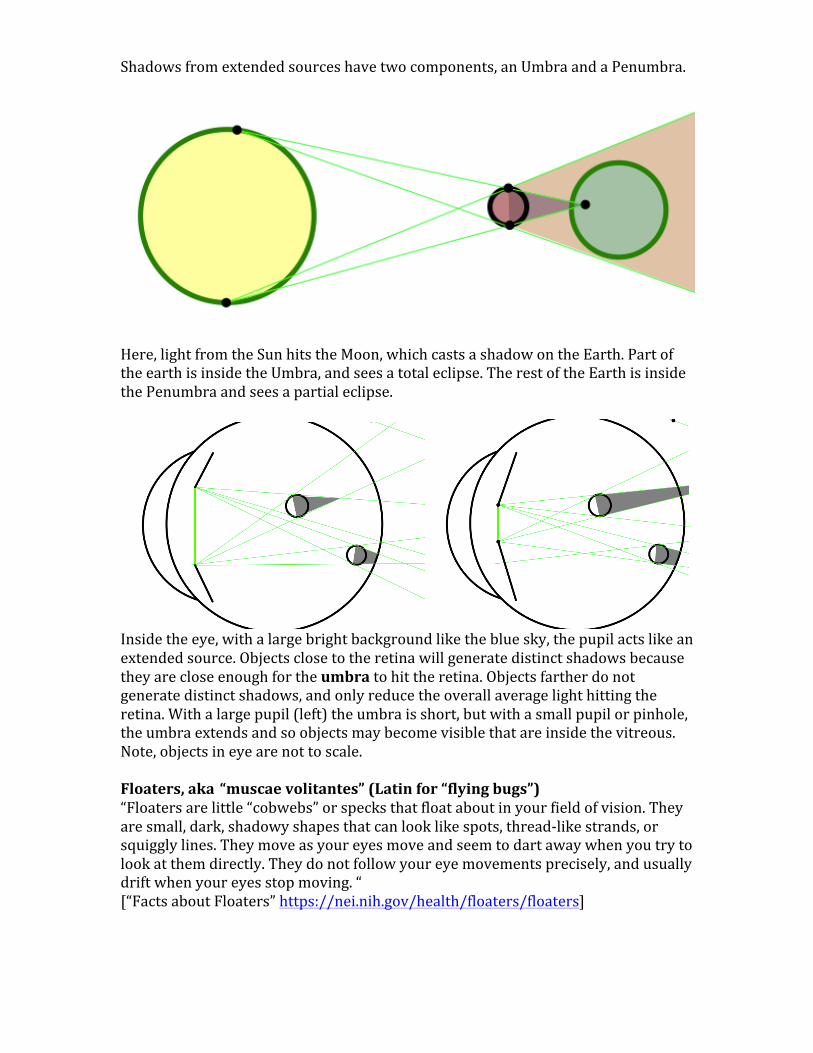
Shadowsfromextendedsourceshavetwocomponents,anUmbraandaPenumbra.
Here,lightfromtheSunhitstheMoon,whichcastsashadowontheEarth.PartoftheearthisinsidetheUmbra,andseesatotaleclipse.TherestoftheEarthisinsidethePenumbraandseesapartialeclipse.
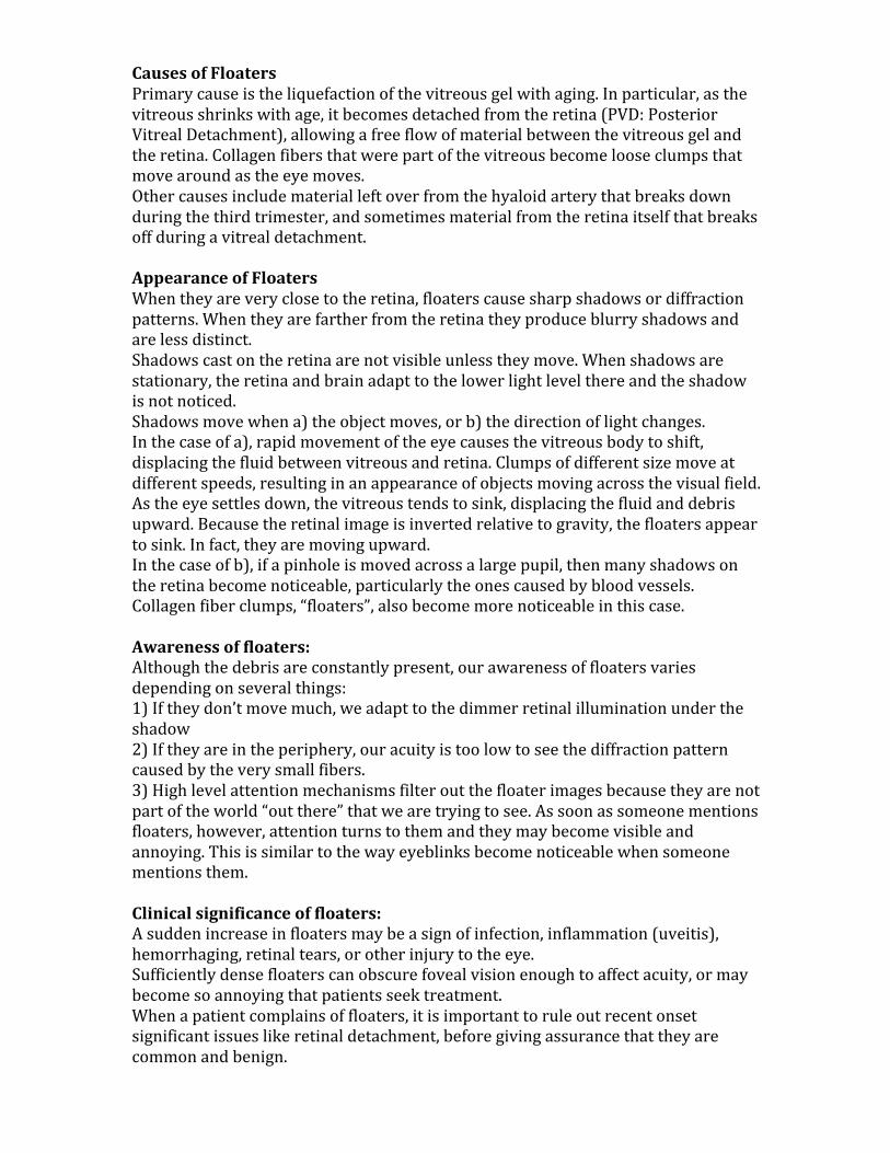
Insidetheeye,withalargebrightbackgroundlikethebluesky,thepupilactslikeanextendedsource.Objectsclosetotheretinawillgeneratedistinctshadowsbecausetheyarecloseenoughfortheumbratohittheretina.Objectsfartherdonotgeneratedistinctshadows,andonlyreducetheoverallaveragelighthittingtheretina.Withalargepupil(left)theumbraisshort,butwithasmallpupilorpinhole,theumbraextendsandsoobjectsmaybecomevisiblethatareinsidethevitreous.Note,objectsineyearenottoscale.Floaters,aka“muscaevolitantes”(Latinfor“flyingbugs”)“Floatersarelittle“cobwebs”orspecksthatfloataboutinyourfieldofvision.Theyaresmall,dark,shadowyshapesthatcanlooklikespots,thread-likestrands,orsquigglylines.Theymoveasyoureyesmoveandseemtodartawaywhenyoutrytolookatthemdirectly.Theydonotfollowyoureyemovementsprecisely,andusuallydriftwhenyoureyesstopmoving.“[“FactsaboutFloaters”https://nei.nih.gov/health/floaters/floaters]
Page 3
movie“Floater_WhiteBloodCells.mp4”
https://upload.wikimedia.org/wikipedia/commons/b/b2/Floaters.pngSimulatedimageoffloatersfromtheWikimediaCommons,astheyappearagainstabrightbluesky.Notethattheysometimesdonotappearinsharpfocus.
Page 4
CausesofFloatersPrimarycauseistheliquefactionofthevitreousgelwithaging.Inparticular,asthevitreousshrinkswithage,itbecomesdetachedfromtheretina(PVD:PosteriorVitrealDetachment),allowingafreeflowofmaterialbetweenthevitreousgelandtheretina.Collagenfibersthatwerepartofthevitreousbecomelooseclumpsthatmovearoundastheeyemoves.Othercausesincludematerialleftoverfromthehyaloidarterythatbreaksdownduringthethirdtrimester,andsometimesmaterialfromtheretinaitselfthatbreaksoffduringavitrealdetachment.AppearanceofFloatersWhentheyareveryclosetotheretina,floaterscausesharpshadowsordiffractionpatterns.Whentheyarefartherfromtheretinatheyproduceblurryshadowsandarelessdistinct.Shadowscastontheretinaarenotvisibleunlesstheymove.Whenshadowsarestationary,theretinaandbrainadapttothelowerlightlevelthereandtheshadowisnotnoticed.Shadowsmovewhena)theobjectmoves,orb)thedirectionoflightchanges.Inthecaseofa),rapidmovementoftheeyecausesthevitreousbodytoshift,displacingthefluidbetweenvitreousandretina.Clumpsofdifferentsizemoveatdifferentspeeds,resultinginanappearanceofobjectsmovingacrossthevisualfield.Astheeyesettlesdown,thevitreoustendstosink,displacingthefluidanddebrisupward.Becausetheretinalimageisinvertedrelativetogravity,thefloatersappeartosink.Infact,theyaremovingupward.Inthecaseofb),ifapinholeismovedacrossalargepupil,thenmanyshadowsontheretinabecomenoticeable,particularlytheonescausedbybloodvessels.Collagenfiberclumps,“floaters”,alsobecomemorenoticeableinthiscase.Awarenessoffloaters:Althoughthedebrisareconstantlypresent,ourawarenessoffloatersvariesdependingonseveralthings:1)Iftheydon’tmovemuch,weadapttothedimmerretinalilluminationundertheshadow2)Iftheyareintheperiphery,ouracuityistoolowtoseethediffractionpatterncausedbytheverysmallfibers.3)Highlevelattentionmechanismsfilteroutthefloaterimagesbecausetheyarenotpartoftheworld“outthere”thatwearetryingtosee.Assoonassomeonementionsfloaters,however,attentionturnstothemandtheymaybecomevisibleandannoying.Thisissimilartothewayeyeblinksbecomenoticeablewhensomeonementionsthem.Clinicalsignificanceoffloaters:Asuddenincreaseinfloatersmaybeasignofinfection,inflammation(uveitis),hemorrhaging,retinaltears,orotherinjurytotheeye.Sufficientlydensefloaterscanobscurefovealvisionenoughtoaffectacuity,ormaybecomesoannoyingthatpatientsseektreatment.Whenapatientcomplainsoffloaters,itisimportanttoruleoutrecentonsetsignificantissueslikeretinaldetachment,beforegivingassurancethattheyarecommonandbenign.
Page 5
Treatmentforfloaters:Mostcasesarenottreated.Patientsaretolditisanaturalpartofagingandthereisnosimpleproceduretoremovethem.Moreseverecasescanbetreatedwithvitrectomy.Removalofthevitreousbodyresolvesthefloaters,butbringswithitothercomplicationslikecataractandpossibleretinaldetachment.“Floaterectomy”isatermusedtodescribepartialvitrectomytoresolvefloaters.“FloaterectomyVersusConventionalParsPlanaVitrectomyForVitreousFloaters”http://www.djo.harvard.edu/site.php?url=/physicians/oa/1004SomedoctorsmaytryYAGlasertreatmenttovaporizethefibers,orenzymestobreakthemup.WhiteBloodCells:Whitebloodcellsarelargeenoughtocompletelyfillthesmallcapillariesintheretina.Sincetheydisplacethesmallerredbloodcells,whichabsorblightandcastashadow,andsincetheyaremostlywater,theymakethebloodvesseltemporarilyclear.Inretinalimaging,whenawhitebloodcellmovesthroughweareabletoseethephotoreceptorsbehindthecapillary.Visually,theymayappearassmallwhitespotsmovinginthevisualfield.ThisiscalledScheerer’sphenomenon.Wikipediahasanicesimulationofhowtheyappearagainstabrightsky.https://en.wikipedia.org/wiki/Blue_field_entoptic_phenomenonhttps://upload.wikimedia.org/wikipedia/commons/7/70/Blue_field_entoptic_phenomenon_animation.gifTounderstandwhytheyappearassmallwhitespots,watchthedemoWBCsim.mp4.Fixatethecenterspotuntilthedarkbandfades,andthemovinggapinthebandwillappearasawhitespot.Foramoreelaborateversionofthis,watchthedemoTroxlerDemo3Hz.mp4.Spotsmovearoundacircle,appearingfirstasgapsintheringthenasobjectsofoppositecolor.PurkinjeTree:ThePurkinjetreeisashadowofthesuperficialretinalvasculature.Itisnormallynotvisiblebecauseofadaptationandcentralinattention.Thereisalsoevidenceoflocalamblyopiaforcellsshadowedbythebloodvessels.AdamsDL,HortonJC."Shadowscastbyretinalbloodvesselsmappedinprimaryvisualcortex."Science2002;298:572-6.
Page 6
ThisfigurefromAdamsandHortonshowsthattherearefewercellsincortexdevotedtotherighteyeinlocationsthatareshadowedbybloodvessels.Dataarefromasquirrelmonkey.ThePurkinjetreebecomesvisiblewhenthelightsourcemoves.Thiscanbeachievedbyviewingwithapinholepupil,whenthenaturalpupilislarge.Movingthepinholeacrossthenaturalpinholemakestheshadowsofthevesselsmoveenoughthattheybecomevisible.AbettermethodtovisualizethePurkinjetreeistoilluminatetheeyethroughthesclera.Placeapenlightagainsttheclosedlidnearthelimbusandmoveitinsmallcircles.Thiswillmaketheshadowsfallonreceptorsthatarenotusuallyshadowed,andthetreebecomesmorevisible.Themorepinpointthelightsource,thesharpertheshadowswillbe,andthussmallerbloodvesselscanbeseen.
Page 7
Phosphenes:Phosphenesareperceivedflashesoflightthatarisebecauseofinternalactivityintheretinaorhighervisualsystem.Phosphenescanbecausedbymechanicalactionontheretina,asoccursduringvitreousdetachment.Moore’slightningstreaksareflashesoflightthatresemblelightningbolts.Theyusuallyappearinthetemporalvisualfield,andarefairlycommoninmiddleageandolderpatientsastheyexperiencePosteriorVitrealDetachment(PVD).Thevitreousputstractionontheretinaandthemechanicalactioncausesthecellstodischarge.FlickPhosphenesareproducedbyrapid,large,saccadicmovementsoftheeye.Theyaremorecommonlynoticedbyolderpatients,suggestingthatthepartiallyorfullydetachedvitreousmaybetuggingand/orbumpingontheretina.Theyaremostnoticeablewhendarkadapted.PressurePhosphenesareproducedbydirectmechanicalstimulationoftheeye.Closeyoureye,turnyoureyetowardthenose,andpressonthetemporalsidethroughyourlid.Youwillseeaspotappearinyournasalvisualfield.
FromFresco,Ophthalmology,1998
TheplotontheleftshowsacorrelationbetweenthepressurerequiredtoproduceaphospheneandtheintraocularpressuremeasuredwithGoldmanntonometry.Inthephoto,asimplepressuresensorispushedagainsttheeyeuntilaphospheneisseen.
ElectricalPhosphenesareproducedbyelectricalstimulationoftheretinaorcortex.ThesearesometimesnoticedbysubjectsbeingfittedwithEOG(EloctroOculoGram)electrodeswhentheimpedenceisbeingtested.Examinersrunasmallamountofcurrentthroughtheelectrodestomakesurethecontactisgood,andthiscurrentstimulatestheretina.CorticalPhosphenesarelikewiseproducedbyelectricalstimulationofthecortex.AnewtechniquecalledTranscranialMagneticStimulationisgaininginterestasatoodforstudyingcorticalfunction,andthissometimesproducesflashesoflight.Someneurologistshaveexperimentedwithimplantedelectrodearraysonthevisualcortexinordertoproducephosphenesinblindpatients.Stimulationelectrode
Page 8
arraysimplantedintheeyearecurrentlyunderdevelopmentaswell.ThemostadvancedoftheseistheARGUSIIfromSecondSight
ARGUSIIelectrodearray.FromLuo & Cruz (2016) Prog. Ret. Eye. Res.
Left: A patient wearing the apparatus that
records video and transmits it by radio into the implanted ARGUS II device.
Above: An image of the photoreceptor array, with a white circle to indicate the relative size of one electrode in the ARGUS II device. The spatial resolution of current implant devices is
extremely poor.
InthecontextofPerception,theimportantthingaboutPhosphenesisthattheyareperceivedaslight,andcanbeusedtorecognizeforms.ThisisanexampleofMüller’slawofspecificnerveenergies.Activationoftheopticnervefibersisperceivedaslightbecausethosenervesareassociatedwithvision.
Page 9
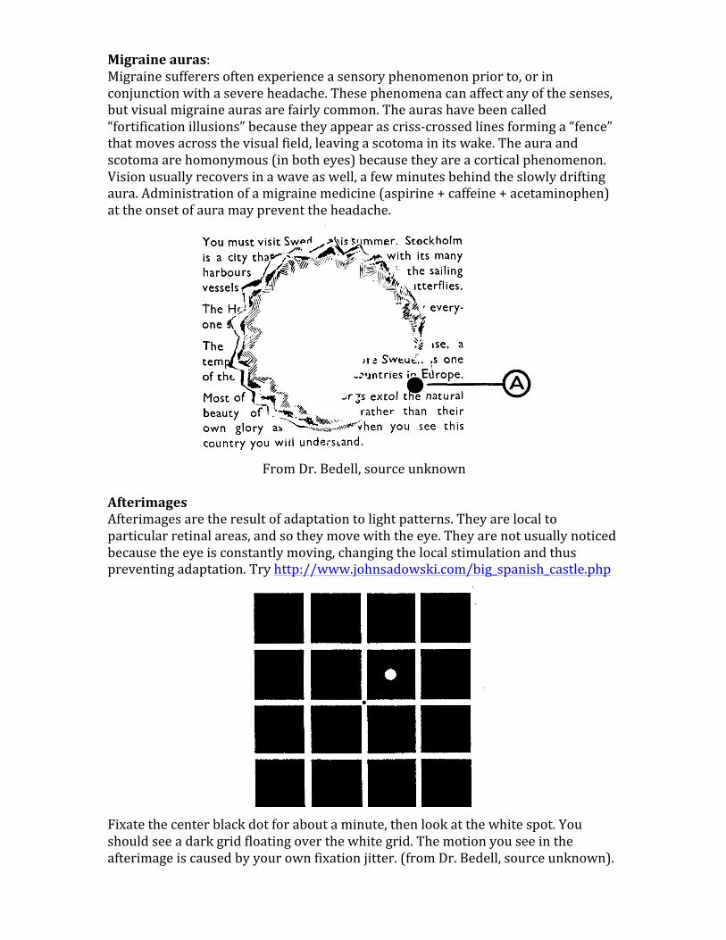
Migraineauras:Migrainesufferersoftenexperienceasensoryphenomenonpriorto,orinconjunctionwithasevereheadache.Thesephenomenacanaffectanyofthesenses,butvisualmigraineaurasarefairlycommon.Theaurashavebeencalled“fortificationillusions”becausetheyappearascriss-crossedlinesforminga“fence”thatmovesacrossthevisualfield,leavingascotomainitswake.Theauraandscotomaarehomonymous(inbotheyes)becausetheyareacorticalphenomenon.Visionusuallyrecoversinawaveaswell,afewminutesbehindtheslowlydriftingaura.Administrationofamigrainemedicine(aspirine+caffeine+acetaminophen)attheonsetofauramaypreventtheheadache.
FromDr.Bedell,sourceunknown
AfterimagesAfterimagesaretheresultofadaptationtolightpatterns.Theyarelocaltoparticularretinalareas,andsotheymovewiththeeye.Theyarenotusuallynoticedbecausetheeyeisconstantlymoving,changingthelocalstimulationandthuspreventingadaptation.Tryhttp://www.johnsadowski.com/big_spanish_castle.php
Fixatethecenterblackdotforaboutaminute,thenlookatthewhitespot.Youshouldseeadarkgridfloatingoverthewhitegrid.Themotionyouseeintheafterimageiscausedbyyourownfixationjitter.(fromDr.Bedell,sourceunknown).
Page 10
Usingafterimagesto“tag”thefoveas,totestforAnomalousRetinalCorrespondence
Inthistest,eacheyeisflashedtoputanafterimageinthemiddleofthefovea.Withbotheyesopen,mostpeoplewillthenseethetwoafterimagesinthesamedirection(upperleftdiagram).PeoplewithAnomalousRetinalCorrespondence(ARC)donotnecessarilyseetheirtwofoveasasrepresentingthesamedirectionwhenbotheyesareopen.Eventhoughthetargetsareonthefoveasofbotheyes,theydonotperceivethemasoverlapping.AbrightmovinglightleavesatrailbehinditthatcomesfromVisualPersistenceandLightAdaptation.Inthesketchbelow,abrightgreenspotleavesbehindashortgreenstreak(fromvisiblepersistence)andbehindthatisa longlastingafterimage(fromlightadapation).Afterimageslastlongerifthebackgroundlightchanges,makingitmoreobviousthatoneregionoftheretinaisadapteddifferentlyfromanother.Movetheeyebackandforthfromabrightareatoadimarea,andtheafterimagewillkeepreappearing,thenfading.
Page 11
Maxwell’sspotThemacularpigmentoverlyingthefoveaselectivelyabsorbsbluelight.Theretinalareacoveredbymacularpigmentisroughlythesamesizeasthefovealpit.Lightcomesfromthebottomandpassesthroughthispigmentbeforereachingthephotoreceptors.Undertherightviewingconditions,weseeadarkspotcenteredonthepointoffixation.Thespotishardtonoticebecauseitmoveswiththeeyeandbecauseweadapttothedifferenceincolor/brightness.
Wavelength (nm)
Leftimage:FromSnodderly1984.Crosssectionofamonkeyretinaintheregionofthefovea.Theupperimagewasmadewithgreenlightillumination,thelowerimagewithbluelight.Thedarkappearanceinthelowerimagecomesfromthemacularpigmentabsorbingthebluelight.Rightimage:Pigmentabsorbancevs.wavelength,withdatafromvariousstudies.Maxwell’sspotismosteasilyseenwithalternatingillumination.Trystaringatthegreensquareforafewsecondstoadaptyourlongandmiddlecones,thenlookattheblue.Youshouldseeadarkspotintheareaaroundyourfixationpoint.Thespotisusuallyalittlebiggerthanyourthumbatarmslength.
Maxwell’sspotissometimesusedtodiagnoseeccentricfixation.Ifapatientseesthespotoffcenterfromthepointoffixation,itindicatestheyarenotusingthecentralfoveaforfixation.AsimilartechniqueusesHaidinger’sbrushes.Theserefertoawindmillpatternthatappearswhenviewingpolarizedbluelight.Ifthewindmillisnotcenteredonthefixationpoint,itindicateseccentricfixation.


![Realistic Soft Shadows by Penumbra-Wedges Blending · Penumbra-wedges X + Specular & diffuse Visibility buffer Modulated spec+diff Ambient Final image. Penumbra-wedges [3/4] Penumbra-wedges](https://static.documents.pub/doc/80x56/5f543a4c0135c76e2b226697/realistic-soft-shadows-by-penumbra-wedges-penumbra-wedges-x-specular-diffuse.jpg)


