23
RADIOLOGY Or, Why do you want to x ray my cat?
| Date post: | 22-Dec-2015 |
| Category: |
Documents |
| Upload: | clemence-dean |
| View: | 219 times |
| Download: | 2 times |

RADIOLOGYOr,
Why do you want to x ray my cat?

Let’s start with the basics.
How does this stuff work anyway? Is it magic?

The Electromagnetic Spectrum
Radio: Radio stations transmit music on radio waves. Radio waves are also emitted by stars and gases in space.
Microwave: Microwaves cook your popcorn, but are also used by astronomers to learn about the structure of nearby galaxies.
Infrared: Night vision goggles pick up infrared light from objects with heat. In space, infrared light helps us map the dust between
stars.Visible: Our eyes detect visible light. Fireflies, light bulbs, and
stars all emit visible light.Ultraviolet: Ultraviolet radiation is emitted by the Sun and is the
reason skin tans and burns. X-ray: The veterinarian uses X-rays to image your pet, and airport
security uses them to see through your bag (and your clothes). Gamma ray: Doctors use gamma-ray imaging to see inside your
body. The biggest gamma-ray generator of all is the Universe.


So who started this?
Wilhelm Conrad Röntgen (27 March 1845 – 10 February 1923) was a German physicist, who, on 8 November 1895, produced and detected electromagnetic radiation in a wavelength range today known as X-rays or Röntgen rays, an achievement that
earned him the first Nobel Prize in Physics in 1901.

The first radiographic image ever was of Röntgen’s wife’s left hand, clearly showing her wedding
ring.

How do we get an image?
The heart of an X-ray generator is the X-ray tube. The X-ray tube contains a cathode, which directs a stream of electrons into a
vacuum, and an anode, which collects the electrons and evacuates the heat generated. When the electrons hit the
target, about 1% of the resulting energy is emitted as X-rays, the remaining 99% released as heat.

Densities
1. Air2. Fat3. Soft tissue4. Bone5. Metal
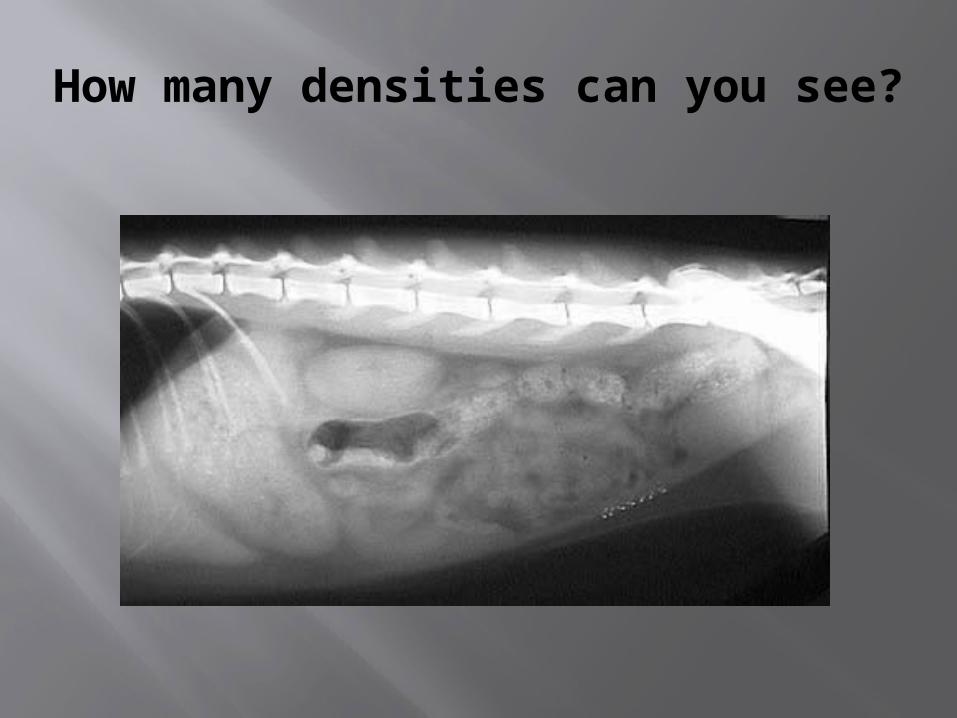
How many densities can you see?

Analog vs. CR Radiography
Analog is traditional film x rays. Much like photography, film is exposed to light to make an image. In radiography, the film is held
between two screens in a cassette. The screens emit light when exposed to x rays and an image of whatever is between the x ray generator (i.e. your cat) and the cassette is captured. The film is developed with chemicals like traditional black and white film.

C(omputed) R(adiography)
With CR systems, the film, intensifying screen, and cassette used in analog systems are replaced with an imaging plate. The
plate has a similar look and feel to a traditional film cassette.With CR, the feel of acquiring an image is similar to analog. As with film, the CR cassette is placed and an exposure is made.The cassette is then loaded into an imaging reader. Inside the
reader, the plate is exposed to lasers that interpret the information on the plate and translate it into a radiographic
image. The imaging plate is then erased by white light inside the imaging reader and is ready to use again.

So why do I need an x ray?

It’s not just about bones.

Sometimes it is…
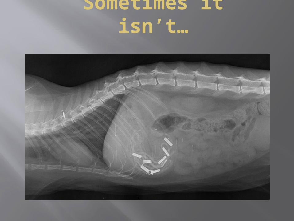
Sometimes it isn’t…

Don’t let your cat play with hair ties (or string, or rubber bands, or silk flowers, or…)

What else can radiographs show us?
Radiographic images can tell the veterinarian a lot about your cat’s health. By comparing body structures to known normals, much can be learned about what’s going on inside your cat.

Normal Cat Abdomen

What’s wrong with this picture?

A word about radiation safety

Radiation is everywhere
We live in a radiation filled environment. Cosmic rays bombard us from outer space, radon is
present in our homes, and radionuclides in the stone constructing our buildings and even the
food we eat give off radiation. On average, we can get 360mrem of radiation a year just from living
on earth.

Time, Distance, Shielding
The principle of ALARA is designed to help radiology technicians and their patients to keep their radiation exposure As Low As
Reasonably Achievable. This is achieved by keeping exposure times as short as possible,
keeping as far away from the source as possible and wearing lead protective
equipment to shield themselves.

The End


