Contents lists available at SciVerse ScienceDirect
Biochimica et Biophysica Acta
j ourna l homepage: www.e lsev ie r .com/ locate /bbamcr
ORAI3 silencing alters cell proliferation and cell cycle progression via c-myc pathwayin breast cancer cells
Malika Faouzi a,b, Philippe Kischel a, Frédéric Hague a, Ahmed Ahidouch a,c,⁎, Nazim Benzerdjeb a,d,Henri Sevestre a,d, Reinhold Penner b, Halima Ouadid-Ahidouch a,⁎⁎a University of Picardie Jules Verne, UFR of Sciences, Laboratory of Cellular and Molecular Physiology EA 4667, SFR CAP-SANTE (FED 4231), Amiens, Franceb Queen's Center for Biomedical Research, The Queen's Medical Center, 1356 Lusitana St., UH Tower 8th Floor, Honolulu, HI 96813, USAc University of Ibn-Zohr, UFR Sciences, Biology Department, Agadir, Moroccod University of Picardie Jules Verne, Amiens Hospital, Anatomy and Pathology Department, Tumor Bank of Picardie, Amiens, France
Members of the Orai family are highly selective calcium ion channels that play an important role instore-operated calcium entry. Among the three known Orai isoforms, Orai3 has gained increased attention,notably for its emerging role in cancer. We recently demonstrated that Orai3 channels are over-expressedin breast cancer (BC) biopsies, and involved specifically in proliferation, cell cycle progression and survivalof MCF-7 BC cells. Here, we investigate the downstream signaling mechanisms affected by Orai3 silencing,leading to the subsequent functional impact specifically seen in MCF-7 cancer cells. We report a correlationbetween Orai3 and c-myc expression in tumor tissues and in the MCF-7 cancer cell line by demonstratingthat Orai3 down-regulation reduces both expression and activity of the proto-oncogene c-myc. This is likelymediated through the MAP Kinase pathway, as we observed decreased pERK1/2 levels and cell-cycle arrestin G1 phase after Orai3 silencing. Our results provide strong evidence that the c-myc proto-oncogene isinfluenced by the store-operated calcium entry channel Orai3 through the MAP kinase pathway. This connec-tion provides new clues in the downstream mechanism linking Orai3 channels and proliferation, cell cycleprogression and survival of MCF-7 BC cells.
The recent discovery of the Orai channels as the pore forming unitsof the Ca2+ selective CRAC channels opened a new era in the field ofstore-operated calcium entry (SOCE) [1]. Three distinct Orai isoformshave been described to date (Orai1, Orai2 and Orai3), with Orai3being “special” in this family, notably because of its exclusive presencein mammals [2] and its receptivity to pharmacological modulation [3].CRAC channels are known for their physio-pathological roles [1,4–6],but their involvement in BC is only emerging [7–9]. For instance, wehave recently reported that Orai3 channels are over-expressed in BCbiopsies, and have also shown that these channels are involved inproliferation, cell cycle progression and survival of BC cells by regulatingtheG1 phase andG1/S transition regulator proteins [9].Moreover, theseeffects are specific to cancer cells, since down-regulation of Orai3 chan-nels does not affect either cell proliferation or cell survival of normal
d calcium entry; TMAs, Tissue
33 322827644.33 322827644.
. Ahidouch),
l rights reserved.
breast cells [9]. How Orai3 impacts such essential processes of thecancer cells' life remains elusive, and we therefore aimed at deciphering,at least in part, the mechanisms linking Orai3 and the above describedcellular effects.
The ubiquitous SOCE pathway is not only necessary to refill internalcalcium stores, but also for activating downstream signaling cascades[10,11]. Thus, on the assumption that at least one transcription factorwas involved, we directed our interest to the c-myc pathway thathas been implicated, just like Orai3, in processes controlling cell cycleprogression, proliferation and apoptosis [12,13]. The proto-oncogenec-myc encodes a transcription factor of the helix–loop–helix/leucinezipper protein family, and is known to be regulated by calcium [14]and several calcium-dependent signaling pathways such as the MAPkinase and the calcineurin/NFAT pathways [15–17].
Most human cancers display enhanced c-myc expression and/orderegulated c-myc activity [18–20]. In BC, several studies have shownthat between 50% and 100% of BC cases display increased c-myc proteinlevels (see [21] for review). Over-expression of c-myc is associatedwithreduced relapse-free and overall survival in BC patients [22,23]. Inter-estingly, down-regulation of c-myc in BC cells induces cell cycle arrestand apoptosis [24,25]. C-myc has been shown to be the key factor inG1 progression and G1/S transition phases in many cell types [26,27].In fact, c-myc positively regulates the expression and/or activity of
753M. Faouzi et al. / Biochimica et Biophysica Acta 1833 (2013) 752–760
cyclins (D1,D2, E, A), cyclin-dependent kinases (CDKs) 4 and 2 [28], andadditionally suppresses cyclin-dependent kinase inhibitors (CDKIs)such as p15, p21 and p27 [13]. These effects are strikingly similar tothe effects seen with Orai3 down-regulation, which also alters cyclinsD1 and E, CDKs 4 and 2, the cyclin-dependent kinase inhibitorp21Waf1/Cip1, and the tumor-suppressing protein p53 [9].
Therefore,wehypothesized thatOrai3 channelsmight be anupstreamregulator of the c-myc pathway, and we investigated the role of c-mycin the Orai3-induced differential effect on proliferation and cell cycleprogression in BC cells and normal breast cells. Using qRT-PCR and tissuemicroarray, we found that expression levels of Orai3 and c-myc arepositively correlated. We observed that Orai3 down-regulation inducedcell cycle arrest in G1 phase through the c-myc pathway. We also foundthat simultaneous down-regulation of both Orai3 and c-myc proteinshadno additive or synergic effects on either BC cell proliferation or surviv-al. Finally, since the MAP Kinase pathway is known to regulate c-mycprotein expression and activity, we investigated the effect of Orai3down-regulation on ERK1/2 phosphorylation and found that the phos-phorylation level of ERK1/2 significantly decreased after silencing Orai3channels.
Our results therefore suggest that Orai3 channels are amongstupstream effectors of the oncogenic c-myc pathway and constitutekey players in BC cells such as the MCF-7 cell line. Importantly, basedon the differential effect seen between normal and BC cells, Orai3could represent a selective target for breast cancer treatment.
2. Materials and methods
2.1. Tissue microarrays
Cancerous breast tissue was obtained from fresh surgical specimens.Consent forms (approved by the University Hospital of Amiens) weresigned by the patients before surgery to allow the use of a portion of tis-sue samples for research purposes. Samples of breast adenocarcinoma,as well as non-tumoral tissues from the same patient were obtainedfrom women having undergone operations at the Amiens hospitals.
Immunohistochemical staining was performed on formalin-fixedparaffin-embedded (FFPE) blocks with a Roche Ultra immunostainer,using antibodies against Orai3 (1:100, HPA015022, Sigma PrestigeAntibody) and c-myc (1:100, N-262, Santa-Cruz BioTechnologies).This was followed by the avidin–biotin–peroxidase complex tech-nique. Reactions were developed using a chromogenic reaction inDAB (diamino-3,3'benzidinetetrahydrochloride) substrate solution(DAB, Sigma Fast). Counterstaining was carried out with hematoxylinsolution. Micrograph acquisitions were performed using a digitalcamera connected to a Zeiss microscope.
Immunostaining in the tumor tissue was determined by subjectivevisual scoring of the brown stain. Two operators independentlyevaluated antigen expression. Scoring of the intensity of the stainingwas performed according to an arbitrary scale with steps of 0, 1, 2,and 3 where “0” was considered to be absence of staining, “1” consid-ered weak staining, “2”was considered asmoderately positive staining,and “3” was considered to be strong staining. A negative control wasperformed using the same technique without primary antibody.
The expression of Orai3 and c-myc was also assessed in 30additional human invasive ductal adenocarcinoma specimens usingTissue Microarray (TMAs). Briefly, four-micrometer-thick sections offormalin-fixed paraffin-embedded tissue samples were taken fromthe FFPE block.
2.2. Immunofluorescence
Immunofluorescent analyses were performed on tissues followinga published protocol [29]. Antibodies used were anti-Orai3 (1:100,HPA015022, Sigma Prestige Antibody), anti-c-myc (9E10, 1:500, SantaCruz Biotechnology, Inc., Heidelberg, Germany), goat anti rabbit IgG
DyLight 549 conjugated (Thermo, Rockford, IL, USA), and Alexa Fluor488 goat anti-mouse IgG (Molecular Probes, Eugene, OR, USA). Nucleiwere stained with DAPI (4′,6-diamidino-2-phenylindole, 1.43 μM). Im-ages were obtained using a Zeiss Axiovert 200 microscope equippedwith ApoTome system (Zeiss, Le Pecq, France).
2.3. Cell culture
MCF-7 BC cells were grown in Eagle's Minimum Essential Mediumsupplemented with 5% fetal calf serum (Lonza, Levallois-Perret,France), 2 mM L-glutamine, and 0.06% HEPES. The immortalizedhuman mammary epithelial cell line MCF-10A was grown in DMEM/F12 medium, composed of Dulbecco's modified Eagle's medium/nutrient mixture F12 supplemented with 5% fetal calf serum, 20 ng/mlepidermal growth factor, 10 mg/ml-1 insulin, 0.5 mg/ml hydrocortisone,and 100 ng/ml cholera toxin (Sigma-Aldrich, St-Quentin Fallavier,France). All cell lines were grown in a 5% CO2-humidified incubatorat 37 °C.
2.4. Transfection
Transfection of cells was performed using the nucleofection tech-nology (Amaxa, Köln, Germany), as previously described [30]. Cells(1×10⁶) were transfected either with 5 μg scrambled siRNA as a con-trol (siGENOME non-targeting siRNA, Dharmacon Research Inc.,Chicago, IL, referred in the text to as “si-CTL”), or with siRNAs directedagainst an Orai isoform (Orai1 or Orai3), or with siRNA directedagainst c-myc, or simultaneously with both si-Orai and si-c-myc. Tothis end, siRNA were combined and nucleofected according to themanufacturer's protocol.
2.5. Real-time quantitative PCR
Extraction of total RNA from cell lines or biopsies and real-timePCR were performed as previously described [9]. The relative amountof Orai3 and c-myc mRNAs in breast cancer cells were normalized tothe endogenous control (ß-actin) and compared to the referencesample (normal breast cells) using the Pfaffl method [31]:
Ratio ¼Etarget
� �Δcttarget normalcells−cancercellsð Þ
Erefð ÞΔctref normalcells−cancercellsð Þ
2.6. Western blot analyses
Whole-cell lysates were preparedwith RIPA buffer containing a pro-tease inhibitor cocktail (Sigma-Aldrich). Proteinswere separated by de-naturing SDS–PAGE, and transferred onto nitrocellulose membranes.The primary antibodies used were: anti-Orai3 (1:100, HPA015022,Sigma Prestige Antibody), anti-c-myc (N-262, 1:500, Santa Cruz Bio-technology Inc., Heidelberg, Germany), anti-ERK1/2 and anti-pERK1/2(1:1000, Cell Signaling Tech., Danvers, MA). Secondary antibodieswere coupled to horseradish peroxidase. Actin antibody (1/2000,Santa Cruz Biotechnology) was used for loading control experiments.Bound antibodies were visualized using ECL chemiluminescentsubstrate (GE Healthcare, Saclay, France) and quantified using theBio-Rad image acquisition software (Quantity One) associated withthe ChemiDoc XRS imager system (Bio-Rad Laboratories).
2.7. c-myc activity measurement
Nuclear extracts were obtained using the Marligen Biosciences Kit.Briefly 72 h after transfection, cells were collected by centrifugationand washed in PBS. Cells were then allowed to swell in CompleteHypotonic Cell Lysis Buffer, and lysis was facilitated by the addition
754 M. Faouzi et al. / Biochimica et Biophysica Acta 1833 (2013) 752–760
of the Detergent Solution. The cell nuclei were collected by gentlecentrifugation, and the cytoplasm was removed. The nuclear pelletwas washed twice in Complete Nuclear Wash Buffer and extracted
by addition of Complete Extraction Buffers. The nuclear extract wasclarified by centrifugation, and the protein concentration was deter-mined using a Bradford assay. Direct quantitation of c-myc binding
755M. Faouzi et al. / Biochimica et Biophysica Acta 1833 (2013) 752–760
activity was then performed on a 96-well plate using Myc-Max activ-ity measurement Kit (Marligen Biosciences).
2.8. Cell viability and mortality
Cell viability and mortality were measured 72 h post-transfectionusing TrypanBlue assay. After transfectionwith siRNAs directed againstOrai3 or c-myc or with non-targeting siRNA, MCF-7 cells were grownin 35 mm Petri dishes at a density of 1×10⁵ cells for 72 h. Cell countwas assessed using the standard Malassez cell method. Briefly, cellswere removed by trypsinization and diluted in Trypan Blue solution.Cell counts were performed six times and the results were expressedas the percentage of viable or dead cells normalized to control.
2.9. Cell-cycle analysis
For the measurement of cellular DNA content, only adherentcells were collected at 72 h post-transfection. Cells were pelleted,re-suspended in PBS/EDTA, treated with 20 mg/ml RNaseA (Sigma-Aldrich) and stained with 50 mg/ml of propidium iodide (Sigma-Aldrich). Samples were then analyzed on an Elite Beckman Coulterflow cytometer. The percentage of cells in different phases was cal-culated using WinMDI 2.8 and Cylchred software.
2.10. Determination of p42/p44 MAP Kinase/extracellular signal regulatedkinase (ERK1/2) by western blotting
The level of phosphorylation of ERK1/2 treated with either si-CTLor si-Orai3 was examined in MCF-7 cells. Forty-eight hours aftertransfection, MCF-7 cells were deprived of FCS for 24 h. The mediumwas then changed to a fresh medium, either without FCS or with 5%FCS. Cells were incubated for 20 minutes in this medium before pro-tein extraction.
2.11. Statistical analyses
Values are expressed as mean±SEM. Statistical analysis of thedata was performed using appropriate ANOVA, Mann–Whitney orpaired t-tests. Differences were considered significant at pb0.05.
3. Results
3.1. Correlation between Orai3 and c-myc expression in tumor tissuesand in BC cell lines
In our previous study, we showed that Orai3 is over-expressed in77% of the studied tumor samples [9]. Because of this, and the factthat c-myc expression is frequently amplified in breast cancer, weassessed the expression levels of c-myc mRNA in the same BC sam-ples previously used for the evaluation of Orai3 expression. Wefound that c-myc was over-expressed (over-expression being definedas a two-fold or greater mRNA level when compared to the expres-sion levels found in normal adjacent breast tissue) in 9 cases out of13 (69.2%, Supplementary Fig. 1). Statistical analysis based on theSpearman's correlation coefficient (Fig. 1A) indicated a positive corre-lation between Orai3 and c-myc mRNA expression (R=0.857). Theexpression of Orai3 and c-myc was also evaluated in 30 additionalhuman invasive adenocarcinoma specimens using TMAs (Fig. 1B).
Fig. 1. Correlation between Orai3 and c-myc genes expression in breast cancer. A: Spearmancoefficient is equal to 0.857. B: Mean score of Orai3 and c-myc staining obtained on the adOrai3 and c-myc expression in cancerous and matched non-tumoral human breast cancer tiwere subjected to immunoperoxidase staining, as described in Materials and methods. OrigMerging of both Orai3 and c-myc staining is shown in δ, and DAPI colored nuclei are showbreast cell line MCF-10A taken as reference for both genes (n=4). mRNAs are normalized
Here too, considering over-expression is at least a two-fold higherexpression, Orai3 and c-myc were over-expressed in 70% (21 out of30 cases) and 80% (24 out of 30 cases) cases respectively (pb0.001by Mann–Whitney Rank Sum Test). Moreover, of the 21 samplesexhibiting high expression of Orai3 (score≥2+), a high c-mycstaining (score≥2+) was found in all samples (100%). Statisticalanalysis based on the Spearman's correlation coefficient indicatedagain a positive correlation between Orai3 and c-myc protein expres-sion (R=0.895). A representative immuno-histochemistry is shownin Fig. 1C. Double immuno-fluorescence was also performed to assessco-overexpression of Orai3 and c-myc in FFPE tissues from the BCpatients. A representative immuno-fluorescence is shown in Fig. 1D.Although some cells express either predominantly c-myc or Orai3, amajority of cells show concomitant over-expression of both proteins.
Together, these results indicate that Orai3 and c-myc are stronglycorrelated in BC tissues.
To test the involvement of c-myc in Orai3-dependent cell prolifera-tion and/or survival, we first studied c-myc mRNA expression levelsin both normal and cancer breast cells. As shown in Fig. 1E, expressionof c-myc, as assessed by RT-qPCR, is higher in the MCF-7 cancer cellline than in the non-cancerous MCF-10A cell line. Remarkably, thisover-expression (2.2±0.5 fold over control) is very similar to the Orai3over-expression seen in these cancer cells (2.4±0.2 fold over control).
3.2. Orai3 down-regulation reduces c-myc expression and activity
Wenext silenced Orai3 channelswith small interfering RNAs in bothcell lines. As can be seen in Fig. 2A, siRNAs against c-myc were effectivein both normal and cancer cell lines. Moreover, si-Orai3 was able todown-regulate c-myc mRNA expression only in MCF-7 cancerouscells. Therefore, we assessed the extent of down-regulation at the pro-tein level, again at 72 h post-transfection, using Western blotting. Thec-myc expression is shown in Fig. 2B and densitometric analyses ofc-myc bands are presented in Fig. 2C. The results at the protein levelare consistent with those obtained at the mRNA level, c-myc proteinlevels being strongly reduced only in MCF-7 cells tranfected withsi-Orai3 (43.1±8.7% of the control value, pb0.05). Interestingly, we ob-served no significant difference in c-myc expression levels betweensi-Orai3 treated MCF-10A cells and MCF-10A control cells.
We next sought to evaluate the c-myc/Max activity in normal andcancer cells. Indeed, it is known that c-myc belongs to the Myc/Max/Mad network that is composed of a group of transcription factors,whose interactions result in transcriptional activation or repressionof their target genes [12,32]. Max is the protein partner of c-myc,whose heterodimerization allows binding to specific DNA sequenceslocated in c-myc target genes and permits the recruitment of tran-scriptional co-activators. This process results in the activation of thec-myc downstream genes, particularly those involved in cell cycleprogression and proliferation. In this context, we measured theMyc/Max complex DNA-binding, which reflects c-myc activity. In BCcells, Orai3 silencing resulted in a highly significant decrease ofc-myc activity (3.6±0.5% vs. 100±30.3% in the control, pb0.001,Fig. 2D). In contrast, the effect observed in MCF-10A cells was notstatistically significant (54±21% vs. 100±37.7% in control, Fig. 2D).
To know whether reduction in c-myc expression and activityare really specific to Orai3 down-regulation or could also be observedwith silencing of another member of the Orai protein family, we chooseto down-regulate Orai1, also present in MCF-7 cells [8]. The results are
's correlation between Orai3 mRNA and c-myc mRNA extracted from 13 patients: the Rditional cohort of 30 patients represented on the TMA. C: Representative examples ofssues, as assessed by immunohistochemistry. Paraffin sections of human breast tissuesinal magnification: ×400. D: Immunofluorescent staining of c-myc (β) and Orai3 (γ).n in α. E: Relative transcript levels of Orai3 and c-myc in MCF-7 cells, with the normalto ß-actin, as described in Materials and methods.
Fig. 2. Orai3 down-regulation affects c-myc protein expression and activity. A: Relative transcript levels of c-myc in MCF-10A and MCF-7 cells after transfection with either si-c-mycor si-Orai3, with a non-targeting si-RNA (si-CTL) taken as reference for both genes (n=3). B: Representative western blot showing the effects of cell transfection with si-control(si-CTL) or si-Orai3 on the expression of c-myc in both MCF-7 and MCF-10A cell lines. C: Quantification of c-myc levels using densitometric analyses of c-myc protein expressionshown in B (n=4). D: c-myc activity measured in cells transfected with si-Orai3, normalized to c-myc activity measured under control condition in MCF-7 cells (n=9) and inMCF-10A cells (n=6). Values are reported as mean±SD, *pb0.05; ***pb0.001 vs. control; ns: not significant.
756 M. Faouzi et al. / Biochimica et Biophysica Acta 1833 (2013) 752–760
illustrated in Supplementary Fig. 2. Using a reporter plasmid, enhancedc-myc promoter activity was found when Orai1 was down-regulated(226±11.17% vs. si-CTL, data not shown). Consistently, si-Orai1 in-duced an upregulation of c-myc mRNA (180±35.7%, SupplementaryFig. 2A). Quantitation of c-myc binding activity was also performedusing the Myc–Max activity measurement kit: again, the activity ofsi-Orai1 treated cells was increased to 183.4±5.3% when compared tothe si-CTL condition (Supplementary Fig. 2B).
Taken together, these results showed that Orai3 down-regulationreduced both c-myc protein expression and activity levels exclusivelyin BC cells, since no statistically significant effect was detected innormal cells. While, silencing Orai1 led to the opposite effect in BCcells (increase of c-myc mRNA expression and activity).
3.3. c-myc-dependent proliferation and survival are regulated by Orai3in BC cells
Given the results obtained for c-myc expression and activity,we next investigated the contribution of c-myc to Orai3-dependent
proliferation and survival. Cells were transfected with different siRNAsindependently (siCTL and siRNAs directed against Orai3 or c-myc) orcombined (si-Orai3+si-c-myc). At 72 h post-transfection, a TrypanBlue assay was performed to assess the number of viable cells and thepercentage of cell mortality for each condition. As expected, Orai3 andc-myc silencing significantly decreased BC cell viability to 34.6±4.1%and 34.7±2.9% of the control, respectively (pb0.001, Fig. 3A) andincreased cell mortality (16.5±1.3% in si-Orai3 transfected cells,16.1±0.6% in si-c-myc transfected cells vs. 6.3±0.5% in control cells,pb0.001, Fig. 3B). Interestingly, the effects of si-Orai3 and si-c-myc oncell viability and mortality were not additive when cells were simulta-neously treated with both siRNAs (Fig. 3A and B).We also checked thepercentage of apoptotic cells after siRNA treatments. As shown inSupplementary Fig. 3, apoptosis increased significantly in si-Orai3,si-c-myc and both si-Orai3 and si-c-myc conditions. Interestingly,values for apoptosis in si-Orai3, as well as in si-c-myc treated cells(either alone or combinedwith si-Orai3)were not statistically different.
InMCF-10A cells, Orai3 down-regulation had no effect on either cellviability or cell mortality. In these cells, c-myc silencing significantly
Fig. 3. c-myc involvement in Orai3-dependent proliferation and survival. A: Effects of Orai3 and c-myc silencing on MCF-7 cell viability. The cell viability was measured 72 h posttransfection, and all values were normalized as percentage of si-CTL. B: Percentages of cell mortality obtained in MCF-7 cells transfected with si-Orai3 and/or si-c-myc for 72 h.C: Effects of Orai3 and c-myc silencing on MCF-10A cell viability at 72 h post-transfection. Values were normalized as percentage of siCTL. D: Effects observed on MCF-10A mortalityafter transfection with si-Orai3 and/or si-c-myc for 72 h. Values are reported as mean±SEM of triplicate experiments, ***pb0.001 vs. control; ns: not significant.
757M. Faouzi et al. / Biochimica et Biophysica Acta 1833 (2013) 752–760
decreased the cell viability to 40±3.8% of the control (pb0.001, Fig. 3C),but had no effect on cell mortality (Fig. 3D). The effects of combinedsi-Orai3 and si-c-myc treatments were not statistically different fromthose given by si-c-myc alone on MCF-10A with respect to viability ormortality (Fig. 3C and D). These results are further evidence of the in-volvement of c-myc in the Orai3-dependent cell proliferation and sur-vival of breast cancer cells.
3.4. Orai3 down-regulation induced cell arrest in G1 phase through thec-myc pathway
We have previously shown that Orai3 silencing induced cellcycle arrest in the G1 phase [9]. To determine the contribution ofc-myc to this effect, we investigated the cell cycle distribution ofcells transfected with siRNAs against Orai3 and/or c-myc. At 72 hpost-transfection, cell cycle analysis was performed by flow cytome-try. Fig. 4A shows the cell cycle distribution of MCF-7 BC cells. Consis-tent with previous observations [9], Orai3 silencing led to cell cyclearrest, with a significant accumulation of cells in the G0/G1 phase(76.8±3.1% vs. 55.6±1% for the control condition, pb0.01). There
was a decrease in the cell percentage in both S (16±2.8% vs. 31±0.8% for the control condition) and G2/M (7.3±0.3% vs. 13.5±0.3%for the control condition) phases. When c-myc was down-regulated,we observed a significant increase of cells in the G0/G1 phase (69±0.7% vs. 55.6±1% for the control condition, pb0.001), a significantdecrease in the cell percentage in S phase (18.4±0.7% vs. 31±0.8%for the control condition) and no significant effect on G2/Mphase (12.6±0.1% vs.13.5±0.3% for the control condition). Cellsco-transfected with both siRNAs showed no significant additive ef-fect, and the cell distribution under this condition was similar tothat observed in cells transfected with si-Orai3 alone. As expected,cell cycle distribution of MCF-10A cells was unchanged in cells withdown-regulated Orai3 when compared to the control condition(Fig. 4B). In contrast, c-myc silencing resulted in cell cycle arrest,with a significant accumulation of cells in the G0/G1 phase (69.5±4.6% vs. 59.5±2.9% for the control condition, pb0.05), and a decreasein the cell percentage in G2/M phase (17.4±3.9% vs. 24.1±3.3% forthe control condition, pb0.05), while no significant effect wasdetected in S phase. When combined, si-Orai3 and si-c-myc inducedthe same effects as si-c-myc alone.
Fig. 4. c-myc involvement in Orai3-dependent cell cycle progression. A: Cell cycle distribution of MCF-7 cells transfected with siRNAs against Orai3 (si-Orai3) and/or c-myc(si-c-myc). B: Cell cycle analysis of MCF-10A cells in the same experimental conditions. The cell cycle analysis was performed at 72 h post-transfection. Insets show raw datafrom the FACS acquisition software. Values are reported as mean±SEM of triplicate experiments, *pb0.05;**pb0.01; ***pb0.001 vs. control; ns: not significant.
758 M. Faouzi et al. / Biochimica et Biophysica Acta 1833 (2013) 752–760
3.5. Orai3 down-regulation affects the c-myc pathway, likely via the MAPKinase pathway
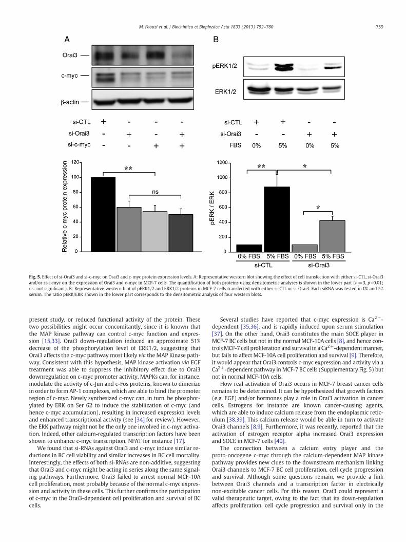
We evaluated c-myc protein expression levels after either individ-ual or concomitant down-regulation of Orai3 and c-myc (Fig. 5A).While separate transfections of either si-Orai3 or si-c-myc induced adecrease of c-myc protein expression to a similar extent (approxi-mately 40% decrease, pb0.01), the combined transfection of bothsiRNAs had no additive effects (ca. 50% down-regulation, without sta-tistical significance with respect to individual siRNAs).
We sought to determine which pathway was most likely involvedin the c-myc down-regulation induced by Orai3 silencing. The MAPKinase pathway is known to control the c-myc promoter. We there-fore investigated the level of phosphorylation of ERK1/2 in MCF-7cells treated with either si-control (si-CTL) or si-Orai3. Stimulationwith 5% serum induced approximately a 8.8 fold increase of theERK1/2 phosphorylation in cells treated with si-CTL (Fig. 5B). Insi-Orai3 treated cells, this phosphorylation level was enhanced onlyby a factor 4, hence showing 51% decrease of the phosphorylationlevel of ERK1/2 when compared to si-CTL treated cells (Fig. 5B).Moreover, the~40% decrease of the c-myc promoter activity observedin the presence of the si-Orai3 was almost fully restored by applica-tion of EGF (100 nM, Supplementary Fig. 4). These results providesupportive evidence that Orai3 specifically affects the c-myc pathway,most likely via the MAP Kinase pathway.
4. Discussion
The present study was prompted by the recent demonstrationthat, in MCF-7 cancer cells, SOCE was mediated by Orai3 [8]. Wenow provide compelling evidence that the early-response genec-myc, which is known for its implication in cell cycle progression,proliferation and survival [12,13], is involved in the cell cycle arrestand apoptosis seen in MCF-7 cancer cells following down-regulationof Orai3. We also demonstrate that Orai3 and c-myc are bothup-regulated in BC tissues.
We have previously shown that Orai3 was over-expressed in 77% ofthe studied cases [9].We now show that c-myc is also over-expressed in9 out of these 13 cases, being in good agreement with previouslyreported studies (see [21] for review). Importantly, using TMA on 30BC tissues, we found that for each cancer tissue showing c-mycover-expression, there is a concomitant up-regulation of Orai3. Thisup-regulation is also found in the cancer cell line MCF-7 when com-pared to the normal cell line MCF-10A.
We also established that Orai3 down-regulation specifically re-duces c-myc expression and activity, Orai1 down-regulation havingopposite effects. These opposite effects could be due either to differ-ent subcellular localization of the channels, or interaction with differ-ent protein complexes involved in c-myc regulation. Reduction ofc-myc activity by Orai3 silencing can be due either to the obvious sub-sequent reduction of c-myc expression levels demonstrated in the
Fig. 5. Effect of si-Orai3 and si-c-myc on Orai3 and c-myc protein expression levels. A: Representative western blot showing the effect of cell transfection with either si-CTL, si-Orai3and/or si-c-myc on the expression of Orai3 and c-myc in MCF-7 cells. The quantification of both proteins using densitometric analyses is shown in the lower part (n=3, pb0.01;ns: not significant). B: Representative western blot of pERK1/2 and ERK1/2 proteins in MCF-7 cells transfected with either si-CTL or si-Orai3. Each siRNA was tested in 0% and 5%serum. The ratio pERK/ERK shown in the lower part corresponds to the densitometric analysis of four western blots.
759M. Faouzi et al. / Biochimica et Biophysica Acta 1833 (2013) 752–760
present study, or reduced functional activity of the protein. Thesetwo possibilities might occur concomitantly, since it is known thatthe MAP kinase pathway can control c-myc function and expres-sion [15,33]. Orai3 down-regulation induced an approximate 51%decrease of the phosphorylation level of ERK1/2, suggesting thatOrai3 affects the c-myc pathway most likely via the MAP Kinase path-way. Consistent with this hypothesis, MAP kinase activation via EGFtreatment was able to suppress the inhibitory effect due to Orai3downregulation on c-myc promoter activity. MAPKs can, for instance,modulate the activity of c-Jun and c-Fos proteins, known to dimerizein order to form AP-1 complexes, which are able to bind the promoterregion of c-myc. Newly synthesized c-myc can, in turn, be phosphor-ylated by ERK on Ser 62 to induce the stabilization of c-myc (andhence c-myc accumulation), resulting in increased expression levelsand enhanced transcriptional activity (see [34] for review). However,the ERK pathway might not be the only one involved in c-myc activa-tion. Indeed, other calcium-regulated transcription factors have beenshown to enhance c-myc transcription, NFAT for instance [17].
We found that si-RNAs against Orai3 and c-myc induce similar re-ductions in BC cell viability and similar increases in BC cell mortality.Interestingly, the effects of both si-RNAs are non-additive, suggestingthat Orai3 and c-myc might be acting in series along the same signal-ing pathways. Furthermore, Orai3 failed to arrest normal MCF-10Acell proliferation, most probably because of the normal c-myc expres-sion and activity in these cells. This further confirms the participationof c-myc in the Orai3-dependent cell proliferation and survival of BCcells.
Several studies have reported that c-myc expression is Ca2+-dependent [35,36], and is rapidly induced upon serum stimulation[37]. On the other hand, Orai3 constitutes the main SOCE player inMCF-7 BC cells but not in the normal MCF-10A cells [8], and hence con-trolsMCF-7 cell proliferation and survival in a Ca2+-dependentmanner,but fails to affect MCF-10A cell proliferation and survival [9]. Therefore,it would appear that Orai3 controls c-myc expression and activity via aCa2+-dependent pathway inMCF-7 BC cells (Supplementary Fig. 5) butnot in normal MCF-10A cells.
How real activation of Orai3 occurs in MCF-7 breast cancer cellsremains to be determined. It can be hypothesized that growth factors(e.g. EGF) and/or hormones play a role in Orai3 activation in cancercells. Estrogens for instance are known cancer-causing agents,which are able to induce calcium release from the endoplasmic retic-ulum [38,39]. This calcium release would be able in turn to activateOrai3 channels [8,9]. Furthermore, it was recently, reported that theactivation of estrogen receptor alpha increased Orai3 expressionand SOCE in MCF-7 cells [40].
The connection between a calcium entry player and theproto-oncogene c-myc through the calcium-dependent MAP kinasepathway provides new clues to the downstream mechanism linkingOrai3 channels to MCF-7 BC cell proliferation, cell cycle progressionand survival. Although some questions remain, we provide a linkbetween Orai3 channels and a transcription factor in electricallynon-excitable cancer cells. For this reason, Orai3 could represent avalid therapeutic target, owing to the fact that its down-regulationaffects proliferation, cell cycle progression and survival only in the
760 M. Faouzi et al. / Biochimica et Biophysica Acta 1833 (2013) 752–760
MCF-7 BC cells. Although it is generally accepted that ion channelsrepresent attractive targets for human cancer therapy, either fortherapeutic or diagnostic purposes, efficient molecules have yet tobe designed to target ion channels in breast cancer [41]. Since Ca2+
channels are easily and directly accessible via the bloodstream,Orai3 could have potential for antibody-targeted therapeutic strate-gies, as recently proposed for TRP ion channels [42] or voltage-gatedpotassium channels [43].
Supplementary data to this article can be found online at http://dx.doi.org/10.1016/j.bbamcr.2012.12.009.
Acknowledgments
This study was supported by the Region Picardie, and by theMinistère de l'Education Nationale (France) and la Ligue NationaleContre le Cancer (SEPTENTRION). We thank Alexandre Patrice fromthe Laboratory of Pr. Pattou F. (Thérapie Cellulaire du Diabète, InsermU859, Lille) for technical assistance with the Luminex plate reader.
References
[1] S. Feske, Y. Gwack, M. Prakriya, S. Srikanth, S.-H. Puppel, B. Tanasa, P.G. Hogan,R.S. Lewis, M. Daly, A. Rao, A mutation in Orai1 causes immune deficiency by ab-rogating CRAC channel function, Nature 441 (2006) 179–185.
[2] T.J. Shuttleworth, Orai3 — the ‘exceptional’ Orai? J. Physiol. 590 (2012) 241–257.[3] R. Schindl, J. Bergsmann, I. Frischauf, I. Derler, M. Fahrner, M. Muik, R. Fritsch, K.
Groschner, C. Romanin, 2-Aminoethoxydiphenyl borate alters selectivity of Orai3channels by increasing their pore size, J. Biol. Chem. 283 (2008) 20261–20267.
[4] I.F. Abdullaev, J.M. Bisaillon, M. Potier, J.C. Gonzalez, R.K. Motiani, M. Trebak,Stim1 and Orai1 mediate CRAC currents and store-operated calcium entry impor-tant for endothelial cell proliferation, Circ. Res. 103 (2008) 1289–1299.
[5] C. El Boustany, M. Katsogiannou, P. Delcourt, E. Dewailly, N. Prevarskaya, A.-S.Borowiec, T. Capiod, Differential roles of STIM1, STIM2 and Orai1 in the controlof cell proliferation and SOCE amplitude in HEK293 cells, Cell Calcium 47 (2010)350–359.
[6] J.M. Bisaillon, R.K. Motiani, J.C. Gonzalez-Cobos, M. Potier, K.E. Halligan, W.F.Alzawahra, M. Barroso, H.A. Singer, D. Jourd'heuil, M. Trebak, Essential role forSTIM1/Orai1-mediated calcium influx in PDGF-induced smooth muscle migra-tion, Am. J. Physiol. Cell Physiol. 298 (2010) C993–C1005.
[7] S. Yang, J.J. Zhang, X.-Y. Huang, Orai1 and STIM1 are critical for breast tumor cellmigration and metastasis, Cancer Cell 15 (2009) 124–134.
[8] R.K. Motiani, I.F. Abdullaev, M. Trebak, A novel native store-operated calciumchannel encoded by Orai3: selective requirement of Orai3 versus Orai1 in estrogenreceptor-positive versus estrogen receptor-negative breast cancer cells, J. Biol.Chem. 285 (2010) 19173–19183.
[9] M. Faouzi, F. Hague, M. Potier, A. Ahidouch, H. Sevestre, H. Ouadid-Ahidouch,Down-regulation of Orai3 arrests cell-cycle progression and induces apoptosisin breast cancer cells but not in normal breast epithelial cells, J. Cell. Physiol.226 (2011) 542–551.
[10] S. Feske, Calcium signalling in lymphocyte activation and disease, Nat. Rev.Immunol. 7 (2007) 690–702.
[11] J.W. Putney, Capacitative calcium entry: from concept to molecules, Immunol.Rev. 231 (2009) 10–22.
[12] S. Pelengaris, M. Khan, G. Evan, c-MYC: more than just a matter of life and death,Nat. Rev. Cancer 2 (2002) 764–776.
[13] S.K. Oster, C.S.W. Ho, E.L. Soucie, L.Z. Penn, The myc Oncogene: Marvelously Com-plex, in: Advances in Cancer Research, volume 84, Academic Press, 2002, pp. 81–154.
[14] H.S. Cross, W. Hulla, W.-M. Tong, M. Peterlik, Growth regulation of human colonadenocarcinoma-derived cells by calcium, vitamin D and epidermal growthfactor, J. Nutr. 125 (1995) 2004S–2008S.
[15] I. Wierstra, J. Alves, The c-myc Promoter: Still MysterY and Challenge, in: F.V.W.George, K. George (Eds.), Advances in Cancer Research, volume 99, AcademicPress, 2008, pp. 113–333.
[16] A. Köenig, T. Linhart, K. Schlengemann, K. Reutlinger, J. Wegele, G. Adler, G.Singh, L. Hofmann, S. Kunsch, T. Büch, E. Schäfer, T.M. Gress,M.E. Fernandez–Zapico,V. Ellenrieder, NFAT-induced histone acetylation relay switch promotes c-myc-dependent growth in pancreatic cancer cells, Gastroenterology 138 (2010)1189–1199.
[17] M. Buchholz, A. Schatz, M. Wagner, P. Michl, T. Linhart, G. Adler, T.M. Gress, V.Ellenrieder, Overexpression of c-myc in pancreatic cancer caused by ectopicactivation of NFATc1 and the Ca2+/calcineurin signaling pathway, EMBO J. 25(2006) 3714–3724.
[18] J.A. Nilsson, J.L. Cleveland, Myc pathways provoking cell suicide and cancer, Oncogene22 (2003) 9007–9021.
[19] A. Albihn, J.I. Johnsen, M. Arsenian Henriksson, MYC in Oncogenesis and as aTarget for Cancer Therapies, in: F.V.W. George, K. George (Eds.), Advances inCancer Research, volume 107, Academic Press, 2010, pp. 163–224.
[20] R. Ponzielli, S. Katz, D. Barsyte-Lovejoy, L.Z. Penn, Cancer therapeutics: targetingthe dark side of Myc, Eur. J. Cancer 41 (2005) 2485–2501.
[21] D.J. Liao, R.B. Dickson, c-Myc in breast cancer, Endocr. Relat. Cancer 7 (2000)143–164.
[22] E.M.J.J. Berns, J.G.M. Klijn, W.L.J. van Putten, I.L. van Staveren, H. Portengen, J.A.Foekens, c-myc Amplification is a better prognostic factor than HER2/neu ampli-fication in primary breast cancer, Cancer Res. 52 (1992) 1107–1113.
[23] C. Schlotter, U. Vogt, U. Bosse, B. Mersch, K. Wabetamann, C-myc, not HER-2/neu,can predict recurrence and mortality of patients with node-negative breastcancer, Breast Cancer Res. 5 (2003) R30–R36.
[24] P.H. Watson, R.T. Pon, R.P.C. Shiu, Inhibition of c-myc expression byphosphorothioate antisense oligonucleotide identifies a critical role for c-myc inthe growth of human breast cancer, Cancer Res. 51 (1991) 3996–4000.
[25] R. Bergan, F. Hakim, G. Schwartz, E. Kyle, R. Cepada, J. Szabo, D. Fowler, R. Gress, L.Neckers, Electroporation of synthetic oligodeoxynucleotides: a novel techniquefor ex vivo bone marrow purging, Blood 88 (1996) 731–741.
[26] C. Schorl, J.M. Sedivy, Loss of protooncogene c-Myc function impedes G1 phaseprogression both before and after the restriction point, Mol. Biol. Cell 14 (2003)823–835.
[27] M.-J. Huang, Y.-c. Cheng, C.-R. Liu, S. Lin, H.E. Liu, A small-molecule c-Myc inhibitor,10058-F4, induces cell-cycle arrest, apoptosis, andmyeloid differentiation of humanacute myeloid leukemia, Exp. Hematol. 34 (2006) 1480–1489.
[28] B. Amati, K. Alevizopoulos, J. Vlach, Myc and the cell cycle, Front. Biosci. 3 (1998)d250–d268.
[29] D. Robertson, K. Savage, J.S. Reis-Filho, C.M. Isacke, Multiple immunofluorescencelabelling of formalin-fixed paraffin-embedded (FFPE) tissue, BMC Cell Biol. 9(2008) 13.
[30] A.S. Borowiec, F. Hague, N. Harir, S. Guenin, F. Guerineau, F. Gouilleux, M.Roudbaraki, K. Lassoued, H. Ouadid-Ahidouch, IGF-1 activates hEAG K(+) channelsthrough an Akt-dependent signaling pathway in breast cancer cells: role in cellproliferation, J. Cell. Physiol. 212 (2007) 690–701.
[31] M.W. Pfaffl, A new mathematical model for relative quantification in real-timeRT-PCR, Nucleic Acids Res. 29 (2001) e45.
[32] C. Grandori, S.M. Cowley, L.P. James, R.N. Eisenman, The Myc/Max/Mad networkand the transcriptional control of cell behavior, Annu. Rev. Cell Dev. Biol. 16 (2000)653–699.
[33] R. Sears, G. Leone, J. DeGregori, J.R. Nevins, Ras enhances myc protein stability,Mol. Cell 3 (1999) 169–179.
[34] A.G. Turjanski, J.P. Vaque, J.S. Gutkind, MAP kinases and the control of nuclearevents, Oncogene 26 (2007) 3240–3253.
[35] I. Quesada, J.M. Rovira, F. Martin, E. Roche, A. Nadal, B. Soria, Nuclear KATPchannels trigger nuclear Ca2+ transients that modulate nuclear function, Proc.Natl. Acad. Sci. 99 (2002) 9544–9549.
[36] M.G. Todorova, E. Fuentes, B. Soria, A. Nadal, I. Quesada, Lysophosphatidic acidinduces Ca2+ mobilization and c-Myc expression in mouse embryonic stemcells via the phospholipase C pathway, Cell. Signal. 21 (2009) 523–528.
[37] M. Dean, R.A. Levine, W. Ran, M.S. Kindy, G.E. Sonenshein, J. Campisi, Regulationof c-myc transcription and mRNA abundance by serum growth factors and cellcontact, J. Biol. Chem. 261 (1986) 9161–9166.
[38] C. Szatkowski, J. Parys, H. Ouadid-Ahidouch, F. Matifat, Inositol 1,4,5-trisphosphate-induced Ca2+ signalling is involved in estradiol-induced breast cancer epithelialcell growth, Mol. Cancer 9 (2010) 156.
[39] H.T. Chang, S.S. Hsu, J.S. Chen, K.L. Lin, J.L. Wang, H.H. Cheng, Y.C. Lu, B.P. Jiann, C.P.Liu, J.K. Huang, J.H. Yeh, A.J. Chiang, W.C. Chen, C.R. Jan, Effect of diethylstilbestrolon Ca2+ handling and cell viability in human breast cancer cells, Chin. J. Physiol.46 (2003) 187–192.
[40] R.K. Motiani, X. Zhang, K.E. Harmon, R.S. Keller, K. Matrougui, J.A. Bennett, M.Trebak, Orai3 is an estrogen receptor α-regulated Ca2+ channel thatpromotes tumorigenesis, FASEB J. (in press), http://dx.doi.org/10.1096/fj.12-213801(September 19, 2012).
[41] G.R. Monteith, D. McAndrew, H.M. Faddy, S.J. Roberts-Thomson, Calcium andcancer: targeting Ca2+ transport, Nat. Rev. Cancer 7 (2007) 519–530.
[42] A.F. Pla, D. Avanzato, L. Munaron, I.S. Ambudkar, Ion channels and transportersin cancer. 6. Vascularizing the tumor: TRP channels as molecular targets, Am.J. Physiol. Cell Physiol. 302 (2012) C9–C15.
[43] H. Wulff, N.A. Castle, L.A. Pardo, Voltage-gated potassium channels as therapeutictargets, Nat. Rev. Drug Discov. 8 (2009) 982–1001.