J Clin Exp Dent. 2021;13(5):e433-9. Colonization of the oral microbiota by L. brevis and L. plantarum in healthy volunteers
e433
Journal section: Periodontology Publication Types: Research
Oral colonization by Levilactobacillus brevis KABPTM-052 and Lactiplantibacillus plantarum KABPTM-051: A Randomized, Double-Blinded,
Placebo-Controlled Trial (Pilot Study)
José Nart 1,2, Sara Jiménez-Garrido 2, Anaïs Ramírez-Sebastià 2, Erola Astó 3, David Buj 3, Pol Huedo 3, Jordi Espadaler 3
1 Department of Periodontology, Universitat Internacional de Catalunya, Barcelona, Spain2 Nart Dental Clinic, Barcelona, Spain3 AB-Biotics SA, Barcelona, Spain
Correspondence:Department of Research and DevelopmentAB-Biotics SA, ESADE CreapolisAv. De la Torre Blanca, 5708172 Sant Cugat del Vallès, Barcelona, Spain [email protected]
Received: 28/08/2020Accepted: 02/11/2020
Abstract Background: To determine the oral colonization capacity of the strains Levilactobacillus brevis KABPTM-052 (CECT 7480) and Lactiplantibacillus plantarum KABPTM-051 (CECT 7481) in healthy subjects.Material and Methods: This randomized, double-blinded, placebo-controlled study included 40 volunteers (22 fe-males, 18 males; age range 18-55 years) with healthy gingiva or mild gingivitis, allocated to receiving probiotic chewing gum (n=20) or placebo (n=20) b.i.d for 6 weeks. At baseline and after 6 weeks of treatment, a periodontics specialist collected saliva samples to assess probiotic colonization by qPCR, and analysed dental plaque, gingival index and dental probing pocket depth in Community Periodontal Index (CPI) teeth subset. Protocol was registered as NCT03540498.Results: Treatment compliance was high (99%). Both L. brevis and L. plantarum were detected in the oral micro-biota at baseline. After 6 weeks, volunteers receiving probiotic showed a significant increase of both L. brevis (p = 0.017) and L. plantarum (p = 0.004) versus placebo. This effect remained significant after adjusting for gender and gingival index at baseline. In the probiotic group, reduction in plaque index significantly correlated to higher levels of L. brevis (rho = 0.57, p = 0.022) but not of L. plantarum at study endpoint, and the number of subjects with dental plaque was reduced during intervention (7 of 17, p = 0.016). No such effects were observed in the placebo group. No adverse drug reactions were reported.Conclusions: Levilactobacillus brevis KABPTM-052 and Lactiplantibacillus plantarum KABPTM-051 colonize the buccal microbiota of healthy volunteers, and higher colonization by L. brevis positively correlated to reduction in dental plaque.
J Clin Exp Dent. 2021;13(5):e433-9. Colonization of the oral microbiota by L. brevis and L. plantarum in healthy volunteers
e434
IntroductionOral diseases such as gingivitis are amongst the most prevalent human diseases and are caused by microbial molecules derived from the accumulation of dental pla-que (1,2). The progression of gingivitis may result in periodontitis, a more serious inflammatory disease that may lead to tooth loss (1). Since the ethology of these diseases is clearly polymicrobial (2), antimicrobial the-rapies may not be effective and, therefore, alternative strategies are required. One promising preventive strategy relies on the use of probiotics (3-8). Probiotics are live microorganisms which, when administered in adequate amounts, confer a health benefit for the host (9). A number of potential benefits arising from the use of probiotics have been demonstrated, including increased resistance to infec-tious diseases (10,11), alleviation of lactose intolerance (12), prevention and treatment of various gut diseases (7), prevention and treatment of vaginal and urogenital infections (14), and reduction of serum cholesterol con-centration (15,16). Several clinical studies have evalua-ted the effect of different strains of probiotics on oral health parameters, reporting conflicting results, thus su-ggesting that some, but not all strains, may exert a bene-ficial effect (7). Of note, studies have mostly focused on subjects with significant gingival condition, while use of probiotics as preventive treatment in subjects with healthy gingiva or mild gingivitis remains poorly docu-mented. Desirable traits of probiotics for oral use include de-monstrating antimicrobial activity against oral pathogens and displaying a favourable safety profile (e.g. hetero-fermentative metabolism resulting in reduced acidifica-tion, lack of transmissible antibiotic resistance genes). The strains Levilactobacillus brevis KABPTM-052 (CECT 7480) and Lactiplantibacillus plantarum KABPTM-051 (CECT 7481), now being renamed as Levilactobacillus brevis and Lactoplantibacillus plantarum (17), have previously shown inhibitory activity against Porphyro-monas gingivalis, Treponema denticola and Fusobacte-rium nucleatum in vitro, a low acidogenic activity, lack of transmissible antibiotic resistances genes and good in vitro potential for oral colonization (18). However, these strains have been tested in a randomized trial in subjects with gingivitis (5), as well in a randomized trial in sub-jects with oral pain, but their true capacity to colonize the oral cavity in vivo has not been assessed.To further investigate the health benefits of the strains L. brevis KABPTM-052 and L. plantarum KABPTM-051 we designed and performed a double-blinded, place-bo-controlled study. The aim of this study is to validate the ability of these two strains to colonise the oral tissues and, secondarily, to investigate their potential benefits in subjects with healthy gingiva or mild gingivitis.
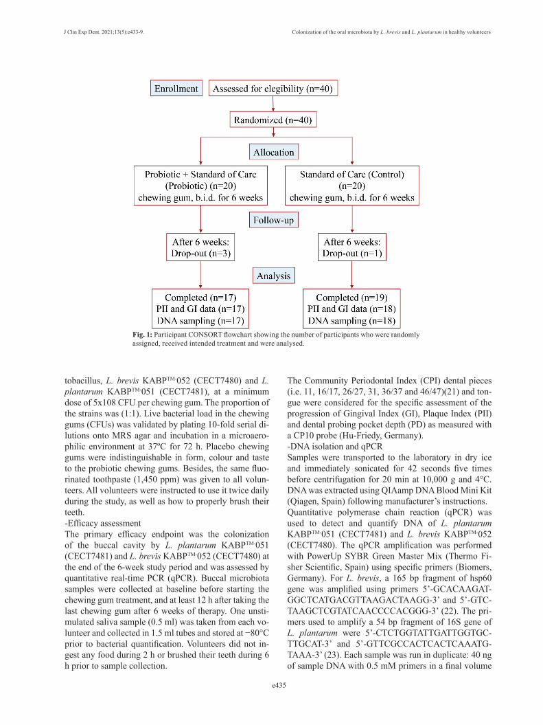
Material and MethodsForty healthy volunteers within age range 18-55 years old were recruited for the double-blind randomized pla-cebo-controlled clinical trial. The study was carried out in Clínica Universitària d’Odontologia (Universitat In-ternacional de Catalunya, Barcelona, Spain) during the period October 2016 to February 2017, in accordance with the Declaration of Helsinki, adhered to the CON-SORT 2010 statement (www.consort-statement.org). The protocol was approved by the Ethics Committee of Clínica Universitària d’Odontologia (Universitat Inter-nacional de Catalunya, Barcelona, Spain) and the study was registered as NCT03540498 on ClinicalTrials.gov on May 07, 2018.-Study PopulationHealthy non-smoker subjects of both sexes, within the 18-55 years old age range and with at least 20 natural teeth were selected. Exclusion criteria included: preg-nant or lactating women; subjects suffering from chronic illness (e.g. diabetes, renal problems, cancer); allergies to the ingredients of the products; antibiotic or probiotic treatment in previous 8 and 4 weeks, respectively; gin-gival index (GI) ≥ 1.5 (19), dental plaque index (PII) ≥ 2.0 (20), dental pocket depth ≥ 5 mm or more than two untreated caries; subjects under orthodontic treatment; use of bactericidal mouthwashes (e.g. chlorhexidine) in previous 4 weeks.-Study DesignThe study was designed as a randomized, double-blind, placebo-controlled, 6-week intervention clinical trial. Volunteers were randomly allocated through a compu-ter-generated random list and received coded product boxes containing blisters of either probiotic or placebo chewing gums accordingly. Identical chewing gums in identical blisters containing the allocated treatment (pro-biotic or placebo) were produced and coded by AB-Bio-tics SA (Barcelona, Spain), and the code was delivered to periodontics specialists after the last volunteer left the study. Therefore, both volunteers and periodontics specialists were blinded to the actual treatment given to each volunteer. A single examiner collected the fo-llowing parameters at baseline and at the end of the 6-week intervention: saliva sample to assess probiotic colonization, gingival index (GI), plaque index (PII) and dental probing pocket depth (PD). A CONSORT flow chart explaining the design of the study is presented in Figure 1. -TreatmentsVolunteers had to take two chewing gums a day, in sepa-rate moments of the day, for a total of 6 weeks. Chewing gums had to be taken at least 1 hour after the previous meal and 1 hour before the following meal and had to be chewed for at least 15-20 minutes each. Probiotic chewing gums contained two different strains of lac-
J Clin Exp Dent. 2021;13(5):e433-9. Colonization of the oral microbiota by L. brevis and L. plantarum in healthy volunteers
e435
Fig. 1: Participant CONSORT flowchart showing the number of participants who were randomly assigned, received intended treatment and were analysed.
tobacillus, L. brevis KABPTM-052 (CECT7480) and L. plantarum KABPTM-051 (CECT7481), at a minimum dose of 5x10⁸ CFU per chewing gum. The proportion of the strains was (1:1). Live bacterial load in the chewing gums (CFUs) was validated by plating 10-fold serial di-lutions onto MRS agar and incubation in a microaero-philic environment at 37ºC for 72 h. Placebo chewing gums were indistinguishable in form, colour and taste to the probiotic chewing gums. Besides, the same fluo-rinated toothpaste (1,450 ppm) was given to all volun-teers. All volunteers were instructed to use it twice daily during the study, as well as how to properly brush their teeth. -Efficacy assessmentThe primary efficacy endpoint was the colonization of the buccal cavity by L. plantarum KABPTM-051 (CECT7481) and L. brevis KABPTM-052 (CECT7480) at the end of the 6-week study period and was assessed by quantitative real-time PCR (qPCR). Buccal microbiota samples were collected at baseline before starting the chewing gum treatment, and at least 12 h after taking the last chewing gum after 6 weeks of therapy. One unsti-mulated saliva sample (0.5 ml) was taken from each vo-lunteer and collected in 1.5 ml tubes and stored at −80°C prior to bacterial quantification. Volunteers did not in-gest any food during 2 h or brushed their teeth during 6 h prior to sample collection.
The Community Periodontal Index (CPI) dental pieces (i.e. 11, 16/17, 26/27, 31, 36/37 and 46/47)(21) and ton-gue were considered for the specific assessment of the progression of Gingival Index (GI), Plaque Index (PII) and dental probing pocket depth (PD) as measured with a CP10 probe (Hu-Friedy, Germany).-DNA isolation and qPCRSamples were transported to the laboratory in dry ice and immediately sonicated for 42 seconds five times before centrifugation for 20 min at 10,000 g and 4°C. DNA was extracted using QIAamp DNA Blood Mini Kit (Qiagen, Spain) following manufacturer’s instructions. Quantitative polymerase chain reaction (qPCR) was used to detect and quantify DNA of L. plantarum KABPTM-051 (CECT7481) and L. brevis KABPTM-052 (CECT7480). The qPCR amplification was performed with PowerUp SYBR Green Master Mix (Thermo Fi-sher Scientific, Spain) using specific primers (Biomers, Germany). For L. brevis, a 165 bp fragment of hsp60 gene was amplified using primers 5’-GCACAAGAT-GGCTCATGACGTTAAGACTAAGG-3’ and 5’-GTC-TAAGCTCGTATCAACCCCACGGG-3’ (22). The pri-mers used to amplify a 54 bp fragment of 16S gene of L. plantarum were 5’-CTCTGGTATTGATTGGTGC-TTGCAT-3’ and 5’-GTTCGCCACTCACTCAAATG-TAAA-3’ (23). Each sample was run in duplicate: 40 ng of sample DNA with 0.5 mM primers in a final volume
J Clin Exp Dent. 2021;13(5):e433-9. Colonization of the oral microbiota by L. brevis and L. plantarum in healthy volunteers
e436
of 10 μL. The qPCR conditions were as follows: 95 °C for 7 min, (95 °C for 15 sec, 64 °C for 1 min, 72 °C for 1 min) x 40 cycles. Quantification cycle (Cq) values, the PCR cycle number at which fluorescence rises above the baseline, were determined using the 7500 software v2.0.5 (Applied Biosystems). The correlation between Cq values and CFU/μg DNA was based on standard curves constructed with 10-fold serial dilutions of each bacterial DNA, from 108 CFU/μg to 102 CFU/μg DNA. All assays were developed with a linear quantitative de-tection established by the slope of 3.39 and 3.54 cycles/log decade, r2 of 0.999 and 1.000, and an efficiency of 97.22 % and 91.51 % for L. brevis KABPTM-051 and L. plantarum KABPTM-052, respectively. Measures to avoid carryover DNA were established. -Compliance, product satisfaction and safetyEmpty blisters returned by volunteers were counted to con-firm treatment compliance. Product satisfaction was rated using Likert scales ranging 1 to 9 for taste, aroma, aftertas-te, and global evaluation. Adverse events were monitored following the directives of the Spanish Pharmacovigilance System for standard clinical trials with drugs.-Statistical analysisPrior data on the in vivo colonization capacity of the stra-ins under study was not available at the time of protocol design to undertake a sample size calculation. Therefore, we designed this study with an arbitrary sample size of 40 patients (20 per group). Statistical analysis of probiotic colonization was per-formed on the population that completed the study. For baseline data, between-group comparisons were perfor-med with Student T-test for quantitative variables and Fisher’s exact test for dichotomous variables. Bacterial concentration data (copies of DNA) corresponding to baseline and end of intervention was normalized using a log transform. Change in bacterial concentration be-
tween groups was assessed using a repeated measures general linear model. The same approach was used to assess change in plaque index (PII), gingival index (GI) and pocked depth (PD). No clustering of samples was performed in repeated measures analysis because a single value was analysed per subject and timepoint. Product satisfaction ratings were analysed by means of Mann-Whitney test. Finally, correlation between log-transformed DNA copies and change in PII, GI and PD were tested using Spearman´s rank test, and changes within group in the proportion of subjects with dental plaque were assessed with the exact version of McNe-mar’s test (Lidell’s test). All statistical analyses were performed with SPSS (v.20.0, IBM Corporation), and significance threshold was set at two-sided p = 0.05.
Results-Study PopulationA total of 40 volunteers (20 per group; age range 18-55 years) were enrolled in the study, and 4 of them (10%, 3 in probiotic group and 1 in placebo group, p = 0.605) dropped out from the study and did not attend the fo-llow-up visit. All volunteers had mean gingival index (GI) in the 0 to 1.1 range (i.e. healthy to mild gingivitis), brushed their teeth twice daily on average, and none of them had dental pockets more than 3 mm deep. Ove-rall, there were no statistically significant differences between groups in none of the measured parameters at baseline, although the proportion of women to men was noticeably higher in probiotic group and GI was so-mewhat higher in placebo group (Table 1). Of note, both L. brevis and L. plantarum were detected in the study subjects at baseline, with abundance of L. brevis (log of CFUs per μg of DNA) being lower than that of L. plan-tarum. This observation indicates these bacteria can be found in the oral cavity of healthy subjects.
PROBIOTIC (n=20) PLACEBO (n=20) P-value
Age (median, range) 30.0 (18-55) 29.5 (18-55) 0.595 A
Gender (women, %) 14 (70%) 8 (40%) 0.111B
Tooth brushing/day (mean, SE) 2.25 (0.14) 2.15 (0.13) 0.609 A
Plaque Index (mean, SE) 0.18 (0.03) 0.19 (0.06) 0.803 A
Gingival Index (mean, SE) 0.18 (0.03) 0.29 (0.08) 0.218 A
Pocket Depth, mm (mean, SE) 1.78 (0.12) 1.75 (0.14) 0.895 A
Number of teeth with depth ≥ 4mm 0 (0%) 0 (0%) 1.000 B
log CFUs L. brevis/μg DNA (mean, SE) 4.00 (0.24) 3.96 (0.25) 0.874 A
Subjects positive for L. brevis (n, %) 19 (95%) 19 (95%) 1.000 B
log CFUs L. plantarum/μg DNA (mean, SE) 4.68 (0.13) 4.53 (0.13) 0.401 A
Subjects positive for L. plantarum (n, %) 20 (100%) 20 (100%) 1.000 B
Table 1: Baseline data of enrolled subjects.
A) Student T-test; B) Fisher exact test
J Clin Exp Dent. 2021;13(5):e433-9. Colonization of the oral microbiota by L. brevis and L. plantarum in healthy volunteers
e437
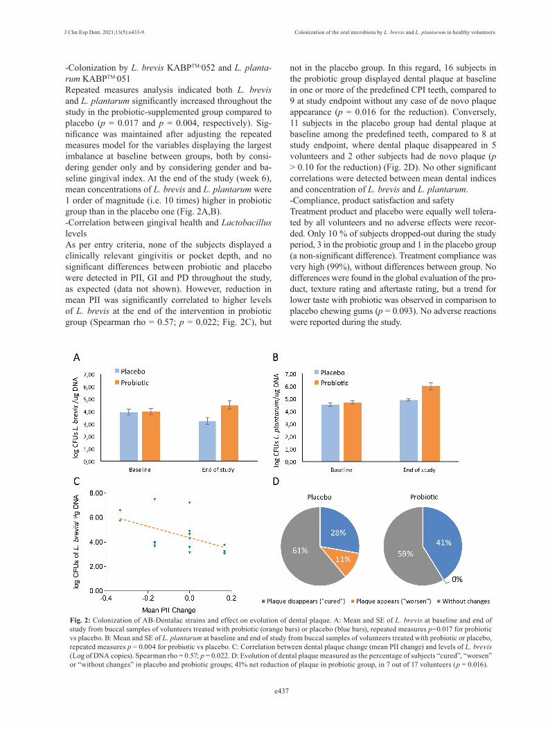
-Colonization by L. brevis KABPTM-052 and L. planta-rum KABPTM-051Repeated measures analysis indicated both L. brevis and L. plantarum significantly increased throughout the study in the probiotic-supplemented group compared to placebo (p = 0.017 and p = 0.004, respectively). Sig-nificance was maintained after adjusting the repeated measures model for the variables displaying the largest imbalance at baseline between groups, both by consi-dering gender only and by considering gender and ba-seline gingival index. At the end of the study (week 6), mean concentrations of L. brevis and L. plantarum were 1 order of magnitude (i.e. 10 times) higher in probiotic group than in the placebo one (Fig. 2A,B). -Correlation between gingival health and Lactobacillus levelsAs per entry criteria, none of the subjects displayed a clinically relevant gingivitis or pocket depth, and no significant differences between probiotic and placebo were detected in PII, GI and PD throughout the study, as expected (data not shown). However, reduction in mean PII was significantly correlated to higher levels of L. brevis at the end of the intervention in probiotic group (Spearman rho = 0.57; p = 0.022; Fig. 2C), but
Fig. 2: Colonization of AB-Dentalac strains and effect on evolution of dental plaque. A: Mean and SE of L. brevis at baseline and end of study from buccal samples of volunteers treated with probiotic (orange bars) or placebo (blue bars), repeated measures p=0.017 for probiotic vs placebo. B: Mean and SE of L. plantarum at baseline and end of study from buccal samples of volunteers treated with probiotic or placebo, repeated measures p = 0.004 for probiotic vs placebo. C: Correlation between dental plaque change (mean PII change) and levels of L. brevis (Log of DNA copies). Spearman rho = 0.57; p = 0.022. D: Evolution of dental plaque measured as the percentage of subjects “cured”, “worsen” or “without changes” in placebo and probiotic groups; 41% net reduction of plaque in probiotic group, in 7 out of 17 volunteers (p = 0.016).
not in the placebo group. In this regard, 16 subjects in the probiotic group displayed dental plaque at baseline in one or more of the predefined CPI teeth, compared to 9 at study endpoint without any case of de novo plaque appearance (p = 0.016 for the reduction). Conversely, 11 subjects in the placebo group had dental plaque at baseline among the predefined teeth, compared to 8 at study endpoint, where dental plaque disappeared in 5 volunteers and 2 other subjects had de novo plaque (p > 0.10 for the reduction) (Fig. 2D). No other significant correlations were detected between mean dental indices and concentration of L. brevis and L. plantarum.-Compliance, product satisfaction and safetyTreatment product and placebo were equally well tolera-ted by all volunteers and no adverse effects were recor-ded. Only 10 % of subjects dropped-out during the study period, 3 in the probiotic group and 1 in the placebo group (a non-significant difference). Treatment compliance was very high (99%), without differences between group. No differences were found in the global evaluation of the pro-duct, texture rating and aftertaste rating, but a trend for lower taste with probiotic was observed in comparison to placebo chewing gums (p = 0.093). No adverse reactions were reported during the study.
J Clin Exp Dent. 2021;13(5):e433-9. Colonization of the oral microbiota by L. brevis and L. plantarum in healthy volunteers
e438
DiscussionPrevious studies have suggested a beneficial role of low acidogenic probiotic strains in colonizing buccal cavity to displace oral pathogens (3,7), although evidence re-mains inconclusive regarding the choice of best strains. In this regard, Montero and colleagues (5) demonstrated that 6-week consumption of a probiotic formula contai-ning Levilactobacillus brevis KABPTM-052 (CECT7480) and Lactiplantibacillus plantarum KABPTM-051 (CECT7481), together with Pediococcus acidilactici KABPTM-053 (CECT8633), was able to reduce the counts of the periodontopathogen Tannerella forsythia in subjects with gingivitis. Similarly, Ferres-Amat and colleagues also demonstrated a reduction in postopera-tive pain against placebo when administering L. brevis KABPTM-052 and L. plantarum KABPTM-051 to subjects undergoing mandibular third molar extraction. Other studies have shown that the treatment with different probiotic strains can help reduce specific pathogens like Streptococcus mutans (24), Prevotella intermedia (25) and Porphyromonas gingivalis (26), as well as the re-duction of caries (27).Despite the above-mentioned effects of some probiotics in the oral microbiome, it stills to be proved whether these probiotic strains build-up in the oral microbiota. The results of this randomized, double-blinded, place-bo-controlled clinical trial show that 6-week supplemen-tation with probiotic strains L. brevis KABPTM-052 and L. plantarum KABPTM-051 significantly increased the concentration of L. brevis and L. plantarum by 1 order of magnitude (1 log) compared to placebo. Of note, both species were found in the oral microbiota of healthy vo-lunteers, as demonstrated by the baseline levels of both lactobacilli in our study. Tooth brushing habits at base-line were identical between groups, all volunteers used the same fluorinated toothpaste during the study and the use of mouthwashes was controlled, further supporting the hypothesis that the differences in the abundance of both L. brevis and L. plantarum were due to the spe-cific probiotic intervention and not to random external factors. Moreover, the significance of the effect against placebo was maintained when adjusting by gender and baseline gingival index, which displayed some imbalan-ce at baseline between groups. This study also confirmed that this probiotic was well tolerated by the volunteers and that no adverse effects were reported in the treat-ment group or placebo group.This study aimed to assess probiotic colonization in sub-jects with healthy gingiva or mild gingivitis, as a first step towards demonstrating their usefulness as a preven-tive therapy. Accordingly, gingival index, plaque index and probing pocket depth were mild at baseline, and no significant differences between groups were noted in the evolution of said indexes during the intervention, as expected. However, an exploratory analysis found oral
cavity colonization by this probiotic was positively co-rrelated with a reduction in mean plaque index. Moreo-ver, the number of subjects displaying dental plaque was significantly reduced by 41 % in probiotic group compa-red to baseline, but no statistically significant difference was observed in placebo group. Previous studies found that L. brevis had a higher affinity for hydroxyapatite teeth surface than L. plantarum (28). These results could explain the observed correlation between a reduction in mean plaque index and higher concentration of L. brevis but not of L. plantarum at study endpoint, as the volun-teers who displayed the largest reduction in mean plaque index were the ones with higher colonization of L. brevis KABPTM-052 (CECT7480) at study endpoint. One limitation of this study is that microbiota was in-vestigated from saliva samples by qPCR analysis. This sampling methodology did not allow for a confirmation of the tissue-specific adhesion of each strain as repor-ted in vitro (18). In addition, qPCR quantification didn’t let us to discriminate between live and dead bacteria. However, since that the last chewing gum was consu-med by volunteers between 12-36 h before the sampling procedure, it is unlikely free DNA could cause the sig-nificant increment of L. plantarum and L. brevis DNA in probiotic due to the continuous wash-out effect of saliva. Moreover, the association between reduced plaque in-dex and increased levels of L. brevis leads us to hypo-thesize that active L. brevis was effectively colonizing oral surfaces of volunteers, in line with previous in vitro findings of L. brevis high adherence to teeth (28). Ano-ther limitation is that qPCR primers were species-speci-fic and amplification of other strains of species L. brevis and L. plantarum cannot be ruled out. In fact, significant detection of both L. brevis and L. plantarum at baseline indicates strains of these species were common in the oral microbiota of the study volunteers. Nevertheless, because of the randomized, placebo-controlled design, lack of differences at baseline between groups, and stan-dardization of oral hygiene habits during the study, the significant increment of the abundance of both L. bre-vis and L. plantarum species in the probiotic-treated group can be attributed to the specific supplementation with strains L. plantarum KABPTM-051 and L. brevis KABPTM-052.In summary, L. plantarum and L. brevis were detected in the saliva of health volunteers in this pilot study, and a 6-week administration of probiotic chewing gums led to a significant increase in both L. plantarum and L. brevis compared to placebo, the difference averaging 1 order of magnitude. As expected, inclusion of healthy subjects prevented the observation of significant effect on plaque and gingival indexes compared to placebo. However, an exploratory analysis identified a reduction in number of subjects with dental plaque in the probiotic group but not the placebo one. In our view, this finding warrants
J Clin Exp Dent. 2021;13(5):e433-9. Colonization of the oral microbiota by L. brevis and L. plantarum in healthy volunteers
e439
additional studies with a larger sample size to confirm the effect of these specific probiotic strains on dental plaque build-up. Such effect could be of interest as an adjunctive preventive treatment for gingivitis, by means of reducing plaque formation. Moreover, to gain insight in the strain’s mechanism of action, analysis by meta-genomic techniques of the complete oral microbiota at various buccal sites pre- and post-intervention would be needed. References1. Peres MA, Macpherson LMD, Weyant RJ, Daly B, Venturelli R, Mathur MR, et al. Oral diseases: a global public health challenge. Lan-cet. 2019;394:249-260.2. Mira A, Simon-Soro A, Curtis MA. Role of microbial communities in the pathogenesis of periodontal diseases and caries. J Clin Periodon-tol. 2017;44:S23-S38.3. Seminario-Amez M, López-López J, Estrugo-Devesa A, Ayu-so-Montero R, Jané-Salas E. Probiotics and oral health: A systematic review. Med Oral Patol Oral Cir Bucal. 2017;22:e282-e288.4. Saha S, Tomaro-Duchesneau C, Tabrizian M, Prakash S. Probiotics as oral health biotherapeutics. Expert Opin Biol Ther. 2012;12:1207-20.5. Montero E, Iniesta M, Rodrigo M, Marín MJ, Figuero E, Herrera D, et al. Clinical and microbiological effects of the adjunctive use of pro-biotics in the treatment of gingivitis: A randomized controlled clinical trial. J Clin Periodontol. 2017;44:708-716.6. Schlagenhauf U, Rehder J, Gelbrich G, Jockel-Schneider Y. Con-sumption of Lactobacillus reuteri - containing lozenges improves pe-riodontal health in navy sailors at sea: A randomized controlled trial. J Periodontol. 2020;91:1328-1338.7. Bustamante M, Oomah BD, Mosi-Roa Y, Rubilar M, Burgos-Díaz C. Probiotics as an Adjunct Therapy for the Treatment of Halitosis, Dental Caries and Periodontitis. Probiotics Antimicrob Proteins. 2020;12:325-334.8. Invernici MM, Salvador SL, Silva PHF, Soares MSM, Casarin R, Palioto DB, et al. Effects of Bifidobacterium probiotic on the treatment of chronic periodontitis: A randomized clinical trial. J Clin Periodon-tol. 2018;45:1198-1210.9. Guarner F, Sanders ME, Eliakim R, Fedorak R, Gangl A, Garisch J, et al. Probiotics and prebiotics. WGO Glob Guidel. 2017.10. Frei R, Akdis M, O’mahony L. Prebiotics, probiotics, synbiotics, and the immune system: Experimental data and clinical evidence. Curr Opin Gastroenterol. 2015;31:153-8.11. Hao Q, Dong BR, Wu T. Probiotics for preventing acute upper respiratory tract infections. Cochrane Database Syst Rev. 2015;2:CD006895.12. Oak SJ, Jha R. The effects of probiotics in lactose intolerance: A systematic review. Crit Rev Food Sci Nutr. 2019;59:1675-1683.13. Guarner F, Perdigon G, Corthier G, Salminen S, Koletzko B, Mo-relli L. Should yoghurt cultures be considered probiotic? Br J Nutr. 2005;93:783.14. Reid G. The development of probiotics for women’s health. Can J Microbiol. 2017;63:269-277.15. Guo Z, Liu XM, Zhang QX, Shen Z, Tian FW, Zhang H, et al. Influence of consumption of probiotics on the plasma lipid profile: a meta-analysis of randomised controlled trials. Nutr Metab Cardiovasc Dis. 2011;21:844-50.16. Fuentes MC, Lajo T, Carrión JM, Cuñé J. Cholesterol-lowering efficacy of Lactobacillus plantarum CECT 7527, 7528 and 7529 in hypercholesterolaemic adults. Br J Nutr. 2013;109:1866-72.17. Zheng J, Wittouck S, Salvetti E, Franz CMAP, Harris HMB, Matta-relli P, et al. A taxonomic note on the genus Lactobacillus: Description of 23 novel genera, emended description of the genus Lactobacillus beijerinck 1901, and union of Lactobacillaceae and Leuconostocaceae. Int J Syst Evol Microbiol. 2020;70:2782-2858. 18. Bosch M, Nart J, Audivert S, Bonachera MA, Alemany AS, Fuen-
tes MC, et al. Isolation and characterization of probiotic strains for improving oral health. Arch Oral Biol. 2012;57:539-49.19. Löe H, Silness J. Periodontal Disease in Pregnancy I. Prevalence and Severity. Acta Odontol Scand. 1963;21:533-51.20. Silness J, Löe H. Periodontal Disease in Pregnancy II. Correla-tion Between Oral Hygiene and Periodontal Condition. Acta Odontol Scand. 1964;22:121-35.21. World Health Organisation. Oral Health Surveys: Basic Methods. WGO; Geneva. 1997. p. 79.22. Herbel SR, Lauzat B, von Nickisch-Rosenegk M, Kuhn M, Muru-gaiyan J, Wieler LH, et al. Species-specific quantification of probiotic lactobacilli in yoghurt by quantitative real-time PCR. J Appl Micro-biol. 2013;115:1402-10.23. Matsuda K, Tsuji H, Asahara T, Matsumoto K, Takada T, Nomoto K. Establishment of an analytical system for the human fecal micro-biota, based on reverse transcription-quantitative PCR targeting of multicopy rRNA molecules. Appl Environ Microbiol. 2009;75:1961-9.24. Burton JP, Drummond BK, Chilcott CN, Tagg JR, Thomson WM, Hale JDF, et al. Influence of the probiotic Streptococcus salivarius strain M18 on indices of dental health in children: A randomized dou-ble-blind, placebo-controlled trial. J Med Microbiol. 2013;62:875-84.25. Kõll P, Mändar R, Marcotte H, Leibur E, Mikelsaar M, Hammars-tröm L. Characterization of oral lactobacilli as potential probiotics for oral health. Oral Microbiol Immunol. 2008;23:139-47.26. Iniesta M, Herrera D, Montero E, Zurbriggen M, Matos AR, Marín MJ, et al. Probiotic effects of orally administered Lactobacillus reu-teri-containing tablets on the subgingival and salivary microbiota in patients with gingivitis. A randomized clinical trial. J Clin Periodontol. 2012;39:736-44.27. Di Pierro F, Zanvit A, Nobili P, Risso P, Fornaini C. Cariogram outcome after 90 days of oral treatment with Streptococcus salivarius M18 in children at high risk for dental caries: Results of a randomized, controlled study. Clin Cosmet Investig Dent. 2015;7:107-13.28. Samot J, Lebreton J, Badet C. Adherence capacities of oral lactoba-cilli for potential probiotic purposes. Anaerobe. 2011;17:69-72.
EthicsThe protocol (code: AB-GUM-2016) was approved by the Ethics Committee of Clínica Universitària d’Odontologia (Universitat Inter-nacional de Catalunya, Barcelona, Spain). The study was conducted in accordance with the Declaration of Helsinki and adhered to the CON-SORT 2010 statement (www.consort-statement.org). Written informed consent was provided before the commencement of the investigation by all participants.
FundingThis study was funded by AB-Biotics SA (Barcelona, Spain), who owns the patent of the two bacterial strains included in this study.
Authors contributionDr Nart and J. Espadaler designed the protocol of the clinical trial. Dr Nart, S. Jiménez-Garrido and A. Ramírez-Sebastià collected the clinical data and buccal samples. E Astó performed the qPCR analysis of the microbial samples. J. Espadaler and D. Buj performed the sta-tistical analysis. Dr. Nart, J. Espadaler, D. Buj and P. Huedo wrote the manuscript. All the authors contributed to the design and interpretation of the study, read and approved the final version to be published.
Conflict of interestEA, DB, PH and JE are full-time employees of AB-Biotics SA. The other authors have no conflict of interest.


![Missa Brevis - 2L · Missa Brevis Wolfgang Plagge [opus 107] for male quartet durata 8:00 composed 2002 version 15.05.2003,!7JA6G1-aeagdh! M-66104-063-7 Contents Missa Brevis Wolfgang](https://static.documents.pub/doc/80x56/604fe9cec8166507435d28d0/missa-brevis-2l-missa-brevis-wolfgang-plagge-opus-107-for-male-quartet-durata.jpg)


