73
Orbit Dr Vibhavari Barhate Dept Of Ophthalmology
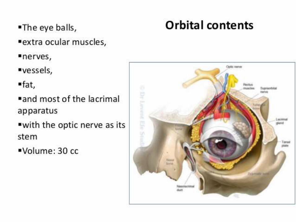
Orbit
Dr Vibhavari Barhate
Dept Of Ophthalmology


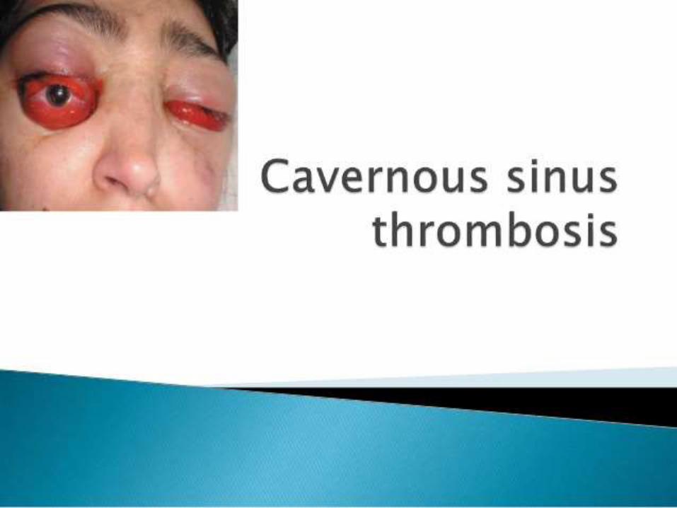

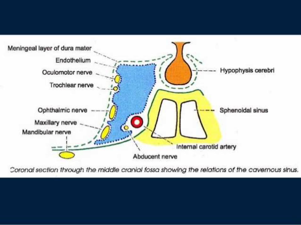
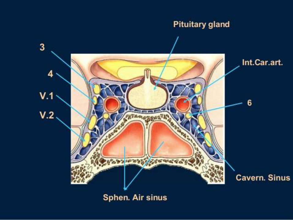
Cavernous sinus thrombosis
• Tributaries of cavernous sinus
• Sup and inf ophthalmic veins
• Labyrinthine vein from middle ear inf petrosal sinus
• Pterygoid plexuse through Middle meningeal veins


Clinical features
• Unilateral initially becomes bilateral >50% cases
• Severe pain along ophthalmic nerve
• Lid oedema,chemosis,congestion,
• Proptosis
• 3rd,4th,6th nerve palsy
• L R palsy earliest sign
• Ophthalmoplegia
contd
• Oedema of mastoid region
• Decreased vision-papilloedema
• other eye becomes infected
• Rigors, vomitting ,sever cerebral symptoms
• Death –meningitis,pulmonary infarction


Signs and symptoms
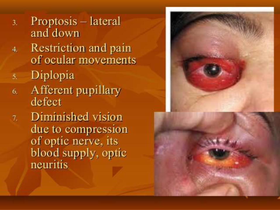
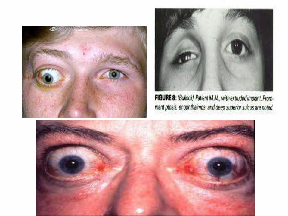
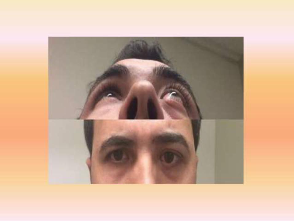
• Proptosis –abnormal protrusion of globe
• Enophthalmos –abnormal retraction of globe
as in Micro-ophthalmos ,pthisis bulbi,blow- out fracture,
• Exophthalmos –proptosis secondary to thyroid eye disease.
(Measured by exophthalmometer)

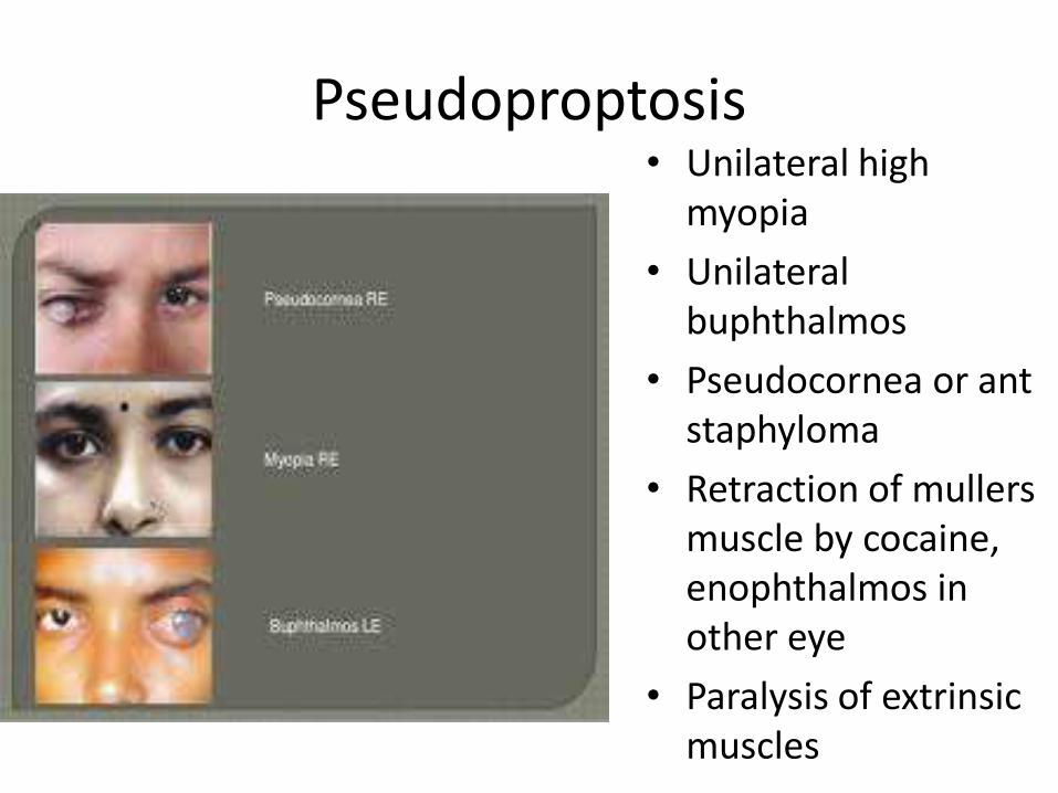
Pseudoproptosis • Unilateral high
myopia
• Unilateral buphthalmos
• Pseudocornea or ant staphyloma
• Retraction of mullers muscle by cocaine, enophthalmos in other eye
• Paralysis of extrinsic muscles

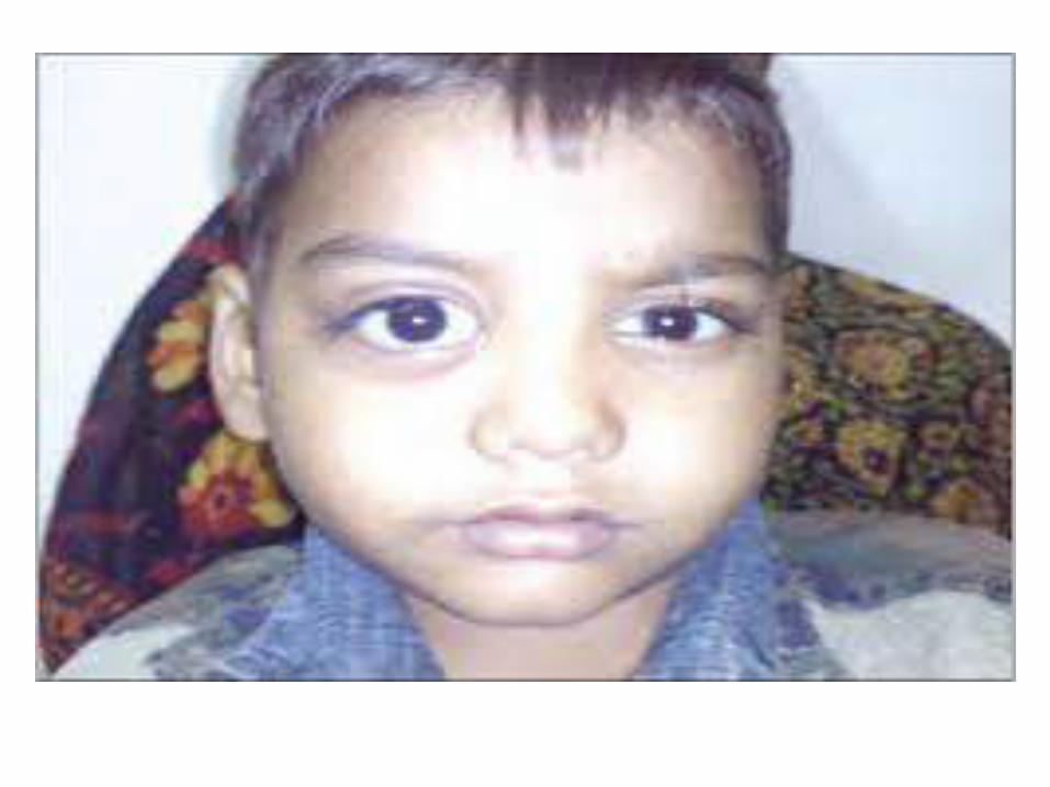
Unilateral proptosis
• Congenital – dermoid , teratoma
• Inflammatory-Orbital cellulitis, Idiopathic orbital inflammatory disease , gumma, sarcoidosis,TB
Thrombosis of orbital vein with or without cavernous sinus thrombosis
• Traumatic-Orbital haemorrhage or emphysema,IO
• Tumours of orbit or its content
• Cystic-parasitic cyst
Arteriovenous aneursym
Bilateral proptosis
• Endocrine exophthalmos –thyroid eye diseases
• Cavernous sinus thrombosis
• Symmetrical orbital tumours (lymphoma ,pseudoleukaemia)
• Developmental -Diminished orbital volume –oxycephaly or tower skull

Axial vs eccentric proptosis
• Axial-eye is pushed centrally forwards,lesion is situated in central space.
• Eccentric-situated elsewhere in orbit, pushes eye in opposite direction.

Pulsatile proptosis• Transmitted vascular pulsation-
Aneurysm of ophthalmic art
Carotid -cavernous fistula
• Transmitted CSF PULSATION-
Absence of grater wing of sphenoid neurofibromatosis
Erosion of orbital roof
Meningocele, meningoencephalocele
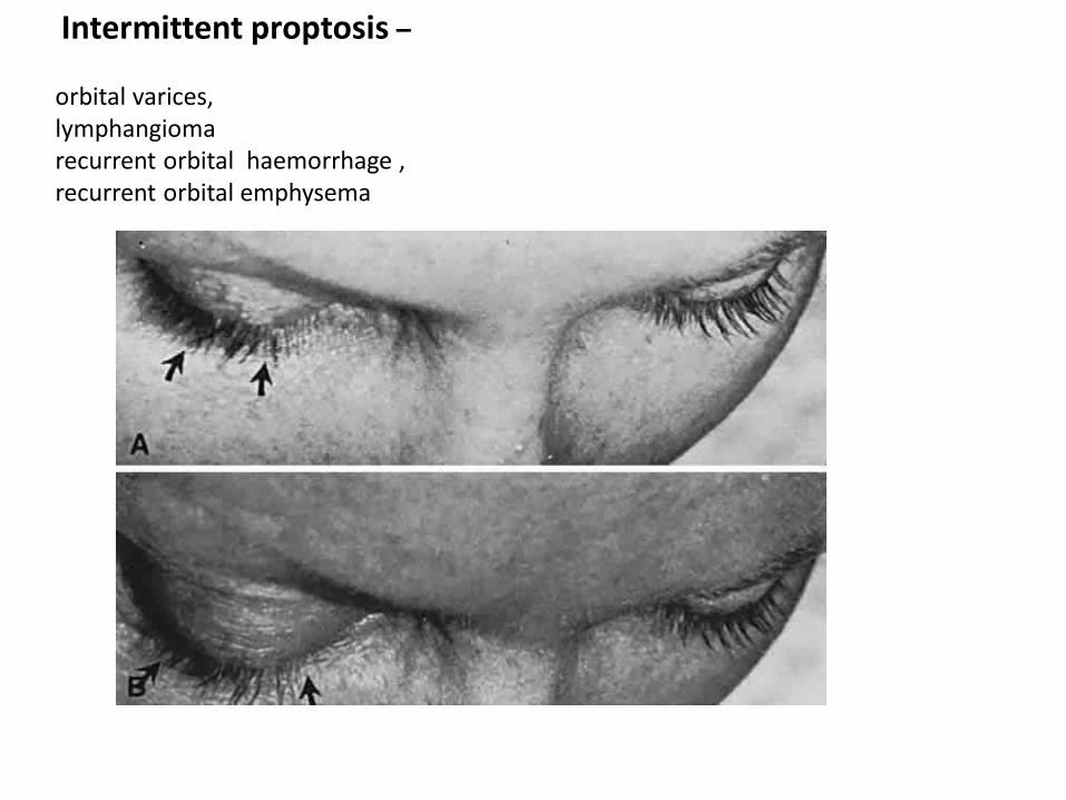
Intermittent proptosis –
orbital varices,lymphangioma recurrent orbital haemorrhage ,recurrent orbital emphysema


Proptosis – Aetiology, Clinical Evaluation, Investigations & Principles of Management :-Endocrinal Exophthalmos. :- Orbital Haemorrhag


contd
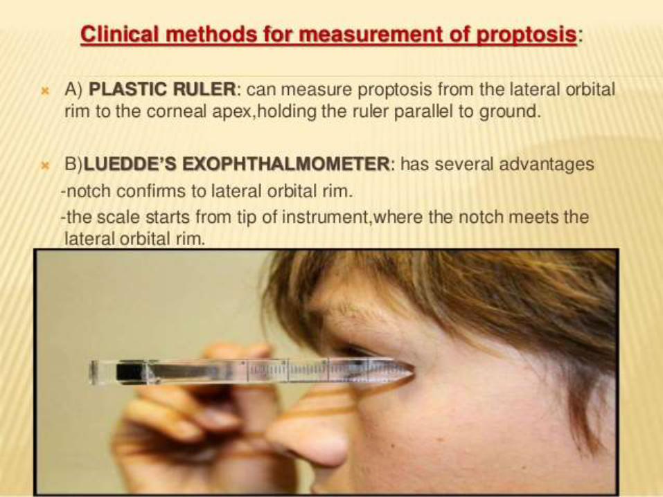
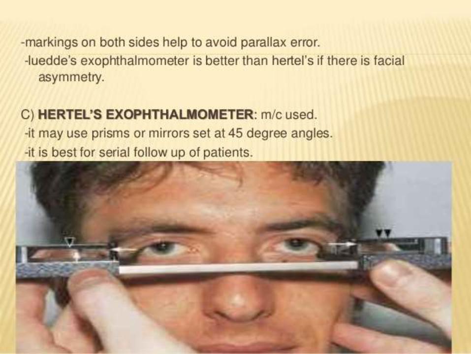
• EXOPHTHALMOMETER –Lueddes ,Hertels
(>21mm or diff of 2mm between two eyes
• inspection of PNS , cranial nerves and systemic examination
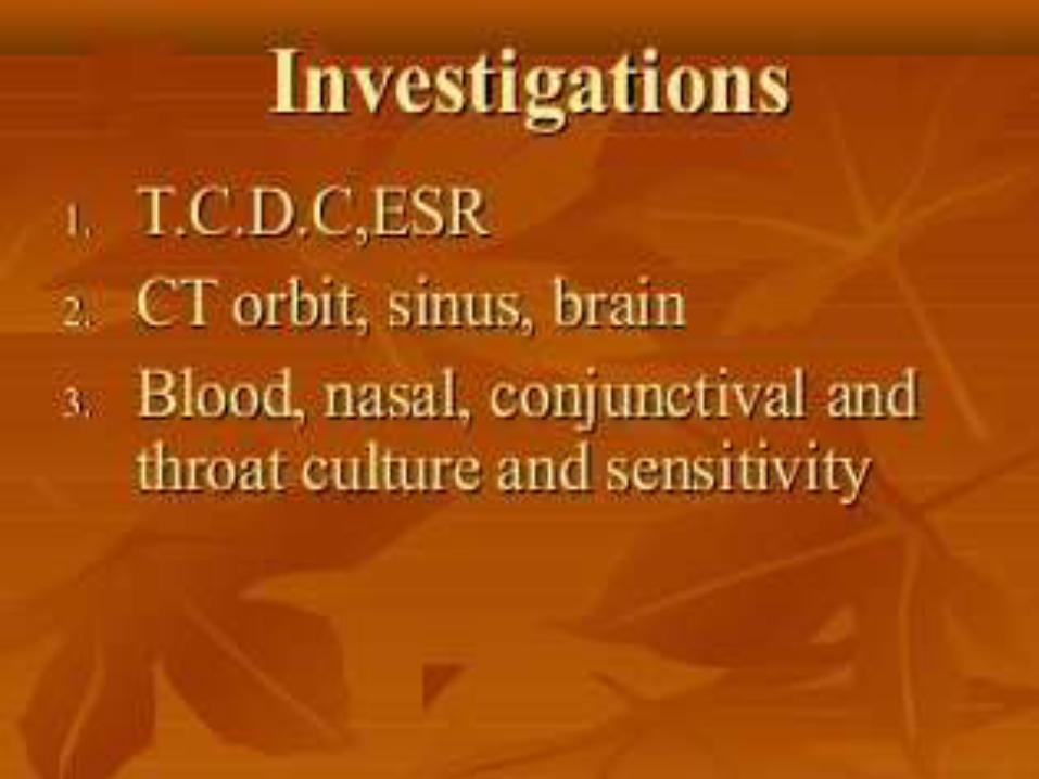
INVESTIGATIONS
• Lab-T3,T4,TSH level, serum ACE level for sarcoidosis
• x ray –P A view for calcification ,F B ,hyperosteosis in meningioma
• Caldwell view –angled PA view for frontal sinus
• Lat view for intracanial lesions
• Waters view for orbital floor fracture

Investigations Contd
• Soft tissue –USG, C T SCAN, MRI
• Orbital vasculature-carotid angiography, digital subtraction angiography and orbital venography
• FNAC,Incisional biopsy ,Excisional biopsy
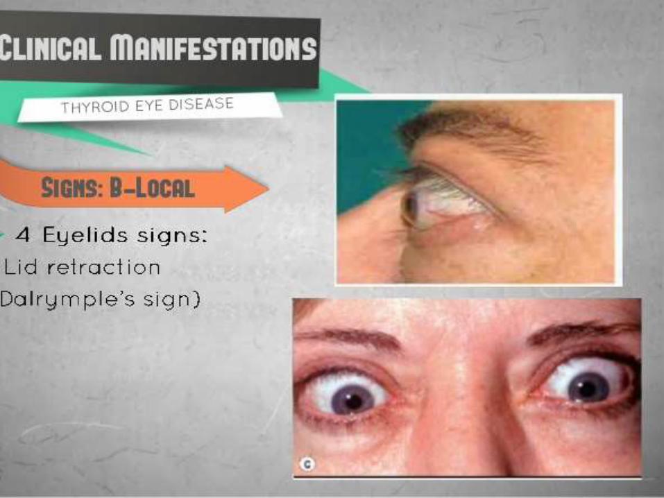
Thyroid ophthalmopathy or dysthyroid eye disease
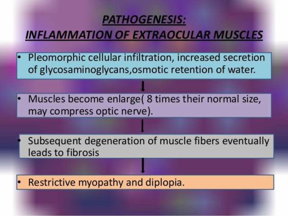
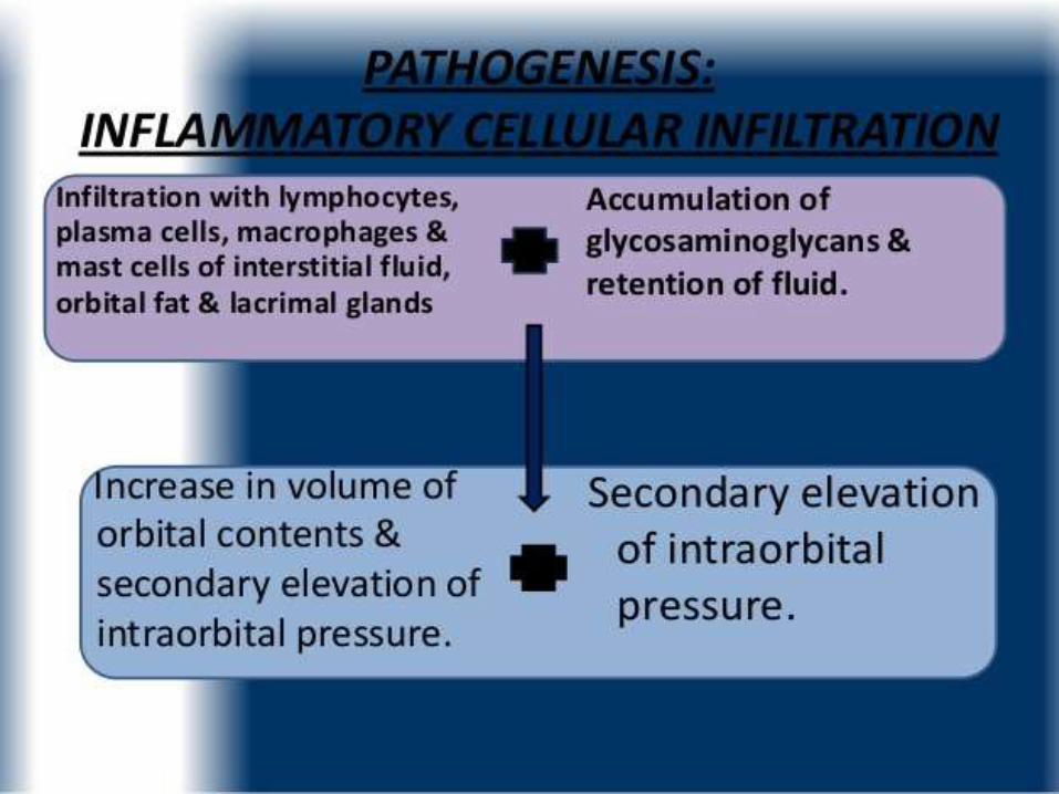
Pathogenesis• Autoimmune reaction directed against orbital
fibroblast and extraocular muscles.
• Anti -TSH receptor antibodies (thyroid stimulating immunoglobulin which mimic TSH) level decides severity of ophthalmopathy, not the level of T3 or T4
• Ophthalmic Graves disease-ophthalmopathy asso with thyrotoxicosis .
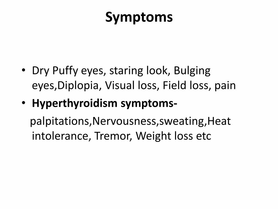
Symptoms
• Dry Puffy eyes, staring look, Bulging eyes,Diplopia, Visual loss, Field loss, pain
• Hyperthyroidism symptoms-
palpitations,Nervousness,sweating,Heat intolerance, Tremor, Weight loss etc
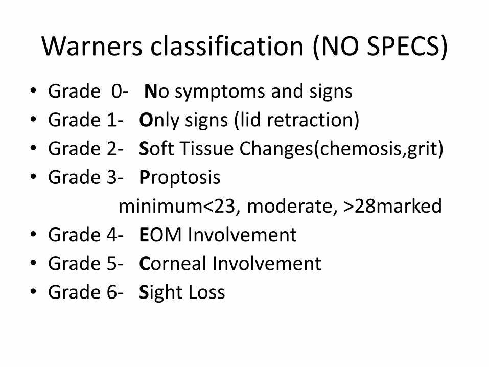
Warners classification (NO SPECS)
• Grade 0- No symptoms and signs
• Grade 1- Only signs (lid retraction)
• Grade 2- Soft Tissue Changes(chemosis,grit)
• Grade 3- Proptosis
minimum<23, moderate, >28marked
• Grade 4- EOM Involvement
• Grade 5- Corneal Involvement
• Grade 6- Sight Loss


Investigations
• Thyroid function test - (pt euthyroid ,hyperthyroid or hypothyroid state )
• B scan
• CT scan
• Forced duction test
treatment• Topical decongestants,lubricants • Medical –thyroxine and oral radioactive iodine therapy
.• Steroids – 40 -60 mg prednisolone orally• Radiotherapy-1000rads from each lateral port • Surgical-tarsorrhaphy ,Orbital Decompression –through
floor by caldwell- Luc approch• Two wall or three wall decompression
Muscle surgery , Canthoplasty, correction of lid retraction etc.


