Orbital lymphoma: diagnostic approach and treatment outcome Eckardt et al. WORLD JOURNAL OF SURGICAL ONCOLOGY Eckardt et al. World Journal of Surgical Oncology 2013, 11:73 http://www.wjso.com/content/11/1/73
Transcript
Orbital lymphoma: diagnostic approach andtreatment outcomeEckardt et al.
WORLD JOURNAL OF SURGICAL ONCOLOGY
Eckardt et al. World Journal of Surgical Oncology 2013, 11:73http://www.wjso.com/content/11/1/73
WORLD JOURNAL OF SURGICAL ONCOLOGY
Eckardt et al. World Journal of Surgical Oncology 2013, 11:73http://www.wjso.com/content/11/1/73
RESEARCH Open Access
Orbital lymphoma: diagnostic approach andtreatment outcomeAndré M Eckardt*, Juliana Lemound, Majeed Rana and Nils-Claudius Gellrich
Abstract
Background: Lymphomas of the orbit and orbital adnexae are rare tumors, comprising only 1% of all non-Hodgkin’slymphoma. The majority of non-Hodgkin’s lymphomas of the orbit are extranodal marginal-zone B-cell lymphomas ofmucosa-associated lymphoid tissue type. Because of nonspecific clinical signs and symptoms, some diagnostic delaymay occur. The purpose of the study was to evaluate the diagnostic approach in orbital lymphomas and to analyzetheir treatment outcome.
Methods: In the period from 2005 to 2012, from a group of 135 patients with tumors of the orbit, we identified 11patients diagnosed with orbital lymphoma. This patient cohort was reviewed retrospectively.
Results: The patient group consisted of 11 patients (seven females, male males) with a median age of 57.7 years(range 42 to 88 years). Orbital swelling, pain and motility impairment were the leading clinical symptoms. Diagnosis wasconfirmed by surgical biopsy. Depending on the anatomic location of the tumor, a surgical biopsy was taken using ablepharoplasty incision, a lateral orbitotomy or a navigation-guided biopsy. The predominant histology was extranodalnon-Hodgkin’s lymphoma of mucosa-associated lymphoid tissue type (82%). All patients underwent complete clinicalstaging. These were clinical stage IEA in seven patients, and stages IIEA (n = 2) and IIIEA (n = 2) in four patients . Patients instage IEA were treated with radiation therapy alone, with radiation doses between 25 and 40 Gy, and patients with stageIIEA received systemic chemotherapy with bendamustin/rituximab. Those two patients diagnosed with diffuse largeB-cell lymphoma and mantle cell lymphoma received systemic chemotherapy according to the R-CHOP protocol.
Conclusions: Owing to unspecific clinical symptoms, some diagnostic delay may occur in orbital lymphoma. Ifunspecific orbital symptoms are present, adequate imaging studies followed by early surgical biopsy will contribute toearly diagnosis. Once diagnosis is established and staging is complete, radiation therapy is the recommended treatmentfor stage IEA patients. Systemic chemotherapy is indicated in selected stage IIEA patients and in patients with stage IIIEAdisease.
Keywords: Orbital lymphoma, Mucosa-associated lymphoid tissue lymphoma, Radiotherapy
BackgroundOrbital lymphomas are rare, comprising only 1% of allnon-Hodgkin’s lymphoma [1]. However, lymphomas arethe most common primary orbital tumor in adults 60 yearsof age and older [2]. Margo and Mulla reported in theirstudy of more than 300 orbital malignancies a 55% rate oflymphomas involving the orbit [3]. The majority of non-Hodgkin’s lymphomas of the orbit and orbital adnexa areextranodal marginal-zone B-cell lymphomas of mucosa-associated lymphoid tissue (MALT)-type lymphomas [4].
* Correspondence: [email protected] of Cranio-Maxillofacial Surgery, Hannover Medical School,Carl-Neuberg-Strasse 1, Hannover 30625, Germany
Although initially described in the stomach and associ-ated with Helicobacter pylori infection, MALT lymph-omas have subsequently been observed to arise in otherepithelial structures, including the thyroid, parotid gland,lung and breast, as well as in the orbit [5-7].Our study aimed at exploring the diagnostic and thera-
peutic approach as well as the clinical course of lymphomasinvolving the orbit.
MethodsA retrospective review of orbital tumors (n = 135) treatedat the Department of Cranio-Maxillofacial Surgery fromJanuary 2005 to April 2012 retrieved 11 cases of non-
Ltd. This is an Open Access article distributed under the terms of the Creativeommons.org/licenses/by/2.0), which permits unrestricted use, distribution, andiginal work is properly cited.
Table 1 Patient and treatment characteristics of orbitallymphomas (n = 11)
Characteristic Number of patients %
Age (years)
Median 57.7
Range 42.5 to 88.7
Gender
Male 4 36
Female 7 64
Clinical symptoms
Periorbital swelling 6 55
Proptosis 5 46
Pain 4 36
Epiphora 2 18
Motility impairment 1 9
Ann Arbor stage
IA 7 64
IIA 2 18
IIIA 2 18
Orbital imaging study
Computed tomography 10 91
Magnetic resonance imaging 8 73
Staging procedure
Chest X-ray 5 46
Chest computed tomography 8 73
Abdominal computed tomography 7 64
Bone marrow biopsy 7 64
Surgical treatment
Surgical biopsy 10 91
Complete surgical removal 1 9
Surgical approach
Transconjunctival 3 27
Blepharoplasty incision 7 64
Coronal/lateral orbitotomy 1 9
Treatment
Radiation 7 64
Chemotherapy 4 36
Eckardt et al. World Journal of Surgical Oncology 2013, 11:73 Page 2 of 6http://www.wjso.com/content/11/1/73
Hodgkin’s lymphomas that presented clinically in theorbital region. All 11 patients had been referred to ourinstitution from the Department of Ophthalmology forfurther diagnosis of an indolent swelling in the or-bital/periorbital region.The initial clinical staging included a computed tom-
ography or magnetic resonance imaging scan of the or-bital/periorbital region to localize the tumor site andextension. Based on imaging findings, a surgical biopsyunder general anesthesia was planned; in selected pa-tients, depending on the anatomic location, the biopsywas taken using a computer-assisted navigation platform(iPlan CMF 3.0; Brainlab, Feldkirchen, Germany). Histo-logic diagnosis was initially performed on H & E-stainedparaffin sections and additional immunohistochemicalstaining performed for further immunologic phenotyping.Once diagnosis was confirmed patients were stagedaccording to the Ann Arbor classification. Followingcomplete clinical staging based on the clinical practice ofthe treating physician, patients were sent to the radiationoncologist or the oncologist for further treatment plan-ning. Post-treatment follow-up visits were organized bythe treating physicians. For the purpose of this analysis,the treating physicians were contacted to obtain the latestfollow-up information of all patients.
ResultsClinical and pathologic featuresEleven patients were included in this analysis. Of the 11patients, seven were women (64%) and four were men(36%), with a median age of 57.7 years (range, 42.5 to88.7 years). The most common presenting signs andsymptoms were periorbital tumor mass in six patients(55%), exophthalmos in five patients (45%), ocular painin four patients (36%), and eye motility and visual re-strictions in one patient (9%). One patient (9%) wasfound to have bilateral disease. The tumor was locatedin the supraorbital region in six patients (55%), theretrobulbar orbital tissue in two patients (18%), and theinfraorbital region in three patients (27%). Table 1 dis-plays patient characteristics, including age, gender, site,diagnostic imaging, and surgical treatment.Orbital computed tomography and/or magnetic reson-
ance imaging scans were used in all patients to evaluatethe extent of the disease and to aid in radiation treat-ment planning.Initial treatment consisted of a surgical biopsy under
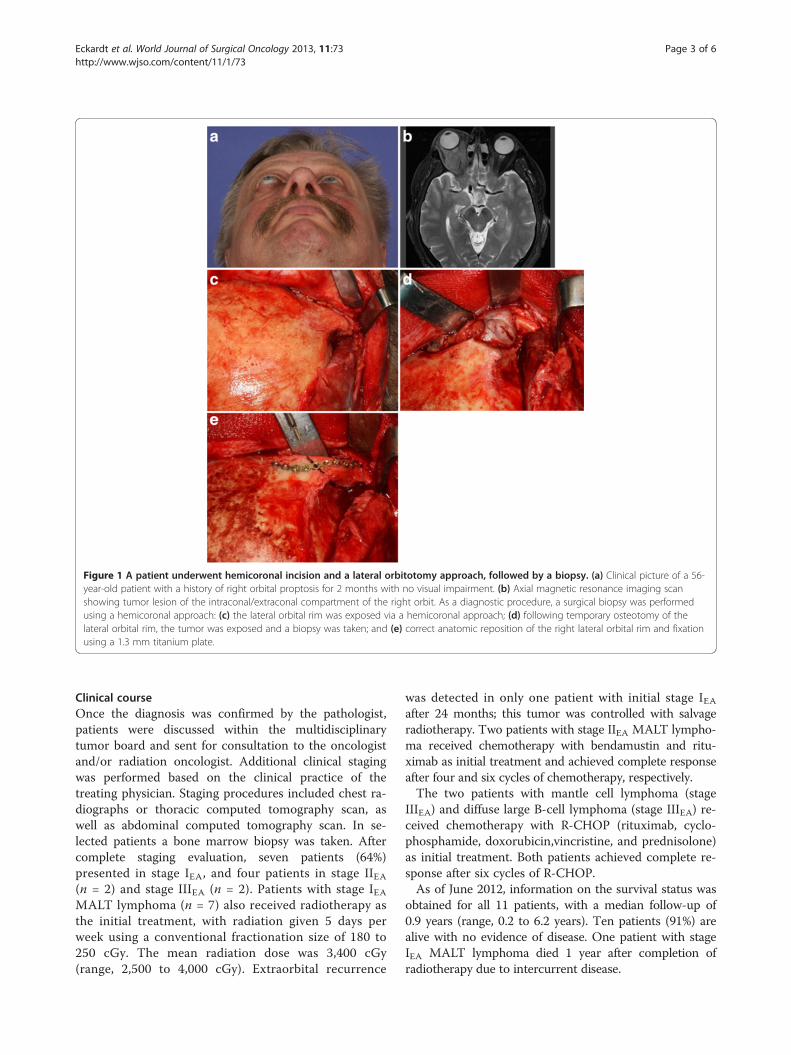
general anesthesia in all patients using various surgicalapproaches. A blepharoplasty incision was used in sevenpatients (64%), combined with navigation-assisted biopsyfor retrobulbar space location in three patients (27%). Atransconjunctival approach was used in three patients(27%). In one patient a biopsy was taken followinghemicoronal incision and a lateral orbitotomy approach
(Figure 1a to e). One patient with involvement of thelacrimal gland received complete surgical removal as theprimary treatment (Figure 2a,b).Histopathological examination together with immuno-
histochemical studies were performed on paraffin sectionsfrom biopsy specimens of all 11 patients. ExtranodalMALT lymphoma was diagnosed in nine patients (82%),and the other two patients were diagnosed with diffuselarge B-cell lymphoma and with recurrence of mantle celllymphoma, respectively.
Figure 1 A patient underwent hemicoronal incision and a lateral orbitotomy approach, followed by a biopsy. (a) Clinical picture of a 56-year-old patient with a history of right orbital proptosis for 2 months with no visual impairment. (b) Axial magnetic resonance imaging scanshowing tumor lesion of the intraconal/extraconal compartment of the right orbit. As a diagnostic procedure, a surgical biopsy was performedusing a hemicoronal approach: (c) the lateral orbital rim was exposed via a hemicoronal approach; (d) following temporary osteotomy of thelateral orbital rim, the tumor was exposed and a biopsy was taken; and (e) correct anatomic reposition of the right lateral orbital rim and fixationusing a 1.3 mm titanium plate.
Eckardt et al. World Journal of Surgical Oncology 2013, 11:73 Page 3 of 6http://www.wjso.com/content/11/1/73
Clinical courseOnce the diagnosis was confirmed by the pathologist,patients were discussed within the multidisciplinarytumor board and sent for consultation to the oncologistand/or radiation oncologist. Additional clinical stagingwas performed based on the clinical practice of thetreating physician. Staging procedures included chest ra-diographs or thoracic computed tomography scan, aswell as abdominal computed tomography scan. In se-lected patients a bone marrow biopsy was taken. Aftercomplete staging evaluation, seven patients (64%)presented in stage IEA, and four patients in stage IIEA(n = 2) and stage IIIEA (n = 2). Patients with stage IEAMALT lymphoma (n = 7) also received radiotherapy asthe initial treatment, with radiation given 5 days perweek using a conventional fractionation size of 180 to250 cGy. The mean radiation dose was 3,400 cGy(range, 2,500 to 4,000 cGy). Extraorbital recurrence
was detected in only one patient with initial stage IEAafter 24 months; this tumor was controlled with salvageradiotherapy. Two patients with stage IIEA MALT lympho-ma received chemotherapy with bendamustin and ritu-ximab as initial treatment and achieved complete responseafter four and six cycles of chemotherapy, respectively.The two patients with mantle cell lymphoma (stage
IIIEA) and diffuse large B-cell lymphoma (stage IIIEA) re-ceived chemotherapy with R-CHOP (rituximab, cyclo-phosphamide, doxorubicin,vincristine, and prednisolone)as initial treatment. Both patients achieved complete re-sponse after six cycles of R-CHOP.As of June 2012, information on the survival status was
obtained for all 11 patients, with a median follow-up of0.9 years (range, 0.2 to 6.2 years). Ten patients (91%) arealive with no evidence of disease. One patient with stageIEA MALT lymphoma died 1 year after completion ofradiotherapy due to intercurrent disease.
Figure 2 A patient with involvement of the lacrimal glandreceived complete surgical removal as primary treatment. (a)Coronal computed tomography scan showing a tumor lesion in theupper right orbit of a 77-year-old woman. (b) The tumor wasexposed using a blepharoplasty approach and completely removedtogether with the lacrimal gland.
Eckardt et al. World Journal of Surgical Oncology 2013, 11:73 Page 4 of 6http://www.wjso.com/content/11/1/73
DiscussionNo specific guidelines currently exist for the manage-ment of MALT lymphomas, except for localized primarygastric MALT lymphoma. Numerous reports confirmthe efficacy of conventional treatment strategies such assurgery, radiotherapy or chemotherapy, alone or in
combination, with no significant survival difference[6,8,9]. Surgery as the only treatment modality shouldnot be administered, because there is obviously a highlikelihood of local relapse after surgery according to pre-vious reports [10]. The difficulty of complete resectionwith preservation of function in the orbit may explainthe high relapse rate.Radiation therapy as the initial treatment has been
reported to be very effective in MALT lymphoma of theorbit [11-28]. Radiotherapy with a dose range of 25 to35 Gy seems to be a standard approach because it pro-vides local control and cure for localized orbital lymph-oma [11,13,28]. Our data support these findings thatexcellent local control as well as survival can be achievedin stage IEA MALT lymphoma of the orbit by radiother-apy alone with a mean dose of 34 Gy. However, evenafter lower radiation dose recommendations in recentpublications, there is still some controversy regardingthe optimal radiation dose for this tumor [17-19]. Onlyone extraorbital relapse (one out of nine MALT lymph-omas, 11%) with skin infiltration in the upper neck de-veloped 2 years after primary radiotherapy with 36 Gy.Radiation doses above 35 Gy resulted in significant latecomplications such as cataract formation or keratitis[28]. It should be noted that lens shielding has to beadded after a dose of 20 Gy [19]. Interestingly severalstudies have reported a relatively high local or distantfailure rate after successful initial radiotherapy. Jenkinsand colleagues reported that 47% of their study populationof 192 patients with orbital MALT lymphoma developedextraorbital recurrence after 5 years [29]. Hasegawa andcolleagues reported a recurrence rate of 25% out of 20 pa-tients who were treated with radiotherapy alone for orbitalMALT lymphoma and developed a relapse in thenonirradiated orbit or at distant sites after a medianfollow-up of 71 months [14]. In view of some conflictingreports it is difficult to draw final conclusions, but allthose sometimes contradictory results point to some vari-ance in the biologic behavior of MALT lymphomas.There is some evidence that combination chemother-
apy is effective in orbital lymphoma [30]. Chemotherapyhas never been systematically evaluated in orbital MALTlymphoma because of excellent local control rates instage IE disease that showed excellent response after pri-mary radiotherapy. Most often, chemotherapy was ad-ministered after either surgery or radiotherapy or wasreserved for patients with advanced disease stages IIIEAand IVEA. With some ongoing controversial discussionas far as the role of chemotherapy in MALT lymphomais concerned, it may be concluded that the developmentof novel systemic strategies particularly for stages IIIEAand IVEA is needed [8,31]. The clinical efficacy of rituxi-mab, a chimeric mAb directed against the B-cell-specificantigen CD20, was first demonstrated in follicular
Eckardt et al. World Journal of Surgical Oncology 2013, 11:73 Page 5 of 6http://www.wjso.com/content/11/1/73
lymphomas, but the use of the antibody has been ex-tended over the last few years to other subtypes of non-Hodgkin lymphomas with promising results, as a singleagent or in combination with chemotherapy [32]. SinceCD20 antigen is expressed on the surface of neoplasticcells in virtually all MALT lymphomas, one can assumethat rituximab is active in this neoplastic disease.Conconi and colleagues could demonstrate clinical activ-ity in extranodal MALT lymphomas in a phase II studyin previously untreated and treated adult patients with aresponse rate of 73% and a median response duration of10.5 months [32]. A recent pilot study assessed the toler-ability and activity of intralesional injection rituximab ina small group of five patients with orbital B-cell lymph-oma, and demonstrated complete remission in two pa-tients and stable disease in two patients [33]. Furtherclinical trials will show some potential synergistic actionbetween rituximab and chemotherapy and will contrib-ute to better define the role of chemotherapy inextranodal MALT lymphomas.
ConclusionMALT lymphomas constitute the majority of orbital andperiorbital non-Hodgkin’s lymphomas. Clinical signs andsymptoms are unspecific, but a slowly growing painlessorbital or periorbital swelling was masking a lymphomain the majority of our patients. From this perspective,the cranio-maxillofacial surgeon plays an integral part inthe multidisciplinary management of patients with or-bital lymphomas. Early surgical biopsy together with ad-equate imaging studies is essential for early andadequate diagnosis of orbital lymphoma. As demon-strated in our patients, radiotherapy is an effective treat-ment option in patients with stage IEA disease resultingin excellent local tumor control and survival. The role ofsystemic chemotherapy in MALT lymphoma is still notwell defined.
ConsentWritten informed consent was obtained from the patientfor publication of this report and any accompanyingimages.
AbbreviationsH & E: Hematoxylin and eosin; mAb: Monoclonal antibody; MALT: Mucosa-associated lymphoid tissue; R-CHOP: Rituximab cyclophosphamide,doxorubicin,vincristine, and prednisolone.
Competing interestsThe authors declare that they have no competing interests.
Authors’ contributionsAME made substantial contributions to conception and design of themanuscript as well as data acquisition. JL and MR have been involved indrafting the manuscript. N-CG was involved in revising the manuscript. Allauthors read and approved the final manuscript.
AcknowledgementsThe article processing charges are funded by the DeutscheForschungsgemeinschaft, ‘Open Access Publizieren’.
Received: 9 August 2012 Accepted: 23 February 2013Published: 18 March 2013
References1. Ahmed S, Shahid RK, Sison CP, Fuchs A, Mehrotra B: Orbital lymphomas: a
clinicopathologic study of a rare disease. Am J Med Sci 2006, 331:79–83.2. Demirci H, Shields CL, Shields JA, Honavar SG, Mercado GJ, Tovilla JC:
Orbital tumors in the older adult population. Ophthalmology 2002,109:243–248.
3. Margo CE, Mulla ZD: Malignant tumors of the orbit, analysis of the Floridacancer registry. Ophthalmology 1998, 105:185–190.
4. Isaacson PG, Wright DH: Malignant lymphoma of mucosal associatedlymphoid tissue: a distinctive type of B cell lymphoma. Cancer 1983,52:1410–1416.
5. White WL, Ferry JA, Harris NL, Grove AS Jr: Ocular adnexal lymphoma. Aclinicopathologic study with identification of lymphomas of mucosaassociated lymphoid tissue type. Ophthalmology 1995, 102:1994–2006.
6. Zinzani PL, Magagnoli M, Galieni P, Martelli M, Poletti V, Zaja F, Molica S,Zaccaria A, Cantonetti AM, Gentilini P, Guardigni L, Gherlinzoni F, RibersaniM, Bendandi M, Albertini P, Tura S: Nongastrointestinal low-grade mucosa-associated lymphoid tissue lymphoma: analysis of 75 patients. J ClinOncol 1999, 17:1254–1258.
7. Ishii Y, Tomita N, Takasi H, Ogusa E, Hattori Y, Matsuura S, Matsumoto C,Takemura S, Kuwabara H, Ishigatsubo Y: Clinical features of extranodalmarginal zone lymphoma of mucosa-associated lymphoid tissue.Hematol Oncol 2012, 30:186–189.
8. Zucca E, Conconi A, Pedrinis E, Cortelazzo S, Motta T, Gospodarowicz MK,Patterson BJ, Ferreri AJ, Ponzoni M, Devizzi L, Giardini R, Pinotti G, Capella C,Zinzani PL, Pileri S, López-Guillermo A, Campo E, Ambrosetti A, Baldini L,Cavalli F, International Extranodal Lymphoma Study Group: Nongastricmarginal zone B-cell lymphoma of mucosa-associated lymphoid tissue.Blood 2003, 101:2489–2495.
9. Thieblemont C, Berger F, Dumontet C, Moullet I, Bouafia F, Felman P, SallesG, Coiffier B: Mucosa-associated lymphoid tissue lymphoma is adisseminated disease in one third of 159 patients analyzed. Blood 2000,95:802–806.
10. Esik O, Ikeda H, Mukai K, Kaneko A: A retrospective analysis of differentmodalities for treatment of primary orbital non-Hodgkin's lymphomas.Radiother Oncol 1996, 38:13.
11. Smitt MC, Donaldson SS: Radiotherapy is successful treatment for orbitallymphoma. Int J Radiat Oncol Biol Phys 1993, 26:59–66.
12. Uno T, Isobe K, Shikama N, Nishikawa A, Oguchi M, Ueno N, Itami J, OhnishiH, Mikata A, Ito H: Radiotherapy for extranodal, marginal zone, B-celllymphoma of mucosa-associated lymphoid tissue originating in theocular adnexa. Cancer 2003, 98:865–871.
13. Lee JL, Kim MK, Lee KH, Hyun MS, Chung HS, Kim DS, Shin SO, Cho HS, BaeSH, Ryoo HM: Extranodal marginal zone B-cell lymphomas of mucosa-associated lymphoid tissue-type of the orbit and ocular adnexa. AnnHematol 2005, 84:13–18.
14. Hasegawa M, Kojima M, Shioya M, Tamaki Y, Saitoh J, Sakurai H, Kitamoto Y,Suzuki Y, Niibe H, Nakano T: Treatment results of radiotherapy formalignant lymphoma of the orbit and histopathologic review accordingto the WHO classification. Int J Radiat Oncol Biol Phys 2003, 57:172–176.
17. Goda JS, Gospodarowicz M, Pintilie M, Wells W, Hodgson DC, Sun A, CrumpM, Tsang RW: Long-term outcome in localized extranodal mucosa-associated lymphoid tissue lymphomas treated with radiotherapy.Cancer 2010, 116:3815.
18. Goda JS, Le LW, Lapperriere NJ, Millar BA, Payne D, Gospodarowicz MK, WellsW, Hodgson DC, Sun A, Simpson R, Tsang RW: Localized orbital mucosa-associated lymphoma tissue lymphoma managed with primary radiationtherapy: efficacy and toxicity. Int J Radiat Oncol Biol Phys 2011, 81:e659.
Eckardt et al. World Journal of Surgical Oncology 2013, 11:73 Page 6 of 6http://www.wjso.com/content/11/1/73
19. Son SH, Choi BO, Kim GW, Yang SW, Hong YS, Choi IB, Kim YS: Primaryradiation therapy in patients with localized orbital marginal zone B-celllymphoma of mucosa-associated lymphoid tissue (MALT lymphoma). IntJ Radiat Oncol Biol Phys 2010, 77:86.
20. Bolek TW, Moyses HM, Marcus RB Jr, Gorden L III, Maiese RL, Almasri NM,Mendenhall NP: Radiotherapy in the management of orbital lymphoma.Int J Radiat Oncol Biol Phys 1999, 44:31.
21. Tsang RW, Gospodarowicz MK, Pintilie M, Wells W, Hodgson DC, Sun A,Crump M, Patterson BJ: Localized mucosa-associated lymphoid tissuelymphoma treated with radiation therapy has excellent clinical outcome.J Clin Oncol 2003, 21:4157.
22. Ejima Y, Sasaki R, Okamoto Y, Maruta T, Azumi A, Hayashi Y, Demizu Y, OtaY, Soejima T, Sugimura K: Ocular adnexal mucosa-associated lymphoidtissue lymphoma treated with radiotherapy. Radiother Oncol 2006, 78:6.
23. Suh CO, Shim SJ, Lee SW, Yang WI, Lee SY, Hahn JS: Orbital marginal zoneB-cell lymphoma of MALT: Radiotherapy results and clinical behaviour.Int J Radiat Oncol Biol Phys 2006, 65:228–233.
24. De Cicco L, Cella L, Liuzzi R, Solla R, Farella A, Punzo G, Tranfa F, Strianese D,Conson M, Bonavolontà G, Salvatore M, Pacelli R: Radiation therapy inprimary orbital lymphoma: a single institution retrospective analysis.Radiat Oncol 2009, 4:60.
25. Zhou P, Ng AK, Silver B, Li S, Hua L, Mauch PM: Radiation therapy fororbital lymphoma. Int J Radiat Oncol Biol Phys 2005, 63:866–871.
26. Le QT, Eulau SM, George TI, Hildebrand R, Warnke RA, Donaldson SS, HoppeRT: Primary radiotherapy for localized orbital MALT lymphoma. Int JRadiat Oncol Biol Phys 2002, 52:657–663.
27. Chao CKS, Lin HS, Deveneni VR, Smith M: Radiation therapy for primaryorbital lymphoma. Int J Radiat Oncol Biol Phys 1995, 31:929–934.
29. Jenkins C, Rose GE, Bunce C, Wright JE, Cree IA, Plowmann N, Lightman S,Moseley I, Norton A: Histological features of ocular adnexal lymphoma(REAL classification) and their association with patient morbidity andsurvival. Br J Ophthalmol 2000, 84:907–913.
30. Galieni P, Polito E, Leccisotti A, Marotta G, Lasi S, Bigazzi C, Bucalossi A,Frezza G, Lauria F: Localized orbital lymphoma. Haematologica 1997,82:436–439.
32. Conconi A, Martinelli G, Thiéblemont C, Ferreri AJ, Devizzi L, Peccatori F,Ponzoni M, Pedrinis E, Dell'Oro S, Pruneri G, Filipazzi V, Dietrich PY, GianniAM, Coiffier B, Cavalli F, Zucca E: Clinical activity of rituximab inextranodal marginal zone B-cell lymphoma of MALT type. Blood 2003,102:2741–2745.
33. Savino G, Battendieri R, Balia L, Colucci D, Larocca LM, Laurenti L, De PaduaL, Blasi MA, Balestrazzi E: Evaluation of intraorbital injection of rituximabfor treatment of primary ocular adnexal lymphoma:a pilot study. CancerSci 2011, 102:1565–1567.
doi:10.1186/1477-7819-11-73Cite this article as: Eckardt et al.: Orbital lymphoma: diagnosticapproach and treatment outcome. World Journal of Surgical Oncology2013 11:73.
Submit your next manuscript to BioMed Centraland take full advantage of:
• Convenient online submission
• Thorough peer review
• No space constraints or color figure charges
• Immediate publication on acceptance
• Inclusion in PubMed, CAS, Scopus and Google Scholar
• Research which is freely available for redistribution
Submit your manuscript at www.biomedcentral.com/submit


![PRIMARY NON-HODGKIN LYMPHOMA OF THE ORBIT ......the patients, presentation is with orbital mass [2]. Common signs at presentation are: palpable mass, pain, exophtalmia, ptosis, dyplopia,](https://static.documents.pub/doc/80x56/614396f46b2ee0265c0224b6/primary-non-hodgkin-lymphoma-of-the-orbit-the-patients-presentation-is.jpg)


