Lateral root formation in plants involves the stimulation ofmature pericycle cells to proliferate and redifferentiate tocreate a new organ. The simple organization of the root ofArabidopsis thaliana allows the development of lateral rootprimordia to be characterized histologically. We havedivided the process of lateral root development into 8 stagesdefined by specific anatomical characteristics and celldivisions. To identify the cell types in the developing pri-mordium we have generated a collection of marker linesthat express β-glucuronidase in a tissue- or cell type-
specific manner in the root. Using these tools we have con-structed a model describing the lineage of each cell type inthe lateral root. These studies show that organization andcell differentiation in the lateral root primordia precede theappearance of a lateral root meristem, with differentialgene expression apparent after the first set of divisions ofthe pericycle.
One of the fundamental questions in developmental biology ishow cells proliferate and organize to form discrete organs.Unlike animals, where organogenesis occurs primarily in theembryo, normal growth in plants involves both embryonic andpostembryonic organogenesis. The primary shoot and rootapical meristems, which are responsible for the growth of theprimary shoot and root, form as part of the developing embryo.After germination, shoot buds are formed in the axils of leafprimordia as the shoot grows (Esau, 1965). These axillaryshoot buds appear to be composed of undifferentiated cells thatcan be triggered to become vegetative meristems, which arethen responsible for the lateral proliferation of the shoot system(Esau, 1965; Steeves and Sussex, 1972). In contrast, nosecondary buds are laid down as the root grows. As a result,the root system must proliferate via meristems formed fromnon-meristematic tissues.
Histological studies have shown that, in angiosperms, lateralroots are derived from the pericycle layer deep within theparent root tissues. There is also a contribution from dividingendodermal cells in some monocots (McCully, 1975). Theinitiation of lateral roots occurs some distance away from theroot apical meristem in the differentiation zone of the root,where the pericycle cells are not actively dividing (Esau,1965). The mature pericycle cells, once stimulated, dediffer-entiate and proliferate to form a lateral root primordium (LRP).The LRP grows through the overlying cell layers of the parentroot and eventually breaks through the epidermis and emerges.At some point during LRP development an active lateral rootapical meristem must form. Little information is availableabout the events involved in either the initiation or develop-ment of the LRP. The pericycle cells that become ‘founder’
cells for the LRP are positioned at the xylem poles, but it isdifficult to predict which pericycle cells along the length of theroot will be recruited to the lateral root program (Charlton,1991).
Development of the LRP involves several stages which maybe regulated by distinct pathways. There is evidence that stim-ulated pericycle cells begin to lose their mature characteristics,or dedifferentiate, even before cell division begins (Foard etal., 1965; McCully, 1975). This suggests that changes in cellsize and shape are separable from cell division in LRPformation. Furthermore, several mutants have recently beenisolated in Arabidopsis thaliana that initiate LRP which fail todevelop beyond a certain point, again indicating multiplephases in the development process (Celenza et al., 1995; Chenget al., 1995). In support of this notion, other studies in Ara-bidopsis have shown that, at a certain stage, the LRP becomescompetent to continue development when explanted tohormone-free media (Laskowski et al., 1995). Based on thesefindings, it has been suggested that LRP formation can bethought of as a two stage process: (1) stimulation of dediffer-entiation and proliferation in the pericycle layer to form theLRP; (2) redifferentiation to form a lateral root meristem,which establishes and perpetuates the organization of thelateral root (Celenza et al., 1995; Cheng et al., 1995;Laskowski et al., 1995).
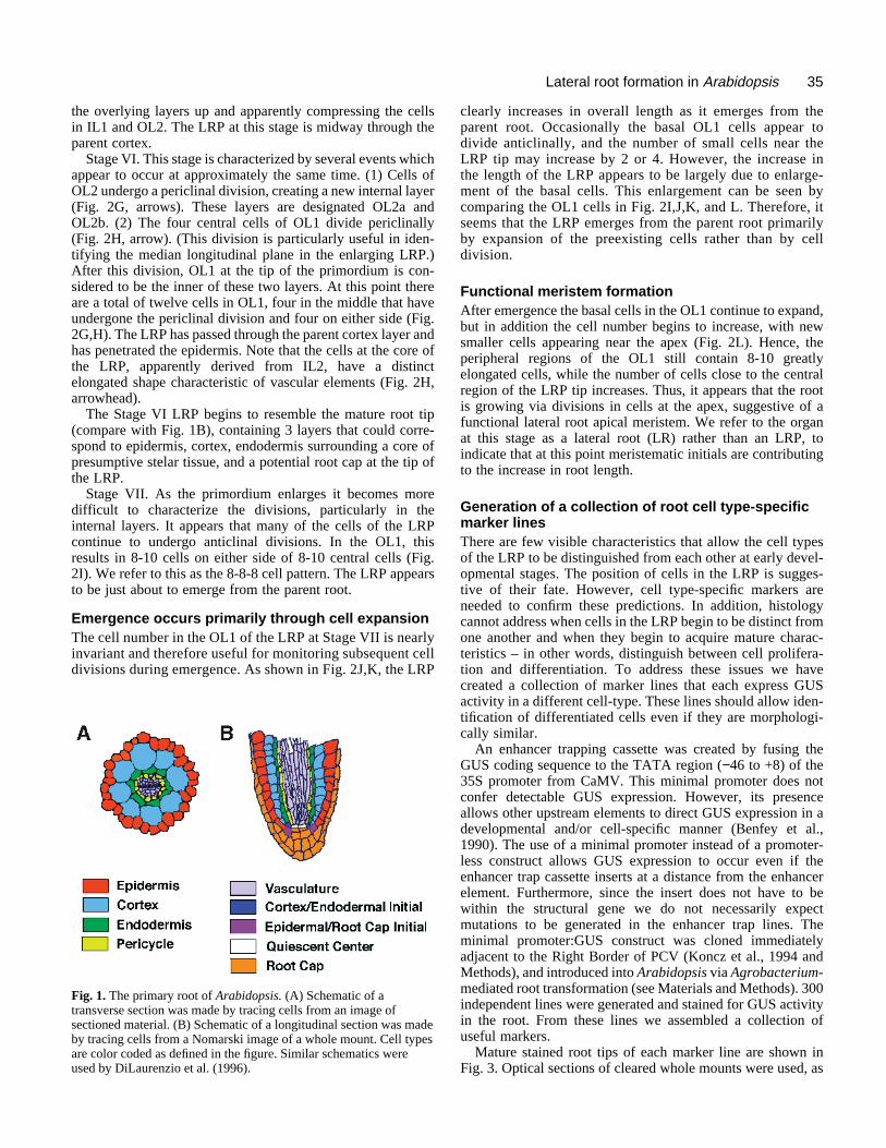
We are attempting to understand these complex develop-mental events in the model dicot Arabidopsis. The root ofArabidopsis provides an excellent system for the study oforgan development. The arrangement of cells in both theprimary and lateral roots is simple and predictable (Dolan etal., 1993). The outer four radial layers, each one cell thick,are (from the outside) the epidermis, cortex, endodermis andpericycle (Fig. 1A). The pericycle forms the outermost layer
34 J. E. Malamy and P. N. Benfey
of the stele, which also contains phloem, xylem, and stelarparenchyma cells (not shown). Each cell type forms verticalfiles of cells that can be traced to meristematic initials in theroot apical meristem (Fig. 1B). The initials are responsiblefor maintaining the cellular organization of the root. Eachtime they divide they regenerate the initial and add one cellto the vertical cell file. There are four sets of meristematicinitials in Arabidopsis roots: one that forms the epidermis andlateral root cap (Fig. 1B); one that forms columella root cap(not shown); one that produces both the cortex and endoder-mal layers (Fig. 1B) and one that produces the cells of thestele (not shown) (Dolan et al., 1993). In the primary root,these initials appear to be established during embryogenesis(Scheres et al., 1994). In contrast, the meristematic initials inthe lateral roots must form postembryonically. In this paperwe use the term root apical meristem to refer to these sets ofmeristematic initials and the actively dividing cells adjacentto them.
As a first step to understanding lateral root patterning anddevelopment, we have taken advantage of the simplicity of theArabidopsis root structure to define anatomically a series ofdiscrete developmental stages in LRP development. We havecomplemented these studies by generating a collection ofmarker lines that express β-glucuronidase (GUS) in a celltype-specific manner in each of the cells of the root. Thisallows the differentiation state of cells to be established basedon molecular characteristics. Furthermore, the marker lineexpression patterns can be used to follow the fates of cells inthe developing LRP. Our results show that the LRP formsthrough a highly ordered series of divisions that generates astructure with a radial organization similar to that of themature root tip. The cells within the LRP become non-identical after the first periclinal division (Stage II) and beginto acquire characteristics of their mature cell fate during theearly stages of LRP development. Based on these findings wepropose a new model of the key stages in lateral rootformation.
MATERIALS AND METHODS
Plant growth conditionsArabidopsis seeds of ecotypes WS, NO-O and Columbia were steril-ized and sown on nutrient agar medium (Murashige and Skoog) platescontaining 4.5% sucrose. Plates were oriented vertically to ease theobservation and removal of intact roots. Growth conditions were 18hours light, 6 hours dark, 22oC.
Histology and histochemistry1- to 3-week old seedlings were stained for GUS activity for up to 3days in the following solution: 1× GUS buffer, 20% methanol, 0.5mg/ml X-Gluc (5-bromo-4-chloro-3-indolyl-β-D-glucuronidase).Addition of methanol greatly improves the specificity and repro-ducibility of staining (Kosugi et al., 1990). Staining solution wasmade fresh from a 10× buffer that was stored in darkness for no morethan one week. 10× GUS buffer: 1 M Tris pH7.5 containing 29 mg/mlNaCl, 6.6 mg/ml K3Fe(CN)6.
For observation of whole mounts, both stained and unstainedroots were transferred to small Petri dishes containing 0.24 N HClin 20% methanol and incubated on a 57°C heat block for 15minutes. This solution was replaced with 7% NaOH, 7% hydroxy-lamine-HCl in 60% ethanol for 15 minutes at room temperature.
(The hydroxylamine can be omitted with no decrease in effectiveclearing). Roots were then rehydrated for 5 minutes each in 40%,20% and 10% ethanol, and infiltrated for 15 minutes in 5% ethanol,25% glycerol. Roots were mounted in 50% glycerol on glass micro-scope slides.
For transverse sections, samples were fixed and embedded in His-toresin as described by DiLaurenzio et al. (1996).
All samples were observed using Nomarski optics on a LeitzLaborlux S microscope. Photographs were taken using a Leitz MPS52camera, and images were scanned into Adobe Photoshop 3.0 to createfigures. In some cases the intensity of the blue color was enhanced.
Enhancer trap linesPlant Cloning Vector (PCV) (Koncz et al., 1994) contains a BamHIsite immediately adjacent to the Right Border sequences. The β-glucuronidase (uidA) coding region fused to the TATA region(−46 to +8) of the Cauliflower Mosaic Virus (CaMV) 35S promoterwere introduced into this site (Benfey et al., 1990). 300 transgeniclines were generated by root transformation of WS and NO-Oecotypes as described by Marton and Browse (1991), and 4 inde-pendent lines from each transformant were screened for GUSactivity in the root.
Cortex and epidermis marker lines were provided by Drs J. Haradaand W. Rerie, respectively. Construction of these lines has beendescribed (Dietrich et al., 1992; Masucci et al., 1996).
RESULTS
Developmental stages of lateral root formationRoots of Arabidopsis seedlings were cleared and observed inwhole mounts in order to define an index of LRP development.The process has been divided into 8 stages (Stage I-VII andEmergence), shown in Fig. 2A-K.
Stage I. The first evidence of LRP initiation is the appear-ance of closely spaced cell walls in the pericycle layer in per-pendicular orientation to the root axis (arrows, Fig. 2A). Anincreased frequency of anticlinal divisions is clearly seen ascompared to the pericycle cells at the opposite side of thestele. In the longitudinal plane, approximately 8-10 ‘short’pericycle cells are formed, which enlarge in a radialdirection.
Stage II. A periclinal division occurs that divides the LRPinto two layers (outer layer (OL) and inner layer (IL)) (Fig.2B). Not all the small pericycle-derived LRP cells appear toparticipate in this division; typically the most peripheral cellsdo not divide. Hence, as the OL and IL cells expand radiallythe domed shape of the LRP begins to appear.
Stage III. The OL divides periclinally, generating a threelayer primordium comprising OL1, OL2 and IL (Fig. 2C).Again, some peripheral cells do not divide, creating outerregions that are one and two cell layers thick. This furtheremphasizes the domed shape of the LRP.
Stage IV. The IL divides periclinally, creating a total of fourcell layers (OL1, OL2, IL1, IL2) (Fig. 2D). At this stage theLRP has penetrated the parent endodermal layer.
Stage V. A central cell in OL1 and OL2 divides anticlinallyto form four small cuboidal cells (Fig. 2E, arrow; cells 5 and6 in Fig. 2F). The cells adjacent to these two cells in the OL1and OL2 also divide (arrows, Fig. 2F), creating an outer layer(OL1) that contains 10-12 cells (Fig. 2F). In addition, cells inIL2 enlarge radially and divide (short arrow, Fig. 2F), pushing
35Lateral root formation in Arabidopsis
the overlying layers up and apparently compressing the cellsin IL1 and OL2. The LRP at this stage is midway through theparent cortex.
Stage VI. This stage is characterized by several events whichappear to occur at approximately the same time. (1) Cells ofOL2 undergo a periclinal division, creating a new internal layer(Fig. 2G, arrows). These layers are designated OL2a andOL2b. (2) The four central cells of OL1 divide periclinally(Fig. 2H, arrow). (This division is particularly useful in iden-tifying the median longitudinal plane in the enlarging LRP.)After this division, OL1 at the tip of the primordium is con-sidered to be the inner of these two layers. At this point thereare a total of twelve cells in OL1, four in the middle that haveundergone the periclinal division and four on either side (Fig.2G,H). The LRP has passed through the parent cortex layer andhas penetrated the epidermis. Note that the cells at the core ofthe LRP, apparently derived from IL2, have a distinctelongated shape characteristic of vascular elements (Fig. 2H,arrowhead).
The Stage VI LRP begins to resemble the mature root tip(compare with Fig. 1B), containing 3 layers that could corre-spond to epidermis, cortex, endodermis surrounding a core ofpresumptive stelar tissue, and a potential root cap at the tip ofthe LRP.
Stage VII. As the primordium enlarges it becomes moredifficult to characterize the divisions, particularly in theinternal layers. It appears that many of the cells of the LRPcontinue to undergo anticlinal divisions. In the OL1, thisresults in 8-10 cells on either side of 8-10 central cells (Fig.2I). We refer to this as the 8-8-8 cell pattern. The LRP appearsto be just about to emerge from the parent root.
Emergence occurs primarily through cell expansionThe cell number in the OL1 of the LRP at Stage VII is nearlyinvariant and therefore useful for monitoring subsequent celldivisions during emergence. As shown in Fig. 2J,K, the LRP
Fig. 1. The primary root of Arabidopsis. (A) Schematic of atransverse section was made by tracing cells from an image ofsectioned material. (B) Schematic of a longitudinal section was madeby tracing cells from a Nomarski image of a whole mount. Cell typesare color coded as defined in the figure. Similar schematics wereused by DiLaurenzio et al. (1996).
clearly increases in overall length as it emerges from theparent root. Occasionally the basal OL1 cells appear todivide anticlinally, and the number of small cells near theLRP tip may increase by 2 or 4. However, the increase inthe length of the LRP appears to be largely due to enlarge-ment of the basal cells. This enlargement can be seen bycomparing the OL1 cells in Fig. 2I,J,K, and L. Therefore, itseems that the LRP emerges from the parent root primarilyby expansion of the preexisting cells rather than by celldivision.
Functional meristem formationAfter emergence the basal cells in the OL1 continue to expand,but in addition the cell number begins to increase, with newsmaller cells appearing near the apex (Fig. 2L). Hence, theperipheral regions of the OL1 still contain 8-10 greatlyelongated cells, while the number of cells close to the centralregion of the LRP tip increases. Thus, it appears that the rootis growing via divisions in cells at the apex, suggestive of afunctional lateral root apical meristem. We refer to the organat this stage as a lateral root (LR) rather than an LRP, toindicate that at this point meristematic initials are contributingto the increase in root length.
Generation of a collection of root cell type-specificmarker linesThere are few visible characteristics that allow the cell typesof the LRP to be distinguished from each other at early devel-opmental stages. The position of cells in the LRP is sugges-tive of their fate. However, cell type-specific markers areneeded to confirm these predictions. In addition, histologycannot address when cells in the LRP begin to be distinct fromone another and when they begin to acquire mature charac-teristics – in other words, distinguish between cell prolifera-tion and differentiation. To address these issues we havecreated a collection of marker lines that each express GUSactivity in a different cell-type. These lines should allow iden-tification of differentiated cells even if they are morphologi-cally similar.
An enhancer trapping cassette was created by fusing theGUS coding sequence to the TATA region (−46 to +8) of the35S promoter from CaMV. This minimal promoter does notconfer detectable GUS expression. However, its presenceallows other upstream elements to direct GUS expression in adevelopmental and/or cell-specific manner (Benfey et al.,1990). The use of a minimal promoter instead of a promoter-less construct allows GUS expression to occur even if theenhancer trap cassette inserts at a distance from the enhancerelement. Furthermore, since the insert does not have to bewithin the structural gene we do not necessarily expectmutations to be generated in the enhancer trap lines. Theminimal promoter:GUS construct was cloned immediatelyadjacent to the Right Border of PCV (Koncz et al., 1994 andMethods), and introduced into Arabidopsis via Agrobacterium-mediated root transformation (see Materials and Methods). 300independent lines were generated and stained for GUS activityin the root. From these lines we assembled a collection ofuseful markers.
Mature stained root tips of each marker line are shown inFig. 3. Optical sections of cleared whole mounts were used, as
36 J. E. Malamy and P. N. Benfey
Fig. 2. Stages of lateral root primordium development. Shown areNomarski images of cleared whole mounts of 2- to 6-week oldroots. (A) Stage I. Arrows point to new cell walls indicatinganticlinal divisions in the pericycle. (B) Stage II. A periclinaldivision has divided the LRP into two layers, outer layer (OL) andinner layer (IL). (C) Stage III. A periclinal division in OL hascreated a total of three layers. (D) Stage IV. A periclinal division inIL creates a fourth layer. (E) Stage Va. Arrow indicates a singleanticlinal division in the center cells of OL1 and OL2. (F) Stage Vb.Arrows indicate two additional anticlinal divisions in OL1 and OL2.The short arrow indicates the region in which cells of the IL2undergo expansion and division, distorting the shape of IL1 andOL2. Cells in the outermost layer are numbered to indicate theconstant organization at this stage. (G) Stage VIa. A periclinaldivision in all but the center cells of OL2 creates a new tier of cells(arrows). The two tiers are designated OL2a and OL2b. (H) StageVIb. Four central cells in OL1 divide periclinally to create anothernew tier at the tip of the primordium (arrow). Numbering shows thatthere are now four cells in OL1 on either side of four divided centralcells. The OL1 is considered to be the inner of these two layers ofdivided central cells. The arrowhead points to the elongated cellsreminiscent of vascular elements. The events shown in G and H maynot necessarily occur sequentially; they are shown in two panels forclarity. (I) Stage VII. All the cells in the OL1 have undergoneanticlinal divisions, as evidenced by the cell shape and the increasednumber of cells in this layer. This gives the characteristic 8-8-8 cellpattern, as indicated by cell numbering. In the central region of thetip of the primordium, numbers are on top of the relevant cells.(J,K) Emerging LRP. Note that the cells in the OL1 are enlarged.The number of cells near the apex has increased slightly, potentiallyindicating the first divisions of meristematic initials. (L) Fullyemerged LR. Note the gross increase in size that has occured in thebasal cells of the OL1. Cell numbering indicates that there are nowmore cells in this layer near the root tip, consistent with the presenceof an active lateral root apical meristem. Bar, 50 µm.
they are particularly useful in identifying the nature of stainingpatterns in three dimensions, especially in the proximity of themeristematic initials which are difficult to visualize in longi-tudinal sections. Transverse sections were also made toconfirm the boundaries of the stained tissues. All of the linesdiscussed in this paper were generated through enhancertrapping, as described above, except for CorAx92 (Dietrich etal., 1992) and EpiGL2 (Masucci et al., 1996), which are trans-genic plants that contain cell type-specific cis-elements fusedto the GUS coding region.
The following lines most clearly define each cell type: Ste05 expresses GUS in the stele, including the pericycle
layer, throughout primary and lateral roots (Fig. 3A). At theroot tip, staining becomes difficult to detect in the elongationand meristematic zones; therefore, it is likely that only differ-entiated stele cells express GUS activity. Expression is alsoseen in the vasculature of aerial parts of the plant. A transversesection is shown in 3B.
End195 expresses in the endodermis of primary and lateralroots (Fig. 3C). Staining can be seen most clearly in cells inthe meristematic zone of the root, although overstaining alsoreveals expression in more mature cells. Staining towards theroot tip includes the first endodermal daughter cell of thecortex/endodermal initial, and may also include the initialitself (asterisk). Expression is also seen at the base of youngleaves and in the stipules. A transverse section is shown inFig. 3D.
End199 expresses in the endodermis of primary and lateralroots, again most clearly in cells in the meristematic zone (Fig.3E). Unlike End195, staining in End199 appears to includeboth the cortex/endodermal initial (asterisk) and, in youngerroots, the cells of the quiescent center. There sometimes alsoappears to be lower level expression throughout the stele.Expression is also observed in young leaf primordia. A trans-verse section is shown in Fig. 3F.
CorAx92. This line was generated by fusing the 5′ and 3′sequences from a cortex-specific gene isolated from oilseedrape to the GUS coding sequence (Dietrich et al., 1992). Rootexpression is strongest in the meristematic zone and is limitedto the cortex layer, extending to but not including thecortex/endodermal initial (asterisk) (Fig. 3G). Staining is alsoapparent in the petioles and leaf blades of expanded leaves. Atransverse section is shown in Fig. 3H.
EpiGL2. This line was generated by fusing the GL2promoter to the GUS coding sequence (Masucci et al., 1996).Expression is seen in the non-hair forming epidermal cells(atrichoblasts), resulting in a striped pattern in the epidermallayer (Masucci et al., 1996; Fig. 3I). Staining is strongest inthe meristematic zone, including the first daughter cell of theepidermal/lateral root cap initial and perhaps the initial(asterisk) as well. Staining is also seen in the trichomes, leafprimordia, and the epidermis of the hypocotyl and leaf petioles.A transverse section is shown in 3J.
CRC219 shows expression in the columella root cap only(Fig. 3K).
LRC244 shows expression in the lateral root cap only (Fig.3L).
Using marker lines to understand LRP organizationThe marker lines described above allow the identification of
specific tissues and cell types in mature roots. The appearanceof staining in specific cells in the LRP of a marker lineindicates that these cells display a gene expression patterncharacteristic of a mature cell type. Hence, it is likely that thesecells represent the progenitors of that tissue. (The develop-mental stage at which staining is first apparent in the LRPdiffers for each marker. This does not necessarily reflect theonset of cell differentiation in the LRP, which may occurearlier, but only the onset of detectable expression of one celltype-specific gene.) We have analyzed staining patterns in theLRP of the marker lines in the context of our histologicalstudies to create a lineage map of each cell type in the lateralroot.
EpidermisThe epidermal marker line EpiGL2 stains 2-4 cells in the OL1of the LRP at Stage VI (Fig. 4A). Staining is not continuousaround the perimeter of the LRP, but excludes the 4 cells atthe center that have undergone a periclinal division. At laterstages, the staining pattern is clearly limited to the peripheralcells in OL1 (Fig. 4B,C).
CortexCortex marker line CorAx92 stains 2-4 cells in the OL2 atStage VII (Fig. 4D; it is difficult to distinguish OL2a and OL2b
37Lateral root formation in Arabidopsis
38 J. E. Malamy and P. N. Benfey
in this image). At emergence, staining appears to extendthroughout the majority of the OL2a (Fig. 4E,F). As in theepidermal marker line, staining does not include the cells inthe center of the layer.
Fig. 3. GUS expressionpatterns in mature root tips ofthe marker lines. Shown areNomarski images of matureroot tips from tissue or celltype-specific marker linesstained for GUS activity.A,C,E,G,I,K and L arecleared whole mounts. Thereis always some difficulty inobserving staining patterns inwhole mounts, as in somecases it is necessary to focusthrough strongly stainedlayers to visualize internallayers. Therefore, transversesections are also shown(B,D,F,H,J). In some casesthe sections are near the roottip; therefore, one or morelayers of lateral root cap cellscan be seen external to theepidermal layer (D,F,H,J).(A) Ste05. Staining is in thevasculature in a matureregion of the root. There isno staining at the root tip (notshown). (B) Transversesection of mature region ofSte05. (C) End195. Stainingis specific to the endodermis,and may extend to thecortex/endodermal initial(asterisk). (D) Transversesection of End195 near theroot tip. The section isslightly oblique, such thatcells from two layers ofendodermis can be seen onthe right side of the image.(E) End199. Staining isstrongest in the endodermis,including the cortex/endodermal initial (asterisk)and queiscent center. Thereappears to be fainter stainingin the stele. (F) Transversesection of End199 near theroot tip. A similar imageappears in DiLaurenzio etal.,1996. (G) CorAx92.Staining is specific to thecortex, extending up to butnot including thecortex/endodermal initial(asterisk). (H) Transversesection of CorAx92 near theroot tip. (I) EpiGL2:GUS.Staining is in theatrichoblasts, a subset of epidermal cells. Staining extends up to and may section of EpiGL2:GUS near the root tip. (K) CRC219. Columella root ca
EndodermisEndodermal marker line End195 initially stains 2-4 cells of theOL2 at Stage V (Fig. 4G). After OL2 undergoes a periclinaldivision staining persists most strongly in cells of the OL2b,
include the epidermal/lateral root cap initial (asterisk). (J) Transversep staining. (L) LRC244. Lateral root cap staining. Bar, 50 µm.
39Lateral root formation in Arabidopsis
excluding the cells in the center of the layer (Fig. 4H,I) Thesingle cell at the tip of the root primordium that abuts the OL2aand OL2b layers also appears to stain.
SteleStele marker line Ste05 stains in the center of the base regionof the emergent LRP (Fig. 4J).
CapLateral root cap marker line LRC244 stains a dome of cells in
Fig. 4. GUS expression indeveloping lateral root primordia inthe marker lines. Shown areNomarski images of lateral rootprimordia from tissue- or cell type-specific marker lines stained for GUSactivity. (A-C) EpiGL2. (A,B) StageVI and VII. Staining appears in OL1cells adjacent to the central four,divided cells. (C) Emergence.Staining is strong in 4 enlarged cellson either side of the divided centralcells. (D-F) CorAx92. (D) Stage VII.Staining first appears in two OL2cells. (E) Emergence. Staining isseen in a file of cells in the OL2. Theregion at the tip of the primordiumdoes not stain. (F) At this stage it isclear that staining is limited to OL2a,the outer tier of cells formed after theperclinal division in OL2 at StageVI. (G-I) End195. (G) Stage V.Staining is seen specifically in twocells in the OL2 at either side of 2-4central cells. (H) Stage VI. Stainingis in four cells of the OL2, two oneither side of two central, unstainedcells. It is likely that the two stainedcells derive from the single stainedcells in Stage V. (I) Stage VII.Staining includes a file of cells. Theshape and position of these cellsconfirms that they are in the OL2blayer, the inner tier formed after thepericlinal division of OL2 at StageVI (compare to CorAx92). Thesingle cell that abuts OL2 and OL2balso appears to stain. (J) Ste05postemergence. Staining is seen inthe vasculature of the primary rootextending into the core of theemerged LRP. (K) LRC244 atemergence. Staining is strongestthroughout the domed outermostlayer of the LRP, surrounding theother cells at the LRP tip. (L)CRC219 postemergence. Staining isapparent in a small number of cells atthe very tip of the primordium. Bar,50 µm.
the outermost layer at the tip of the emergent LRP (Fig. 4K).The stained region does not include the cells stained in theepidermal marker line. As the LRP develops into an LR, asmall number of cells at the very center of the tip are excludedfrom staining in LRC244 (not shown). These unstained cellscorrespond to the cells that stain in the columella root capmarker line, CRC219 (Fig. 4L), suggesting that the two regionsof the root cap may become distinct at this point.
These results support the prediction that by Stage VI theLRP has a radial organization similar to that of the mature root
40 J. E. Malamy and P. N. Benfey
tip. Cell layers appear histologically to be organized into a rootcap and a single layer of epidermis, cortex and endodermis sur-rounding a central stele. Cell type-specific markers for each ofthese cell types are expressed in the predicted layer. Theexpression patterns of GUS in epidermis, cortex and endoder-mal markers lines are summarized in a schematic form in Fig.5. Correlating these expression patterns with the histologicallydefined stages of development (Fig. 2) allows the lineage ofeach cell type to be traced back through the earliest stages ofLRP formation (see Discussion).
Onset of differentiation in the lateral rootprimordiumThe marker lines described in the previous section, EpiGL2,CorAx92 and End195, provide evidence that cells of the LRPare non-identical at early developmental stages, since adjacentcells are showing differential gene expression. Two additionalmarker lines show differential staining at even earlier stages ofLRP development. One of these, LRB10 (lateral root base),does not express GUS in the primary root tip at all (not shown).Staining is apparent in all cells of the LRP at Stage I (Fig. 6A),and Stage II (not shown). However, by Stage IV only the cellsat the periphery of the LRP are still expressing GUS (Fig. 6B).As the LRP develops, these cells continue to stain, althoughless intensely, resulting in a ring of GUS-expressing cells atthe base of the LRP (Fig. 6C-E) and of the LR (not shown).
Another line, End199, presents a different early expressionpattern. Staining is first apparent at Stage II in only the threecentral cells of the OL (Fig. 6F). As the LRP reaches Stage Vthe staining remains strongest in the central 4 cells of OL2 (Fig.6G,H). By Stage VII staining also includes the newly formedOL2b (Fig. 6I), and staining in both this layer and the centralcells persists beyond emergence (Fig. 6J).
The GUS expression patterns of LRB10 and End199 aresummarized schematically in Fig. 7. These expression patternsclearly demonstrate non-identity between LRP cells at veryearly stages, Stage IV in the case of LRB10 and within the OLat Stage II in End199. Staining in postemergent lateral rootsindicates that the cells at the circumference of the LR in LRB10(not shown) and the central cells in the End199 root (Figs 6J,3E) continue to show GUS activity at maturity. This suggeststhat at the early stages of LRP development, cells are not onlybecoming non-identical but are also gaining attributes thatreflect their differentiated cell fates. These observationssuggest a very early onset of differentiation in the cells of theLRP.
DISCUSSION
Organization and cell lineages in lateral rootprimordiaArabidopsis roots have a simplicity of structure that makes itpossible to define developmental stages in lateral rootprimordia by histological observations. The LRP developsthrough a consistent series of divisions, in which a multilay-ered structure is created and then subdivided to produce all ofthe cell layers present in a mature root. The ordered nature ofthese divisions suggests that there is no stage at whichpericycle cells are proliferating in an unorganized manner.
At later developmental stages the organization of the LRP
resembles that of the primary root tip, such that the identityof many of the cells can be tentatively assigned. The creationof tissue- and cell type-specific marker lines makes it possibleto confirm the identity of these cells. The origin of each celltype can then be inferred by tracing its lineage back throughhistologically defined stages. Together, our histologicalstudies and marker line staining patterns lead to predictions ofthe lineage of each cell type in the developing LRP. Althoughthese lineages need to be confirmed by dynamic cell-fateanalysis, we can formulate a working model, as shown in Fig.8. In this model, the epidermis is derived from the OL at StageII, and from the OL1 after the subsequent periclinal division.Characteristic anticlinal divisions in this layer are denotedwith arrows (Va and b, Fig. 8). Cortex and endodermis areboth derived from the Stage II OL, and from OL2 after theStage III division of OL. Cortex and endodermis becomedistinct from each other via a periclinal division at Stage VI(arrow, VIb in Fig. 8). Note that at this and all subsequentstages the putative endodermal and cortical layers abut asingle cell at the tip of the primordium which is in a positionsimilar to that of the cortex/endodermal initial in the matureapical root meristem (shown in dark blue at Stage VIb;compare to Fig. 1B). The cells of the pericycle and steleappear to derive from IL in Stage II. The pericycle probablyarises from IL1 after the periclinal division of IL at Stage IV,while the other stele cells arise from proliferation in IL2. TheIL2 cells enlarge (arrowhead, Va in Fig. 8) and undergoextensive divisions and take on an elongated shape (VIa andb, Fig. 8) characteristic of vascular elements. The root cap isdefined at Stage VI (arrow, VIa in Fig. 8) via a periclinaldivision in OL1.
The cluster of cells at the tip of the LRP which do notexpress GUS activity in the epidermal, cortex or endodermalmarker lines (white in Fig. 8, Stages VI and VII) are situatedto form the quiescent center and potentially contribute to theinitials of the new apical meristem. Of all of our markerlines, only End199 stains in these cells. In fact, End199staining in the putative precursors of these cells at Stage IIis the earliest observed instance of differential expressionwithin the LRP. Fortuitously, we discovered that the GUScassette in End199 is situated approximately 1 kb upstreamof the start of translation of the SCARECROW (SCR) gene(DiLaurenzio et al., 1996). In situ RNA analyses indicatethat the GUS patterns in End199 accurately reflectexpression of the SCR gene (Helariutta and Benfey, unpub-lished). Mutants in the SCR gene are completely lacking oneof the radial layers between the epidermis and pericycle inboth primary and lateral roots, apparently because the peri-clinal division of the cortex/endodermal initial that forms thetwo cell files does not take place (Scheres et al., 1995;DiLaurenzio et al., 1996). The expression of SCR in thecentral cells of the OL at Stage II is consistent with a regu-latory role for SCR in radial organization, and suggests thatthese central cells may play an important role in organizationof the LRP at very early stages.
Cell differentiationEven the earliest stages of LRP development are characterizedby highly consistent periclinal and anticlinal divisions. TheLRP can therefore be thought of as an organized structure frominitiation onwards. However, this does not necessarily indicate
41Lateral root formation in Arabidopsis
that the cells within that structure have unique identities or thatthey have differentiated into specific cell types. The markerlines can provide information about the onset of cell differen-tiation as GUS expression in a subset of LRP cells indicatesthat different molecular events are occurring in these cells ascompared to their neighbors.
As noted above, the earliest observed differential expressionis in End199 at Stage II. Expression is in the central cells ofOL, indicating not only that the two layers of cells in the StageII LRP are non-identical, but also that cells have different iden-tities within a single layer. Early differential staining is alsoseen in LRB10 at Stages IV and in End195 at Stage V.Although in some cases we do not know the identity of thegenes whose expression are reflected in the marker lines, it isclear that the same genes are specifically regulated in themature root. Therefore, it is likely that the onset of stainingindicates that cells are beginning to acquire mature character-istics. Based on these observations, it appears that differen-tiation of LRP cells begins in the earliest stages of LRPformation.
Early activity of the LR apical meristemIt is clear that both organization and cell differentiation in theLRP occur very early in development, and therefore precedethe formation of a lateral root apical meristem. However, in amature root, it is divisions of initials in the meristem thatmaintain the organization of the root by generating orderedfiles of cells of each type. Therefore, at some time in lateralroot formation a meristem must be established to fulfill thisfunction. We define a functional meristem as a group of cellswhich includes a set of stem cell-like initials, cells that divideto regenerate themselves and produce new cells of a particularcell type. There are also some stereotypical patterns of divisionthat are hallmarks of the initials of the mature root apicalmeristem. For example, the epidermal/lateral root cap initialundergoes a periclinal division to generate both epidermal cellsand cells of the lateral root cap. The cortex/enodermal initialundergoes a periclinal division to generate cortex and endo-dermal cell files.
Before Stage V, it is relatively easy to observe the celldivisions in the LRP, and it appears that there are no particu-lar cells that serve as initials. However, at Stage VI divisionsbecome more complex and difficult to follow. Furthermore,between Stage VI and Stage VII the number of small cells atthe tip of the primordium in the OL1 doubles from 4-8, andthe cell number in OL2 appears to undergo a similar increase.It is also at Stages VI and VII that files of cells of specific celltypes can be distinguished in the marker lines. Since many cellsof the LRP are actively dividing at Stages V-VII, the defini-tive question is whether there are any cells that can be identi-fied as initials during these stages.
In the absence of specific markers for initials, it is difficultto resolve this question. For the epidermal, cortical and endo-dermal marker lines, staining is first observed in a smallnumber of cells (2-4 cells in a median longitudinal section). Itis possible that these cells will be the first to function as initials,giving rise to all the cells that will show GUS staining at sub-sequent stages and maintaining themselves in a meristematicstate. Even if this is the case, the putative ‘initials’ in the earlyLRP do not resemble the initials in the mature root tip. In the
OL1, the first cells that stain in the epidermal marker line,EpiGL2, are not derived from a cell that generated both theepidermis and lateral root cap, nor do they appear to undergoa periclinal division to generate these two tissues. Neverthe-less, these cells could form a ‘temporary meristem’, andgenerate the first files of differentiated cells in the LRP atStages VI and VII. Hence, radial patterning would be estab-lished via divisions in an apical meristem. An alternativemodel is that the files of cells that stain in the marker lines dif-ferentiate in response to positional cues, and are derived fromanticlinal and periclinal divisions throughout the LRP. In thisscenario, no active meristem is established until later (seebelow). This is analogous to embryonic root development, inwhich radial organization precedes active meristem formation(Scheres et al., 1994).
A recent study has identified an important stage in earlyLRP development. Laskowski et al. (1995) defined a devel-opmental point after which an excised LRP could continue todevelop in hormone-free media. This functional assaytherefore defined the stage at which the LRP is autonomous.Although it is difficult to compare our stages with thosedescribed in this paper, it appears that the autonomous LRPare somewhere between Stages III and V. Based on the dataof Laskowski et al. (1995) it is clear that the LRP is suffi-ciently organized at this stage to proceed with developmentof an LR if isolated from the parent root. However, this capa-bility does not necessarily mean that a meristem has beenestablished, but only that the isolated LRP can direct theappropriate set of cell divisions to eventually generate ameristem.
Apical meristem activity during and after LRPemergenceThe number of cells in the OL of the developing LRPchanges only slightly, if at all, from Stage VII until afteremergence. If there is already a functional meristem duringthis stage, it is not highly active. As the LRP increases inlength the basal-most cells become grossly enlarged. Hence,emergence of the LRP from the parent root and the initialgrowth of the LR occur primarily by cell expansion. This isconsistent with studies in Vicia faba that used [3H]thymidinelabeling to show that LRP emergence was accompanied bya period of low mitotic activity, which increased again post-emergence (McCully, 1975). The period of low mitoticactivity during emergence is suggestive of growth throughexpansion of existing cells. Together, these findings supportthe idea that divisions in meristematic initials are not theprimary cause of elongation during LRP emergence from theprimary root. After this point, the number of cells in the outerlayer near the tip begins to increase dramatically while basalcell numbers remain constant, strongly indicating the acti-vation of a meristem that directs growth from this stageonward.
The idea that emergence of the LRP is not dependent on anactive apical meristem is consistent with the findings of Chenget al. (1995), who have isolated a mutant, rml-1, that initiatesLRPs which emerge from the parent root and then cease todevelop. If these mutants are unable to form an active rootapical meristem, as suggested by the authors, they would beexpected to arrest at just this developmental stage, with
42 J. E. Malamy and P. N. Benfey
Fig. 5. Schematic representation of staining in EpiGL2, CorAx92and End195. These diagrams correspond to the images in Fig. 4 withsome additional stages. The diagrams at the far right show stainingpatterns in a mature root tip.
emergence being driven by cell expansion. The same interpre-tation might be applied to the alf-3 mutant isolated by Celenzaet al. (1995). It would be interesting to determine if the lateralroots of rml-1 and alf-3 arrest at the 8-8-8 cell pattern seenfrom Stage VII until root meristem activity commences. (rml-1 lateral roots reportedly arrest with 17 cells in the epidermalcell file, whereas we predict 12-14 cells per file, potentiallycoinciding with their observations). It is possible that all theevents up to and including LRP emergence have to proceedcorrectly for a root apical meristem to be formed and become
Fig. 6. GUS expression in lateral root primordia of LRB10 andEnd199. Shown are Nomarski images of developing LRP. These twolines show differential GUS expression at very early stages of LRPdevelopment. (A-E) LRB10. (A) Stage I. Staining is ubiquitousthroughout the early primordium. (B) Stage IV. Staining begins to berestricted to the periphery of the LRP. (C) Stage V. Restriction ofstaining to the peripheral cells is clearer at this stage. (D) Stage VI.Staining becomes limited to a ring of cells at the base of the LRP.(E) Emergence. Staining is fainter than at earlier stages, but remainsas a clear ring around the base of the emerged root. (F-J) End199.(F) Stage II. Staining is restricted to three cells in the center of theOL. (G) Stage III. Staining is strongest in the central cells of OL1and OL2. (H) Stage V. Staining has become restricted to the centralcells of OL2. There may also be weaker staining in other regions ofthe LRP. (I) Stage VII. Staining is now not only in the central cellsbut also in the OL2b layer. (J) Emergence. Staining persists in apattern similar to that seen in mature lateral roots of End199,extending through the OL2b layer and the central cells near the tip ofthe primordium. Bar, 50 µm.
43Lateral root formation in Arabidopsis
Fig. 8. Model of LRP development in Arabidopsis. Stages areindicated above each diagram. Images were created by tracing celloutlines from Fig. 2. Color coding shows the putative derivation ofeach tissue from Stage I through Stage VII, based on the informationfrom the histological studies and the marker lines. Colors correspondto the same cell-types as in Fig. 1. Note that by Stage VI6 all theradial pattern elements of the primary root are present in the LRP.The cluster of white cells near the LRP tip at Stages VI and VIIcannot be clearly identified, but the position and lack of staining ofthese cells in differentiated cell-specific marker lines suggest thatthey develop into initials and quiescent center. White cells at thebase of the LRP could not be identified. See text for further details.
Fig. 7. Schematic representation of the GUS expression patterns inLRB10 and End199. The diagrams correspond to the images shownin Fig. 6.
active, and that a defect in any part of the LRP developmentprocess would result in a rml-1/alf-3 -like phenotype.
Key stages in lateral root formationTaken together with previous work, our studies suggest thatlateral root formation can be divided into the following majorstages:
(1) Stimulation and dedifferentiation of pericycle cells.(2) Ordered cell divisions and cell differentiation to generate
a highly organized LRP, which may include a group of cellsthat function as an apical meristem.
(3) Emergence via cell expansion.(4) Activation of the lateral root meristem to allow continued
growth of the organized lateral root.
Lateral root development may recapitulateembryonic root formationIt is interesting to compare the development of the LRP withthe development of the embryonic root. In both cases, a highlyorganized structure with differentiated cells is established firstand meristematic initials then set aside (Scheres, 1994; thispaper). Germination could be regarded as analogous to LRPemergence. In both cases, once organization is establishedthere is a period during which growth is driven primarily bycell expansion (Cheng et al., 1995; this paper). After thisperiod, growth occurs via divisions in the new root meristem.
The notion that embryonic and lateral root organization areestablished via the same molecular mechanism is supported byradial organization mutants such as scarecrow, short-root andfass (Scheres et al., 1995) and the meristem activation mutantrml-1, (Cheng et al., 1995) in which a similar defect is seen inboth primary and lateral roots. However, the alf-3 and alf-4mutant phenotypes appear only in the lateral root (Celenza etal., 1995), suggesting that there are unshared mechanisms.Studies are in progress to define the embryonic expressionpatterns of the marker lines to further compare the embryonicand postembryonic developmental processes. Preliminary dataindicate that many genes expressed during early LRP devel-
opment are also expressed during embryonic root develop-ment. If this should indeed prove to be the case, lateral rootswould provide an easily observed, accessible model forembryonic development of roots as well as a system for thestudy of postembryonic organogenesis.
The authors would like to thank J. Martinssons, Yi Zhang and P.Chugh for technical assistance, J. Harada, W. Rerie and J. Schifelbeinfor sharing their marker lines, and L. DiLaurenzio, Y. Helariutta, J.Jun, J. Lim, A. Morikami, L. Pysh, K. Seeley, B. Scheres, G. Schin-delman and J. Wysocka-Diller for valuable discussions and criticalreading of the manuscript. Funding for this project was provided byPioneer Hi-Bred International, Inc., Des Moines, IA, the NYU Tech-nology Transfer Fund, and NIH Grant # GM43778. J. E. M. wassupported by a fellowship from the Damon Runyon-Walter WinchellCancer Research Fund.
44 J. E. Malamy and P. N. Benfey
REFERENCES
Benfey, P. N., Ren, L. and Chua, N. H. (1990). Tissue-specific expressionfrom CaMV 35S enhancer subdomains in early stages of plant development.EMBO J. 9, 1677-1684.
Celenza, J. L. Jr., Grisafi, P. L. and Fink, G. R. (1995). A pathway for lateralroot formation in Arabidopsis thaliana. Genes Dev. 9, 2131-2142.
Charlton, W. A. (1991). Lateral Root Initiation in Plant Roots – The HiddenHalf (ed. Waisel, Y, Eshel, A. and Kafkafi, U.), pp. 107-128. New York:Marcel Dekker, Inc.
Cheng, J., Seeley, K. A. and Sung, Z. R. (1995). rml-1 and rml-2, Arabidopsisgenes required for cell proliferation at the root tip. Plant Physiol. 107, 365-376.
DiLaurenzio, L., Wysocka-Diller, J., Malamy, J., Pysh, L., Helariutta, Y.,Freshour, G., Hahn, M. G., Feldmann, K. A. and Benfey, P. N. (1996).The SCARECROW gene regulates an asymmetric cell division that isessential for generating the radial organization of the Arabidopsis root. Cell86, 423-433.
Dietrich, R. A., Radke, S. E. and Harada, J. J. (1992). Downstream DNAsequences are required to activate a gene expressed in the root cortex ofembryos and seedlings. Plant Cell 4, 1371-1382.
Dolan, L., Janmaat, K., Willemsen, V., Linstead, P., Poethig, S., Roberts,K. and Scheres, B. (1993). Cellular organization of the Arabidopsis thalianaroot. Development 119, 71-84.
Esau, K. (1965) Plant Anatomy. New York: John Wiley and Sons, Inc. Foard, D. E., Haber, A. H. and Fishman, T. N. (1965). Initiation of lateral
root primordia without completion of mitosis and without cytokinesis inuniseriate pericycle Amer. J. Bot. 52, 580-590.
Koncz, C., Martini, N., Szabados, L., Hrouda, M., Bachmair, A. and Schell,J. (1994). Specialized vectors for gene tagging and expression studies. In
Plant Molecular Biology Manual Vol. B2 (ed. Gelvin, S. B. and Schilperoot,R. A.), pp. 1-22. Dordrecht, The Netherlands: Kluwer Academic Press.
Kosugi, S., Ohashi, Y., Nakajima, K. and Arai, Y. (1990). An improvedassay for β-glucuronidase in transformed cells: methanol almost completelysuppresses a putative endogenous β-glucuronidase activity. Plant Sci. 70,133-140.
Laskowski, M. J., Williams, M. E., Nusbaum, H. C. and Sussex, I. M.(1995). Formation of lateral root meristems is a two stage process.Development 121, 3303-3310.
Marton, L. and Browse, J. (1991). Facile transformation of Arabidopsis.Plant Cell Reports 10, 235-239.
Masucci, J. D., Rerie, W. G., Foreman, D. R., Zhang, M., Galway, M. E.,Marks, M. D. and Schiefelbein, J. W. (1996). The homeobox geneGLABRA-2 is required for position dependent cell differentiation in the rootepidermis of Arabidopsis thaliana. Development 122, 1253-1260.
McCully, M. E. (1975). The development of lateral roots in The Developmentand Function of Roots (eds. J. G. Torrey and D. T. Clarkson), pp. 105-124. :Academic Press: New York.
Scheres, B., Wolkenfelt, H., Willemsen, V., Terlouw, M., Lawson, E., Dean,C. and Weisbeek, P. (1994). Embryonic origin of the Arabidopsis primaryroot and root meristem initials. Development 120, 2475-2487.
Scheres, B., Di Laurenzio, L., Willemsen, V., Hauser, M-T., Janmaat, K.,Weisbeek, P. and Benfey, P. N. (1995). Mutations affecting the radialorganization of the Arabidopsis root display specific defects throughout theembryonic axis. Development 121, 53-62.
Steeves, T. A. and Sussex, I. (1972). Patterns in Plant Development.Englewood Cliffs, NJ: Prentice-Hall, Inc.