19
Organization of the Motor System A. Closed-loop 1. triggered directly by sensory input 2. reflexive QuickTime™ and a TIFF (Uncompressed) decompressor are needed to see this picture.
| Date post: | 21-Dec-2015 |
| Category: |
Documents |
| View: | 213 times |
| Download: | 0 times |

Organization of the Motor SystemA. Closed-loop
1. triggered directly by sensory input2. reflexive
QuickTime™ and aTIFF (Uncompressed) decompressor
are needed to see this picture.

Organization of the Motor SystemB. Open-loop
1. triggered by a sensory cue or voluntary desire2. volitional, originating in cerebral cortex
QuickTime™ and aTIFF (Uncompressed) decompressor
are needed to see this picture.
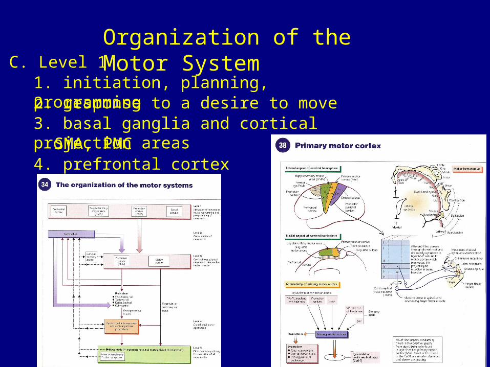
Organization of the Motor SystemC. Level 1
1. initiation, planning, programming2. response to a desire to move3. basal ganglia and cortical projection areas
SMA, PMC4. prefrontal cortex

Organization of the Motor SystemC. Level 1
5. damage to basal ganglia and cortical projection sitesa. does not produce weaknessb. can result in abnormal involuntary movements
chorea: irregular, rapid, uncontrolled, involuntary, excessive movement that seems to move randomly from one part of the body to another.
dystonia: sustained muscle contractions cause twisting and repetitive movements or abnormal postures.
ballismus: jerky or shaking movements of the arms or legs, especially such movements occurring in chorea.
c. Parkinson’s disease

Organization of the Motor SystemD. Level 2
1. cerebellum2. coordination of movements3. is a comparator4. stores a lot of muscle memory
5. damage- so motor pathways can “remember” how to perform actions
does not cause weaknessloss of coordination

Organization of the Motor SystemD. Level 3
1. origin of descending motor pathwaysa. corticospinals/pyramidal tractsb. extrapyramidal tracts
- originate from subcortical structures
2. damage- still receive an input from the primary motor cortex (MsI)
- weakness- increased tone- hyperreflexia

Organization of the Motor SystemD. Level 4
1. spinal cord interneurons- some mediate spinal cord reflexes
2. central pattern generators- spinal interneurons capable of generating their own inputs to motor neurons (independent of any input)

Organization of the Motor SystemD. Level 5
1. lower motor neurons- is the output to the skeletal muscle
2. receives input from the muscle spindle and Golgi tendon organs3. center for simple stretch reflexes of muscles4. damage
- weakness- wasting- hypotonia- weak or areflexic QuickTime™ and a
TIFF (Uncompressed) decompressorare needed to see this picture.


QuickTime™ and aTIFF (Uncompressed) decompressor
are needed to see this picture.
Ia afferent nerves (from spindle)

QuickTime™ and aTIFF (Uncompressed) decompressor
are needed to see this picture.
QuickTime™ and aTIFF (Uncompressed) decompressor
are needed to see this picture.

Bag 1: rate changeBag 2: absolute length

Lateral = distal (typically)Ventral = axial & proximal
Ventral anterior horn = extensor musclesDorsal anterior horn = flexor muscles
Propriospinals connect the motor neuron poolsPropriospinals also connect the CPGs
Humans cannot typically locomote in the absence of significant supraspinal inputs (no fictive locomotion)

QuickTime™ and aTIFF (Uncompressed) decompressor
are needed to see this picture.


QuickTime™ and aTIFF (Uncompressed) decompressor
are needed to see this picture.
QuickTime™ and aTIFF (Uncompressed) decompressor
are needed to see this picture.
QuickTime™ and aTIFF (Uncompressed) decompressor
are needed to see this picture.

QuickTime™ and aTIFF (Uncompressed) decompressor
are needed to see this picture.
Remember: almost all motor tracts are crossed.


