Background and Objectives: The evaluation of left ventricular (LV) diastolic function in patients with atrial fibrillation (AF) is challenging. This study aimed to investigate the efficacy of the diagnostic algorithm for LV diastolic dysfunction (LVDD) in the current guidelines and to evaluate the association between increased left atrial pressure (LAP) and LV diastolic parameters.Methods: One hundred and twenty-four patients with non-valvular AF and a preserved LV ejection fraction who had the same rhythm status on echocardiography and LAP measurements during catheter ablation were included. LV diastolic function was classified as normal, indeterminate, or LVDD according to the recent guidelines. Increased LAP was defined as mean LAP (mLAP) ≥15 mmHg.Results: The mLAP was not different among the normal, indeterminate, and LVDD groups. However, the prevalence of increased LAP was higher in the LVDD group. Among the LV diastolic parameters, only medial E/e′ was independently associated with mLAP in the whole study population. In patients with persistent AF (PeAF), E/e′ and e′ were significantly associated with mLAP, whereas in paroxysmal AF (PAF), mLAP was not associated with the LV diastolic parameters but with left atrial conduit function.Conclusions: In general, increased LAP is known to be closely related with LVDD. However, the algorithm for LVDD from recent guidelines does not reflect well the increased LAP in AF patients. The diastolic parameters may aid in estimating the increased LAP in PeAF but may only have limited value for assessing increased LAP in PAF.
Atrial fibrillation (AF) is the most common arrhythmia. Its prevalence increasing as the population is aging1) and is significantly higher during heart failure, regardless of the left ventricular ejection fraction (LVEF) status.2) Left ventricular diastolic dysfunction (LVDD) shares common risk factors with AF, including age, diabetes, hypertension, and obesity.3)4) The prevalence of LVDD increases with age, and it is more likely that a patient has both AF
Int J Heart Fail. 2020 Jan;2(1):55-65https://doi.org/10.36628/ijhf.2020.0003pISSN 2636-154X·eISSN 2636-1558
Original Article
Received: Jan 4, 2020Revised: Jan 23, 2020Accepted: Jan 27, 2020
Correspondence toSeong-Mi Park, MD, PhDDivision of Cardiology, Department of Internal Medicine, Korea University Anam Hospital, 73 Goryeodae-ro, Seongbuk-gu, Seoul 02841, Korea.E-mail: [email protected]
ORCID iDsMi-Na Kim https://orcid.org/0000-0001-6589-5122Seong-Mi Park https://orcid.org/0000-0002-6710-685XHee-Dong Kim https://orcid.org/0000-0003-1432-2257Dong-Hyuk Cho https://orcid.org/0000-0001-8480-9082Jaemin Shim https://orcid.org/0000-0001-8251-1522Jong-il Choi https://orcid.org/0000-0001-6617-508XWan Joo Shim https://orcid.org/0000-0002-2467-3336
Mi-Na Kim , MD, PhD, Seong-Mi Park , MD, PhD, Hee-Dong Kim , MD, Dong-Hyuk Cho , MD, PhD, Jaemin Shim , MD, PhD, Jong-il Choi , MD, PhD, Young Hoon Kim, MD, PhD, and Wan Joo Shim , MD, PhD
Division of Cardiology, Department of Internal Medicine, Korea University Anam Hospital, Seoul, Korea
Assessment of the Left Ventricular Diastolic Function and Its Association with the Left Atrial Pressure in Patients with Atrial Fibrillation
Conflict of InterestThe authors have no financial conflicts of interest.
Author ContributionsConceptualization: Kim MN, Shim WJ; Data curation: Kim MN, Park SM, Kim HD, Cho DH, Shim J, Choi JI, Kim YH; Formal analysis: Kim MN, Shim WJ; Investigation: Shim WJ; Methodology: Park SM, Shim WJ; Supervision: Park SM; Validation: Kim MN; Writing - original draft: Kim MN; Writing - review & editing: Park SM.
and LVDD. Furthermore, AF and LVDD also mutually influence each other's development, exacerbation, and outcomes.5-7)
The evaluation of LVDD in AF is quite complicated. Left atrial (LA) enlargement, a parameter of LVDD, is limited in estimating LVDD in AF because LA enlargement can also be derived from AF. Furthermore, the echocardiographic parameters, which are recommended by the guidelines for evaluating LV diastolic function in AF patients, such as the deceleration time (DT) of the pulmonary vein diastolic velocity and the ratio of the peak E wave velocity to flow propagation velocity,8) may not be routinely measured in daily clinical practice.
This study aimed to investigate the efficacy of the diagnostic algorithm for LVDD in the current guidelines8) for detecting increased left atrial pressure (LAP) in AF patients and to identify the best predictor for increased LAP from among the well-known diastolic parameters in daily practice.
METHODS
Study populationThe data of 124 patients with non-valvular AF and preserved LVEF (≥50%) were retrospectively assessed in this study. The patients were scheduled to undergo catheter ablation for AF. In all patients, trans-thoracic echocardiography was performed within 24 hours before catheter ablation, and LAP was measured immediately after a trans-septal puncture. The rhythm status on echocardiography and LAP measurements were same in all patients. In patients with paroxysmal AF (PAF), 67 patients were in sinus rhythm (SR) during echocardiography and measurement of LAP. And 11 patients with PAF were in AF during examinations. In patients with persistent AF (PeAF), 8 patients were in SR and 38 patients were in AF during echocardiography and measurement of LAP. Hypertension, diabetes, dyslipidemia and heart failure were defined if the patient was previously diagnosed or taken related medication. The exclusion criteria were as follows: 1) presence of valvular stenosis or regurgitation of moderate grade or higher grade; 2) cardiomyopathies; 3) congenital heart disease; 4) thyroid disease; 5) uncontrolled hypertension; 6) history of any cardiac surgery and cardiac device insertion; and 7) significant systemic illness. This study was approved by the Institutional Review Board of Korea University College of Medicine (IRB number 2017AN0194).
Trans-thoracic echocardiographyTrans-thoracic echocardiography was performed for all patients, using commercially available machines (Vivid 7 and 9; General Electric Medical Health, Waukesha, WI, USA, and iE33; Philips Medical, Andover, MA, USA). The echocardiographic examination was conducted according to the recommendations of the American Society of Echocardiography.9) The LV and LA size were measured, and the LV mass was calculated using the cube formula proposed by the American Society of Echocardiography that was modified by Devereux et al. The LV mass index (LVMI) was calculated by dividing the LV mass by the body surface area (BSA). The LVEF was measured using the modified biplane Simpson's method. The LA volume was evaluated using the modified biplane Simpson's method, and the LV volume index (LAVI) was calculated by dividing the LV volume by the BSA. The LA emptying fraction (LAeF) was calculated as:
Left Ventricular Diastolic Dysfunction in Atrial Fibrillation
The peak early (E) and late (A) diastolic mitral inflow velocities and DT were measured by pulsed-wave Doppler in the apical 4-chamber view. The systolic (s′) and early (e′) and late (a′) diastolic mitral tissue velocities were measured at the medial and lateral mitral annulus from the apical 4-chamber view using pulsed-wave tissue Doppler imaging. E/e′ was also calculated. Tricuspid regurgitation (TR) velocity was measured using continuous-wave Doppler in the right ventricular modified apical 4-chamber view. Each measurement including Doppler data was performed over at least 5 consecutive beats, and the average was obtained.
Classification of LVDDAll patients were classified as normal, indeterminate, and LVDD according to a recent guideline from the American Society of Echocardiography and the European Association of Cardiovascular Imaging.8) Diastolic dysfunction was diagnosed according to the following indices: an averaged E/e′ >14, septal e′ velocity <7 cm/s or lateral e′ velocity <10 cm/s, TR velocity >2.8 m/s, and LAVI >34 mL/m2. If a patient satisfied one of the aforementioned parameters, the patient was classified as normal. If a patient satisfied 2 of these parameters, the patient was classified in the indeterminate group, and the patient who satisfied 3 or more of these criteria was allocated to the LVDD group.
Measurement of LAPAll patients fasted overnight before LAP measurement. For AF catheter ablation, a fluoroscopy-guided double transseptal puncture was performed using the Brockenborough technique. After a trans-septal sheath (Schwartz left 1; St. Jude Medical Inc., Minnetonka, MN, USA) was advanced into the left atrium, a 6-F pigtail catheter (A&A Medical Device Inc., Seongnam, Korea) was inserted into the left atrium through the trans-septal sheath, and the LAP was measured just after the trans-septal puncture. The LAP was measured at the end of expiration by computer-generated wave during SR or AF which was heart rhythm of patient at the time of procedure. It was averaged over 5 consecutive beats at the end of expiration. Increased LAP was defined as mean LAP ≥15 mmHg, which is considered a criterion for LA hypertension.10)
Statistical analysisQuantitative data are expressed as the mean±standard deviation and categorical data as number or percentage. The categorical data were analyzed using χ2 test or Fisher's exact test. Unpaired Student's t-test was used to compare the continuous variables between the 2 groups. Analysis of variance was performed to compare the continuous variables among 3 groups, and post hoc analysis was performed. The analysis of covariance was used to compare patients with and without increased LAP after adjusting for the related factors of LAP, including age, hypertension, diabetes, coronary artery disease, stroke, heart rate, rhythm status, and AF type and duration. Univariate linear regression was used to evaluate the association between the mean LAP and diastolic echocardiographic parameters. Variables with p<0.1 in the univariate model were selected for multivariate analysis to identify the factors independently associated with the mean LAP. A significant value was obtained if the 95% confidence interval (CI) exceeded 1, and the p value was <0.05. The predictive power of the diastolic parameters for increased LAP was tested by receiver operating characteristic (ROC) curve analysis. All analyses, except the comparison of the ROC curves, were performed using the SPSS 25.0 software program (SPSS, Chicago, IL, USA). The ROC curves were compared by MedCalc for Windows (version 19.1.3; MedCalc Software, Mariakerke, Belgium).
Left Ventricular Diastolic Dysfunction in Atrial Fibrillation
RESULTS
Baseline characteristicsThe mean age of the study population was 56.7±11.1 years. Males constituted 81.5% of the study population (105 patients). Seventy-eight patients (62.9%) had PAF. The most common comorbidity was dyslipidemia (42.7%), followed by hypertension (37.1%). When the patients were classified for LVDD, 9 patients (7.3%) constituted the LVDD group, and 36 patients (29.0%) were classified in the indeterminate group. The baseline characteristics regarding the LVDD type are presented in Table 1. The patients with LVDD and indeterminate diastolic function were older than patients with normal diastolic function. The prevalence of diabetes was higher in patients with LVDD. The prevalence of hypertension, dyslipidemia, coronary artery disease, heart failure, and previous stroke or transient ischemic attack were not different between the groups. And there was no difference in medication between the groups (Supplementary Table 1).
LAP according to the criteria for left ventricular diastolic functionThe mean LAP of the study population was 12.1±4.1 mmHg, which was close to the upper limit of the normal range of the LAP. Forty-two patients (33.9%) had an increased LAP. The mean LAP of patients with LVDD was higher than that of the patients in the other groups, but this difference was not statistically significant (Figure 1). The proportion of patients with increased LAP was higher in the LVDD group than in the other groups and was similar between the normal and indeterminate groups (Figure 2).
Differences in the left ventricular diastolic parameters according to the increased LAPThe baseline characteristics were not different between patients with and without increased LAP (Supplementary Table 2). The LA size and function were not different between patients with and without increased LAP. The E wave velocity for patients with increased LAP was higher than for patients without increased LAP (71.1±17.6 vs. 62.9±15.8, p=0.010). The mean E/e′ was significantly higher in patients with increased LAP (10.0±3.0 vs. 8.33±2.8, p=0.002). The other diastolic parameters, including DT, medial and lateral e′, lateral E/e′, and maximal TR velocity, were not different between the groups. After adjusting for the clinical factors associated with the LAP and LV diastolic function (age, sex, hypertension, diabetes, coronary artery disease, and stroke), heart rate and rhythm status during echocardiography and LAP measurement, AF type and duration, E wave velocity and medial E/e′ were statistically significantly different between patients with and without increased LAP (Table 2).
Left Ventricular Diastolic Dysfunction in Atrial Fibrillation
10
2
6
0
4
8
12
16
14
18*p=0.121
Indeterminate LVDDNormal
11.7±4.112.5±4.2
14.6±3.5
p=0.228 p=0.490
p=0.157
Figure 1. Difference in the mean left atrial pressure between the groups showing diastolic dysfunction. LVDD = left ventricular diastolic dysfunction. *The p by analysis of variance between patients with normal, indeterminate, and LVDD. The p by post hoc analysis between 2 groups.
p=0.05
Indeterminate
No increased LAP Increased LAP
LVDD
%
Normal
0 20 40 60 80 100
57 (72.2%) 22 (27.8%)
22 (61.1%) 14 (38.9%)
3 (33.3%) 6 (66.7%)
Figure 2. Association between increased left atrial pressure and left ventricular diastolic dysfunction in the study population. LAP = left atrial pressure; LVDD = left ventricular diastolic dysfunction.
Table 2. Comparison of left ventricular diastolic parameters between patients with and without increased left atrial pressure
Patients without increased LAP (n=82)
Patients with increased LAP (n=42)
p value
LA diameter (mm) 40.7±5.4 41.7±5.6 0.338LA volume index (mL/m2) 42.5±15.2 43.2±14.2 0.813LA emptying fraction (%) 35.5±12.9 34.6±12.3 0.713LVMI 91.4±16.2 88.0±16.4 0.272E (cm/s) 62.9±15.8 71.1±17.6 0.010DT (msec) 181.9±39.6 182.5±52.5 0.945e′ medial (cm/s) 8.03±2.20 7.42±1.90 0.123e′ lateral (cm/s) 11.7±3.4 11.4±3.1 0.646E/e′ medial 8.33±2.80 10.00±3.00 0.002E/e′ lateral 5.80±2.60 6.47±1.80 0.132TR Vmax (m/s) 2.29±2.20 2.09±0.60 0.566Values are presented as mean±standard deviation.DT = deceleration time; LA = left atrial; LAP = left atrial pressure; LVMI = left ventricular mass index; TR Vmax = tricuspid regurgitation maximal velocity.
The LVMI, E, and medial E/e′ were correlated with LAP, as indicated by the univariate linear regression analysis. Multivariate linear regression analysis revealed that only medial E/e′ was significantly associated with LAP (Table 3). The predictive value of E/e′ for increased LAP was modest (c-statistics, 0.681; 95% CI, 0.583–0.778; p=0.001); however, E/e′ was the strongest predictive parameter for increased LAP compared to the other echocardiographic diastolic parameters (Figure 3).
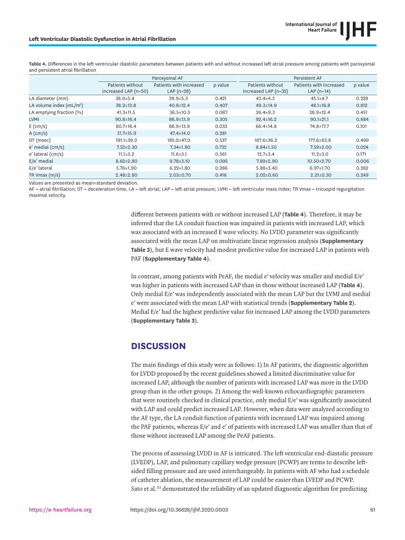
Association of LVDD and LAP according to the type of AFThe mean LAP was not different between patients with PAF and PeAF (12.2±4.5 vs. 12.0±3.4; p=0.742). The baseline characteristics were not different between patients with PAF or PeAF. PAF patients with increased LAP had a higher E wave velocity than PAF patients without increased LAP. A statistical trend that LAeF, representative of LA reservoir function, was impaired in patients with increased LAP was observed. However, A wave velocity was not
Left Ventricular Diastolic Dysfunction in Atrial Fibrillation
Table 3. Relationship between echocardiographic parameters and the mean left atrial pressureMultiple linear regression β 95% CI p valueLVEF −0.193 −0.40, 0.15 0.068LVMI −0.038 −0.09, 0.01 0.146LA volume index −0.018 −0.08, 0.05 0.572LA emptying fraction −0.007 −0.07, 0.08 0.845E −0.074 −0.17, 0.02 0.125DT −0.004 −0.03, 0.17 0.725e′ medial 0.758 −0.03, 1.54 0.920E/e′ medial 0.932 0.32, 1.55 0.003TR Vmax 0.103 −0.29, 0.50 0.608The correlation of the mean LAP calculated after adjusting for the clinical related factors of LV diastolic function (age, sex, hypertension, diabetes, coronary artery disease, and stroke), heart rate and rhythm status on echocardiography, and AF type and duration.AF = atrial fibrillation; CI = confidence interval; DT = deceleration time, LA = left atrial; LAP = left atrial pressure; LV = left ventricular; LVEF = left ventricular ejection fraction; LVMI = left ventricular mass index; TR Vmax = tricuspid regurgitation maximal velocity.
100
20
60
0
40
80
10020 60100-Specificity
Sens
itivi
ty
40 80
E/e′
Medial e′LAVI
Lateral e′TR Vmax
Figure 3. Receiver operating characteristic curves for the echocardiographic parameters for predicting increased left atrial pressure. LAVI = left atrial volume index; TR Vmax = tricuspid regurgitation maximal velocity.
different between patients with or without increased LAP (Table 4). Therefore, it may be inferred that the LA conduit function was impaired in patients with increased LAP, which was associated with an increased E wave velocity. No LVDD parameter was significantly associated with the mean LAP on multivariate linear regression analysis (Supplementary Table 3), but E wave velocity had modest predictive value for increased LAP in patients with PAF (Supplementary Table 4).
In contrast, among patients with PeAF, the medial e′ velocity was smaller and medial E/e′ was higher in patients with increased LAP than in those without increased LAP (Table 4). Only medial E/e′ was independently associated with the mean LAP but the LVMI and medial e′ were associated with the mean LAP with statistical trends (Supplementary Table 2). Medial E/e′ had the highest predictive value for increased LAP among the LVDD parameters (Supplementary Table 3).
DISCUSSION
The main findings of this study were as follows: 1) In AF patients, the diagnostic algorithm for LVDD proposed by the recent guidelines showed a limited discriminative value for increased LAP, although the number of patients with increased LAP was more in the LVDD group than in the other groups. 2) Among the well-known echocardiographic parameters that were routinely checked in clinical practice, only medial E/e′ was significantly associated with LAP and could predict increased LAP. However, when data were analyzed according to the AF type, the LA conduit function of patients with increased LAP was impaired among the PAF patients, whereas E/e′ and e′ of patients with increased LAP was smaller than that of those without increased LAP among the PeAF patients.
The process of assessing LVDD in AF is intricated. The left ventricular end-diastolic pressure (LVEDP), LAP, and pulmonary capillary wedge pressure (PCWP) are terms to describe left-sided filling pressure and are used interchangeably. In patients with AF who had a schedule of catheter ablation, the measurement of LAP could be easier than LVEDP and PCWP. Sato et al.11) demonstrated the reliability of an updated diagnostic algorithm for predicting
Left Ventricular Diastolic Dysfunction in Atrial Fibrillation
Table 4. Differences in the left ventricular diastolic parameters between patients with and without increased left atrial pressure among patients with paroxysmal and persistent atrial fibrillation
increased LVEDP. The recent diagnostic algorithm for LVDD had better accuracy for detecting increased LVEDP and better discriminative value for the prognosis of the patients than the previous recommendations for diagnosing LVDD. The recently updated algorithm for LVDD was for patients with normal SR and was not intended for AF patients.8) However, the echocardiographic parameters recommended for evaluating the diastolic function in AF patients were not routinely checked in practice. Therefore we evaluated the reliability of the recently reported diagnostic algorithm, which would be relatively easily applicable in AF patients. As AF itself causes LA dilation, the LAVI could be less discriminative and insufficient to assess LVDD in the AF patients. Approximately 50% of the normal patients and all intermediate and LVDD patients had increased LAVI (LAVI>34 mL/m2) in this study (Supplementary Table 5). Therefore, the LA volume parameter has limited value for assessing LVDD, and evaluating LVDD with the remaining 3 indices may be less discerning.
In this study, increased LAP was associated with impaired LA conduit function in PAF patients. Previous studies reported that a worsened LA conduit function was closely associated with the recurrence of AF after cardioversion.12)13) LA fibrosis, assessed using late gadolinium enhancement by cardiac magnetic resonance, was significantly associated with impaired LA conduit function.14) The LA conduit function is affected not only by LA remodeling but also by LV early filling impairment; therefore, it reflects atrioventricular coupling. In this study, the ventricular parameters of LVDD were not different between the patients with and without increased LAP, and LA remodeling by AF may be associated with increased LAP in PAF patients. The increased E wave velocity might be due to decreased distensibility of the left atrium and increased LA stiffness. Therefore, estimating LAP with conventional echocardiographic LVDD parameters might be limited in PAF.
In contrast, an increased LAP was independently associated with the echocardiographic LVDD parameters in patients with PeAF. LVDD, which is caused by impaired ventricular relaxation, loss of restoring force, and increased LV diastolic stiffness, leads to an increase in the LVEDP and LAP. In AF, the LAP increases and is uncoupled from the LVEDP because of decreased atrioventricular coupling and reduced LA compliance by the impaired LA function due to the loss of atrial contraction.15-17) Therefore, E/e′, which is representative of LVEDP, may be less accurate in estimating the LAP in patients with AF. Previous studies reported that E/e′ was associated functional capacity,18) exercise intolerance,19) hospitalization for heart failure,20) and poor prognosis20)21) in AF. In this study, E/e′ was significantly associated with the LAP in patients with PeAF.
There are several limitations of this study. As the patients of this study underwent catheter ablation for AF, selection bias may be present. Catheter ablation was not performed in patients who had a significantly enlarged left atrium. Patients with an earlier stage of AF may have been enrolled, and patients with an advanced stage of AF were not included in this study. The results of this study might not be generalizable to heart failure with preserved ejection fraction (HFpEF) patients with AF. The second limitation was that we did not measure the LVEDP. The LAP and LVEDP are the same in most cases, except under special conditions. There may be a difference in the LAP and LVEDP in patients with AF.22) However, the LVEDP only provides information about LV compliance and does not provide information about the LA compensation for the LVDD and LV compliance. Therefore, the LAP, which is determined by LA remodeling and compensation in response to chronic pressure overload from the LV, may be strongly associated with symptom development and exercise tolerance in patients with HFpEF and AF. The third limitation was that the echocardiography and measurement
Left Ventricular Diastolic Dysfunction in Atrial Fibrillation
of LAP were not performed simultaneously. Therefore, the relationship between LAP and echocardiographic parameter might be inaccurate. During echocardiography, E and e′ velocities were not measured at the same time and the R-R interval was not adjusted. Therefore E/e′ might be inaccurate. But in majority of patients, ventricular rate was not significantly changed during echocardiography. The last limitation was the lacks of laboratory including data on the B-type natriuretic peptide (BNP) or N-terminal pro BNP (NT-pro BNP), cardiac enzyme levels and further echocardiographic data, like LA strain. As the study population was scheduled to undergo catheter ablation, these analyses were not routinely assessed in these patients.
In conclusion, the evaluation of LVDD in AF is challenging. The algorithms for LVDD detection in the recent guidelines do not reflect well the increased LAP in AF patients. Although medial E/e′ was a single sensitive marker for increased LAP in the whole study population, this finding was only valid for patients with PeAF. In PAF, the routine echocardiographic LVDD parameters had limited value for estimating increased LAP. Further studies with larger study populations and more detailed considerations regarding the LA and LV function, like strain, are required to evaluate the diagnostic algorithm for LVDD in AF patients.
SUPPLEMENTARY MATERIALS
Supplementary Table 1Medication of study population
Click here to view
Supplementary Table 2Baseline characteristics of the patients with and without increased LAP
Click here to view
Supplementary Table 3Association between the echocardiographic parameters and mean left atrial pressure in patients with paroxysmal or persistent atrial fibrillation
Click here to view
Supplementary Table 4Predictive value of diastolic parameters for increased left atrial pressure in patients with paroxysmal or persistent atrial fibrillation
Click here to view
Supplementary Table 5Prevalence of each index in the 2016 LVDD diagnostic algorithm in each LVDD group
1. Rahman F, Kwan GF, Benjamin EJ. Global epidemiology of atrial fibrillation. Nat Rev Cardiol 2014;11:639-54. PUBMED | CROSSREF
2. Batul SA, Gopinathannair R. Atrial fibrillation in heart failure: a therapeutic challenge of our times. Korean Circ J 2017;47:644-62. PUBMED | CROSSREF
3. Lau DH, Nattel S, Kalman JM, Sanders P. Modifiable risk factors and atrial fibrillation. Circulation 2017;136:583-96. PUBMED | CROSSREF
4. Shammas RL, Khan NU, Nekkanti R, Movahed A. Diastolic heart failure and left ventricular diastolic dysfunction: what we know, and what we don't know! Int J Cardiol 2007;115:284-92. PUBMED | CROSSREF
5. Caldwell JC, Mamas MA. Heart failure, diastolic dysfunction and atrial fibrillation; mechanistic insight of a complex inter-relationship. Heart Fail Rev 2012;17:27-33. PUBMED | CROSSREF
6. Ishikawa S, Sugioka K, Sakamoto S, et al. Relationship between tissue Doppler measurements of left ventricular diastolic function and silent brain infarction in patients with non-valvular atrial fibrillation. Eur Heart J Cardiovasc Imaging 2017;18:1245-52. PUBMED | CROSSREF
7. Kosiuk J, Breithardt OA, Bode K, et al. The predictive value of echocardiographic parameters associated with left ventricular diastolic dysfunction on short- and long-term outcomes of catheter ablation of atrial fibrillation. Europace 2014;16:1168-74. PUBMED | CROSSREF
8. Nagueh SF, Smiseth OA, Appleton CP, et al. Recommendations for the evaluation of left ventricular diastolic function by echocardiography: an update from the American Society of Echocardiography and the European Association of Cardiovascular Imaging. Eur Heart J Cardiovasc Imaging 2016;17:1321-60. PUBMED | CROSSREF
9. Lang RM, Badano LP, Mor-Avi V, et al. Recommendations for cardiac chamber quantification by echocardiography in adults: an update from the American Society of Echocardiography and the European Association of Cardiovascular Imaging. J Am Soc Echocardiogr 2015;28:1-39.e14. PUBMED | CROSSREF
10. Markowitz SM. Left atrial hypertension in atrial fibrillation: dealing with the pressure. JACC Clin Electrophysiol 2017;3:470-2. PUBMED | CROSSREF
11. Sato K, Grant ADM, Negishi K, Cremer PC, Negishi T, Kumar A, et al. Reliability of updated left ventricular diastolic function recommendations in predicting elevated left ventricular filling pressure and prognosis. Am Heart J 2017;189:28-39 PUBMED | CROSSREF
12. Degiovanni A, Boggio E, Prenna E, et al. Association between left atrial phasic conduit function and early atrial fibrillation recurrence in patients undergoing electrical cardioversion. Clin Res Cardiol 2018;107:329-37. PUBMED | CROSSREF
13. Giubertoni A, Boggio E, Ubertini E, et al. Atrial conduit function quantitation precardioversion predicts early arrhythmia recurrence in persistent atrial fibrillation patients. J Cardiovasc Med (Hagerstown) 2019;20:169-79. PUBMED | CROSSREF
14. Habibi M, Lima JA, Khurram IM, et al. Association of left atrial function and left atrial enhancement in patients with atrial fibrillation: cardiac magnetic resonance study. Circ Cardiovasc Imaging 2015;8:e002769. PUBMED | CROSSREF
15. Melenovsky V, Hwang SJ, Redfield MM, Zakeri R, Lin G, Borlaug BA. Left atrial remodeling and function in advanced heart failure with preserved or reduced ejection fraction. Circ Heart Fail 2015;8:295-303. PUBMED | CROSSREF
16. Dernellis JM, Stefanadis CI, Zacharoulis AA, Toutouzas PK. Left atrial mechanical adaptation to long-standing hemodynamic loads based on pressure-volume relations. Am J Cardiol 1998;81:1138-43. PUBMED | CROSSREF
17. Dickinson MG, Lam CS, Rienstra M, et al. Atrial fibrillation modifies the association between pulmonary artery wedge pressure and left ventricular end-diastolic pressure. Eur J Heart Fail 2017;19:1483-90. PUBMED | CROSSREF
18. Lee SH, Jung JH, Choi SH, et al. Exercise intolerance in patients with atrial fibrillation: clinical and echocardiographic determinants of exercise capacity. J Am Soc Echocardiogr 2005;18:1349-54. PUBMED | CROSSREF
19. Punjani S, Wu WC, Cohen S, Sharma SC, Choudhary G. Echocardiographic indices of diastolic function relate to functional capacity and quality of life in ambulatory men with atrial fibrillation. J Am Soc Echocardiogr 2011;24:533-540.e3. PUBMED | CROSSREF
20. Hsu PC, Lee WH, Chu CY, et al. The ratio of early mitral inflow velocity to global diastolic strain rate as a useful predictor of cardiac outcomes in patients with atrial fibrillation. J Am Soc Echocardiogr 2014;27:717-25. PUBMED | CROSSREF
21. Park SJ, Lee SC, Jang SY, et al. E/e′ ratio is a strong prognostic predictor of mortality in patients with non-valvular atrial fibrillation with preserved left ventricular systolic function. Circ J 2011;75:2350-6. PUBMED | CROSSREF
22. Reddy YN, El-Sabbagh A, Nishimura RA. Comparing pulmonary arterial wedge pressure and left ventricular end diastolic pressure for assessment of left-sided filling pressures. JAMA Cardiol 2018;3:453-4. PUBMED | CROSSREF