Dermatoglyphics as a Novel Method for Assessing Intelligence Quotient in Children Aged 5–11 Years: A Cross-sectional StudyNamratha Tharay1, SVSG Nirmala2, Venkata N Bavikati3, Sivakumar Nuvvula4
Ab s t r Ac t Aim: To measure the IQ and record dermatoglyphic patterns of children including intellectually disabled aged between 5 years and 11 years and to correlate them.Materials and methods: The present study was a cross-sectional study conducted in two private schools in the city corporation limits. A total of 300 children aged between 5 years and 11 years were equally allocated into three groups based on IQ using covariate adaptive randomization. IQ of the children was measured using Raven’s Coloured Progressive Matrices. Bilateral palmar and finger prints were obtained on A4-size papers by ink method using rolling technique. Prints thus obtained were analyzed for dermatoglyphic variables using magnification lens and are read based on Cummins and Midlo classification for fingertip patterns and Bali and Chaube classification for palmar flexion creases. The data were entered and statistically analyzed. For statistical significance, a two-tailed probability value of less than 0.05 was taken as significant.Results: The current study suggests a relationship between different fingertip patterns of digits of I, II, III, and IV of left and right hand and also digit V of right hand with various levels of IQ.Conclusion: Hence, dermatoglyphics can be considered as a preliminary noninvasive approach for the determination of IQ. Also, it plays an imperative role, especially in distinguishing genetic intellectual disabilities.Keywords: Children, Dermatoglyphics, Fingerprint patterns, Intellectually disabled children, IQ.International Journal of Clinical Pediatric Dentistry (2020): 10.5005/jp-journals-10005-1793
In t r o d u c t I o n Dermatoglyphics is an indispensable part of forensic odontology and has numerous applications in areas of science, such as anthropology, criminology, cytogenetic studies, and also in dentistry. “Dermatoglyphics” derives its denotation from the Greek literature “Derma” denotation skin and “glyphic” which means carvings.1 As to define dermatoglyphics, also well known as “epidermal ridge configurations” are the characteristics of the ridged skin on the fingertips, palms, toes, and soles of primates (including human beings) and some other mammals.2
Dermal ridge dif ferentiation takes place early in fetal development and remains constant throughout the life. By sixth prenatal month, epidermal ridge patterns will be completed, when the glandular folds are fully molded and after the sweat gland secretion and keratinization have commenced. At this time, the shapes on the skin surface begin to reflect the underlying patterns.3
The subsequent ridge silhouettes are genetically determined and persuaded by environmental forces.1 Any variations in the dermatoglyphic patterns may be the consequence of early prenatal disturbances that are prejudiced by numerous environmental factors such as hypoxia, viral infections, delayed growth factors, and maternal alcohol exposure, in turn affects the size, symmetry, and number of the epidermal ridges.4
In the field of psychology, one of the greatest endeavors was the measurement of intelligence which not only plays a significant role in one’s individual’s life but also in the society. When compared with high IQ individuals, low IQ individuals will have hitches in acquisition, thinking, and processing of new information and knowledge. There will be necessity of extra care, education, and medical services, and the need for care will persist throughout
the life among individuals with low IQ who has associated genetic abnormalities as they fail to attain personnel independence, which is thought to be 50% prenatal in origin.4
IQ and dermatoglyphics have same origin, as neural tissue and epidermis are embryonic ectodermal derivatives, and the embryological–fetal developmental takes place in the second trimester of intrauterine life. Evidence suggests that dermatoglyphics can be used as an adjunct to other diagnostic methods in finding syndromes of genetic origin.
As they take the same origin in the intrauterine life, it could be beneficial if dermatoglyphic variables can estimate IQ range. And it is easier to record the dermatoglyphic patterns for measuring IQ than measuring IQ by using traditional methods. So, the aim of
1Department of Pediatric and Preventive Dentistry, AME’s Dental College and Hospital, Raichur, Karnataka, India2,4Department of Pediatric and Preventive Dentistry, Narayana Dental College, Nellore, Andhra Pradesh, India3Department of Orthodontics and Dentofacial Orthopedics, AME’s Dental College and Hospital, Raichur, Karnataka, IndiaCorresponding Author: Namratha Tharay, Department of Pediatric and Preventive Dentistry, AME’s Dental College and Hospital, Raichur, Karnataka, India, Phone: +91 9704058513, e-mail: [email protected] to cite this article: Tharay N, Nirmala SVSG, Bavikati VN, et al. Dermatoglyphics as a Novel Method for Assessing Intelligence Quotient in Children Aged 5–11 Years: A Cross-sectional Study. Int J Clin Pediatr Dent 2020;13(4):355–360.Source of support: NilConflict of interest: None
International Journal of Clinical Pediatric Dentistry, Volume 13 Issue 4 (July–August 2020)356
the study is to compare the dermatoglyphic pattern to IQ of the children, and the null hypothesis was formulated, i.e., neither IQ nor dermatoglyphic patterns are related to each other.
MAt e r I A l s A n d Me t h o d s This is a cross-sectional study that was conducted in the schools, Red Cross Society present in City Corporation limits after the institutional ethical clearance was obtained (D138407006). The schools were selected randomly. Whereas the Red Cross Society was selected purposefully and the data were collected from December 2013 to November 2014. The inclusion criteria being the children aged between 5 years and 11 years, with informed consent by the institutions and with various intelligence levels, whereas the exclusion criteria being individuals with blindness, upper limb disabilities, and with definitely negative behavior.
Sample Size DeterminationSample size was estimated by taking 95% power and 5% margin of error (1-ß), and a total of 300 children were considered as appropriate sample size to fulfill the purpose of the study. Each group comprised of 100 children, and the children are assigned into their respective groups using covariant adaptive randomization.
MethodologyThe IQ was measured by Raven’s colored progressive matrices, a nonverbal intelligence scale. Based on IQ scores, the subjects are divided into 3 groups as follows:Group I: Subjects with IQ greater than 120Group II: Subjects with IQ between 70-120Group III: Subjects with IQ less than 70
Bilateral palmar and finger prints of the children were obtained on the A4-size papers using ink method. Prior to recording the patterns, it is required to eliminate sweat, oil, or dirt from the skin surface of the hands in order to increase the quality of the prints. This was achieved by cleaning the hands with soap and water and wiping with a cloth. The dried palms of the children were placed on the ink pad with a mild pressure, and the excess ink was wiped off with the help of a gauze piece. Then, the bilateral palmar prints was recorded by placing the palm on the paper with moderate pressure and also the individual fingertip prints of both the hands were recorded separately in the same manner, as the palmar prints were recorded using rolling technique on the same paper. The unclear prints were discarded, and the prints were repeated until clear prints were obtained (Fig. 1). After recording the dermatoglyphic
patterns, the palms and fingers of the child are gently wiped with cotton damped with ethyl alcohol followed by washing the hands with Dettol® solution and water. Prints thus obtained were analyzed for the following parameters using magnification of power *2 and are read based on Cummins and Midlo classification for fingertip patterns and by Bali and Chaube 1971 classification for palmar flexion creases. Dermatoglyphic variables read are presence of arches (A), loops, and whorls (W) for fingertip patterns, ATD angle, and palmar flexion for palms.
Statistical AnalysisThe data were entered in the Microsoft excel spread sheet 2010. Crosstabs were used for comparison of fingertip patterns and creases within the group, and difference between the three groups were assessed using Chi-square analysis. Mean and standard deviations were calculated for left and right ATD angle, and the differences between means were tested with one-way analysis of variance followed by post hoc Tukey test. Statistical significance was set at p value <0.05.
re s u lts A total of 384 children aged 5–11 years who met the eligibility criteria were evaluated. Based on IQ, 300 children were included into the present study (Flowchart 1).
Demographic DataAnalysis of data revealed that out of 300 children, 158 were boys and 142 were girls. In group I, there were equal distributions, whereas in group II, 55 boys and 45 girls; and in group III, 53 boys and 47 girls were included. The mean age in group I is 9.46 years, 8.74 years for group II, and 6.48 years for group III, respectively (Table 1).
Fig. 1: Recorded palmar and fingerprints
Flowchart 1: Study Model
Dermatoglyphics and IQ
International Journal of Clinical Pediatric Dentistry, Volume 13 Issue 4 (July–August 2020) 357
Fingertip Pattern AnalysisThe results were analyzed for individual fingers by comparing the three groups (comparisons were made between groups I and II, II and III, and I and III) and also comparing the right with the left for fingertip patterns between the groups by cross tabs and Chi-square test, respectively. The most common type of fingertip pattern was ulnar loops, whereas the presence of radial loops (RL) rare being. Statistical significance was present between different fingertip patterns of digits of I, II, and IV of right and left hand and also digit V of right hand with various levels of IQ (p value < 0.05).
Individual Digit AnalysisDigit IWhen digit I is compared on right and left side for the four fingertip patterns in among three groups, statistically significant difference were found when group I and III were compared regarding the prevalence of A on the left side of digit I (p value = 0.007) which had high number of A in group I whereas A was absent in group III. When groups II and III were compared, statistically significant difference was found on right side for ulnar loop (UL) (p value = 0.03) where group III had high UL, and for A and W, both the sides showed statistically significant difference [AR (p value = 0.03), WR (p value = 0.001) and WL (p value = 0.001)] and highly statistically significant AL (p value = 0.004), where group II had high number of A and group III had high W. Rest of the comparisons were not statistically significant. In digit I, UL were the most prevalent patterns in the three groups with the highest being in the group III. For group I and III, AL and for group II and III, AR, AL, ULR, WR and WL digit patterns can be used for estimation of IQ (Table 2).
Digit IIComparing the right and left digits, group I and II, revealed that when group I is compared to group II, statistically significant difference was found in the left and right RL (p value = 0.01), i.e., in group I, RL were absent. When group I was compared to group III, left digit II showed statistically significant difference (p value = 0.01), i.e., group I had high number of A than group III. In case of RL, statistically significant difference was observed when groups I and II and groups I and III were compared for both right and left digits. [RLRI&II (p value = 0.04), RLLI&II (p value = 0.04), RLRI&III (p value = 0.01) and RLLI&III (p value = 0.02)]. When groups II and III were compared, only left UL was found to be statistically significant (p value = 0.01). The most prevalent fingertip patterns on digit II were UL. For group I and II – RLR and RLL, group I and III – AL, RLR and RLL and for group II and III – ULR can be used for the assessment of IQ (Table 3).
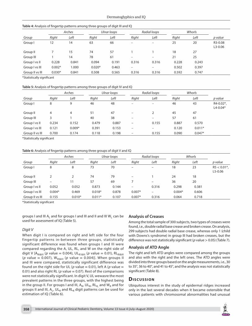
Digit IIIWhen digit III was compared on right and left side, statistically significant difference was found when groups I and III and groups II and III were compared regarding the prevalence of A on the right side of digit III [ARI&III (p = 0.002), ARII&III (p value = 0.03)]. When groups I and III were compared, statistically significant difference was found on the right side for UL (p value = 0.02) where group III had high UL. In digit I, UL was the most prevalent patterns in the three groups with the highest being in the group III. For group I and III, AR and ULR for group II and III, AR can be used for estimation of IQ (Table 4).
Digit IVWhen digit IV was analyzed on right and left sides, statistically significant difference was found regarding the prevalence of A on the left side of digit IV when groups I and III were compared [ALI&III (p value = 0.002)]. Furthermore, statistically significant difference was found on left side for W in both the comparison between groups I and III and groups II and III [WLI&III (p = 0.01), WLII and III (p = 0.04)]. In digit IV, W was the most prevalent patterns and for
Table 1: Demographic distribution among three groups
Group I Group II Group IIIBoys:Girls 50:50 55:45 53:47Age 9.46 (1.11) 8.74 (1.02) 6.48 (1.16)
Table 2: Analysis of fingertip patterns among three groups of digit I and IQ
Group
Arches Ulnar loops Radial loops Whorls
p valueRight Left Right Left Right Left Right LeftGroup I 5 7 60 60 – – 35 34 R1-0.03*,
L1-0.01*Group II 7 8 68 66 1 2 24 24Group III 1 – 53 55 – – 46 46Group I vs II 0.552 0.788 0.239 0.380 0.316 0.155 0.088 0.119Group I vs III 0.097 0.007* 0.318 0.474 – – 0.113 0.083Group II vs III 0.030* 0.004* 0.030* 0.112 0.316 0.155 0.001* 0.001*
*Statistically significant
Table 3: Analysis of fingertip patterns among three groups of digit II and IQ
Groups
Arches Ulnar loops Radial loops Whorls
p valueRight Left Right Left Right Left Right LeftGroup I 20 28 40 36 – – 40 36 R2-0.01*
L2-0.01*Group II 12 17 52 48 4 4 32 30Group III 18 14 35 47 6 5 41 35Group I vs II 0.123 0.063 0.089 0.086 0.043* 0.043* 0.239 0.367Group I vs III 0.718 0.015* 0.465 0.114 0.013* 0.024* 0.885 0.883Group II vs III 0.235 0.558 0.015* 0.887 0.516 0.733 0.186 0.450
*Statistically significant
Dermatoglyphics and IQ
International Journal of Clinical Pediatric Dentistry, Volume 13 Issue 4 (July–August 2020)358
groups I and III AL and for groups I and III and II and III WL can be used for assessment of IQ (Table 5).
Digit VWhen digit I is compared on right and left side for the four f ingertip patterns in-between three groups, statistically significant difference was found when groups I and III were compared regarding the A, UL, RL, and W on the right side of digit V [ARI&III (p value = 0.004), ULR1&III (p value = 0.01), RLRI&III (p value = 0.007), WRI&III (p value = 0.004)]. When groups II and III were compared, statistically significant difference was found on the right side for UL (p value = 0.01), left A (p value = 0.01) and also right RL (p value = 0.07). Rest of the comparisons were not statistically significant. In digit V, UL wewasre the most prevalent patterns in the three groups, with the highest being in the group II. For groups I and III, AR, ULR, RLR, and WR and for groups II and III, AL, ULR and RLR digit patterns can be used for estimation of IQ (Table 6).
Analysis of CreasesAmong the total sample of 300 subjects, two types of creases were found, i.e., double radial base crease and broken crease. On analysis, 299 subjects had double radial base crease, whereas only 1 (child with Downs’s syndrome) in group III had broken creases, but the difference was not statistically significant (p value > 0.05) (Table 7).
Analysis of ATD AngleThe right and left ATD angles were compared among the groups and also with the right and the left ones. The ATD angles were divided into three groups based on the angle measurements, i.e., 30 to 35°, 36 to 40°, and 41 to 45°, and the analysis was not statistically significant (Table 8).
dI s c u s s I o n Ubiquitous interest in the study of epidermal ridges increased only in the last several decades when it became ostensible that various patients with chromosomal abnormalities had unusual
Table 4: Analysis of fingertip patterns among three groups of digit III and IQ
Group
Arches Ulnar loops Radial loops Whorls
p valueRight Left Right Left Right Left Right LeftGroup I 12 14 63 66 – – 25 20 R3-0.08
L3-0.06Group II 7 15 74 57 1 1 18 27Group III 1 14 78 61 – – 21 25Group I vs II 0.228 0.841 0.094 0.191 0.316 0.316 0.228 0.243Group I vs III 0.002* 1.000 0.020* 0.463 – – 0.502 0.397Group II vs III 0.030* 0.841 0.508 0.565 0.316 0.316 0.592 0.747
*Statistically significant
Table 5: Analysis of fingertip patterns among three groups of digit IV and IQ
Group
Arches Ulnar loops Radial loops Whorls
p valueRight Left Right Left Right Left Right LeftGroup I 8 9 46 48 – – 46 43 R4-0.02*,
L4-0.04*Group II 4 4 51 47 – 2 45 47Group III 3 1 40 38 – – 57 61Group I vs II 0.234 0.152 0.479 0.887 – 0.155 0.887 0.570Group I vs III 0.121 0.009* 0.391 0.153 – – 0.120 0.011*Group II vs III 0.700 0.174 0.118 0.198 – 0.155 0.090 0.047*
*Statistically significant
Table 6: Analysis of fingertip patterns among three groups of digit V and IQ
Group
Arches Ulnar loops Radial loops Whorls
p valueRight Left Right Left Right Left Right LeftGroup I 8 8 73 70 – – 18 23 R5 < 0.01*,
L5-0.06Group II 2 2 74 79 – 1 24 18Group III – 11 57 69 7 – 36 20Group I vs II 0.052 0.052 0.873 0.144 – 0.316 0.298 0.381Group I vs III 0.004* 0.469 0.018* 0.878 0.007* – 0.004* 0.606Group II vs III 0.155 0.010* 0.011* 0.107 0.007* 0.316 0.064 0.718
*Statistically significant
Dermatoglyphics and IQ
International Journal of Clinical Pediatric Dentistry, Volume 13 Issue 4 (July–August 2020) 359
ridge configurations.5 Specifically, these ridge patterns make decent material for genetic studies because, unlike stature and body weight, they are not prejudiced by age or postnatal environmental factors and have the advantage of remaining unalterable throughout the life and therefore can be compared among individuals of different ages, especially in the diagnosis aberrations of genetic origin.1
“Absence of any correlation between dermatoglyphics and IQ” was the null hypothesis of the present study. As a result, this study sought to rule out the hypothesis by comparing differences between the relative frequencies of finger patterns among the 3 groups with different levels of IQ. Unto the extent of our knowledge, this is the first study to compare the fingertip patterns of all the ten fingers with IQ. Support for this correlation came from the remarks that a substantial part of learning difficulties in genetically inherited syndromes like trisomy 21 have been documented as having abnormal dermatoglyphic characteristics and also the dermal ridges are thought to be related to fetal development, which counterparts the development of the central nervous system.6 Furthermore, dermal ridges are predisposed by factors, such as maternal psychological stress, anticonvulsants,7 or alcohol8 ingested by the pregnant mother during fetal development,. Therefore, correlation may exist between the dermatoglyphics and IQ.
In the present study, the most recurrent dermatoglyphic patterns observed on different fingers were ulnar loops followed by whorls. Despite the fact that the frequencies of the digital patterns in the normal population as established by studies differ round the world, it has also been described that ulnar loops and whorls are the most common finger patterns,9 which confirms the results of this study.
A correlation between dermatoglyphics and IQ had already been reported but confined to particular digits.4,6,10 The results of the present study show statistically significant association of right digit I, II, IV, and V, while in the left hand, digits I, II, and IV had significant association with various levels of IQ. These results were in concomitant with Najafi6 and Tornjova‐Randelova11 who reported
the association between some dermatoglyphic patterns detected on the right digit II in normal ones, second and fourth digits among children with visual, auditory, and mental insufficiency and normal children, respectively. The constraint of this association to these digits might be related to the differing evolutionary olden times of the different digits and their innervations.11
Furthermore, in the present study, arches are the most common finding in group III which is similar to the study by Rosa et al.4 Increased arches, a simple fingerprint pattern, and increased radial loops, an unusual pattern, have been found in children predominantly with learning difficulties. Also, significant increase in abnormal flexion creases has also been identified in individuals with learning difficulties which was similar to the present study. Presence of abnormal palmer flexion creases is definitely associated with learning disabilities accompanying with chromosomal abnormalities, especially Down syndrome, i.e., chromosomal aberrations.
As the ATD angle decreases, there will be increase in the level of IQ as reported by Cesarik et al.12 This finding is in contrary to the observations of the present study, where there is no significance in the ATD angle. To some extent this may be because in the latter study three different levels of IQ were correlated but in the former one, the comparison was only between the superior intelligence and normal adolescence. However, when the two groups’ (group I and II) were compared, no significance was found.
In order to prevent the development of risk indicators in children with the presence of risk factors, Cvjeticanin and Polovina13 suggested that palmar and fingerprints be taken in the immediate postnatal period. Another study by Dar et al.14 concluded that rare features might specify an “at-risk” infant if dermatography was completed during the routine examination of the newborn. The results of their study testify a certain diagnostic and prognostic worth of dermatoglyphic features. Furthermore, presence of abnormal palmar flexion creases definitely indicates the children with intellectual disability.
The limitation of the present study is that the gender-based differences were not evaluated.
Table 7: Analysis of palmar flexion creases
Group
Total p valueI II IIICreases Broken creases Count 0 0 1 1 0.367
% within group 0.0 0.0 1.0 0.3SRBC Count 100 100 99 299
% within group 100.0 100.0 99.0 99.7Total Count 100 100 100 300
% within group 100.0 100.0 100.0 100.0*Statistically significant
Table 8: One-way ANOVA analysis of ATD angle
Sum of squares df Mean square F p valueRATD Between groups 35.248 2 17.624 0.986 0.374
Within groups 5306.299 297 17.866Total 5341.547 299
LATD Between groups 41.097 2 20.548 1.113 0.330Within groups 5485.500 297 18.470Total 5526.597 299
Dermatoglyphics and IQ
International Journal of Clinical Pediatric Dentistry, Volume 13 Issue 4 (July–August 2020)360
co n c lu s I o n Dermatoglyphics can be used as an adjunct in the estimation of IQ, which becomes more easier and economical method, especially in case of children with chromosomal abnormalities. In pediatric dentistry, it would be better if we apply behavior guidance techniques depending on IQ of the child, where the child can understand and accepts the treatment procedure.
Ac k n ow l e d g M e n tAll the authors made equal contribution in the study.
re f e r e n c e s 1. Schaumann B, Alter M. Dermatoglyphics in Medical Disorders.
Springer Verlag, New York 1976. 2. Aronson J. When I use a word…Fingerprints. Br Med J 1997;315(7113):i.
DOI: 10.1136/bmj.315.7113.0i. 3. Holt SB. Genetics of dermal ridges: sib-pair correlations for total finger
ridge count. Ann Hum Genet 1957;21(4):352–362. DOI: 10.1111/j.1469-1809.1972.tb00204.x.
4. Rosa A, Gutiérrez B, Guerra A, et al. Dermatoglyphics and abnormal palmar flexion creases as markers of early prenatal stress in children with idiopathic intellectual disability. J Intellect Disabil Res 2001; 45(Pt 5):416–423. DOI: 10.1046/j.1365-2788.2001.00351.x.
5. Venkatesh E, Bagewadi A, Keluskar V, et al. Palmar dermatoglyphics in oralleukoplakia and oral squamous cell carcinoma patients. Journal of Indian Academy of Oral Medicine and Radiology 2008;20(3):94–99. DOI: 10.4103/0972-1363.52774.
6. Najafi M. Association between finger patterns of digit II and intelligence quotient level in adolescence. Iran J Pediatr 2009;19: 277–284.
7. Andermann E, Danski I, Andermann F, et al. Dermatoglyphic changes and minor congenital malformations associated with maternal use of anticonvulsant drugs during pregnancy Dam M, Gram I, Perry JK, ed. Advances in Epileptology: 12th Epilepsy International Symposium. New York, NY: Raven Press; 1981. pp. 613–620.
8. Qazi QH, Masakawa A, McGann B, et al. Dermatoglyphic abnormalities in the fetal alcohol syndrome. Teratology 1980;21(2):157–160. DOI: 10.1002/tera.1420210204.
9. Reed T. Dermatoglyphics in medicine problems and use in suspected chromosome abnormalities. Am J Med Genet 1981;8(4):411–429. DOI: 10.1002/ajmg.1320080407.
10. Kiran K, Rai K, Hegde AM. Dermatoglyphics as a noninvasive diagnostic tool in predicting mental retardation. J Int Oral Health 2010;2:96–100.
11. Tornjova‐Randelova SG. Some aspects on the dermatoglyphics of normal and defective children in Bulgaria. Anthropol Anz 1994;52(4):351–355.
12. Cesarik M, Bozicevic D, Milicic J, et al. Quantitative dermatoglyphic analysis in persons with superior intelligence. Coll Antropol 1996;20:413–418.
13. Cvjeticanin M, Polovina A. Quantitative analysis of digitopalmar dermatoglyphics in male children with central nervous system lesion by quantification of clinical parameters of locomotor disorder. Acta Med Croatica 1999;53(1):5–10.
14. Dar H, Bolchinsky D, Jaffe M, et al. Routine analysis of dermatoglyphics and palmar creases in children with developmental disorders. Dev Med Child Neurol 1978;20(6):735–737. DOI: 10.1111/j.1469-8749.1978.tb15304.x.