Int J Clin Exp Pathol 2016;9(5):5004-5014 www.ijcep.com /ISSN:1936-2625/IJCEP0023375 Original Article Genome-wide analysis of differential methylation in pancreatic cancer Fan Yin 1* , Huan Wang 1* , Yuehua Men 2 , Jidong Li 3 , Minggen Hu 3 , Qingfang Li 1 1 Department of Cancer Center, General Hospital of PLA, 28 Fuxing Road, Beijing 100853, China; 2 Department of Dermatology, Peking University Third Hospital, 49 Huayuan North Road, Beijing 100191, China; 3 Department of Surgical Oncology, Cancer Center, Chinese People’s Liberation Army (PLA) General Hospital, 28 Fuxing Road, Beijing 100853, China. * Equal contributors. Received January 6, 2016; Accepted March 20, 2016; Epub May 1, 2016; Published May 15, 2016 Abstract: Pancreatic cancer as a fatal malignant tumor is difficult in diagnosis and treatment. Effective biomarkers are demanded in clinical practice. Up to now, there is little known about the DNA methylation signatures across the whole genome scale in pancreatic cancer. In this study, comparison of differential mathylation sites was performed between pancreatic cancer tissues and pericarcinous tissues using Infinium Human Methylation 450 Beadchips. A total of 24,417 CpG sites representing 9,589 genes were identified between two cohorts. Of the 24,417 CpG sites, 14,721 (60%, 14,721 of 24,417) CpG sites were hypomethylated and 9,705 (40%, 9,705 of 24,417) CpG sites were hypermethylated. GO (Gene Ontology) and KEGG analysis were implemented to systematically characterize the significant differential methylated genes between pancreatic cancer tissues and pericarcinous tissues. In addition, we further screened 51 genes with aberrant methylation, which were the most likely candidate methylation markers within the scale of global differential methylation profiling. GO and KEGG analysis indicated these genes owning a wide range of functions. The identification of differential methylated genes in this study provides information valu- able to the in-depth study of pancreatic cancer. Keywords: Pancreatic cancer, DNA methylation, CpG sites, KEGG, GO Introduction The estimated incidence of pancreatic cancer in the United States was 37,700 cases, and an estimated 34,300 patients died from the dis- ease in 2008 [1]. The overall 5-year survival rate among patients with pancreatic cancer is <5% [2]. Pancreatic cancer is more common in elderly persons than in younger persons, and less than 20% of patients present with local- ized, potentially curable tumors. Several envi- ronmental factors have been implicated, but evidence of a causative role exists only for tobacco use. The risk of pancreatic cancer in smokers is 2.5 to 3.6 times that in nonsmokers [3]. Some studies have shown an increased incidence of pancreatic cancer among patients with a history of diabetes or chronic pancreati- tis, and there is also evidence that chronic cir- rhosis, a high-fat, high-cholesterol diet, and previous cholecystectomy are associated with an increased incidence [4, 5]. More recently, an increased risk has been observed among patients with blood type A, B, or AB as com- pared with blood type O [6]. In some patients, pancreatic cancer develops as part of a well defined cancer-predisposing syndrome for which germ-line genetic alterations are known. In addition, in some families with an increased risk of pancreatic cancer, a genetic rather than an environmental cause is suspected [7]. Epigenetics is defined as the study of mitotically or meiotically heritable variations in gene func- tion that cannot be explained by changes in DNA sequence [8]. Recently, attention to its role in pancreatic cancer has recently increased. DNA methylation has gained much recent inter - est for its role in cancer biology. Aberrant pat- terns of DNA methylation are known to be asso- ciated with carcinogenesis and to affect the regulation of genome stability and gene tran- scription [9]. Genome wide studies of CpG islands have uncovered thousands of loci where differential methylation can segregate pancre- atic tumor tissue from normal tissue [10, 11]. Despite this progress, the combination research of changes in DNA methylation in pancreatic
Transcript
Int J Clin Exp Pathol 2016;9(5):5004-5014www.ijcep.com /ISSN:1936-2625/IJCEP0023375
Original ArticleGenome-wide analysis of differential methylation in pancreatic cancer
1Department of Cancer Center, General Hospital of PLA, 28 Fuxing Road, Beijing 100853, China; 2Department of Dermatology, Peking University Third Hospital, 49 Huayuan North Road, Beijing 100191, China; 3Department of Surgical Oncology, Cancer Center, Chinese People’s Liberation Army (PLA) General Hospital, 28 Fuxing Road, Beijing 100853, China. *Equal contributors.
Received January 6, 2016; Accepted March 20, 2016; Epub May 1, 2016; Published May 15, 2016
Abstract: Pancreatic cancer as a fatal malignant tumor is difficult in diagnosis and treatment. Effective biomarkers are demanded in clinical practice. Up to now, there is little known about the DNA methylation signatures across the whole genome scale in pancreatic cancer. In this study, comparison of differential mathylation sites was performed between pancreatic cancer tissues and pericarcinous tissues using Infinium Human Methylation 450 Beadchips. A total of 24,417 CpG sites representing 9,589 genes were identified between two cohorts. Of the 24,417 CpG sites, 14,721 (60%, 14,721 of 24,417) CpG sites were hypomethylated and 9,705 (40%, 9,705 of 24,417) CpG sites were hypermethylated. GO (Gene Ontology) and KEGG analysis were implemented to systematically characterize the significant differential methylated genes between pancreatic cancer tissues and pericarcinous tissues. In addition, we further screened 51 genes with aberrant methylation, which were the most likely candidate methylation markers within the scale of global differential methylation profiling. GO and KEGG analysis indicated these genes owning a wide range of functions. The identification of differential methylated genes in this study provides information valu-able to the in-depth study of pancreatic cancer.
Keywords: Pancreatic cancer, DNA methylation, CpG sites, KEGG, GO
Introduction
The estimated incidence of pancreatic cancer in the United States was 37,700 cases, and an estimated 34,300 patients died from the dis-ease in 2008 [1]. The overall 5-year survival rate among patients with pancreatic cancer is <5% [2]. Pancreatic cancer is more common in elderly persons than in younger persons, and less than 20% of patients present with local-ized, potentially curable tumors. Several envi-ronmental factors have been implicated, but evidence of a causative role exists only for tobacco use. The risk of pancreatic cancer in smokers is 2.5 to 3.6 times that in nonsmokers [3]. Some studies have shown an increased incidence of pancreatic cancer among patients with a history of diabetes or chronic pancreati-tis, and there is also evidence that chronic cir-rhosis, a high-fat, high-cholesterol diet, and previous cholecystectomy are associated with an increased incidence [4, 5]. More recently, an increased risk has been observed among patients with blood type A, B, or AB as com-
pared with blood type O [6]. In some patients, pancreatic cancer develops as part of a well defined cancer-predisposing syndrome for which germ-line genetic alterations are known. In addition, in some families with an increased risk of pancreatic cancer, a genetic rather than an environmental cause is suspected [7].
Epigenetics is defined as the study of mitotically or meiotically heritable variations in gene func-tion that cannot be explained by changes in DNA sequence [8]. Recently, attention to its role in pancreatic cancer has recently increased. DNA methylation has gained much recent inter-est for its role in cancer biology. Aberrant pat-terns of DNA methylation are known to be asso-ciated with carcinogenesis and to affect the regulation of genome stability and gene tran-scription [9]. Genome wide studies of CpG islands have uncovered thousands of loci where differential methylation can segregate pancre-atic tumor tissue from normal tissue [10, 11]. Despite this progress, the combination research of changes in DNA methylation in pancreatic
cancer tissues, pericarcinous tissues remains unexplored.
In this study, we have employed a global meth-ylation profiling platform in this work to compre-hensively survey a large scale of CpG sites in pancreatic cancer genome. We compared the DNA methylation profiles of the pancreatic tumors and pericarcinous tissues in order to unravel methylation markers for diagnostic pur-poses. The results suggested that 24,417 CpG sites representing 9,589 genes were detected, among which, 14,721 (60%, 14,721 of 24,417) CpG sites were hypomethylated and 9,705 (40%, 9,705 of 24,417) CpG sites were hyper-methylated. We then performed GO (Gene Ontology) and KEGG analysis to systematically characterize the differential methylated genes between pancreatic cancer tissues and peri-carcinous tissues. In addition, we further screened 51 genes with aberrant methylation, which were the most likely candidate methyla-tion markers within the scale of global differen-tial methylation. GO and KEGG analysis indicat-ed these genes owning a wide range of functions.
Materials and methods
Subjects and haematoxylin eosin (H&E) stain-ing
Six patients with pancreatic cancer (2 males and 4 females, mean age: 58.83±14.95 y), without radiation, chemotherapy and immuno-therapy treatment, were recruited from General Hospital of PLA in China. The diagnosis of pan-creatic cancer was made by at least two experi-enced oncologists. Sample collection accorded to the following criterions: 1) the minimum diameter of tumor was greater than 2 cm. Meanwhile, pancreatic cancer was identified by haematoxylin and eosin (H&E) staining and the ratio of cancer cells in the whole cells section was over 80%. 2) Tissue adjacent to cancer was collected as far as possible from the can-cer tissue in order to avoid the mistake sam-pling. Pancreatic cancer tissue and tissue adja-cent to cancer of each patient were collected and stored in liquid nitrogen immediately for DNA extraction and staining. All specimens were subjected to autolysis for 4 to 8 h and then snap-frozen at -80°C until use in analysis. DNA was extracted from 25 mg samples of the tissue specimens using the QIAamp DNA Mini
Kit (Qiagen) according to the manufacturer’s instructions. The DNA yield and purity were de- termined spectrophotometrically (NanoDrop® ND1000; Thermo Fisher Scientific Inc., Wal- tham, MA, USA) and by gel electrophoresis, respectively. DNA of sample was stored at -20°C for further study. Haematoxylin Eosin (H&E) staining were prepared according to the method of [12, 13]. Samples were fixed in 10% buffered formalin, and embedded in paraffin. Three to five micrometer thick sections were stained with hematoxylin (Sigma H 3136) for 10 min and with eosin (Sigma E 4382) for 1 min to establish the diagnosis areas. The Research Ethics Committee of General Hospital of PLA approved the collection of tissue samples for research. Written informed consent was obtained from individuals in order to collect the tissues during surgery.
DNA methylation methods
Bisulfite conversion of 500 ng genomic DNA was performed using the EZ DNA methylation kit (Zymo Research). DNA methylation level was assessed according to the manufacturer’s instructions using Infinium-HumanMethyla- tion450 Beadchips (Illumina Inc.). The technical schemes, the accuracy, and the high reproduc-ibility of this array have been described in previ-ous papers [14]. Quantitative measurements of DNA methylation were determined for 485,577 CpG dinucleotides, which covered 99% of the RefSeq genes and were distributed across the whole gene regions, including promoter, gene body, and 30-untranslated regions (UTRs). They also covered 96% of CGIs from the UCSC data-base with additional coverage in CGI shores (0-2 kb from CGI) and CGI shelves (2-4 kb from CGI). Detailed information on the contents of the array is available in the Infinium Hu- manMethylation450 User Guide and Human-Methylation450 manifest (www.illumina.com) and in recent papers [15]. DNA methylation data were analyzed with the methylation analy-sis module within the BeadStudio software (Illumina Inc.). DNA methylation status of the CpG sites was calculated as the ratio of the sig-nal from a methylated probe relative to the sum of both methylated and unmethylated probes. This value, known as b, ranges from 0 (com-pletely unmethylated) to 1 (fully methylated). For intra-chip normalization of probe intensi-ties, colored balance and background correc-tions in every set of ten samples from the same
DNA methylation profile of the pancreatic cancer
5006 Int J Clin Exp Pathol 2016;9(5):5004-5014
chip were performed using internal control probes. X chromosome CpG sites in the CGIs in the AR gene in this array as well as the internal control probes were checked to validate the DNA methylation measurements.
Statistical methods
Surrogate variable analysis was used to identify CpG loci showing significant differences in DNA methylation between pancreatic cancer tissues and pericarcinous tissues [16]. This analysis is useful in clinical studies, where a large number of clinical variables, including known and unknown factors, have a complicated joint impact on microarray data, as applied in previ-ous studies [17]. A false discovery rate (FDR) correction was applied at the 0.05 level for mul-tiple testing. A paired t-test was used to assess the significance of DNA methylation differences between pancreatic cancer tissue and tissue adjacent to cancer. p values <0.01 were con-sidered significant differential methylation of DNA methylation differences between two groups.
Bioinformatics
The biological processes, molecular functions, and cellular components of the identified differ-entially methylated genes were examined by using the KOBAS software to perform gene ontology (GO) annotation (http://kobas.cbi.pku.edu.cn/home.do) [18]. GO enrichment analysis of differentially methylated genes was imple-mented by GORILLA (http://cbl-gorilla.cs.tech-nion.ac.il/) [19], in which gene length bias was
corrected. GO terms with corrected p-value less than 10-4 were considered significantly enriched by differential methylated genes. KEGG is a database resource for understand-ing high-level functions and utilities of the bio-logical system, such as the cell, the organism and the ecosystem, from molecular-level infor-mation, especially large-scale molecular datas-ets generated by genome sequencing and other high-through put experimental technologies (http://www.genome.jp/kegg/). We used KOBA- S software to test the statistical enrichment of differential methylated genes in KEGG pathways.
Results
Histopathologic features
For each patient, one pancreatic cancer tissue and one pericarcinous tissue were collected respectively. Twelve tissues, including six pan-creatic cancer tissues and six pericarcinous tis-sues, were gathered from six patients accord-ing our rigorous set of criteria as described in method section (Table 1). Then all of the tis-sues were implemented haematoxylin and eosin (H&E) staining to test the accurate rate of sampling, the results indicated that pericarci-nous tissues of patients 2 and patients 4 can-not reach the standers that we set were exclud-ed from this study. Figure 1 showed two exam-ples of pancreatic cancer tissues and two peri-carcinous tissues after Haematoxylin Eosin (H&E) staining using optical microscope with different magnification. The islands of tumor cells were surrounded by an abundant muci-nous stroma with separation of the tumor cells from the surrounding stroma (clefting). Pro- minent increased vascularity was also noted, as well as solar elastosis and variable amounts of predominantly mononuclear inflammatory cells with scattered neutrophils.
Diagnostic differences in DNA methylation between pancreatic cancer tissues with peri-carcinous tissues
DNA methylation levels were compared be- tween six pancreatic cancer tissues with four pericarcinous tissues control subjects using Infinium HumanMethylation450 Bead Chips. Of 485,577 CpG sites, significant diagnostic dif-ferences in DNA methylation were observed at 24,417 CpG sites representing 9,589 genes at
Table 1. Haematoxylin and eosin (H&E) staining analysis of pancreatic cancer tissue and tissue adjacent to cancer of six patients
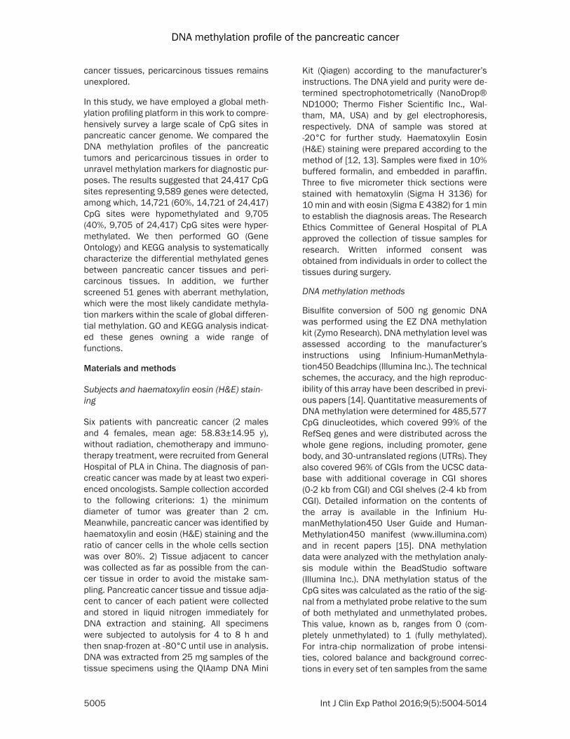
FDR 5% correction (Figure 2). Of 24,417 CpG sites with significant diagnostic differences in DNA methylation, 14,721 (60%, 14,721 of 24,417) CpG sites were hypomethylated and 9,705 (40%, 9,705 of 24,417) CpG sites were hypermethylated. Functional distribution of 9,705 hypermethylated CpG sites suggested that 42% of these sites were located in promot-er regions, 37% of these sites were located in gene bodies, 16% of these sites were located in intergenic regions and 5% of these sites were located in the 3’-untranslated regions (UTRs). Furthermore, sublocation analysis of 4032 CpG sites in promoter region with hypermethylated indicated that 35% of these sites were located in regions from -200 to -1,500 nt upstream of the transcription start site (TSS1500), 26% of these sites were located in regions from -200 nt upstream to the TSS itself (TSS200), 24% of these sites were located in 1st Exon regions and 15% of these sites were located in the
5’-untranslated regions (UTRs). These hyper-methylated CpG sites were mostly located in gene bodies and promoter regions. Meanwhile, Functional distribution of 14,721 hypomethyl-ated CpG sites suggested that 22% of these sites were located in promoter regions, 39% of these sites were located in gene bodies, 35% of these sites were located in intergenic regions and 4% of these sites were located in 3’UTR regions. Furthermore, sublocation analysis of 14,721 hypomethylated CpG sites in promoter regions indicated that 50% of these sites were located in TSS1500 regions, 16% of these sites were located in TSS200 regions, 13% of these sites were located in 1st Exon regions and 21% of these sites were located in 5’UTR regions. These hypomethylated CpG sites were mostly located in gene bodies, promoter regions and intergenic regions. The results above seem to be conflictive with the previous studies, which showed that gene promoter areas of actively
Figure 1. Aa. Pericarcinous tissue with 100 times magnification. Ab. Pericarcinous tissue with 200 times magni-fication. Ba. Pancreatic cancer tissue with 100 times magnification. Bb. Pancreatic cancer tissue with 200 times magnification.
DNA methylation profile of the pancreatic cancer
5008 Int J Clin Exp Pathol 2016;9(5):5004-5014
transcribed genes are largely unmethylated so as to be accessible to transcription factors. Based on the research object, we speculated that lots of transcription factor must be modi-fied and detained from transcription so that they can adapt to the cellular abnormalities as cancer.
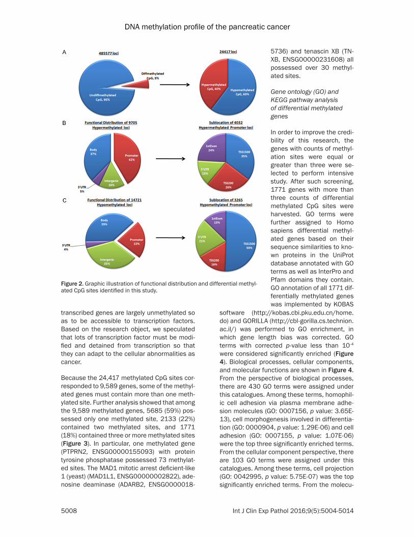
Because the 24,417 methylated CpG sites cor-responded to 9,589 genes, some of the methyl-ated genes must contain more than one meth-ylated site. Further analysis showed that among the 9,589 methylated genes, 5685 (59%) pos-sessed only one methylated site, 2133 (22%) contained two methylated sites, and 1771 (18%) contained three or more methylated sites (Figure 3). In particular, one methylated gene (PTPRN2, ENSG00000155093) with protein tyrosine phosphatase possessed 73 methylat-ed sites. The MAD1 mitotic arrest deficient-like 1 (yeast) (MAD1L1, ENSG00000002822), ade-nosine deaminase (ADARB2, ENSG0000018-
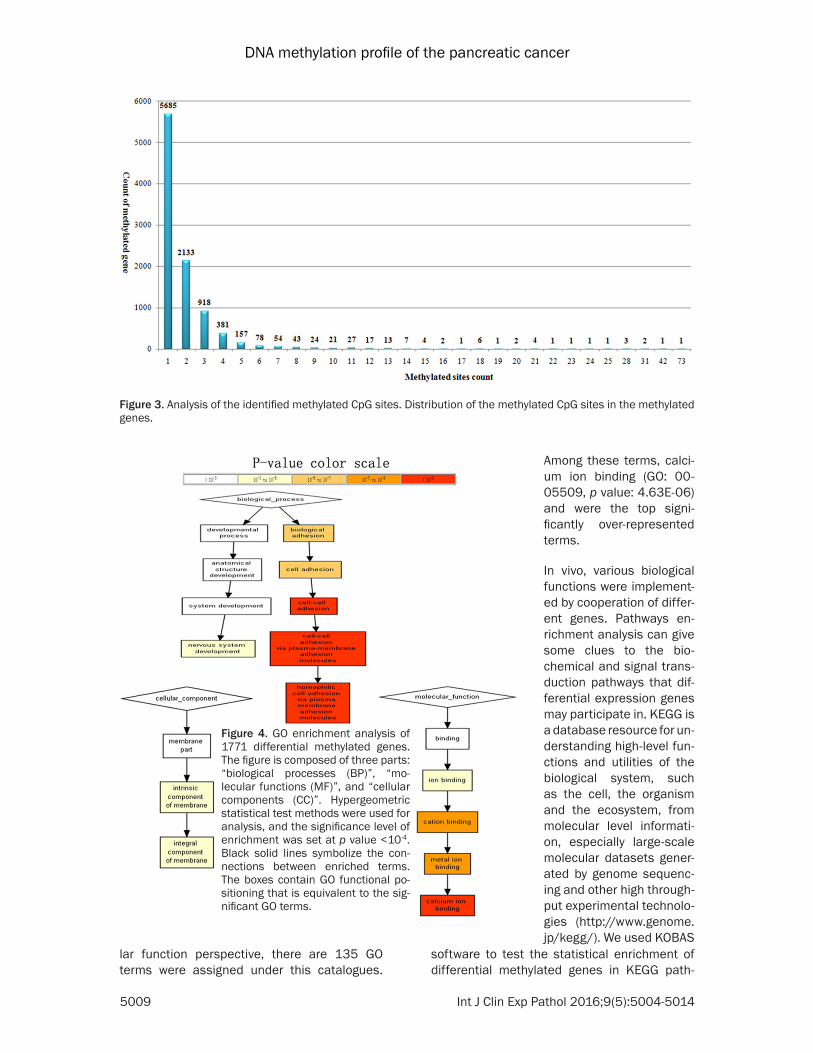
software (http://kobas.cbi.pku.edu.cn/home.do) and GORILLA (http://cbl-gorilla.cs.technion.ac.il/) was performed to GO enrichment, in which gene length bias was corrected. GO terms with corrected p-value less than 10-4 were considered significantly enriched (Figure 4). Biological processes, cellular components, and molecular functions are shown in Figure 4. From the perspective of biological processes, there are 430 GO terms were assigned under this catalogues. Among these terms, homophil-ic cell adhesion via plasma membrane adhe-sion molecules (GO: 0007156, p value: 3.65E-13), cell morphogenesis involved in differentia-tion (GO: 0000904, p value: 1.29E-06) and cell adhesion (GO: 0007155, p value: 1.07E-06) were the top three significantly enriched terms. From the cellular component perspective, there are 103 GO terms were assigned under this catalogues. Among these terms, cell projection (GO: 0042995, p value: 5.75E-07) was the top significantly enriched terms. From the molecu-
Figure 2. Graphic illustration of functional distribution and differential methyl-ated CpG sites identified in this study.
5736) and tenascin XB (TN- XB, ENSG00000231608) all possessed over 30 methyl-ated sites.
Gene ontology (GO) and KEGG pathway analysis of differential methylated genes
In order to improve the credi-bility of this research, the genes with counts of methyl-ation sites were equal or greater than three were se- lected to perform intensive study. After such screening, 1771 genes with more than three counts of differential methylated CpG sites were harvested. GO terms were further assigned to Homo sapiens differential methyl-ated genes based on their sequence similarities to kno- wn proteins in the UniProt database annotated with GO terms as well as InterPro and Pfam domains they contain. GO annotation of all 1771 dif-ferentially methylated genes was implemented by KOBAS
DNA methylation profile of the pancreatic cancer
5009 Int J Clin Exp Pathol 2016;9(5):5004-5014
lar function perspective, there are 135 GO terms were assigned under this catalogues.
Figure 3. Analysis of the identified methylated CpG sites. Distribution of the methylated CpG sites in the methylated genes.
Figure 4. GO enrichment analysis of 1771 differential methylated genes. The figure is composed of three parts: “biological processes (BP)”, “mo-lecular functions (MF)”, and “cellular components (CC)”. Hypergeometric statistical test methods were used for analysis, and the significance level of enrichment was set at p value <10-4. Black solid lines symbolize the con-nections between enriched terms. The boxes contain GO functional po-sitioning that is equivalent to the sig-nificant GO terms.
software to test the statistical enrichment of differential methylated genes in KEGG path-
Among these terms, calci-um ion binding (GO: 00- 05509, p value: 4.63E-06) and were the top signi- ficantly over-represented terms.
In vivo, various biological functions were implement-ed by cooperation of differ-ent genes. Pathways en- richment analysis can give some clues to the bio-chemical and signal trans-duction pathways that dif-ferential expression genes may participate in. KEGG is a database resource for un- derstanding high-level fun- ctions and utilities of the biological system, such as the cell, the organism and the ecosystem, from molecular level informati- on, especially large-scale molecular datasets gener-ated by genome sequenc-ing and other high through-put experimental technolo-gies (http://www.genome.jp/kegg/). We used KOBAS
DNA methylation profile of the pancreatic cancer
5010 Int J Clin Exp Pathol 2016;9(5):5004-5014
ways [20]. In this study, 1771 differential meth-ylated genes involve 261 pathways. Figure 5 shows the results of pathways enrichment, it clearly display that phosphatidylinositol signal-ing system were the top enriched term. 22 dif-ferential methylated genes that identified in our study participate in this pathway. Moreover, it is worth noting that arrhythmogenic right ventric-ular cardiomyopathy (ARVC), oxytocin signaling pathway, ascorbate and aldarate metabolism and Focal adhesion were also significant enriched in this study. The pathways mentioned above were adopted with the function that can-cer played.
Candidate genes regulated by aberrant DNA methylation
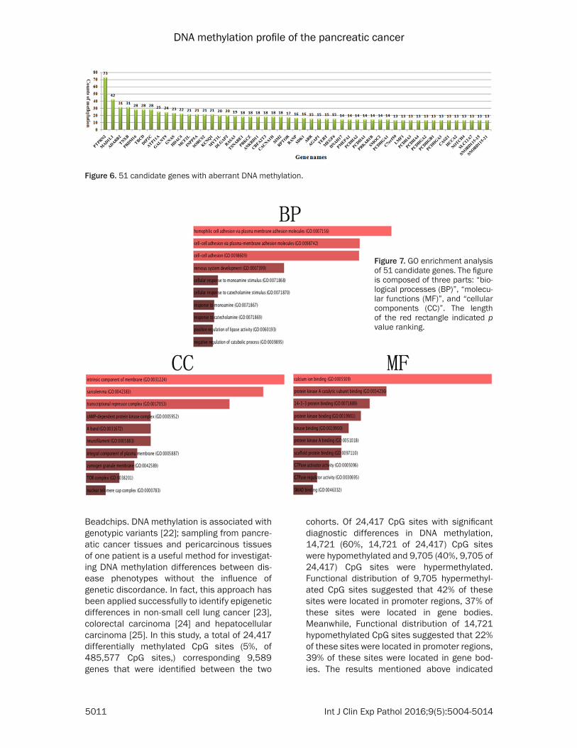
To pinpoint the candidate genes regulated by aberrant DNA methylation in pancreatic cancer, we focused on the genes of discordant catego-ries in Figure 6. Genes with count of methyla-tion was equal or greater than 13 were selected to perform further study (p value <0.05). After such screening, 51 genes with equal or more than 13 counts of methylated sites were har-vested. Functional descriptions of these genes suggested they owned a wide range. For exam-ple, PTPRN2 gene as the most significant meth-ylated item possessed 73 methylated CpG sites, which was identified as protein tyrosine phosphatase, MAD1L1 gene as the second sig-nificant methylated item possessed 42 methyl-ated CpG sites, which was identified as MAD1
31224, p value: 0.063) was the top significantly enriched terms. From the molecular function perspective, calcium ion binding (GO: 0005509, p value: 0.00011) and protein kinase A cata-lytic subunit binding (GO: 0034236, p value: 0.038) was the top two significantly enriched terms. Meanwhile, KEGG analysis indicated that Insulin signaling pathway (hsa04910, p value: 0.01), Circadian entrainment (hsa04713, p value: 0.036) and Platelet activation (hsa- 04611, p value: 0.06) were the most enriched in this study.
Discussion
Feinberg and Vogelstein were the first to asso-ciate differences in DNA methylation status to cancer in the early 80’ of the previous century [21]. Since then, it has been an explosion of research regarding aberrant DNA methylation of various diseases. Recently developed genome scale methods for mapping DNA meth-ylation across whole genomes have resulted in translation of basic discoveries. Therefore, aberrant methylation of genes which were associated with diseases, including types of cancer, Prader-Willi, Angelman and so on, were introduced into routine clinical application.
In this study, genome-wide DNA methylation profiling was conducted in pancreatic cancer tissues and pericarcinous tissues (six pancre-atic cancer tissues and four pericarcinous tis-sues) using Infinium HumanMethylation450
Figure 5. KEGG enrichment analysis of all the 1771 differential methylated genes identified in this study. The significance level of enrichment was set at p value <0.01.
mitotic arrest deficient-like 1. GO analysis of these 51 genes revealed function po- sitioning from three different viewpoints (Figure 7). From the perspective of biological processes, homophilic cell adhesion via plasma mem-brane adhesion molecules (GO: 0007156, p value: 1.27E-08), cell-cell adhesion via plasma-membrane adhe-sion molecules (GO: 0098- 742, p value: 1.34E-07) and nervous system develop-ment (GO: 0007399, p value: 0.026) were the top three significantly enriched terms. From the cellular component perspective, intrinsic compo-nent of membrane (GO: 00-
DNA methylation profile of the pancreatic cancer
5011 Int J Clin Exp Pathol 2016;9(5):5004-5014
Beadchips. DNA methylation is associated with genotypic variants [22]; sampling from pancre-atic cancer tissues and pericarcinous tissues of one patient is a useful method for investigat-ing DNA methylation differences between dis-ease phenotypes without the influence of genetic discordance. In fact, this approach has been applied successfully to identify epigenetic differences in non-small cell lung cancer [23], colorectal carcinoma [24] and hepatocellular carcinoma [25]. In this study, a total of 24,417 differentially methylated CpG sites (5%, of 485,577 CpG sites,) corresponding 9,589 genes that were identified between the two
cohorts. Of 24,417 CpG sites with significant diagnostic differences in DNA methylation, 14,721 (60%, 14,721 of 24,417) CpG sites were hypomethylated and 9,705 (40%, 9,705 of 24,417) CpG sites were hypermethylated. Functional distribution of 9,705 hypermethyl-ated CpG sites suggested that 42% of these sites were located in promoter regions, 37% of these sites were located in gene bodies. Meanwhile, Functional distribution of 14,721 hypomethylated CpG sites suggested that 22% of these sites were located in promoter regions, 39% of these sites were located in gene bod-ies. The results mentioned above indicated
Figure 6. 51 candidate genes with aberrant DNA methylation.
Figure 7. GO enrichment analysis of 51 candidate genes. The figure is composed of three parts: “bio-logical processes (BP)”, “molecu-lar functions (MF)”, and “cellular components (CC)”. The length of the red rectangle indicated p value ranking.
DNA methylation profile of the pancreatic cancer
5012 Int J Clin Exp Pathol 2016;9(5):5004-5014
that hypomethylated CpG sites seem to be more prevalent in the whole genome. On the contrary, hypermethylated CpG sites can be more easily indentified in promoter regions. As shown in Figure 2, aberrant DNA methylation in pancreatic cancer was mostly observed at CpG sites in the gene bodies and promoter regions. The present study demonstrated that altered DNA methylation in pancreatic cancer tissues occurred at CpG sites not only in the CGIs but also in CGI shores and CGI shelves. This is con-sistent with previous research that is methyla-tion of these regions can represses transcrip-tion. For example, Irizarry et al. demonstrated that altered DNA methylation in cancer occurred in CGI shores rather than in the CGIs, and DNA methylation changes in CGI shores were strong-ly related to gene expression [26]. In addition, we had noticed that numerous differential CpG sites were located in gene bodies. It is still unknown about the mechanism that how these differential CpG sites can have impact on gene functions. Shann et al. demonstrated the cor-relation between intragenic hypomethylation and gene silencing in cancer cell lines [27], and Ball et al. demonstrated that gene body DNA methylation in highly expressed genes is a con-sistent phenomenon in human cells [28]. Recently, it became apparent that CGIs in gene bodies act as alternative promoters [29, 30] and that tissue-specific or cell type-specific CGI methylation is prevalent in gene bodies [30, 31]. GO enrichment analysis suggested that significant function differences were identified between pancreatic cancer tissues with peri-carcinous tissues. Moreover, KEGG analysis indicated several important pathways can be retrieved in this study. For example, previous studies suggested that phosphatidylinositol 3-kinases in the most enriched item of phos-phatidylinositol signaling system (hsa04070) play important role PI3K/Akt/mTOR signaling pathway, which has impact on proliferation and activation of tumor cell [32, 33], and this path-way is becoming to research highlights of tar-geted drug. Moreover, we have noticed that focal adhesion kinase (FAK) which belongs to Focal adhesion (hsa04510) pathway. FAK is a focal adhesion-associated protein kinase involved in cellular adhesion (how cells stick to each other and their surroundings) and spread-ing processes (how cells move around) [34]. It has been shown that when FAK was blocked, breast cancer cells became less metastatic
due to decreased mobility [35]. FAK is phos-phorylated in response to integrin engagement, growth factor stimulation, and the action of mitogenic neuropeptides [36, 37].
There are several limitations to the present study. First, the sample size was not large. Replication studies will be needed in larger samples. Second, the analyzed CpG sites were limited in number, although the 450 K microar-ray is one of the most powerful and cost-effec-tive tools currently available for assessing methylation changes. Third, it is not possible to differentiate methylation from 5-hydroxymeth-ylation of cytosine, which also plays a critical role in gene regulation [38]. In summary, aber-rant DNA methylation in pancreatic cancer tis-sues was identified at numerous CpG sites across the whole genome in using two indepen-dent sets of samples. Of the differently methyl-ated CpG sites in the CGIs, most of them were located in the promoter regions. These findings support the hypothesis that altered DNA meth-ylation could be involved in the pathophysiology of pancreatic cancer. In this study, we analyzed the genome-wide DNA methylation profiles of human somatic tissues. Although the number of analyzed individuals was limited, the analy-sis was sufficient to provide DNA methylation distribution patterns across different genomic regions that were largely in agreement with pat-terns previously observed by previous studies. The methylome data alone was sufficient for correctly distinguishing between all the ten tis-sues studied, collectively demonstrating that tissues are characterized by distinctive methyl-ation patterns that reflect their tissue-specific functions. Our study provoked the question, of how differential methylated CpG sites mecha-nistically contribute to the gene functions, especially for the numerous methylation regions that were found in gene body areas. In addition, it remains unclear, however, how the gene body differential methylated CpG sites may function as regulators of gene expression, and this question should be addressed in the future epigenetic studies.
Conclusion
Previous studies have demonstrated that DNA methylation play important roles in the regula-tion of developmental processes of several types cancers. In this study, a total of 24,417
DNA methylation profile of the pancreatic cancer
5013 Int J Clin Exp Pathol 2016;9(5):5004-5014
CpG sites representing 9,589 genes that were common between these two cohorts were iden-tified. Of the 24,417 CpG sites, 14,721 (60%, 14,721 of 24,417) CpG sites were hypomethyl-ated and 9,705 (40%, 9,705 of 24,417) CpG sites were hypermethylated. Then GO (Gene Ontology) and KEGG analysis were implement-ed to systematically characterize the significant differential methylated genes between pancre-atic cancer tissues and pericarcinous tissues. In addition, we further screened 51 genes with aberrant methylation, which were the most like-ly candidate methylation markers within the scale of global differential methylation profiling. GO and KEGG analysis indicated these genes owning a wide range of functions. The identifi-cation of differential methylated genes in this study provides information valuable to the in-depth study of pancreatic cancer. Moreover, the results of this study will not only further our understanding of the differential methylated genes, but will also help to enhance methylome studies of pancreatic cancer.
Acknowledgements
This study was supported by the National Natural Science Foundation of China (No: 811- 70494), Natural Science Foundation of Beijing (No: 7162176) and Beijing Nova program (Z121107002512122).
Disclosure of conflict of interest
None.
Address correspondence to: Dr. Qingfang Li, De- partment of Cancer Center, General Hospital of PLA, 28 Fuxing Road, Beijing 100853, China. Tel: +86-10-66937592; Fax: +86-10-66937592; E-mail: [email protected]
References
[1] Jemal A, Siegel R, Ward E, Hao Y, Xu J, Murray T, Thun MJ. Cancer statistics, 2008. CA Cancer J Clin 2008; 58: 71-96.
[3] Hassan MM, Bondy ML, Wolff RA, Abbruzzese JL, Vauthey JN, Pisters PW, Evans DB, Khan R, Chou TH, Lenzi R, Jiao L, Li D. Risk factors for pancreatic cancer: case-control study. Am J Gastroenterol 2007; 102: 2696-707.
[4] Batty GD, Kivimaki M, Morrison D, Huxley R, Smith GD, Clarke R, Marmot MG, Shipley MJ. Risk factors for pancreatic cancer mortality:
extended follow-up of the original Whitehall Study. Cancer Epidemiol Biomarkers Prev 2009; 18: 673-5.
[5] Landi S. Genetic predisposition and environ-mental risk factors to pancreatic cancer: A re-view of the literature. Mutat Res 2009; 681: 299-307.
[6] Wolpin BM, Chan AT, Hartge P, Chanock SJ, Kraft P, Hunter DJ, Giovannucci EL, Fuchs CS. ABO blood group and the risk of pancreatic cancer. J Natl Cancer Inst 2009; 101: 424-31.
[7] Tersmette AC, Petersen GM, Offerhaus GJ, Falatko FC, Brune KA, Goggins M, Rozenblum E, Wilentz RE, Yeo CJ, Cameron JL, Kern SE, Hruban RH. Increased risk of incident pancre-atic cancer among first-degree relatives of pa-tients with familial pancreatic cancer. Clin Cancer Res 2001; 7: 738-44.
[8] Petronis A, Gottesman II, Crow TJ, DeLisi LE, Klar AJ, Macciardi F, McInnis MG, McMahon FJ, Paterson AD, Skuse D, Sutherland GR. Psychiatric epigenetics: a new focus for the new century. Mol Psychiatry 2000; 5: 342-6.
[9] Alvarez H, Opalinska J, Zhou L, Sohal D, Fazzari MJ, Yu Y, Montagna C, Montgomery EA, Canto M, Dunbar KB, Wang J, Roa JC, Mo Y, Bhagat T, Ramesh KH, Cannizzaro L, Mollenhauer J, Thompson RF, Suzuki M, Meltzer SJ, Melnick A, Greally JM, Maitra A, Verma A. Widespread hy-pomethylation occurs early and synergizes with gene amplification during esophageal car-cinogenesis. PLoS Genet 2011; 7: e1001356.
[10] Vincent A, Omura N, Hong SM, Jaffe A, Eshleman J, Goggins M. Genome-wide analy-sis of promoter methylation associated with gene expression profile in pancreatic adeno-carcinoma. Clin Cancer Res 2011; 17: 4341-54.
[11] Nones K, Waddell N, Song S, Patch AM, Miller D, Johns A, Wu J, Kassahn KS, Wood D, Bailey P, Fink L, Manning S, Christ AN, Nourse C, Kazakoff S, Taylor D, Leonard C, Chang DK, Jones MD, Thomas M, Watson C, Pinese M, Cowley M, Rooman I, Pajic M; APGI, Butturini G, Malpaga A, Corbo V, Crippa S, Falconi M, Zamboni G, Castelli P, Lawlor RT, Gill AJ, Scarpa A, Pearson JV, Biankin AV, Grimmond SM. Genome-wide DNA methylation patterns in pancreatic ductal adenocarcinoma reveal epi-genetic deregulation of SLIT-ROBO, ITGA2 and MET signaling. Int J Cancer 2014; 135: 1110-8.
[12] Chan JK. The wonderful colors of the hematox-ylin-eosin stain in diagnostic surgical patholo-gy. Int J Surg Pathol 2014; 22: 12-32.
[13] Fischer AH, Jacobson KA, Rose J, Zeller R. Hematoxylin and eosin staining of tissue and cell sections. CSH Protoc 2008; 2008: pdb. prot 4986.
[14] Dedeurwaerder S, Defrance M, Calonne E, Denis H, Sotiriou C, Fuks F. Epigenomics 2011; 3: 771-84.
[15] Sandoval J, Heyn H, Moran S, Serra-Musach J, Pujana MA, Bibikova M, Esteller M. Validation of a DNA methylation microarray for 450,000 CpG sites in the human genome. Epigenetics 2011; 6: 692-702.
[16] Leek JT, Storey JD. Capturing heterogeneity in gene expression studies by surrogate variable analysis. PLoS Genet 2007; 3: 1724-35.
[17] Numata S, Ye T, Hyde TM, Guitart-Navarro X, Tao R, Wininger M, Colantuoni C, Weinberger DR, Kleinman JE, Lipska BK. DNA methylation signatures in development and aging of the human prefrontal cortex. Am J Hum Genet 2012; 90: 260-72.
[18] Xie C, Mao X, Huang J, Ding Y, Wu J, Dong S, Kong L, Gao G, Li CY, Wei L. KOBAS 2.0: a web server for annotation and identification of en-riched pathways and diseases. Nucleic Acids Res 2011; 39: W316-22.
[19] Eden E, Navon R, Steinfeld I, Lipson D, Yakhini Z. GOrilla: a tool for discovery and visualization of enriched GO terms in ranked gene lists. BMC Bioinformatics 2009; 10: 48.
[20] Mao X, Cai T, Olyarchuk JG, Wei L. Automated genome annotation and pathway identification using the KEGG Orthology (KO) as a controlled vocabulary. Bioinformatics 2005; 21: 3787-3793.
[21] Feinberg AP, Vogelstein B. Hypomethylation distinguishes genes of some human cancers from their normal counterparts. Nature 1983; 301: 89-92.
[22] Numata S, Ye T, Hyde TM, Guitart-Navarro X, Tao R, Wininger M, Colantuoni C, Weinberger DR, Kleinman JE, Lipska BK. DNA methylation signatures in development and aging of the human prefrontal cortex. Am J Hum Genet 2012; 90: 260-72.
[23] Wang LG, Ni Y, Su BH, Mu XR, Shen HC, Du JJ. MicroRNA-34b functions as a tumor suppres-sor and acts as a nodal point in the feedback loop with Met. Int J Oncol 2013; 42: 957-62.
[24] Jin S, Mu Y, Wang X, Liu Z, Wan L, Xiong Y, Zhang Y, Zhou L, Li L. Overexpressed RACK1 is positively correlated with malignant degree of human colorectal carcinoma. Mol Biol Rep 2014; 41: 3393-9.
[25] Wang Z, Zhang L, Zhang D, Sun R, Wang Q, Liu X. Glycolysis inhibitor 2-deoxy-D-glucose sup-presses carcinogen-induced rat hepatocar-cinogenesis by restricting cancer cell metabo-lism. Mol Med Rep 2015; 11: 1917-24.
[26] Irizarry RA, Ladd-Acosta C, Wen B, Wu Z, Montano C, Onyango P, Cui H, Gabo K, Rongione M, Webster M, Ji H, Potash JB, Sabunciyan S, Feinberg AP. The human colon cancer methylome shows similar hypo- and hy-permethylation at conserved tissue-specific CpG island shores. Nat Genet 2009; 41: 178-86.
[27] Shann YJ, Cheng C, Chiao CH, Chen DT, Li PH, Hsu MT. Genome-wide mapping and character-ization of hypomethylated sites in human tis-sues and breast cancer cell lines. Genome Res 2008; 18: 791-801.
[28] Ball MP, Li JB, Gao Y, Lee JH, LeProust EM, Park IH, Xie B, Daley GQ, Church GM. Targeted and genome-scale strategies reveal gene-body methylation signatures in human cells. Nat Biotechnol 2009; 27: 361-8.
[29] Illingworth RS, Gruenewald-Schneider U, Webb S, Kerr AR, James KD, Turner DJ, Smith C, Harrison DJ, Andrews R, Bird AP. Orphan CpG islands identify numerous conserved promot-ers in the mammalian genome. PLoS Genet 2010; 6: e1001134.
[30] Maunakea AK, Nagarajan RP, Bilenky M, Ballinger TJ, D’Souza C, Fouse SD, Johnson BE, Hong C, Nielsen C, Zhao Y, Turecki G, Delaney A, Varhol R, Thiessen N, Shchors K, Heine VM, Rowitch DH, Xing X, Fiore C, Schillebeeckx M, Jones SJ, Haussler D, Marra MA, Hirst M, Wang T, Costello JF. Conserved role of intragenic DNA methylation in regulating alternative promot-ers. Nature 2010; 466: 253-7.
[31] Deaton AM, Webb S, Kerr AR, Illingworth RS, Guy J, Andrews R, Bird A. Cell type-specific DNA methylation at intragenic CpG islands in the immune system. Genome Res 2011; 21: 1074-86.
[32] Yu G, Huang B, Chen G, Mi Y. Phospha- tidylethanolamine-binding protein 4 promotes lung cancer cells proliferation and invasion via PI3K/Akt/mTOR axis. J Thorac Dis 2015; 7: 1806-1816.
[33] Tang H, Li RP, Liang P, Zhou YL, Wang GW. miR-125a inhibits the migration and invasion of liver cancer cells via suppression of the PI3K/AKT/mTOR signaling pathway. Oncol Lett 2015; 10: 681-686.
[34] Blackshaw SE, Kamal DJ, Lackie JM. The dic-tionary of cell and molecular biology. 3rd edi-tion. San Diego: Academic Press; 1999. ISBN 0-12-432565-3.
[35] Leslie M. Physorg: When cancer cells can’t let go. J Cell Biol 2009; 185: 178.
[36] Chen R, Kim O, Li M, Xiong X, Guan JL, Kung HJ, Chen H, Shimizu Y, Qiu Y. Regulation of the PH-domain-containing tyrosine kinase Etk by focal adhesion kinase through the FERM do-main. Nat Cell Biol 2001; 3: 439-44.
[37] Eliceiri BP, Puente XS, Hood JD, Stupack DG, Schlaepfer DD, Huang XZ, Sheppard D, Cheresh DA. Src-mediated coupling of focal ad-hesion kinase to integrin alpha (v) beta5 in vascular endothelial growth factor signaling. J Cell Biol 2002; 157: 149-60.
[38] Bhutani N, Burns DM, Blau HM. DNA demethyl-ation dynamics. Cell 2011; 146: 866-72.