Int J Clin Exp Pathol 2017;10(4):4321-4329 www.ijcep.com /ISSN:1936-2625/IJCEP0048267 Original Article microRNA-433 inhibits gastric cancer cell proliferation and migration and promotes apoptosis by targeting JNK1 Yifeng Zhao 1,2 , Xiujuan Li 3 , Xianghui He 1 , Yuping Zhang 4 , Haixia Qiao 5 , Shuguang Li 6 , Mingxia Li 7 1 Department of General Surgery, Tianjin Medical University General Hospital, Tianjin 300052, China; 2 Depart- ment of General Surgery, The First Affiliated Hospital of Hebei North University, Hebei 075000, China; Depart- ments of 3 Pathology, 4 Pathophysiology, 5 Microbiology, Hebei North University, Hebei 075000, China; Departments of 6 Gastrointestinaloma Surgery, 7 Endocrinology, The First Affiliated Hospital of Hebei North University, Hebei 075000, China Received January 6, 2017; Accepted February 20, 2017; Epub April 1, 2017; Published April 15, 2017 Abstract: Gastric cancer (GC) is the most frequent gastrointestinal tumor in adults and is the most lethal form of human cancer. A number of microRNAs (miRNAs) have been implicated in cell cycle progression, growth, apoptosis, angiogenesis and metastasis in GC. To define novel modulators that regulate susceptibility to tumorgenesis, we fo- cused on miRNA-433. Quantitative RT-PCR was employed to investigate the level of miRNA-433 in GC cell lines and normal human gastric mucosal epithelial cells (GES-1). Luciferase reporter gene assays were performed to identify the interaction between miR-433 and 3’UTR of Jun N-terminal kinase (JNK1) mRNA. In vitro cell proliferation, cell cycle, apoptosis assays, cell migration, and invasion assays were performed by CCK-8, flow cytometry and transwell assay, respectively, to elucidate biological effects of miRNA-433. The protein level of JNK1, Bax, Bcl-2, Caspase-3, PCNA, MMP-2 and MMP-9 was determined by Western blotting. We found that miR-433 was down-regulated in GC cell lines compared with normal gastric GES-1 cells. Restoring miR-433 expression in GC cells dramatically sup- pressed cells proliferation, invasion and migration. Importantly, our data showed that miR-433 down-regulated the expression of JNK1 through directly targeting its 3’UTR. In conclusion, miRNA-433 is potentially involved in GC progression and metastasis by targeting JNK1, which may provide a novel potential target for diagnostic and thera- peutic applications in GC. Keywords: Gastric cancer, miR-433, JNK1, MKN-45 Introduction Cancer is a universal public health problem because of its high incidence and mortality rate. Currently, gastric cancer (GC) is the fourth most common human malignant disease and the second leading cause of cancer-related deaths worldwide [1], with an overall 5-year sur- vival of approximately 20% in patients with late- stage GC [2]. Advances in treatment of this dis- ease are likely to come from a fuller under- standing of its biology and behavior. Eating habits and an increase in Helicobacter pyloriin- fection are important causes for GC [3]. Data from several studies show that various genetic alterations affect the downstream signal trans- duction pathways involved in the control of cell growth and differentiation and confer a tremen- dous survival and growth advantage to GC cells, thus cause tumorigenesis and progression of GC [4, 5]. However, markers for tumorigenesis and progression of gastric cancer have not yet been discovered and specific therapeutic tar- gets have not been identified. MicroRNAs (miRNA) are a class of small RNA molecules involved in regulating gene function through specifically binding to the 3’untranslat- ed region (3’UTR) of target mRNAs for transla- tion and degradation [6]. A growing body of evi- dence indicates that miRNAs are implicated in the pathogenesis of a variety of human diseas- es, notably cancers function as oncogenes or tumor suppressors [7, 8]. Several recent stud- ies conducted in human specimens point to a physiological function of microRNA-433 (miR-
Transcript
Int J Clin Exp Pathol 2017;10(4):4321-4329www.ijcep.com /ISSN:1936-2625/IJCEP0048267
Original Article microRNA-433 inhibits gastric cancer cell proliferation and migration and promotes apoptosis by targeting JNK1
1Department of General Surgery, Tianjin Medical University General Hospital, Tianjin 300052, China; 2Depart-ment of General Surgery, The First Affiliated Hospital of Hebei North University, Hebei 075000, China; Depart-ments of 3Pathology, 4Pathophysiology, 5Microbiology, Hebei North University, Hebei 075000, China; Departments of 6Gastrointestinaloma Surgery, 7Endocrinology, The First Affiliated Hospital of Hebei North University, Hebei 075000, China
Received January 6, 2017; Accepted February 20, 2017; Epub April 1, 2017; Published April 15, 2017
Abstract: Gastric cancer (GC) is the most frequent gastrointestinal tumor in adults and is the most lethal form of human cancer. A number of microRNAs (miRNAs) have been implicated in cell cycle progression, growth, apoptosis, angiogenesis and metastasis in GC. To define novel modulators that regulate susceptibility to tumorgenesis, we fo-cused on miRNA-433. Quantitative RT-PCR was employed to investigate the level of miRNA-433 in GC cell lines and normal human gastric mucosal epithelial cells (GES-1). Luciferase reporter gene assays were performed to identify the interaction between miR-433 and 3’UTR of Jun N-terminal kinase (JNK1) mRNA. In vitro cell proliferation, cell cycle, apoptosis assays, cell migration, and invasion assays were performed by CCK-8, flow cytometry and transwell assay, respectively, to elucidate biological effects of miRNA-433. The protein level of JNK1, Bax, Bcl-2, Caspase-3, PCNA, MMP-2 and MMP-9 was determined by Western blotting. We found that miR-433 was down-regulated in GC cell lines compared with normal gastric GES-1 cells. Restoring miR-433 expression in GC cells dramatically sup-pressed cells proliferation, invasion and migration. Importantly, our data showed that miR-433 down-regulated the expression of JNK1 through directly targeting its 3’UTR. In conclusion, miRNA-433 is potentially involved in GC progression and metastasis by targeting JNK1, which may provide a novel potential target for diagnostic and thera-peutic applications in GC.
Keywords: Gastric cancer, miR-433, JNK1, MKN-45
Introduction
Cancer is a universal public health problem because of its high incidence and mortality rate. Currently, gastric cancer (GC) is the fourth most common human malignant disease and the second leading cause of cancer-related deaths worldwide [1], with an overall 5-year sur-vival of approximately 20% in patients with late-stage GC [2]. Advances in treatment of this dis-ease are likely to come from a fuller under-standing of its biology and behavior. Eating habits and an increase in Helicobacter pyloriin-fection are important causes for GC [3]. Data from several studies show that various genetic alterations affect the downstream signal trans-duction pathways involved in the control of cell growth and differentiation and confer a tremen-
dous survival and growth advantage to GC cells, thus cause tumorigenesis and progression of GC [4, 5]. However, markers for tumorigenesis and progression of gastric cancer have not yet been discovered and specific therapeutic tar-gets have not been identified.
MicroRNAs (miRNA) are a class of small RNA molecules involved in regulating gene function through specifically binding to the 3’untranslat-ed region (3’UTR) of target mRNAs for transla-tion and degradation [6]. A growing body of evi-dence indicates that miRNAs are implicated in the pathogenesis of a variety of human diseas-es, notably cancers function as oncogenes or tumor suppressors [7, 8]. Several recent stud-ies conducted in human specimens point to a physiological function of microRNA-433 (miR-
433) in human diseases. miR-433 was decreased in hepatocellular carcinoma and inhibited cell proliferation by targeting p21 acti-vated kinase (PAK4) [9]. Decreased expression of miNA-433 is associated with the prognosis of ovarian cancer [10] and inhibits cell migra-tion and invasion via targeting Notch1 [11]. miR-433 was down-regulated in GC patients [12] and low expression of miR-433 had poorer survival than did patients with high expression of miR-433 [13]. However, the role of miR-433 in GC remains largely unknown.
Protein kinases are the largest family of enzymes encoded by the human genome and have emerged as one of the most popular drug target class in the pharmaceutical industry [14]. Jun N-terminal kinase (JNK) is a major MAPK pathway, which is called stress activated protein kinase pathway and is often deregulat-ed in cancers [15]. The JNK kinase family includes three proteins (JNK1, JNK2 and JNK3). The early findings that the transforming actions of several oncogenes could be JNKs depen-dent, suggested that the JNK signaling could contribute to the cellular transformation that supports cancer development [16, 17]. JNK1 is activated in primary hepatocellular carcinomas (HCC) and is associated with both a poor dis-ease prognosis and expression of progenitor cell biomarkers [18], and absence of JNK1 impaired liver cell proliferation and tumor for-mation [19]. In sharp contrast to the tumor-pro-moting role of JNK1 described above, in breast cancer models, the JNK1 has been shown to suppress tumor development [20]. However, the role of JNK1 in GC tumorigenesis is still unknown.
In the present study, miR-433 expression was evaluated by qRT-PCR in 3 GC cell lines, which showed that miR-433 was downregulated in GC cell lines and inversely correlated with JNK1 expression. Our data further demonstrated that miR-433 suppressed GC cell proliferation, invasion and migration and induced cell cycle arrest and apoptosis. More importantly, we demonstrated that forced expression of JNK1 rescued the inhibitory effects of miRNA-433 on GC proliferation, apoptosis, invasion and migra-tion. Therefore, our study suggests that miRNA-433 inhibits GC tumorigenesis by directly tar-geting JNK1.
Materials and methods
Cell culture and transfection
GES-1, BGC-823, MKN-28, and MKN-45 cell lines were obtained from the Institute of Biochemistry and Cell Biology (Shanghai, China). Cells were grown routinely in RPMI-1640 medium (Invitrogen, CA, USA) supple-mented with 10% fetal bovine serum (Gibco, CA, USA) plus 50 U/ml penicillin and streptomy-cin and cultured in a 37°C humidified atmo-sphere of 5% CO2.
Lentiviral production and transduction
Transduction of cells was performed using lipo-fectamine 2000 (Invitrogen Life Technologies, Gaithersburg, MD, USA) according to the manu-facturer’s protocol. Briefly, MKN-45 cells were seeded in 6-well plates at 30-40% confluence 24 h prior to transduction. The pre-miR-433 and JNK1 coding sequencewas cloned into the pLKO.1-C1 lentiviral vector. The constructs were then co-transducted into HEK 293T cells with lentiviral packaging vectors by using lipo-fectamine 2000. Viruses were collected 48 h after transduction and used to infect MKN-45 cells. Empty pLKO.1-C1 lentiviral vector was used as a negative control (Mock).
Luciferase reporter assays
The 3’UTR of human JNK1 gene that were pre-dicted to interact with miR-433 were synthe-sized and inserted into downstream of the fire-fly luciferase gene in the pGL3 vector (Promega, Madison, WI, USA), yielding pGL3-JNK1. MKN-45 cells were seeded in 96-well plates, report-er plasmids were co-transfected with miR-433 expressing vector or negative control (Mock) using the Lipofectamine 2000 method. After 24 h, cells were lysed and activities of firefly luciferase and Renilla luciferase were exam-ined using the Dual-Luciferase Reporter Assay System (Promega, Beijing, China). Firefly lucifer-ase activity was normalized to Renilla lucifer-ase activity.
CCK-8 assay
Cell viability was evaluated using a CCK-8 (Dojindo; Kumamoto, Japan) assay. The viability of MKN-45 cells was assessed at four time points (hour 0, 24, 48, and 72) after being seeded at 3×10³ cells/well into 96-well culture
microRNA-433 targets JNK1
4323 Int J Clin Exp Pathol 2017;10(4):4321-4329
plates with serumfree medium for 24 h. Briefly, cells were incubated in 10% CCK-8 diluted in normal culture medium at 37°C until visual color conversion occurred. The absorbance of each well was measured with a microplate reader set at 450 nM.
Cell cycle and apoptosis assay
Cell cycle progression and apoptosis analysis was performed by flow cytometry. After being centrifuged, the cell pellets were resuspended in 500 μL propidium iodine (PI, 10 μg/mL) con-taining 300 μg/mL RNase (Sigma-Aldrich, USA) with subsequent incubation at 37°C for 30 min. Cell cycle distribution was calculated from 1×10⁴ cells for the FL-2 area using a flow cytom-eter (Beckman Coulter, Brea, CA). For apoptosis assay, 2×10⁵ cells were resuspended in 100 μL of 1×Annexin-binding buffer, 5 μl of Annexin V/FITC and 20 μl of PI solution (20 μg/mL). After incubation, the cells were left at room tempera-ture in the dark for 15 min. Fluorescence was measured with a flow cytometer (Becton, Dickinson and Co., San Jose, CA), and the data were analyzed with Cell Quest software.
Cell migration and invasion assays
Cells were serum-starved for 24 h, and 5×10⁴ cells were seeded on Transwell (Corning, Inc., Corning, NY, USA) inserts. After 48 h, the non-migrated cells were removed from the insert with a cotton swab. The migrated cells were fixed for 10 min in 3.7% (v/v) formaldehyde in PBS before staining with 0.1% crystal violet for
15 min, followed by washing with PBS. Images were taken with a microscope (CX41RF; Olympus Corporation, Tokyo, Japan). The crys-tal violet-stained cells were counted. Cell inva-sion assays were performed as described for the migration assays, except that the Transwell was coated with Matrigel (BD Biosciences). Invading cells were fixed and stained. Images were taken, and cells were counted.
TaqMan RT-PCR for miRNA expression
Total RNA with miRNA was isolated from MKN-45 cells using TRIzol (Takara Inc., Dalian, P.R. China) according to the protocols supplied by the manufacturers. miR-433 was reverse tran-scripted by the looped primer, which binds to six nucleotides at the 3’ portion of miR-433 molecules. Real-time PCR was performed using a TaqMan MicroRNA assay (Invitrogen, USA) on an ABI 7300 Real-Time PCR System (Applied Biosystems) according to the instructions of manufacturer. All mRNA quantification data was normalized to 5S according to the 2-ΔΔCt method. The primer sequences were presented as follows: miR-433 5’-TGCGGTACGGTGAGCCT- GTC-3’ (forward) and 5’-CCAGTGCAGGGTCCGA- GGT-3’ (reverse), 5S 5’-CCATACCACCCTGGAAA- CGC-3’ (forward) and 5’-TACTAACCGAGCCCGA- CC-CT-3’ (reverse).
Western blot analysis
Western blot was performed to detect JNK1, Bax, Bcl-2, Caspase-3, PCNA, MMP-2, MMP-9 and GAPDH protein level in MKN-45 cells.
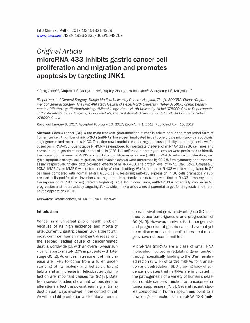
Figure 1. miRNA-433 directly targeted JNK1 in GC cells. A. JNK1 expression in GC cell lines and normal gastric GES-1 cells was measured by Real-time PCR. B. Base-pairing interaction between miRNA-433 seed sequences and JNK1, as predicted by TargetScan. C. Luciferase assay. MKN-45 cells were co-transfected with pGL3-JNK1 and miR-433 expressing vector or negative control (Mock). Firefly luciferase activity was normalized to Renilla lucifer-ase activity. D. JNK1 protein levels were examined in MKN-45 cells transfected with miR-433 expressing vector or mock.**P<0.01 compared with GES-1 cells or Mock; ##P<0.01 compared with miRNA-433.
microRNA-433 targets JNK1
4324 Int J Clin Exp Pathol 2017;10(4):4321-4329
Cultured cells were lysed in RIPA buffer with 1% PMSF, and isolated protein was loaded onto a SDS-PAGE and transferred onto PVDF mem-brane. The membrane was then incubated with antibodies against JNK1, Bax, Bcl-2, Caspase-3, PCNA, MMP-2 and MMP-9 over-night at 4°C, followed by incubation with horse-radish peroxidase-conjugated secondary anti-body at room temperature for 1 h. Signals were detected using ECL Substrates (Millipore, MA, USA). GAPDH was used as a loading control for normalization.
Statistical analysis
All the experiments were similarly done at least three times. All statistical analyses were per-formed using the GraphPad Prism 5 software (GraphPad Software, Inc., LaJolla, CA, USA). Statistical analyses were done by analysis of variance (ANOVA) or Student’s t test and P<0.05 was considered statistically significant.
Results
miR-433 regulates JNK1 expression in MKN-45 cells
We first analyzed miR-433 expression in three GC cell lines, BGC-823, MKN-28 and MKN-45
cell lines and normal GES-1 cells by Real-time PCR (Figure 1A). miR-433 was expressed in lower level in BGC-823, MKN-28 and MKN-45 cell lines compared with GES-1 cells. To explore the regulation mechanism of miRNA-433, TargetScan bioinformatics analysis (www.tar-getscan.org) was employed. TargetScan identi-fied that JNK1 mRNA contained potential bind-ing sites of miRNA-433 (Figure 1B). To confirm JNK1 as a target and regulated by miR-433 in GC cells, JNK1 3’UTR was cloned and inserted into a luciferase reporter vector. The luciferase assay showed that miR-433 significantly sup-pressed luciferase activity containing the JNK1 3’UTR (Figure 1C). Western blot assay showed that, JNK1 was notably decreased after trans-fection of miR-433 into MKN-45 cells. Consistent with these results, a significant up-regulation of JNK1 was observed upon overex-pression of JNK1 in miR-433-transfected MKN-45 cells (Figure 1D). These data suggested that JNK1 might be a direct target of miR-433 in GC cells.
miR-433 represses MKN-45 cell proliferation and induces cell cycle arrest
Then, miR-433 and miR-433+JNK1 expression vector were transfected into MKN-45 cells,
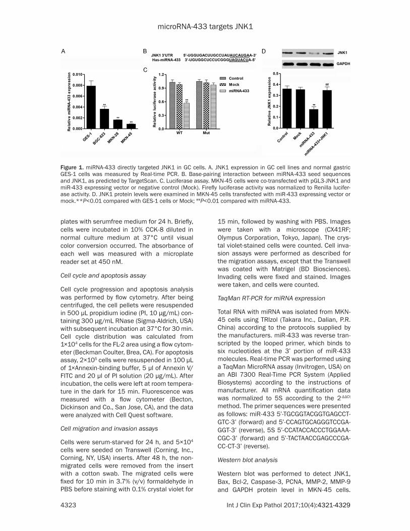
Figure 2. miRNA-433 inhibited MKN-45 cell growth and induced cell cycle arrest. A. Cell viability was determined in MKN-45 cells by CCK-8 assays. B, C. The proportion of cells in each phase of the cell cycle was determined in MKN-45 cells by flow cytometry assay. *P<0.05, **P<0.01 compared with Mock; ##P<0.01 compared with miRNA-433.
microRNA-433 targets JNK1
4325 Int J Clin Exp Pathol 2017;10(4):4321-4329
respectively. 0, 24, 48 and 72 h after transfec-tion, cell proliferation was analyzed using CCK-8 assay. As shown in Figure 2A, miR-433 induced great inhibition on cell proliferation compared with Mock sequence (Mock) in MKN-45 cells. To further validate the cell prolifera-tion inhibition of miR-433, cell cycle was ana-lyzed in MKN-45 cells. Cell cycle analysis showed that overexpression of miR-433 nota-bly increased the rate of G0/G1 and S phase cells and reduced G2/M phase cell population in MKN-45 cells (Figure 2B and 2C). On the contrary, overexpression of JNK1 slightly reduced G0/G1 and S phase population and increased the rate of G2/M phase cells in miR-433-transfected MKN-45 cells. These results indicated that miR-433 in GC cells may inhibit-ed cell proliferation by arresting cell cycle pro-gression in G0/G1 and S phases.
miR-433 induces MKN-45 cell apoptosis
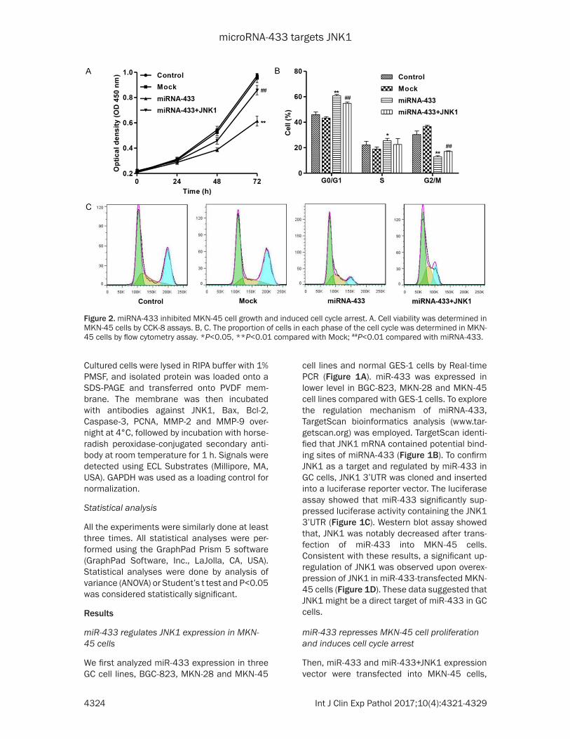
Then, we evaluated the apoptotic function of miR-433 in MKN-45 cells by Annexin V-FITC/PI staining assay. As shown in Figure 3A and 3B, flow cytometry analysis revealed that overex-pression of miR-433 in MKN-45 cells signifi-cantly increased cell apoptosis by 6.27-fold compared to corresponding Mock. While, over-expression of JNK1 in MKN-45 cells reduced cell apoptosis by 54.7% compared with miR-433 overexpression.
miR-433 represses MKN-45 cell invasion and migration
As GC is a type of highly malignant tumor with a potent capacity to invade locally and distant metastasis, we next attempted to explore the effect of miR-433 restoration on GC cell inva-sion and migration. As shown in Figure 4A and 4B, the transwell invasion assays showed that the number of cells that passed through Matrigel-coated membrane into the lower chamber was significantly reduced in the miR-433-transfected MKN-45 cells compared with the mock control-transfected cells (Mock). And ectopic expression of miR-433 using miR-433 expression plasmids significantly attenuated the migration potential of MKN-45 cells (Figure 4C and 4D). However, overexpression of JNK1 in miR-433-transfected MKN-45 cells showed significantly increased invasion and migration potential (Figure 4A-D). These results suggest a role of miR-433 as a potential tumor suppressor.
Effects of miR-433 on apoptosis and metasta-sis-related protein expression
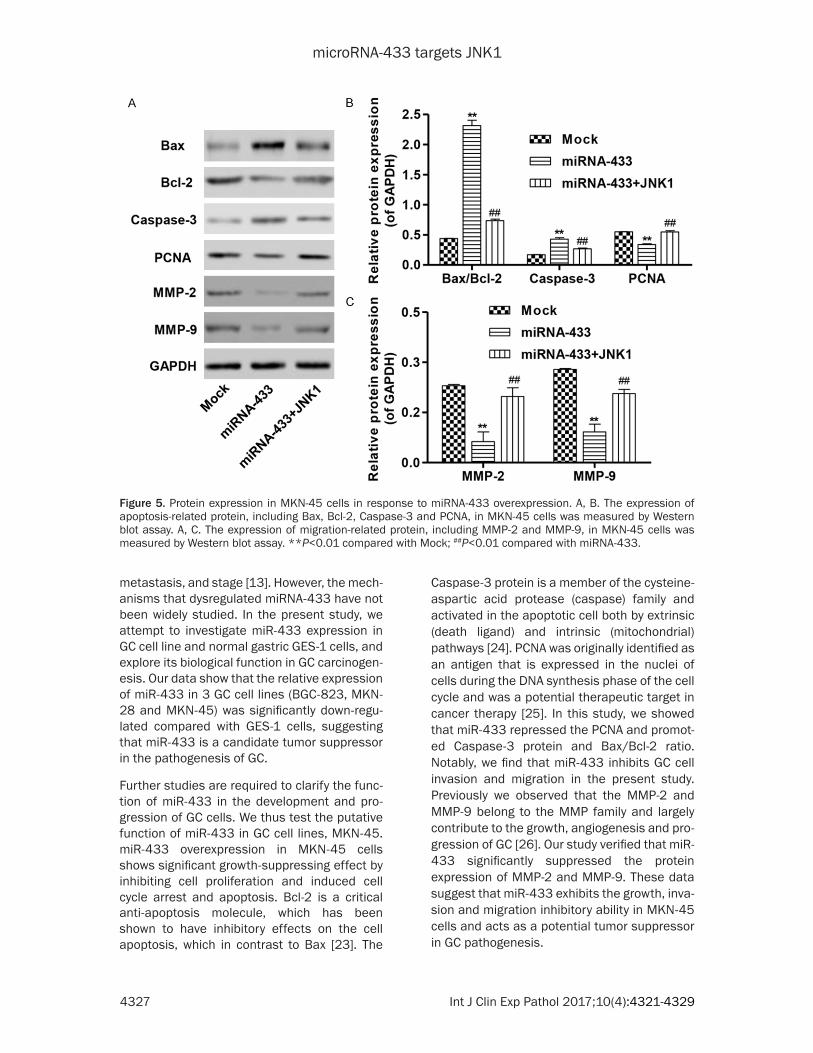
Moreover, four apoptotic factors Bcl-2, Bax, PCNA and Caspase-3 were evaluated by Western blot (Figure 5A and 5B). In MKN-45 cells, overexpression of miR-433 induced down-regulation of PCNA and up-regualtion of
Figure 3. miRNA-433 promoted MKN-45 cell apoptosis. A, B. Cell apoptosis was de-termined in MKN-45 cells by flow cytom-etry assay. **P<0.01 compared with Mock; ##P<0.01 compared with miRNA-433.
microRNA-433 targets JNK1
4326 Int J Clin Exp Pathol 2017;10(4):4321-4329
Bax/Bcl-2 ratio and Caspase-3. Since MMP-2 and MMP-9 play a vital role to facilitate the invasion and migration of GC cells, Western blot was performed to detect the two proteins expression. As shown in Figure 5A and 5C, the expression of MMP-2 and MMP-9 was signifi-cantly reduced in miR-433-transfected MKN-45 cells compared with mock control cells. However, overexpression of JNK1 in MKN-45 cells significantly inhibited changes in protein expression induced by miR-433 (Figure 5A-C). Taken together, these results indicated that miR-433 induced cell apoptosis and inhibited migration in GC cells by targeting JNK1.
Discussion
In the present study, we found that JNK1 could promote GC cells proliferation, invasion and
migration and inhibit apoptosis as an onco-gene, and miRNA-433 acts as a tumor suppres-sor that was inversely correlated with JNK1 expression in GC cells, and could inhibits prolif-eration, invasion and migration and increase apoptosis by regulation of JNK1.
In the last decade, miRNAs have emerged as critical regulators in regulation of cancer cell proliferation, invasion and migration, function as tumor suppressors or promoters of oncogen-esis [21, 22]. However, to the best of our knowl-edge few previous reports have identified the miR-433 involvement in GC carcinogenesis. Low expression of miR-433 was associated with unfavourable outcome of GC patients in overall survival independent of clinical covari-ates, including depth of invasion, lymph-node
Figure 4. miRNA-433 inhibited MKN-45 cell invasion and migration. Cell invasion (A, B) and migration ability (C, D) was determined in MKN-45 cells by Transwell assay. **P<0.01 compared with Mock; ##P<0.01 compared with miRNA-433.
microRNA-433 targets JNK1
4327 Int J Clin Exp Pathol 2017;10(4):4321-4329
metastasis, and stage [13]. However, the mech-anisms that dysregulated miRNA-433 have not been widely studied. In the present study, we attempt to investigate miR-433 expression in GC cell line and normal gastric GES-1 cells, and explore its biological function in GC carcinogen-esis. Our data show that the relative expression of miR-433 in 3 GC cell lines (BGC-823, MKN-28 and MKN-45) was significantly down-regu-lated compared with GES-1 cells, suggesting that miR-433 is a candidate tumor suppressor in the pathogenesis of GC.
Further studies are required to clarify the func-tion of miR-433 in the development and pro-gression of GC cells. We thus test the putative function of miR-433 in GC cell lines, MKN-45. miR-433 overexpression in MKN-45 cells shows significant growth-suppressing effect by inhibiting cell proliferation and induced cell cycle arrest and apoptosis. Bcl-2 is a critical anti-apoptosis molecule, which has been shown to have inhibitory effects on the cell apoptosis, which in contrast to Bax [23]. The
Caspase-3 protein is a member of the cysteine-aspartic acid protease (caspase) family and activated in the apoptotic cell both by extrinsic (death ligand) and intrinsic (mitochondrial) pathways [24]. PCNA was originally identified as an antigen that is expressed in the nuclei of cells during the DNA synthesis phase of the cell cycle and was a potential therapeutic target in cancer therapy [25]. In this study, we showed that miR-433 repressed the PCNA and promot-ed Caspase-3 protein and Bax/Bcl-2 ratio. Notably, we find that miR-433 inhibits GC cell invasion and migration in the present study. Previously we observed that the MMP-2 and MMP-9 belong to the MMP family and largely contribute to the growth, angiogenesis and pro-gression of GC [26]. Our study verified that miR-433 significantly suppressed the protein expression of MMP-2 and MMP-9. These data suggest that miR-433 exhibits the growth, inva-sion and migration inhibitory ability in MKN-45 cells and acts as a potential tumor suppressor in GC pathogenesis.
Figure 5. Protein expression in MKN-45 cells in response to miRNA-433 overexpression. A, B. The expression of apoptosis-related protein, including Bax, Bcl-2, Caspase-3 and PCNA, in MKN-45 cells was measured by Western blot assay. A, C. The expression of migration-related protein, including MMP-2 and MMP-9, in MKN-45 cells was measured by Western blot assay. **P<0.01 compared with Mock; ##P<0.01 compared with miRNA-433.
microRNA-433 targets JNK1
4328 Int J Clin Exp Pathol 2017;10(4):4321-4329
Although the evidences have highlighted the importance of miR-433 as an onco suppressor in GC cells, the precise molecular mechanisms remain largely unclear. To better understand the tumor suppressive effect of miR-433 in GC tumorigenesis, bioinformatics analysis was used and identified JNK1 as a potential target of miR-433. With the 3’UTR luciferase reporter assay, JNK1 was identified as the direct target of miR-433. The increase in miR-433 expres-sion is accompanied by down-regulation of JNK1 expression.
JNK1 has been proved to be activated in response to several stress signals, including tumor necrosis factor and hyperosmotic condi-tions, and is associated with induction of apop-tosis [27] and acts as a crucial regulator of cell migration and epithelial morphogenesis [28]. Recent studies have demonstrated that JNK1 is a critical regulator of gastric tumorigenesis and frequent JNK1 activation in GC [15, 29]. Moreover, JNK activation has been detected in human GC samples and, consistent with this, JNK1 controls both tumor initiation and promo-tion by affecting cell proliferation and ROS pro-duction in a mouse model of GC [30]. In line with previous study, we found that over- expression of JNK1 in GC cells significantly enhanced cell proliferation, invasion and migra-tion and inhibited cell apoptosis induced by miRNA-433.
In conclusion, the present study demonstrated that miRNA-433 could inhibit GC cell prolifera-tion, invasion and migration via targeting JNK1. Further studies are required to fully understand the roles and mechanisms of miRNA-433 and JNK1 in GC, which will be beneficial for the development of therapeutic strategies for GC.
Disclosure of conflict of interest
None.
Address correspondence to: Xianghui He, Depart- ment of General Surgery, Tianjin Medical University General Hospital, Building No. 154 Anshan Road, Heping District, Tianjin 300052, China. Tel: +86-022-60362255; E-mail: [email protected]
References
[1] Torre LA, Bray F, Siegel RL, Ferlay J, Lortet-Tieulent J, Jemal A. Global cancer statistics, 2012. CA Cancer J Clin 2015; 65: 87-108.
[2] Smyth GK. Linear models and empirical bayes methods for assessing differential expression in microarray experiments. Stat Appl Genet Mol Biol 2004; 3: Article3.
[3] Yasui W, Oue N, Sentani K, Sakamoto N, Motoshita J. Transcriptome dissection of gas-tric cancer: identification of novel diagnostic and therapeutic targets from pathology speci-mens. Pathol Int 2009; 59: 121-136.
[4] Chen CN, Chang CC, Lai HS, Jeng YM, Chen CI, Chang KJ, Lee PH, Lee H. Connective tissue growth factor inhibits gastric cancer peritoneal metastasis by blocking integrin alpha3beta1-dependent adhesion. Gastric Cancer 2015; 18: 504-515.
[5] Lee EK, Song KA, Chae JH, Kim KM, Kim SH, Kang MS. GAGE12 mediates human gastric carcinoma growth and metastasis. Int JCancer 2015; 136: 2284-2292.
[6] Cheng CJ, Bahal R, Babar IA, Pincus Z, Barrera F, Liu C, Svoronos A, Braddock DT, Glazer PM, Engelman DM, Saltzman WM, Slack FJ. MicroRNA silencing for cancer therapy target-ed to the tumour microenvironment. Nature 2015; 518: 107-110.
[7] Kim HS, Lee KS, Bae HJ, Eun JW, Shen Q, Park SJ, Shin WC, Yang HD, Park M, Park WS, Kang YK, Nam SW. MicroRNA-31 functions as a tu-mor suppressor by regulating cell cycle and epithelial-mesenchymal transition regulatory proteins in liver cancer. Oncotarget 2015; 6: 8089-8102.
[8] Li BS, Zuo QF, Zhao YL, Xiao B, Zhuang Y, Mao XH, Wu C, Yang SM, Zeng H, Zou QM, Guo G. MicroRNA-25 promotes gastric cancer migra-tion, invasion and proliferation by directly tar-geting transducer of ERBB2, 1 and correlates with poor survival. Oncogene 2015; 34: 2556-2565.
[9] Xue J, Chen LZ, Li ZZ, Hu YY, Yan SP, Liu LY. MicroRNA-433 inhibits cell proliferation in he-patocellular carcinoma by targeting p21 acti-vated kinase (PAK4). Mol Cell Biochem 2015; 399: 77-86.
[10] Wang M, Zhang B, Dai X, Zhang Y, Lian W. Decreased expression of microRNA-433 is as-sociated with the prognosis of epithelial ovari-an cancer. Int J Clin Exp Pathol 2016; 9: 3606-3611.
[11] Liang T, Guo Q, Li L, Cheng Y, Ren C, Zhang G. MicroRNA-433 inhibits migration and invasion of ovarian cancer cells via targeting Notch1. Neoplasma 2016; 63: 696-704.
[12] Luo H, Zhang H, Zhang Z, Zhang X, Ning B, Guo J, Nie N, Liu B, Wu X. Down-regulated miR-9 and miR-433 in human gastric carcinoma. J Exp Clin Cancer Res 2009; 28: 82.
[13] Ueda T, Volinia S, Okumura H, Shimizu M, Taccioli C, Rossi S, Alder H, Liu CG, Oue N,
Yasui W, Yoshida K, Sasaki H, Nomura S, Seto Y, Kaminishi M, Calin GA, Croce CM. Relation between microRNA expression and progres-sion and prognosis of gastric cancer: a microR-NA expression analysis. Lancet Oncol 2010; 11: 136-146.
[14] Zhang J, Yang PL, Gray NS. Targeting cancer with small molecule kinase inhibitors. Nat Rev Cancer 2009; 9: 28-39.
[15] Wagner EF, Nebreda AR. Signal integration by JNK and p38 MAPK pathways in cancer devel-opment. Nat Rev Cancer 2009; 9: 537-549.
[16] Manning AM, Davis RJ. Targeting JNK for thera-peutic benefit: from junk to gold? Nat Rev Drug Discov 2003; 2: 554-565.
[17] Karin M, Gallagher E. From JNK to pay dirt: jun kinases, their biochemistry, physiology and clinical importance. IUBMB Life 2005; 57: 283-295.
[18] Chang Q, Chen J, Beezhold KJ, Castranova V, Shi X, Chen F. JNK1 activation predicts the prognostic outcome of the human hepatocel-lular carcinoma. Mol Cancer 2009; 8: 64.
[19] Chang Q, Zhang Y, Beezhold KJ, Bhatia D, Zhao H, Chen J, Castranova V, Shi X, Chen F. Sustained JNK1 activation is associated with altered histone H3 methylations in human liver cancer. J Hepatol 2009; 50: 323-333.
[20] Cellurale C, Weston CR, Reilly J, Garlick DS, Jerry DJ, Sluss HK, Davis RJ. Role of JNK in a Trp53-dependent mouse model of breast can-cer. PLoS One 2010; 5: e12469.
[21] Lu C, Shan Z, Li C, Yang L. MiR-129 regulates cisplatin-resistance in human gastric cancer cells by targeting P-gp. Biomed Pharmacother 2016; 86: 450-456.
[22] Li C, Zhang K, Chen J, Chen L, Wang R, Chu X. MicroRNAs as regulators and mediators of forkhead box transcription factors function in human cancers. Oncotarget 2016; [Epub ahead of print].
[23] Xiong S, Mu T, Wang G, Jiang X. Mitochondria-mediated apoptosis in mammals. Protein Cell 2014; 5: 737-749.
[24] Ghavami S, Hashemi M, Ande SR, Yeganeh B, Xiao W, Eshraghi M, Bus CJ, Kadkhoda K, Wiechec E, Halayko AJ, Los M. Apoptosis and cancer: mutations within caspase genes. J Med Genet 2009; 46: 497-510.
[25] Wang SC. PCNA: a silent housekeeper or a po-tential therapeutic target? Trends Pharmacol Sci 2014; 35: 178-186.
[26] Zheng H, Takahashi H, Murai Y, Cui Z, Nomoto K, Niwa H, Tsuneyama K, Takano Y. Expressions of MMP-2, MMP-9 and VEGF are closely linked to growth, invasion, metastasis and angiogen-esis of gastric carcinoma. Anticancer Res 2006; 26: 3579-3583.
[27] Zhang S, Shi L, Ma H, Li H, Li Y, Lu Y, Wang Q, Li W. Dihydroartemisinin induces apoptosis in human gastric cancer cell line BGC-823 through activation of JNK1/2 and p38 MAPK signaling pathways. J Recept Signal Transduct Res 2016; 12: 1-7.
[28] Xia Y, Karin M. The control of cell motility and epithelial morphogenesis by Jun kinases. Trends Cell Biol 2004; 14: 94-101.
[29] Mishra P, Senthivinayagam S, Rangasamy V, Sondarva G, Rana B. Mixed lineage kinase-3/JNK1 axis promotes migration of human gas-tric cancer cells following gastrin stimulation. Mol Endocrinol 2010; 24: 598-607.
[30] Shibata W, Maeda S, Hikiba Y, Yanai A, Sakamoto K, Nakagawa H, Ogura K, Karin M, Omata M. c-Jun NH2-terminal kinase 1 is a critical regulator for the development of gastric cancer in mice. Cancer Res 2008; 68: 5031-5039.