The Role of Liver Fructose-1,6-Bisphosphatase in Regulating Appetite and Adiposity Sherley Visinoni, 1 Nurul Fathiah Izzati Khalid, 1 Christos N. Joannides, 1 Arthur Shulkes, 2 Mildred Yim, 2 Jon Whitehead, 3 Tony Tiganis, 4 Benjamin J. Lamont, 1 Jenny M. Favaloro, 1 Joseph Proietto, 1 Sofianos Andrikopoulos, 1 and Barbara C. Fam 1 Liver fructose-1,6-bisphosphatase (FBPase) is a regulatory en- zyme in gluconeogenesis that is elevated by obesity and dietary fat intake. Whether FBPase functions only to regulate glucose or has other metabolic consequences is not clear; therefore, the aim of this study was to determine the importance of liver FBPase in body weight regulation. To this end we performed comprehen- sive physiologic and biochemical assessments of energy balance in liver-specific transgenic FBPase mice and negative control littermates of both sexes. In addition, hepatic branch vagotomies and pharmacologic inhibition studies were performed to confirm the role of FBPase. Compared with negative littermates, liver- specific FBPase transgenic mice had 50% less adiposity and ate 15% less food but did not have altered energy expenditure. The reduced food consumption was associated with increased circulat- ing leptin and cholecystokinin, elevated fatty acid oxidation, and 3- b-hydroxybutyrate ketone levels, and reduced appetite-stimulating neuropeptides, neuropeptide Y and Agouti-related peptide. Hepatic branch vagotomy and direct pharmacologic inhibition of FBPase in transgenic mice both returned food intake and body weight to the negative littermates. This is the first study to identify liver FBPase as a previously unknown regulator of appetite and adiposity and describes a novel process by which the liver participates in body weight regulation. Diabetes 61:1122–1132, 2012 O ver recent years, excessive nutrient intake has been associated with rapidly increasing rates of obesity in both developed and developing so- cieties (1). Despite much effort, the specific biochemical mechanisms involved in body weight regula- tion are not completely understood. Body weight is maintained by a fine balance between food intake and energy expenditure. Under normal con- ditions, energy homeostasis is maintained through a com- plex interaction between peripheral organs and the central nervous system (CNS). Many peripheral signals from white adipose tissue, the gut, and the pancreas are known to regulate body weight (2). The CNS receives these signals and adjusts food intake and energy expenditure accord- ingly. Although not normally considered as one of the classic body weight regulatory organs, indirect evidence has accumulated over the years in a variety of models to suggest a role for the liver in controlling food intake (3–7). Russek (7) was the first to propose that a signal to termi- nate food intake was generated from the liver. This was based on studies demonstrating that direct injection of glucose into the liver of fasted dogs suppressed food in- take more effectively than systemic injection of glucose (7). Direct infusion of free fatty acids (FFAs) into the he- patic portal vein of rats has also demonstrated an in- volvement of the liver in lowering food intake through an increase in liver fatty acid oxidation (FAO) (3,4). Con- versely, the fructose analog 2,5-anhydro-D-mannitol (6) and other metabolic and FAO inhibitors have been reported to stimulate appetite when administered into animals (5,8,9). The gene expressing fructose-1,6-bisphosphatase (FBPase) is one of many genes upregulated in the liver by obesity and fat (10,11). Even though FBPase is known as a regulatory enzyme in gluconeogenesis, a previous study from our laboratory showed that liver-specific FBPase transgenic mice with a physiologic threefold level of overexpression had no change in whole-body glucose tolerance or endog- enous glucose production (12). Surprisingly, the mice con- sistently displayed an approximate 10% reduction in body weight compared with negative littermates (12), leading us to propose that liver FBPase may have a novel role in the control of body weight. We therefore investigated this potential regulatory role of liver FBPase by using our transgenic mouse model that specifically overexpresses FBPase in the liver. We report that overexpression of this liver enzyme leads to the lean body weight phenotype in the transgenic mice by markedly reducing adiposity levels by ;50%. Reductions in food in- take rather than elevated energy expenditure were found to be the contributing factors. The appetite-stimulating neu- ropeptides, neuropeptide Y (NPY) and Agouti-related pep- tide (AgRP), were significantly suppressed, whereas the circulating satiety hormones, cholecystokinin (CCK), and leptin, rose significantly. Elevation of liver FAO via an in- creased flux through the hexosamine biosynthesis pathway (HBP) appears to be the key linking the increase in liver FBPase to reduced food intake and adiposity in our transgenic mouse. RESEARCH DESIGN AND METHODS Animals. Hemizygous transgenic mice (males and females) overexpressing the human liver FBPase gene (FBP-1) specifically in the liver and their age- matched negative littermate controls, both on a C57Bl6/J background, were used unless otherwise stated. Mice were generated, maintained, and geno- typed as described previously (12). All mice were maintained in accordance with guidelines of the Austin Hospital Animal Ethics Committee (AEC#s: A2007/2752 and A2009/03766). Energy balance studies. Mice were individually housed and provided a standard laboratory chow diet (3% fat, 77% carbohydrate, 20% protein) and water ad libitum. Body weights were measured at 4, 8, and 12 weeks and From the 1 Department of Medicine, University of Melbourne, Heidelberg, Victoria, Australia; the 2 Department of Surgery, Austin Health, University of Melbourne, Heidelberg, Victoria, Australia; the 3 Mater Medical Research In- stitute, Brisbane, Queensland, Australia; and the 4 Department of Biochemis- try and Molecular Biology, Monash University, Clayton, Victoria, Australia. Corresponding author: Barbara C. Fam, [email protected]. Received 26 October 2011 and accepted 26 January 2012. DOI: 10.2337/db11-1511 This article contains Supplementary Data online at http://diabetes .diabetesjournals.org/lookup/suppl/doi:10.2337/db11-1511/-/DC1. S.A. and B.C.F. contributed equally to this study. Ó 2012 by the American Diabetes Association. Readers may use this article as long as the work is properly cited, the use is educational and not for profit, and the work is not altered. See http://creativecommons.org/licenses/by -nc-nd/3.0/ for details. 1122 DIABETES, VOL. 61, MAY 2012 diabetes.diabetesjournals.org ORIGINAL ARTICLE
Transcript
The Role of Liver Fructose-1,6-Bisphosphatase inRegulating Appetite and AdipositySherley Visinoni,
1Nurul Fathiah Izzati Khalid,
1Christos N. Joannides,
1Arthur Shulkes,
2
Mildred Yim,2Jon Whitehead,
3Tony Tiganis,
4Benjamin J. Lamont,
1Jenny M. Favaloro,
1
Joseph Proietto,1Sofianos Andrikopoulos,
1and Barbara C. Fam
1
Liver fructose-1,6-bisphosphatase (FBPase) is a regulatory en-zyme in gluconeogenesis that is elevated by obesity and dietaryfat intake. Whether FBPase functions only to regulate glucose orhas other metabolic consequences is not clear; therefore, the aimof this study was to determine the importance of liver FBPase inbody weight regulation. To this end we performed comprehen-sive physiologic and biochemical assessments of energy balancein liver-specific transgenic FBPase mice and negative controllittermates of both sexes. In addition, hepatic branch vagotomiesand pharmacologic inhibition studies were performed to confirmthe role of FBPase. Compared with negative littermates, liver-specific FBPase transgenic mice had 50% less adiposity and ate15% less food but did not have altered energy expenditure. Thereduced food consumption was associated with increased circulat-ing leptin and cholecystokinin, elevated fatty acid oxidation, and 3-b-hydroxybutyrate ketone levels, and reduced appetite-stimulatingneuropeptides, neuropeptide Y and Agouti-related peptide. Hepaticbranch vagotomy and direct pharmacologic inhibition of FBPase intransgenic mice both returned food intake and body weight to thenegative littermates. This is the first study to identify liver FBPaseas a previously unknown regulator of appetite and adiposity anddescribes a novel process by which the liver participates in bodyweight regulation. Diabetes 61:1122–1132, 2012
Over recent years, excessive nutrient intake hasbeen associated with rapidly increasing rates ofobesity in both developed and developing so-cieties (1). Despite much effort, the specific
biochemical mechanisms involved in body weight regula-tion are not completely understood.
Body weight is maintained by a fine balance betweenfood intake and energy expenditure. Under normal con-ditions, energy homeostasis is maintained through a com-plex interaction between peripheral organs and the centralnervous system (CNS). Many peripheral signals from whiteadipose tissue, the gut, and the pancreas are known toregulate body weight (2). The CNS receives these signalsand adjusts food intake and energy expenditure accord-ingly. Although not normally considered as one of theclassic body weight regulatory organs, indirect evidence
has accumulated over the years in a variety of models tosuggest a role for the liver in controlling food intake (3–7).Russek (7) was the first to propose that a signal to termi-nate food intake was generated from the liver. This wasbased on studies demonstrating that direct injection ofglucose into the liver of fasted dogs suppressed food in-take more effectively than systemic injection of glucose(7). Direct infusion of free fatty acids (FFAs) into the he-patic portal vein of rats has also demonstrated an in-volvement of the liver in lowering food intake through anincrease in liver fatty acid oxidation (FAO) (3,4). Con-versely, the fructose analog 2,5-anhydro-D-mannitol (6) andother metabolic and FAO inhibitors have been reported tostimulate appetite when administered into animals (5,8,9).
The gene expressing fructose-1,6-bisphosphatase (FBPase)is one of many genes upregulated in the liver by obesity andfat (10,11). Even though FBPase is known as a regulatoryenzyme in gluconeogenesis, a previous study from ourlaboratory showed that liver-specific FBPase transgenicmice with a physiologic threefold level of overexpressionhad no change in whole-body glucose tolerance or endog-enous glucose production (12). Surprisingly, the mice con-sistently displayed an approximate 10% reduction in bodyweight compared with negative littermates (12), leading usto propose that liver FBPase may have a novel role in thecontrol of body weight.
We therefore investigated this potential regulatory roleof liver FBPase by using our transgenic mouse model thatspecifically overexpresses FBPase in the liver. We reportthat overexpression of this liver enzyme leads to the leanbody weight phenotype in the transgenic mice by markedlyreducing adiposity levels by ;50%. Reductions in food in-take rather than elevated energy expenditure were found tobe the contributing factors. The appetite-stimulating neu-ropeptides, neuropeptide Y (NPY) and Agouti-related pep-tide (AgRP), were significantly suppressed, whereas thecirculating satiety hormones, cholecystokinin (CCK), andleptin, rose significantly. Elevation of liver FAO via an in-creased flux through the hexosamine biosynthesis pathway(HBP) appears to be the key linking the increase in liverFBPase to reduced food intake and adiposity in ourtransgenic mouse.
RESEARCH DESIGN AND METHODS
Animals.Hemizygous transgenic mice (males and females) overexpressing thehuman liver FBPase gene (FBP-1) specifically in the liver and their age-matched negative littermate controls, both on a C57Bl6/J background, wereused unless otherwise stated. Mice were generated, maintained, and geno-typed as described previously (12). All mice were maintained in accordancewith guidelines of the Austin Hospital Animal Ethics Committee (AEC#s:A2007/2752 and A2009/03766).Energy balance studies. Mice were individually housed and provideda standard laboratory chow diet (3% fat, 77% carbohydrate, 20% protein) andwater ad libitum. Body weights were measured at 4, 8, and 12 weeks and
From the 1Department of Medicine, University of Melbourne, Heidelberg,Victoria, Australia; the 2Department of Surgery, Austin Health, University ofMelbourne, Heidelberg, Victoria, Australia; the 3Mater Medical Research In-stitute, Brisbane, Queensland, Australia; and the 4Department of Biochemis-try and Molecular Biology, Monash University, Clayton, Victoria, Australia.
Corresponding author: Barbara C. Fam, [email protected] 26 October 2011 and accepted 26 January 2012.DOI: 10.2337/db11-1511This article contains Supplementary Data online at http://diabetes
.diabetesjournals.org/lookup/suppl/doi:10.2337/db11-1511/-/DC1.S.A. and B.C.F. contributed equally to this study.� 2012 by the American Diabetes Association. Readers may use this article as
long as the work is properly cited, the use is educational and not for profit,and the work is not altered. See http://creativecommons.org/licenses/by-nc-nd/3.0/ for details.
1122 DIABETES, VOL. 61, MAY 2012 diabetes.diabetesjournals.org
subsequently each week until age 22 weeks. Food intake (g/day) was measuredeach week from age 12 to 22 weeks. Subcutaneous, infrarenal, and gonadal fatpads were collected and weighed in the nonfasted state. Voluntary physicalactivity, resting energy expenditure (REE), respiratory quotient (RQ), andlevels of whole-body fat and glucose oxidation were assessed as previouslydescribed (13–16).Hepatic branch vagotomy. Common hepatic branch vagotomy was performedon 16-week-old anesthetizedmice that specifically targets the vagal nerve branch,the major connection between the liver and brain (17). A laparotomy incisionwas made on the ventral midline, and the abdominal wall was opened witha second incision. The common hepatic vagal branch was located (under theliver) and transected, stretching the fascia containing the common hepaticbranch using fine forceps. A similar incision was also made in the sham-operatedmice and the hepatic vagus nerve was located but not transected. The incisionswere sutured, mice were administered saline (1 mL) intraperitoneally to aid inpostsurgery recovery, and were allowed to recover for 1 week. Food intake andbody weight was subsequently measured weekly for 10 weeks.Pharmacologic inhibition of FBPase. FBPase transgenic mice, negativelittermate controls (;16 weeks old), and NZO mice (7 weeks old) were gavageddaily a 5 mg/kg dose of benzoxazole benzene sulfonamide, a commerciallyavailable human-specific FBPase inhibitor (Calbiochem) (18,19), to specificallytarget our construct or vehicle (water) 2 h before the dark cycle (feeding pe-riod) for 10 days. Body weight and food intake measurements were taken daily.On the 10th day of treatment, plasma was collected from anesthetized mice inthe fed state (1 h postdark cycle) via cardiac puncture, the whole brain wascollected, and fat depots were collected and weighed.CCK1 receptor antagonist study. FBPase transgenic mice and negativelittermates (10 weeks old) were injected intraperitoneally with a 300 mg/kgdose of lorglumide, a CCK1 receptor (CCK1R) antagonist (Sigma Aldrich, St.Louis, MO) (20), or saline vehicle at the onset of the dark cycle, and foodintake was recorded 24 h later.3-b-Hydroxybutyrate study. Male C57BL/6J mice (10 weeks old) were sub-cutaneously injected with a 10 mmol/kg dose of DL-3-hydroxybutyric acid (so-dium salt, 98% purity 3-b-hydroxybutyrate[BHB]; MP Biomedicals) (5) or salineat the onset of the dark cycle. Body weight and food intake were measured 1and 2 h thereafter. Mice were left to recover for a week, and BHB adminis-tration was repeated for plasma collection 1 h after administration only.Hormone and metabolite assays. Cardiac punctures were performed onanesthetized transgenic mice and negative littermates (;16 weeks old) in the fedstate (1 h into the dark cycle). Circulating ghrelin was measured using a specificenzyme immunoassay (Phoenix Pharmaceuticals Inc., Belmont, CA), circulatingCCK was measured by an in-house radioimmunoassay (21), and circulatingleptin was measured by radioimmunoassay (Linco Research, St Charles, MO).BHB was measured from cardiac plasma samples collected after an overnightfast using a 3-hydroxybutyrate II reagent kit (Helena Laboratories Australia PtyLtd, Melbourne, VIC, Australia). Fructose-1,6-phosphate (F-1,6-P) and fructose-6-phosphate (F6P) assays were performed as previously described (10).FAO inhibitor study. FBPase transgenic mice and negative littermates(10 weeks old) were gavaged with one dose (10 mg/kg) of etomoxir, a carnitinepalmitoyl-CoA transferase (CPT)-1a inhibitor (Sigma Aldrich, St. Louis, MO)(22) or saline vehicle at the onset of the dark cycle. Food intake was recorded24 h later.O-linked N-acetylglucosamine transferase RL2 Western blotting.
Liver protein homogenates were processed and collected as previously de-scribed (23), and RL2 protein levels, indicative of O-linked N-acetylglucosaminetransferase (OGT) (;135 kDa) (24) were determined by Western blotting aspreviously described (25).Hypothalamic dissection. Hypothalamic sections (arcuate nucleus [ARC])and paraventricular nucleus [PVN, as a control]) were collected in hemizygousmice (;16 weeks old) and in homozygous mice (;24 weeks old), 1 h into thedark cycle as previously described (13,16,26).Measurement of mRNA expression levels in hypothalamus and liver.
RNA was extracted from ARC, PVN, and liver samples using TRIzol (Invitrogen,Mount Waverley, VIC, Australia), treated with DNaseI (Ambion, Scoresby,VIC, Australia), and cDNA was synthesized using 1 mg DNase-treated RNAand random primers with the Promega Reverse Transcription kit (Annandale,NSW, Australia). AgRP and proopiomelanocortin (POMC) primers for SYBR-green real-time PCR were designed across an intron-exon boundary to ensurenonamplification of genomic DNA. POMC primers: forward 59-CTG GCC CTCCTG CTT CAG-39; reverse 59-GGA TGC AAG CCA GCA GGT T-39 (0.3 mmol/Leach). AgRP primers: forward 59-TCC CAG AGT TCC CAG GTC TAA G-39;reverse 59-TAG CAC CTC CGC CAA AGC-39 (0.3 mmol/L each). Primers fora-actin endogenous control were ised as previously described (27). TaqMangene expression assays (Applied Biosystems, Scoresby, VIC, Australia) wereused for OGT (Mm00507300_m1), CPT-1a (Mm00550435_m1), CPT-1c(Mm00463970_m1), peroxisome proliferator–activated receptor (PPAR)a(Mm00440939_m1), peroxisome proliferator–activated receptor-g coactivator-1
(PGC1)-a (Mm01208832_m1), leptin (Mm00434759_m1), and NPY (Mm00445771_m1). 18S primer mix from ABI was used as the endogenous control. ABIsequence detection software and relative quantification using the compara-tive DCt method was used.Statistical analysis. All data are presented as mean 6 SEM. Comparisonsbetween single parameters were measured using one-way ANOVA analysis(Minitab 15, 2007). The trapezoidal rule was used for area under the curve(AUC). For repeated measures, a general linear model (GLM) ANOVA wasused for comparison and a Tukey post hoc t test to determine significance(Minitab 15, 2007). Significance was determined as P , 0.05.
RESULTS
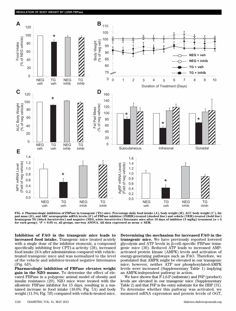
Liver FBPase transgenic mice develop a lean bodyweight phenotype. Male (Fig. 1A)and female (Fig. 1B)hemizygous transgenic mice both weighed ;10% less thantheir age matched negative littermates, evident from8 weeks of age. Male homozygous transgenic mice (Sup-plementary Fig. 1A) and females (data not shown) alsoweighed significantly less. The reduced body weight in thetransgenic mice was associated with a significant re-duction in white adipose tissue mass across all threedepots (subcutaneous, infrarenal, and gonadal) com-pared with the negative male (Fig. 1C) and female (Fig. 1D)littermates.Lean body weight phenotype is associated withreduced food intake. Daily food intake was reduced by;15% in male (Fig. 1E) and female transgenic mice (Fig. 1F)compared with the negative littermates. No differencesin physical activity or resting energy expenditure (REE)were seen with either sex (male: Fig. 1G and H, re-spectively, and female: Fig. 1I [REE data not shown]).Likewise, analysis in another transgenic line overexpressingFBPase in both liver and hypothalamus (27) demonstratedthe same lean phenotype, reduction in food intake, and nochange in energy expenditure (Supplementary Fig. 2A–E).Reduced food intake is associated with suppressionof appetite-stimulating neuropeptides. No differencesin ARC mRNA expression level of POMC (the precursor tothe appetite-suppressing neuropeptide a-MSH) were detec-ted (Fig. 2A). However, the appetite-stimulating neuro-peptides, NPY and AgRP, were significantly reduced by;50% in the ARC of transgenic mice (Fig. 2B and C). PVNsections showed no differences in expression in any of thegenes measured (data not shown).Vagotomized FBPase transgenic mice have anormalized phenotype. Hepatic branch vagotomieswere performed to establish whether the signal to inhibitfood intake in the transgenic mice was vagally mediated.As expected, at 10 weeks after vagotomy recovery, sham-operated transgenic mice ate less food compared withsham-operated negative mice, whereas the vagotomizedtransgenic mice ate more than the sham-operated trans-genic mice (Fig. 3A). This reduced food intake effect inthe transgenic mice was reflected in the significant weightgain after vagotomy (Fig. 3B).Specific pharmacologic inhibition of FBPase reversesthe lean body weight phenotype. Figure 4A–C illustratesthe normalization of food intake in the transgenic miceafter 10 days of FBPase inhibitor treatment with a con-comitant increase in body weight. Inhibitor treatment hadno effect on food intake (Fig. 4A) or body weight (Fig. 4Band C) in the negative mice, implying specificity of theinhibitor to the overexpressed gene. Vehicle-treated trans-genic mice, as expected, had smaller fat depots, whereasinhibitor-treated mice had similar fat depot masses to neg-ative littermates (Fig. 4D). When the appetite-stimulatingneuropeptides were measured, NPY and AgRP mRNA levels
S. VISINONI AND ASSOCIATES
diabetes.diabetesjournals.org DIABETES, VOL. 61, MAY 2012 1123
FIG. 1. Characterization of liver FBPase transgenic (TG) mice. Body weights of male negative (NEG, white circles) and TG (black circles) mice fromhemizygous breeding from age 4 weeks to 22 weeks (NEG: n = 11 and TG: n = 14; AUC *P < 0.001 vs. NEG, GLM ANOVA) (A) and female NEG (whitecircles) and TG (black circles) mice from hemizygous breeding from age 4 weeks to 22 weeks (NEG: n = 13 and TG: n = 10; AUC *P < 0.005 vs. NEG,GLM ANOVA) (B). Fat pad mass of male hemizygous TG mice (black bars) compared with their NEG (white bars) littermates (NEG: n = 14 and TG n =9; *P < 0.02 vs. NEG, one-way ANOVA) (C) and female hemizygous TG mice (black bars) compared with their NEG (white bars) littermates at age 24weeks (NEG: n = 11 and TG: n = 6; *P < 0.05 vs. NEG, one-way ANOVA) (D). Average daily food intake of male hemizygous TG mice (black bars)compared with NEG (white bars) littermates (NEG: n = 21 and TG: n = 14; *P < 0.0001 vs. NEG GLM ANOVA) (E) and of female hemizygous TG mice(black bars) compared with NEG (white bars) littermates (NEG: n = 18 and TG; n = 10; *P< 0.05 vs. NEG, GLM ANOVA) (F). Physical activity (NEG:n = 7 and TG: n = 5) (G) and resting energy expenditure (n = 4 for each group) (H) in male hemizygous transgenic mice (black bars) compared withnegative (white bars) littermates. I: Physical activity measurements of female hemizygous TG (black bars) compared with NEG (white bars) litter-mates (NEG: n = 10 and Tg: n = 4). All data expressed as mean 6 SEM.
REGULATION OF BODY WEIGHT BY LIVER FBPase
1124 DIABETES, VOL. 61, MAY 2012 diabetes.diabetesjournals.org
were both decreased in vehicle-treated transgenic mice,whereas inhibitor-treated mice exhibited normalized levelsto that of the negative littermates (Fig. 4E).Circulating satiety hormones from the gut areincreased in transgenic mice. Levels of the hungerhormone ghrelin remained unchanged (negative: 1300 6343 pg/mL vs. transgenic: 1534 6 562 pg/mL, n = 4),whereas circulating CCK (Fig. 5A) and leptin (Fig. 5B)levels were both elevated ;30% in the transgenic mice.Duodenal CCK peptide concentrations remained unchanged(negative: 436 16 pmol/g vs. transgenic: 356 12 pmol/g, n =6), suggesting CCK secretion rather than peptide synthesiswas enhanced in the transgenic mice. Administration of theCCK1R antagonist, lorglumide, caused the transgenic miceto consume the same amount of food as the negative lit-termates 24 h after injection (Fig. 5C), supporting vagalnerve and CCK1R involvement.The ketone body, BHB, raises circulating CCK levelsin vivo. We have previously shown a positive associationbetween increased levels of the ketone body BHB (a by-product of liver FAO) and increased CCK levels in humans(21). Circulating BHB levels in the transgenic mice weresignificantly higher than in the negative littermates (Fig. 5D).To determine whether BHB could lower food intake bytriggering CCK and/or leptin secretion, studies wereconducted in C57Bl/6J wild-type mice. Food intake was
significantly reduced 1 h after BHB administration com-pared with vehicle-treated mice (Fig. 5E). CCK was sig-nificantly elevated in the BHB-treated mice (Fig. 5F);however, circulating leptin was lower (negative: 2.18 60.26 mmol/mL vs. transgenic: 1.51 6 0.14 mmol/mL, n = 4;P = 0.03).Liver and whole-body FAO is increased in transgenicmice. Measurement of CPT1-a, the rate-limiting enzymeinvolved in liver FAO, demonstrated an approximatetwofold elevation in mRNA expression levels in thetransgenic mice (Fig. 6A), suggestive of increased FAO. Toexclude any hypothalamic contribution, we measuredCPT1-a and CPT1-c (brain-specific isoform) and foundundetectable levels of CPT1-a and no difference in CPT1-clevels (data not shown). Genes regulating fatty acidsynthesis (fatty acid synthase [FASN]), stearoyl-CoA-desaturase (SCD1), and sterol regulatory element bindingtranscription factor-1 (SREBF1) were not significantlydifferent between the transgenic and negative littermatemice (Supplementary Table 1). At a whole-body level, RQwas significantly lower in the transgenic mice (Fig. 6B),suggesting increased fat oxidation, and was verified bythe calculated fat oxidation rate (Fig. 6C). No differencescould be detected in glucose oxidation rates (negative:0.007 6 0.003 mg/min/g vs. transgenic: 0.007 6 0.0005mg/min/g, n = 4).
FIG. 2. Neuropeptide levels of liver FBPase transgenic (TG) mice. ARC neuropeptide mRNA levels in hemizygous TG mice (black bars) comparedwith negative (NEG, white bars) littermates measured by real-time PCR: POMC (A), NPY (B), and AgRP (C) (n = 4 for each group). *P < 0.03 vs.NEG, one-way ANOVA. All data expressed as mean 6 SEM.
FIG. 3. Food intake and body weight in vagotomized (vag) liver FBPase transgenic (tg) mice. Percentage food intake (A) and weight gain (B) fromsham-operated and vagotomized hemizygous TG and negative (neg) littermate mice 10 weeks after vagotomy (n = 4 for each group). *P < 0.05 vs.NEG sham; #P < 0.05 vs. TG sham, one-way ANOVA. All data expressed as mean 6 SEM.
S. VISINONI AND ASSOCIATES
diabetes.diabetesjournals.org DIABETES, VOL. 61, MAY 2012 1125
Inhibition of FAO in the transgenic mice leads toincreased food intake. Transgenic mice treated acutelywith a single dose of the inhibitor etomoxir, a compoundspecifically inhibiting liver CPT1-a activity (28), increasedfood intake 24 h after administration compared with vehicle-treated transgenic mice and was normalized to the levelof the vehicle and inhibitor-treated negative littermates(Fig. 6D).Pharmacologic inhibition of FBPase elevates weightgain in the NZO mouse. To determine the effect of ele-vated FBPase in a polygenic animal model of obesity andinsulin resistance (29), NZO mice were treated with theallosteric FBPase inhibitor for 15 days, resulting in a sus-tained increase in food intake (18.6%; Fig. 7A) and bodyweight (11.5%; Fig. 7B) compared with vehicle-treated mice.
Determining the mechanism for increased FAO in thetransgenic mice. We have previously reported loweredglycolysis and ATP levels in b-cell–specific FBPase trans-genic mice (30). Reduced ATP leads to increased AMP-activated protein kinase (AMPK) levels and activation ofenergy-generating pathways such as FAO. Therefore, wepostulated that AMPK might be elevated in our transgenicmice; however, neither ATP nor phosphorylated-AMPKlevels were increased (Supplementary Table 1) implyingan AMPK-independent pathway in action.
We have shown that F-1,6-P (substrate) and F6P (product)levels are elevated in our transgenic mice (SupplementaryTable 2) and that F6P is the entry substrate for the HBP (31).To determine whether this pathway was activated, wemeasured mRNA expression and protein levels of OGT,
FIG. 4. Pharmacologic inhibition of FBPase in transgenic (TG) mice. Percentage daily food intake (A), body weight (B), AUC body weight (C), fatpad mass (D), and ARC neuropeptide mRNA levels (E) of FBPase inhibitor (INHIB)-treated (dashed line) and vehicle (VEH)-treated (bold line)hemizygous TG (black bars/circles) and negative (NEG, white bars/circles) littermate mice after 10 days of inhibitor (5 mg/kg) treatment (n = 4for each group). *P < 0.05 vs. all groups, one-way ANOVA. All data expressed as mean 6 SEM.
REGULATION OF BODY WEIGHT BY LIVER FBPase
1126 DIABETES, VOL. 61, MAY 2012 diabetes.diabetesjournals.org
the rate-limiting enzyme. Figure 8A demonstrates that mRNAand protein levels of liver OGT were both elevated in thetransgenic mice, but there was no increase in the other rate-limiting enzyme glutamine:F-6-P aminotransferase (GFAT;Supplementary Table 1). A recent article suggested that OGTcan form a physical complex with one of the regulators ofFAO, PGC1-a (32). PGC1-a, has been documented to regu-late CPT1-a, via activation of PPARa (33). When we mea-sured PGC1-a and PPARamRNA in transgenic mouse livers,both were significantly upregulated (Fig. 8B and C re-spectively). The proposed mechanism linking FBPase to theHBP and FAO is illustrated in Fig. 8D, and Fig. 8E illustratesthe final schematic of the proposed working model.
DISCUSSION
Obesity has become a major global health issue partly be-cause its major complication is the development of type 2
diabetes. Paradoxically, several antidiabetes medications,including the thiazolidinediones, sulfonylureas, and insulin,have the undesired side effect of promoting weight gain.Because there are data showing inappropriate elevation ofgluconeogenesis in type 2 diabetes (34), a recent target forglucose-lowering medications has been the gluconeogenicenzyme FBPase (35,36). Liver FBPase was increased inanimal models of obesity and insulin resistance, in patientswith type 2 diabetes (30), and after fat-feeding in mice andrats (10,11). The NZO mouse is a model of obesity, glucoseintolerance, and liver insulin resistance (37) associated withincreased liver FBPase (10,29). After administration of aspecific liver FBPase allosteric inhibitor, we were surprisedto find significant, sustained increases in food intake andbody weight gain compared with vehicle-treated mice.Similarly, administration of another selective FBPase in-hibitor to ZDF rats also resulted in a significant increase inbody weight after 1 month of treatment (36). Together, the
FIG. 5. Increased satiety hormones in livers of FBPase transgenic (TG) mice. Circulating CCK (A) and circulating leptin (B) levels in hemizygousTG (black bars) and negative (NEG, white bars) littermate mice (n = 6). *P < 0.05 vs. NEG, one-way ANOVA. C: Food intake levels after CCK1receptor antagonist (lorglumide [LORG], 300 mg/kg) treatment (n = 5). *P < 0.05 vs. all groups, one-way ANOVA. D: Circulating BHB concen-trations in hemizygous TG (black) and NEG (white) littermate mice (n = 8). *P < 0.05 vs. NEG, one-way ANOVA. Food intake levels at 1 and 2 h(E) and circulating CCK concentration at 1 h (F) after BHB (10 mmol/kg) administration to C57Bl/6J mice (n = 16). *P < 0.05 vs. NEG, one-wayANOVA. All data expressed as mean 6 SEM.
S. VISINONI AND ASSOCIATES
diabetes.diabetesjournals.org DIABETES, VOL. 61, MAY 2012 1127
data suggest that upregulation of liver FBPase may not onlybe important in glucose metabolism but may also play asignificant part in a homeostatic mechanism to control bodyweight in states of excess nutrient intake.
Overexpression of FBPase specifically in the liver of miceresulted in a lean body weight phenotype that was observedconsistently throughout adult life, a finding that has notbeen reported previously. In association with this leanphenotype, there was a dramatic .50% reduction in whiteadipose tissue mass across the three fat depots measured.We did not directly measure lean body mass so cannotdiscount the possibility that such a reduction may also bea contributing factor. Nonetheless, the large decrease inadiposity suggests that liver FBPase may be highly im-portant in regulating adiposity. We found that reducedfood intake, rather than increases in energy expenditure,was strongly associated with the reduction in adiposityand was specific to liver FBPase expression because wehave previously shown no body weight phenotype in micespecifically overexpressing FBPase in the b-cell (30).Reductions in the appetite-stimulating neuropeptides(NPY and AgRP) were the basis of this effect, which wewere able to reverse with pharmacologic inhibition ofFBPase (along with body weight and food intake). Thelack of effect of the inhibitor in the negative littermates isnot surprising, given we are proposing that the action ofFBPase occurs to limit the amount of weight gained inresponse to nutrient overconsumption. Body weight isregulated by a plethora of endogenous hormone/peptidecombinations, and FBPase alone is not critical in thesesituations. Studies in humans with a deficiency in en-dogenous liver FBPase (due to mutations in the FBP1gene) causes a disruption in gluconeogenesis that leads
to recurrent episodes of fasting hypoglycemia and lacticacidosis that is often lethal during the neonatal periodand infancy (38–43). These studies show no evidence ofincreased body weight or appetite in response to reducedFBPase levels and, in fact, show that alternate metabolicconsequences occur when FBPase is nonfunctional. Thisevidence substantiates our hypothesis that increasedliver FBPase does not function in the normal physiologicregulation of body weight but acts only when the systemis exposed to excess nutrients (i.e., fat). Furthermore, ourdata demonstrate a strong link between liver and thehypothalamus to suppress appetite-stimulating hormonesto lower food intake and reduce adiposity, an observationnever before reported.
The vagus nerve serves as the primary line of commu-nication between the liver and the CNS and has both in-hibitory and stimulatory effects on food intake (44,45).Indeed, hepatic branch vagotomies in our transgenic miceincreased food intake and, consequently, body weightgain, demonstrating a requirement for vagal nerve signal-ing. In fact, we found a clear 30% increase in circulatingCCK and leptin levels in the transgenic mice, hormonesknown to act via the vagus nerve, supporting a role for thisnerve in mediating FBPases’ satiety effects. Furthermore,studies using the specific CCK1R antagonist, lorglumide,clearly showed increased food intake in the transgenicmice to similar levels seen in the negative mice. The par-adoxic finding of increased leptin in the transgenic mice issurprising given that leptin is tightly regulated with adi-posity. There is a possibility that FBPase may activatea signal from the liver to adipose tissue to stimulate leptinproduction and secretion, however, this has not been in-vestigated. Nevertheless, our data lend critical support for
FIG. 6. Fat oxidation in liver FBPase transgenic (TG) mice. Liver CPT1-a mRNA levels (A), whole-body RQ (B), and whole-body fat oxidation rates(C) in TG and negative (NEG) littermate mice (n = 4). *P < 0.05 vs. NEG, one-way ANOVA. D: Food intake after FAO inhibition with etomoxir(10 mg/kg) treatment (n = 6). *P < 0.05 vs. all groups, one-way ANOVA. All data expressed as mean 6 SEM.
REGULATION OF BODY WEIGHT BY LIVER FBPase
1128 DIABETES, VOL. 61, MAY 2012 diabetes.diabetesjournals.org
the important and essential involvement of the vagus nervein the altered body weight of the FBPase transgenic mice.
Our group has previously shown that increased levels ofcirculating BHB, a by-product of liver FAO, is linked toelevated CCK levels in humans (21). Our transgenic micealso have elevated circulating BHB levels, and when BHBwas administered to C57BL/6J mice, food intake decreased,which was associated with higher circulating CCK con-centration. These data not only support a positive linkbetween BHB and CCK levels as seen in humans (21) butalso provide direct evidence that ketones are effective inlowering food intake through CCK stimulation. However,the mechanism(s) by which BHB regulates CCK productionand secretion is not understood. The increases seen inCPT1-a, its regulators PGC1-a and PPARa (33,46), and theelevated food intake after etomoxir inhibition, support theBHB data and the overall finding of increased FAO, and areconsistent with the increase seen in whole-body fat oxida-tion (RQ and fat oxidation rates). Our data demonstrate forthe first time that increased FAO plays a key role in thereduced appetite of our liver-specific FBPase transgenicmice.
In seeking the reason for increased FAO, we consideredalterations in ATP levels and induction of AMPK, known to
be intimately involved in FAO. Our previous character-ization of the b-cell– specific FBPase transgenic miceshowed reduced glycolysis and pancreatic ATP levels (30).A reduction in ATP levels may also be occurring in theliver-specific FBPase transgenic mice to increase liverFAO (and generate more ATP) via AMPK activation. How-ever, no differences were found when liver ATP andphospho-AMPK levels were assessed, implying an AMPK-independent pathway. The HBP has been proposed asanother cellular sensor of energy availability in muscleand fat (31,47,48), with the main enzyme, OGT, acting asa catalytic subunit for the target of many substrates (32).The transgenic mice have increased glycerol gluconeo-genesis and circulating glycerol levels (27) leading to el-evated F6P levels, the substrate for the HBP and theproduct of the reaction that FBPase catalyses, and to-gether with elevated OGT levels, implies increased HBPflux. Recently, PGC1-a has been shown to form a com-plex with OGT (32) and was significantly elevated in ourtransgenic mice. PGC1-a works with PPARa to mediateCPT1-a transcriptional activation (33,46) and thus acti-vate FAO, which as our data clearly show, does occur.Thus, the action of liver FBPase that appears to regulatefood intake in our transgenic mice is likely due to an
FIG. 7. Pharmacologic inhibition of FBPase in the NZO mouse. Food intake (absolute g/day) and AUC of food intake on 15 mg/kg inhibitortreatment over 7 days (panel inset) (A) and body weight (B) of NZO mice treated with FBPase inhibitor (black circles) or with vehicle (whitecircles) for 15 days (n = 5 per group). *P < 0.05 vs. NZO vehicle-treated mice, GLM ANOVA. All data expressed as mean 6 SEM. (A high-qualitycolor representation of this figure is available in the online issue.)
S. VISINONI AND ASSOCIATES
diabetes.diabetesjournals.org DIABETES, VOL. 61, MAY 2012 1129
FIG. 8. Mechanism for FBPase increasing liver FAO and regulating food intake. A: Liver OGT mRNA and OGT Western blot protein levels intransgenic (TG) and negative (NEG) littermate mice using RL2 antibody (n = 4). *P < 0.05 vs. NEG. Liver PGC1-a mRNA levels (B) andPPARa mRNA levels (C) as measured by real-time PCR (n = 4). *P < 0.05 vs. NEG, one-way ANOVA. All data expressed as mean 6 SEM.D: Schematic of mechanism by which FBPase increases FAO in the liver. We propose that elevated F6P increases flux through the HBP (asevidenced by an increase in OGT levels), triggering an upregulation of the FAO regulator, PGC1-a that in turn activates PPARa to activatethe rate-limiting enzyme in liver FAO, CPT1-a. E: Schematic of mechanism for the overall action of liver FBPase as a regulator of appetiteand adiposity. We propose that liver FBPase is upregulated by obesity and/or fat in the diet. This increase in FBPase in the liver appears to betriggering an increase in FAO from the liver, as indicated by a higher concentration of circulating BHB in the TG mice compared with the NEGlittermates. BHB then stimulates CCK secretion (which may act synergistically with leptin), which acts on the CCK1R on the vagus nerve to
REGULATION OF BODY WEIGHT BY LIVER FBPase
1130 DIABETES, VOL. 61, MAY 2012 diabetes.diabetesjournals.org
increased flux through the HBP because its activationleads to increased CPT1-a expression (via PGC-1a andPPARa upregulation) and an overall increase in FAO.
In conclusion, we show that a specific upregulation ofFBPase in the liver leads to increased FAO, overproductionof BHB, stimulation of CCK and leptin release, and thegeneration of a vagal signal leading to reduction in theappetite stimulating hormones, NPY and AgRP, and a sub-sequent reduction in food intake. We propose that in-creased expression of liver FBPase after nutrient excess,such as a high-fat diet (10,11), is a novel negative feedbackmechanism developed to limit weight gain in response toan abundance of dietary fat. Furthermore, our findingprovides strong evidence that any drug to successfullyinhibit FBPase will lead to weight gain. Unlike the insulin-sensitizing thiazolidinediones that increase subcutaneousbut decrease visceral fat deposition (49), the use of FBPaseinhibitors would also likely increase all fat depots, poten-tially worsening metabolic control for patients with type 2diabetes. We have clearly demonstrated that liver FBPaseshould be viewed not only as a mediator of glucose me-tabolism but also as an important regulator of appetite andadiposity.
ACKNOWLEDGMENTS
This work was funded by the National Health and MedicalResearch Council of Australia (APP# 566784).
No potential conflicts of interest relevant to this articlewere reported.
S.V. performed all of the experiments, collated andanalyzed all of the collected data, and wrote and criti-cally reviewed the manuscript. N.F.I.K. coperformed andanalyzed the inhibitor and BHB studies. C.N.J. performedand analyzed the CPT1-a and CPT1-c mRNA expressionanalyses and reviewed the manuscript. A.S. reviewed themanuscript. A.S. and M.Y. performed CCK analysis (circu-lating and peptide levels). J.W. performed the ATP analysisand reviewed manuscript. T.T. performed the AMPKphosphorylation Western blotting and reviewed manu-script. B.J.L. performed the initial characterization of themice, provided scientific input, and reviewed manuscript.J.M.F. provided scientific input into mouse generationand critically reviewed the manuscript. J.P. contributed tothe study design, intellectual input, and critical revision ofthe manuscript. S.A. codirected the study, contributed tothe study concept and design and interpretation of thedata, and provided critical revision of the manuscript forintellectual content. B.C.F. obtained funding for the work,designed and codirected the studies, provided training andsupervision for the animal physiology work, collated,interpreted, and statistically analyzed data, and providedcritical revision and intellectual input of the manuscript.B.C.F. is the guarantor of this work and, as such, had fullaccess to all the data in the study and takes responsibilityfor the integrity of the data and the accuracy of the dataanalysis.
The authors thank the following individuals from theDepartment of Medicine (Austin Health), University ofMelbourne, for their excellent technical assistance: ZhengRuan, Rebecca Sgambellone, Christian Rantzau, Amy Blair,
Cassie Bush, Kavi Jayatileka, and Therese Boehm. Theauthors also thank Dr Daniela M. Sartor (Department ofMedicine [Austin Health], Clinical Pharmacology and Ther-apeutics Unit, VIC, Australia) for supplying the lorglumidefor the CCK1R antagonist experiment.
REFERENCES
1. Peters JC. Combating obesity: challenges and choices. Obes Res 2003;11(Suppl.):7S–11S
2. Schwartz MW, Woods SC, Porte D Jr, Seeley RJ, Baskin DG. Central ner-vous system control of food intake. Nature 2000;404:661–671
3. Jambor de Sousa UL, Arnold M, Langhans W, Geary N, Leonhardt M.Caprylic acid infusion acts in the liver to decrease food intake in rats.Physiol Behav 2006;87:388–395
4. Jambor de Sousa UL, Benthem L, Arsenijevic D, et al. Hepatic-portal oleicacid inhibits feeding more potently than hepatic-portal caprylic acid inrats. Physiol Behav 2006;89:329–334
5. Langhans W, Wiesenreiter F, Scharrer E. Different effects of subcutaneousD,L-3-hydroxybutyrate and acetoacetate injections on food intake in rats.Physiol Behav 1983;31:483–486
6. Rawson NE, Blum H, Osbakken MD, Friedman MI. Hepatic phosphatetrapping, decreased ATP, and increased feeding after 2,5-anhydro-D-mannitol. Am J Physiol 1994;266:R112–R117
7. Russek M. Participation of hepatic glucoreceptors in the control of intakeof food. Nature 1963;197:79–80
8. Horn CC, Friedman MI. Metabolic inhibition increases feeding and brainFos-like immunoreactivity as a function of diet. Am J Physiol 1998;275:R448–R459
9. Horn CC, Friedman MI. Methyl palmoxirate increases eating behavior andbrain Fos-like immunoreactivity in rats. Brain Res 1998;781:8–14
10. Andrikopoulos S, Proietto J. The biochemical basis of increased hepaticglucose production in a mouse model of type 2 (non-insulin-dependent)diabetes mellitus. Diabetologia 1995;38:1389–1396
11. Song S, Andrikopoulos S, Filippis C, Thorburn AW, Khan D, Proietto J.Mechanism of fat-induced hepatic gluconeogenesis: effect of metformin.Am J Physiol Endocrinol Metab 2001;281:E275–E282
12. Visinoni S, Fam BC, Blair A, et al. Increased glucose production in miceoverexpressing human fructose-1,6-bisphosphatase in the liver. Am JPhysiol Endocrinol Metab 2008;295:E1132–E1141
13. Fam BC, Morris MJ, Hansen MJ, et al. Modulation of central leptin sensi-tivity and energy balance in a rat model of diet-induced obesity. DiabetesObes Metab 2007;9:840–852
14. Mangiafico SP, Lim SH, Neoh S, et al. A primary defect in glucose pro-duction alone cannot induce glucose intolerance without defects in insulinsecretion. J Endocrinol 2011;210:335–347
15. Rana K, Fam BC, Clarke MV, Pang TP, Zajac JD, Maclean HE. Increasedadiposity in DNA binding-dependent androgen receptor knockout malemice associated with decreased voluntary activity and not insulin re-sistance. Am J Physiol Endocrinol Metab 2011;301:E767–E778
16. Wong N, Fam BC, Cempako GR, et al. Deficiency in interferon-gammaresults in reduced body weight and better glucose tolerance in mice. En-docrinology 2011;152:3690–3699
17. Tordoff MG, Rawson N, Friedman MI. 2,5-anhydro-D-mannitol acts in liverto initiate feeding. Am J Physiol 1991;261:R283–R288
18. Lai C, Gum RJ, Daly M, et al. Benzoxazole benzenesulfonamides as allo-steric inhibitors of fructose-1,6-bisphosphatase. Bioorg Med Chem Lett2006;16:1807–1810
19. von Geldern TW, Lai C, Gum RJ, et al. Benzoxazole benzenesulfonamidesare novel allosteric inhibitors of fructose-1,6-bisphosphatase with a dis-tinct binding mode. Bioorg Med Chem Lett 2006;16:1811–1815
20. Lo CM, Zhang DM, Pearson K, et al. Interaction of apolipoprotein AIV withcholecystokinin on the control of food intake. Am J Physiol Regul IntegrComp Physiol 2007;293:R1490–R1494
21. Chearskul S, Delbridge E, Shulkes A, Proietto J, Kriketos A. Effect ofweight loss and ketosis on postprandial cholecystokinin and free fatty acidconcentrations. Am J Clin Nutr 2008;87:1238–1246
22. Horn CC, Ji H, Friedman MI. Etomoxir, a fatty acid oxidation inhibitor,increases food intake and reduces hepatic energy status in rats. PhysiolBehav 2004;81:157–162
send a signal to the brain to inhibit the appetite stimulating neuropeptides, AgRP and NPY, and in turn reduce food intake and body weight.The bold arrows indicate the proposed mechanism, and the dotted arrows show direct effects. (A high-quality color representation of thisfigure is available in the online issue.)
S. VISINONI AND ASSOCIATES
diabetes.diabetesjournals.org DIABETES, VOL. 61, MAY 2012 1131
23. Andrikopoulos S, Rosella G, Kaczmarczyk SJ, Zajac JD, Proietto J. Impairedregulation of hepatic fructose-1,6-biphosphatase in the New Zealand Obesemouse: an acquired defect. Metabolism 1996;45:622–626
24. Konrad RJ, Tolar JF, Hale JE, Knierman MD, Becker GW, Kudlow JE.Purification of the O-glycosylated protein p135 and identification asO-GlcNAc transferase. Biochem Biophys Res Commun 2001;288:1136–1140
25. Zraika S, Dunlop M, Proietto J, Andrikopoulos S. The hexosamine bio-synthesis pathway regulates insulin secretion via protein glycosylation inmouse islets. Arch Biochem Biophys 2002;405:275–279
27. Lamont BJ, Visinoni S, Fam BC, et al. Expression of human fructose-1,6-bisphosphatase in the liver of transgenic mice results in increasedglycerol gluconeogenesis. Endocrinology 2006;147:2764–2772
28. Weis BC, Cowan AT, Brown N, Foster DW, McGarry JD. Use of a selectiveinhibitor of liver carnitine palmitoyltransferase I (CPT I) allows quantifi-cation of its contribution to total CPT I activity in rat heart. Evidence thatthe dominant cardiac CPT I isoform is identical to the skeletal muscleenzyme. J Biol Chem 1994;269:26443–26448
29. Andrikopoulos S, Rosella G, Gaskin E, et al. Impaired regulation of hepaticfructose-1,6-bisphosphatase in the New Zealand obese mouse model ofNIDDM. Diabetes 1993;42:1731–1736
30. Kebede M, Favaloro J, Gunton JE, et al. Fructose-1,6-bisphosphataseoverexpression in pancreatic beta-cells results in reduced insulin secre-tion: a new mechanism for fat-induced impairment of beta-cell function.Diabetes 2008;57:1887–1895
31. Wang J, Liu R, Hawkins M, Barzilai N, Rossetti L. A nutrient-sensingpathway regulates leptin gene expression in muscle and fat. Nature 1998;393:684–688
32. Housley MP, Rodgers JT, Udeshi ND, et al. O-GlcNAc regulates FoxO ac-tivation in response to glucose. J Biol Chem 2008;283:16283–16292
33. Song S, Zhang Y, Ma K, et al. Peroxisomal proliferator activated receptorgamma coactivator (PGC-1alpha) stimulates carnitine palmitoyltransferaseI (CPT-Ialpha) through the first intron. Biochim Biophys Acta 2004;1679:164–173
34. Consoli A, Nurjhan N, Capani F, Gerich J. Predominant role of gluconeo-genesis in increased hepatic glucose production in NIDDM. Diabetes 1989;38:550–557
35. Erion MD, van Poelje PD, Dang Q, et al. MB06322 (CS-917): A potent andselective inhibitor of fructose 1,6-bisphosphatase for controlling gluco-neogenesis in type 2 diabetes. Proc Natl Acad Sci USA 2005;102:7970–7975
36. van Poelje PD, Potter SC, Chandramouli VC, Landau BR, Dang Q, ErionMD. Inhibition of fructose 1,6-bisphosphatase reduces excessive endoge-nous glucose production and attenuates hyperglycemia in Zucker diabeticfatty rats. Diabetes 2006;55:1747–1754
37. Veroni MC, Proietto J, Larkins RG. Evolution of insulin resistance in NewZealand obese mice. Diabetes 1991;40:1480–1487
38. Baker L, Winegrad AI. Fasting hypoglycaemia and metabolic acidosis as-sociated with deficiency of hepatic fructose-1,6-diphosphatase activity.Lancet 1970;2:13–16
39. el-Maghrabi MR, Lange AJ, Jiang W, et al. Human fructose-1,6-bisphosphatase gene (FBP1): exon-intron organization, localization tochromosome bands 9q22.2-q22.3, and mutation screening in subjects withfructose-1,6-bisphosphatase deficiency. Genomics 1995;27:520–525
40. Faiyaz-Ul-Haque M, Al-Owain M, Al-Dayel F, et al. Novel FBP1 gene mu-tations in Arab patients with fructose-1,6-bisphosphatase deficiency. EurJ Pediatr 2009;168:1467–1471
41. Kikawa Y, Inuzuka M, Jin BY, et al. Identification of genetic mutations inJapanese patients with fructose-1,6-bisphosphatase deficiency. Am J HumGenet 1997;61:852–861
42. Matsuura T, Chinen Y, Arashiro R, et al. Two newly identified genomicmutations in a Japanese female patient with fructose-1,6-bisphosphatase(FBPase) deficiency. Mol Genet Metab 2002;76:207–210
43. Moon S, Kim JH, Han JH, et al. Novel compound heterozygous mutationsin the fructose-1,6-bisphosphatase gene cause hypoglycemia and lacticacidosis. Metabolism 2011;60:107–113
44. Date Y, Shimbara T, Koda S, et al. Peripheral ghrelin transmits orexigenicsignals through the noradrenergic pathway from the hindbrain to the hy-pothalamus. Cell Metab 2006;4:323–331
45. Reidelberger RD, Hernandez J, Fritzsch B, Hulce M. Abdominal vagalmediation of the satiety effects of CCK in rats. Am J Physiol Regul IntegrComp Physiol 2004;286:R1005–R1012
46. Napal L, Marrero PF, Haro D. An intronic peroxisome proliferator-activated receptor-binding sequence mediates fatty acid induction of thehuman carnitine palmitoyltransferase 1A. J Mol Biol 2005;354:751–759
47. Hawkins M, Angelov I, Liu R, Barzilai N, Rossetti L. The tissue concen-tration of UDP-N-acetylglucosamine modulates the stimulatory effect ofinsulin on skeletal muscle glucose uptake. J Biol Chem 1997;272:4889–4895
48. Hawkins M, Barzilai N, Liu R, Hu M, Chen W, Rossetti L. Role of the glu-cosamine pathway in fat-induced insulin resistance. J Clin Invest 1997;99:2173–2182
49. Festuccia WT, Blanchard PG, Turcotte V, et al. Depot-specific effects ofthe PPARgamma agonist rosiglitazone on adipose tissue glucose uptakeand metabolism. J Lipid Res 2009;50:1185–1194
REGULATION OF BODY WEIGHT BY LIVER FBPase
1132 DIABETES, VOL. 61, MAY 2012 diabetes.diabetesjournals.org