Tolbutamide Controls Glucagon Release From Mouse Islets Differently Than Glucose Involvement of K ATP Channels From Both a-Cells and d-Cells Rui Cheng-Xue, 1 Ana Gómez-Ruiz, 1 Nancy Antoine, 1 Laura A. Noël, 1 Hee-Young Chae, 1 Magalie A. Ravier, 2 Fabrice Chimienti, 3 Frans C. Schuit, 4 and Patrick Gilon 1 We evaluated the role of ATP-sensitive K + (K ATP ) channels, so- matostatin, and Zn 2+ in the control of glucagon secretion from mouse islets. Switching from 1 to 7 mmol/L glucose inhibited glucagon release. Diazoxide did not reverse the glucagonostatic effect of glucose. Tolbutamide decreased glucagon secretion at 1 mmol/L glucose (G1) but stimulated it at 7 mmol/L glucose (G7). The reduced glucagon secretion produced by high concen- trations of tolbutamide or diazoxide, or disruption of K ATP chan- nels (Sur1 2/2 mice) at G1 could be inhibited further by G7. Removal of the somatostatin paracrine influence (Sst 2/2 mice or pretreatement with pertussis toxin) strongly increased gluca- gon release, did not prevent the glucagonostatic effect of G7, and unmasked a marked glucagonotropic effect of tolbutamide. Glu- cose inhibited glucagon release in the absence of functional K ATP channels and somatostatin signaling. Knockout of the Zn 2+ trans- porter ZnT8 (ZnT8 2/2 mice) did not prevent the glucagonostatic effect of glucose. In conclusion, glucose can inhibit glucagon re- lease independently of Zn 2+ ,K ATP channels, and somatostatin. Closure of K ATP channels controls glucagon secretion by two mechanisms, a direct stimulation of a-cells and an indirect in- hibition via somatostatin released from d-cells. The net effect on glucagon release results from a balance between both effects. Diabetes 62:1612–1622, 2013 G lucose homeostasis is supported in a complex manner by the endocrine pancreas, which contains different cell types that respond met- abolically to the circulating glucose concentra- tion. Oppositely acting closed feedback loops of control have been identified between glucose and the hypergly- cemic hormone glucagon on the one hand, and between glucose and the hypoglycemic hormone insulin on the other hand. The importance of this duality of secretion of both hormones was suggested by the idea that both lack of insulin and chronic hyperglucagonemia are needed to trigger overt diabetes (1). Although there is recent re- newed interest in the pancreatic a-cell, the exact molec- ular and cellular mechanisms by which glucose inhibits glucagon secretion are still poorly understood and hotly debated. One area of discussion is whether glucose con- trols a-cell activity directly or indirectly through the other cell types in the islets of Langerhans (2). A direct effect of glucose on a-cells was first proposed as a result of studies on purified rat a-cells (3), but the underlying mechanisms are still disputed. The most documented hypothesis attributes a key role to ATP- sensitive K + (K ATP ) channels (4–6), which are highly expressed in a-cells, as in b-cells, and possess the same subunit composition, i.e., the pore-forming subunit Kir6.2 and the sulfonylurea receptor SUR1 (7–9). In b-cells, the closure of K ATP channels by acceleration of glucose me- tabolism depolarizes the plasma membrane, leading to opening of voltage-dependent Ca 2+ channels and to an increase of the free cytosolic Ca 2+ concentration ([Ca 2+ ] c ), which triggers insulin release. The a-cells possess a dif- ferent equipment of voltage-dependent channels than do b-cells. It has been proposed that at low glucose, the a-cell K ATP current is already small, and the plasma membrane is partially depolarized, displaying action potentials that in- volve voltage-dependent channels. Hence [Ca 2+ ] c is high and glucagon secretion is stimulated. At high glucose, a further closure of K ATP channels depolarizes the plasma membrane to a potential at which low-threshold voltage- dependent channels inactivate, leading to a decreased amplitude of action potentials, Ca 2+ influx, and eventually exocytosis (4,5). This model is, however, challenged by some reports indicating that glucose hyperpolarizes rather than depolarizes the plasma membrane (7,10–12). Three other hypotheses of direct inhibition of a-cells by glucose suggest a glucose-induced control of a depolarizing store- operated current (10,13), a hyperpolarizing current carried by the Na + pump (14), or AMP-activated protein kinase (15). Another hypothesis of direct control proposes that glucose does not inhibit but rather stimulates a-cells by mechanisms similar to those present in b-cells (8,16–18). The stimulatory action of glucose observed in these stud- ies with isolated a-cells suggests that the glucagonostatic effect of glucose in intact islets is mediated by indirect inhibitory paracrine factor from b-cells or d-cells. Several factors have been suggested, such as insulin (2), Zn 2+ co- released with insulin after its vesicular accumulation by the ZnT8 transporter (8,17), or somatostatin (SST) (19). From the 1 Pôle d’Endocrinologie, Diabète et Nutrition, Institut de Recherche Expérimentale et Clinique, Université Catholique de Louvain, Brussels, Belgium; the 2 Institut de Génomique Fonctionnelle, CNRS UMR-5203, INSERM U661, Universités de Montpellier 1 et 2, Montpellier, France; 3 Mellitech, Grenoble, France; and the 4 Gene Expression Unit, Department of Molecular and Cellular Medicine, Katholieke Universiteit Leuven, Leuven, Belgium. Corresponding author: Patrick Gilon, [email protected]. Received 19 March 2012 and accepted 15 December 2012. DOI: 10.2337/db12-0347 This article contains Supplementary Data online at http://diabetes .diabetesjournals.org/lookup/suppl/doi:10.2337/db12-0347/-/DC1. R.C.-X., A.G.-R., N.A., and L.A.N contributed equally to this study. Ó 2013 by the American Diabetes Association. Readers may use this article as long as the work is properly cited, the use is educational and not for profit, and the work is not altered. See http://creativecommons.org/licenses/by -nc-nd/3.0/ for details. See accompanying commentary, p. 1391. 1612 DIABETES, VOL. 62, MAY 2013 diabetes.diabetesjournals.org ORIGINAL ARTICLE
Transcript
Tolbutamide Controls Glucagon Release FromMouse Islets Differently Than GlucoseInvolvement of KATP Channels From Both a-Cellsand d-CellsRui Cheng-Xue,
1Ana Gómez-Ruiz,
1Nancy Antoine,
1Laura A. Noël,
1Hee-Young Chae,
1
Magalie A. Ravier,2Fabrice Chimienti,
3Frans C. Schuit,
4and Patrick Gilon
1
We evaluated the role of ATP-sensitive K+ (KATP) channels, so-matostatin, and Zn2+ in the control of glucagon secretion frommouse islets. Switching from 1 to 7 mmol/L glucose inhibitedglucagon release. Diazoxide did not reverse the glucagonostaticeffect of glucose. Tolbutamide decreased glucagon secretion at1 mmol/L glucose (G1) but stimulated it at 7 mmol/L glucose(G7). The reduced glucagon secretion produced by high concen-trations of tolbutamide or diazoxide, or disruption of KATP chan-nels (Sur12/2 mice) at G1 could be inhibited further by G7.Removal of the somatostatin paracrine influence (Sst2/2 miceor pretreatement with pertussis toxin) strongly increased gluca-gon release, did not prevent the glucagonostatic effect of G7, andunmasked a marked glucagonotropic effect of tolbutamide. Glu-cose inhibited glucagon release in the absence of functional KATPchannels and somatostatin signaling. Knockout of the Zn2+ trans-porter ZnT8 (ZnT82/2 mice) did not prevent the glucagonostaticeffect of glucose. In conclusion, glucose can inhibit glucagon re-lease independently of Zn2+, KATP channels, and somatostatin.Closure of KATP channels controls glucagon secretion by twomechanisms, a direct stimulation of a-cells and an indirect in-hibition via somatostatin released from d-cells. The net effecton glucagon release results from a balance between both effects.Diabetes 62:1612–1622, 2013
Glucose homeostasis is supported in a complexmanner by the endocrine pancreas, whichcontains different cell types that respond met-abolically to the circulating glucose concentra-
tion. Oppositely acting closed feedback loops of controlhave been identified between glucose and the hypergly-cemic hormone glucagon on the one hand, and betweenglucose and the hypoglycemic hormone insulin on theother hand. The importance of this duality of secretion ofboth hormones was suggested by the idea that both lack
of insulin and chronic hyperglucagonemia are needed totrigger overt diabetes (1). Although there is recent re-newed interest in the pancreatic a-cell, the exact molec-ular and cellular mechanisms by which glucose inhibitsglucagon secretion are still poorly understood and hotlydebated. One area of discussion is whether glucose con-trols a-cell activity directly or indirectly through the othercell types in the islets of Langerhans (2).
A direct effect of glucose on a-cells was first proposedas a result of studies on purified rat a-cells (3), but theunderlying mechanisms are still disputed. The mostdocumented hypothesis attributes a key role to ATP-sensitive K+ (KATP) channels (4–6), which are highlyexpressed in a-cells, as in b-cells, and possess the samesubunit composition, i.e., the pore-forming subunit Kir6.2and the sulfonylurea receptor SUR1 (7–9). In b-cells, theclosure of KATP channels by acceleration of glucose me-tabolism depolarizes the plasma membrane, leading toopening of voltage-dependent Ca2+ channels and to anincrease of the free cytosolic Ca2+ concentration ([Ca2+]c),which triggers insulin release. The a-cells possess a dif-ferent equipment of voltage-dependent channels than dob-cells. It has been proposed that at low glucose, the a-cellKATP current is already small, and the plasma membrane ispartially depolarized, displaying action potentials that in-volve voltage-dependent channels. Hence [Ca2+]c is highand glucagon secretion is stimulated. At high glucose,a further closure of KATP channels depolarizes the plasmamembrane to a potential at which low-threshold voltage-dependent channels inactivate, leading to a decreasedamplitude of action potentials, Ca2+ influx, and eventuallyexocytosis (4,5). This model is, however, challenged bysome reports indicating that glucose hyperpolarizes ratherthan depolarizes the plasma membrane (7,10–12). Threeother hypotheses of direct inhibition of a-cells by glucosesuggest a glucose-induced control of a depolarizing store-operated current (10,13), a hyperpolarizing current carriedby the Na+ pump (14), or AMP-activated protein kinase(15). Another hypothesis of direct control proposes thatglucose does not inhibit but rather stimulates a-cells bymechanisms similar to those present in b-cells (8,16–18).The stimulatory action of glucose observed in these stud-ies with isolated a-cells suggests that the glucagonostaticeffect of glucose in intact islets is mediated by indirectinhibitory paracrine factor from b-cells or d-cells. Severalfactors have been suggested, such as insulin (2), Zn2+ co-released with insulin after its vesicular accumulation bythe ZnT8 transporter (8,17), or somatostatin (SST) (19).
From the 1Pôle d’Endocrinologie, Diabète et Nutrition, Institut de RechercheExpérimentale et Clinique, Université Catholique de Louvain, Brussels,Belgium; the 2Institut de Génomique Fonctionnelle, CNRS UMR-5203,INSERMU661, Universités de Montpellier 1 et 2, Montpellier, France; 3Mellitech,Grenoble, France; and the 4Gene Expression Unit, Department of Molecularand Cellular Medicine, Katholieke Universiteit Leuven, Leuven, Belgium.
Corresponding author: Patrick Gilon, [email protected] 19 March 2012 and accepted 15 December 2012.DOI: 10.2337/db12-0347This article contains Supplementary Data online at http://diabetes
.diabetesjournals.org/lookup/suppl/doi:10.2337/db12-0347/-/DC1.R.C.-X., A.G.-R., N.A., and L.A.N contributed equally to this study.� 2013 by the American Diabetes Association. Readers may use this article as
long as the work is properly cited, the use is educational and not for profit,and the work is not altered. See http://creativecommons.org/licenses/by-nc-nd/3.0/ for details.
See accompanying commentary, p. 1391.
1612 DIABETES, VOL. 62, MAY 2013 diabetes.diabetesjournals.org
However, their involvement in the glucagonostatic effectof glucose is again debated.
In the current study, we have studied islets isolated fromwild-type and genetically modified mouse strains to reas-sess the role of KATP channels, paracrine SST, and para-crine Zn2+ in the glucagonostatic effect of glucose. Wefound that glucose and the KATP channel blocker, tolbu-tamide (Tolb), have distinct effects, and that glucose cancontrol glucagon release independently of KATP channels,SST, and Zn2+. Tolb influences glucagon secretion by twomechanisms, a direct stimulation of a-cells and an indirectinhibition by SST released from d-cells.
RESEARCH DESIGN AND METHODS
Animals. Several mouse models were used: Sur12/2 (lacking functional KATP
channels) (20) and C57BL/6 (Sur1+/+) mice, Sst2/2 (21) and Sst+/+ mice (CBA/Ca 3 C57BL/10 F1 mice used as controls of Sst2/2 mice to have the samegenetic background) (19), and ZnT82/2 and ZnT8+/+ mice (both strainsobtained from heterozygous ZnT8+/2 mice) (22). The study was approved byour Commission d’Ethique d’Experimentation Animale.Preparation and solutions. Islets were isolated with collagenase and cul-tured overnight in RPMI 1640 medium containing 7 mmol/L glucose (G7) and10% heat-inactivated fetal calf serum. The medium (pH 7.4) used for allexperiments contained (in mmol/L): 120 NaCl, 4.8 KCl, 2.5 CaCl2, 1.2 MgCl2, 24NaHCO3, 1 mg/mL BSA, and various test agents as indicated. It was gassedwith O2:CO2 (94:6%). To stimulate glucagon release, a 6 mmol/L amino acidmixture (2 mmol/L alanine, 2 mmol/L glutamine, and 2 mmol/L arginine) waspresent in most perifusion experiments. RO280450 was from Axon Medchem (theNetherlands), SST-14 was from Bachem, and pertussis toxin was from Tocris.Insulin, glucagon, and somatostatin secretion experiments. Batches of100 to 500 islets were perifused at 37°C, at a flow rate of 0.5 mL/min, withvarious test solutions. Insulin (homemade assay) (23), glucagon (Millipore),and SST (Euro Diagnostica) were measured by radioimmunoassay.Presentation of results. The results are presented as mean traces (6SE) ofexperiments with islets obtained from at least three different preparations.Statistical significance of differences was evaluated by paired or unpairedStudent t test.
RESULTS
Except for the experiments illustrated in Fig. 8, all peri-fusion experiments were performed in the presence of a6 mmol/L amino acid mixture to stimulate glucagon se-cretion (23). This allows an easier detection of an in-hibitory effect of glucose.Glucose must be metabolized to inhibit glucagonsecretion. In the presence of 2 mmol/L glucose, glucagonsecretion was high. Increasing the concentration to G7 re-versibly inhibited glucagon release and stimulated insulinsecretion (Fig. 1A). Addition of RO280450, a glucokinaseactivator (24), to a medium containing 2 mmol/L glucosemimicked the glucagonostatic and insulinotropic effects ofG7 (Fig. 1B). By contrast, addition of 6 mmol/L 3-O-methyl-D-glucose, a nonmetabolizable glucose analog, to a mediumcontaining 1 mmol/L glucose (G1) did not reproduce theglucagonostatic effect of glucose (Fig. 1C).Effects of glucose on glucagon secretion in thepresence and absence of functional KATP channels. Totest whether the glucagonostatic effect of glucose requiresa modulation of KATP channels, we compared the effects ofG7 on islets from Sur1+/+ and Sur12/2mice. In Sur1+/+ islets,switching from G1 to G7 reversibly inhibited glucagon re-lease (Fig. 2A). In Sur12/2 islets perifused with G1, glucagonsecretion was much lower than in Sur1+/+ islets (Fig. 2A; P,0.05). This difference was not attributable to a difference inthe glucagon content of the islets, which was similar inSur1+/+ and Sur12/2 islets (726 6 75 vs. 628 6 116 pg/islet,respectively). Application of G7 to Sur12/2 islets inhibitedglucagon release. Expression of secretion as a percentage of
release in G1 revealed that the extent of the inhibition was;50% lower in Sur12/2 than in Sur1+/+ islets (Fig. 2B).
We next tested the effect of glucose on glucagon secretionfrom Sur1+/+ islets in conditions in which KATP channelswere rendered pharmacologically insensitive to glucose af-ter their maximal closure or opening with, respectively, 500mmol/L Tolb or 250 mmol/L diazoxide (Dz). In the presenceof Tolb and G1 (Fig. 2C), glucagon secretion was lower thanin the absence of the KATP channel blocker (Fig. 2A; 0.32 60.02 [n = 3] vs. 0.68 6 0.1 pg/islet/min [n = 5]). Applying G7induced a small and reversible inhibition of secretion (Fig.2C). In the presence of Dz and G1, glucagon secretion wasdrastically reduced (0.043 6 0.002 pg/islet/min; n = 3; Fig.2C). Surprisingly, G7 still was able to reversibly suppressglucagon release (Fig. 2C, inset). As expected, neither Tolbnor Dz affected glucagon secretion from Sur12/2 islets inthe presence of G1 or G7 (not shown).
FIG. 1. Glucose (G) metabolism leads to inhibition of glucagon secre-tion from mouse islets. Islets from C57Bl/6 mice were perifused in thepresence of alanine, glutamine, and arginine (2 mmol/L each, mix AA).A: The G concentration was changed between 2 and 7 mmol/L whenindicated. B: 10 mmol/L RO280450, a glucokinase activator, was addedto a medium containing 1 mmol/L G as indicated. C: 6 mmol/L 3-O-methyl-D-glucose (3-O-MG) was added to a medium containing 1 mmol/LG as indicated. Traces are means6 SE for three experiments with isletsfrom different preparations.
R. CHENG-XUE AND ASSOCIATES
diabetes.diabetesjournals.org DIABETES, VOL. 62, MAY 2013 1613
These experiments suggest that at least part of theglucagonostatic effect of glucose does not require KATPchannels.
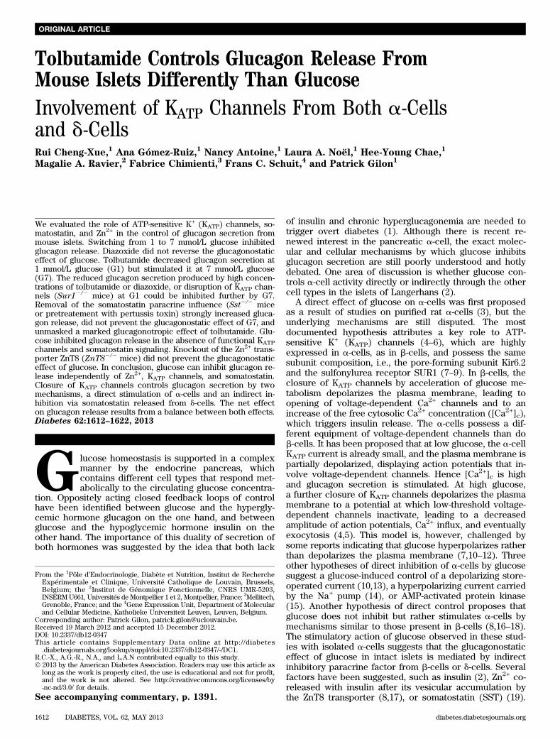
Effect of KATP channel modulators on glucagonsecretion from C57Bl/6 mice. Addition of 500 mmol/LTolb to a medium containing G1 reversibly inhibited glu-cagon secretion (Fig. 3A). The effect of the sulfonylureawas dose-dependent, being modest at 10 mmol/L (26% ofinhibition; P , 0.01) and strong at 50 mmol/L (66% of in-hibition; P , 0.01; Fig. 3C). Surprisingly, 500 mmol/L Tolbstimulated glucagon secretion when applied in G7 (Fig.3B). A concentration of at least 50 mmol/L Tolb was re-quired to see this effect (48% of stimulation; P , 0.05; Fig.3D). It has been suggested that glucose concentrationshigher than G7 paradoxically stimulate glucagon secretion(25). If this results from an additional closure of KATPchannels, then this could be compatible with the stimula-tory effect of Tolb. However, we found that an increasefrom G7 to 30 mmol/L glucose inhibited glucagon secretion(Fig. 3G) and did not reproduce the stimulatory effect ofTolb. Therefore, glucose and Tolb exert distinct effects onglucagon secretion.
We tested the effect of Dz. Addition of 250 mmol/L Dz inG1 strongly inhibited glucagon release (Fig. 3A). The in-hibitory effect was already robust at 50 mmol/L of the drug(48% of inhibition; P , 0.01; Fig. 3E). We also checkedwhether any of the tested Dz concentrations could re-verse the glucagonostatic effect of glucose; 250 mmol/L Dzstrongly inhibited glucagon secretion in G7 (Fig. 3B), anda weak, but nonsignificant, inhibition was observed at 50mmol/L (Fig. 3F). Importantly, lower concentrations ofDz never reversed the inhibitory effect of G7 (Fig. 3F),suggesting that glucose inhibited glucagon secretion in-dependently from KATP channel closure.Effect of glucose on hormone secretion of islets fromSst+/+ and Sst2/2
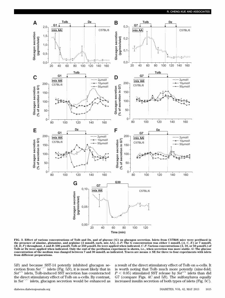
mice. The role of SST in the control ofglucagon secretion by glucose was studied using Sst+/+ andSst2/2 mice. As expected, Sst2/2 islets lack immunoreac-tive SST (Supplementary Fig. 1) and do not secrete de-tectable amounts of SST. In G1, glucagon secretion wassignificantly (P , 0.05) higher in Sst2/2 than in Sst+/+ islets(Fig. 4A). The difference was larger when secretion wasexpressed as percentage of content (0.24% 6 0.06 vs.0.056% 6 0.01; P , 0.05) because the glucagon contentwas lower in Sst2/2 than in Sst+/+ islets (664 6 53 pg/islet[n = 18] vs. 820 6 53 pg/islet [n = 23], respectively;P , 0.05). This suggests that SST exerts a strong tonicinhibition on glucagon release. Switching from G1 to G7inhibited glucagon secretion from both Sst+/+ and Sst2/2
islets, which demonstrates that SST alone is not re-sponsible for the inhibition of glucagon secretion by glu-cose (Fig. 4A). However, the inhibition was less sustainedin Sst2/2 than in Sst+/+ islets, supporting a possible in-volvement of SST in the glucagonostatic effect of glucose(Fig. 4B). G7 stimulated SST release from Sst+/+ islets (Fig.4C) and triggered a larger insulin secretion from Sst2/2 thanSst+/+ islets (Fig. 4D; 23.43 6 5.12 [n = 3] vs. 8.28 6 0.45 pg/min/islet [n = 5]). This latter observation was not attribut-able to reduced insulin content (47 6 16 vs. 586 19 ng/isletin Sst+/+ and Sst2/2 islets, respectively) and suggests thatSST exerts an inhibitory paracrine control on insulin releaseduring glucose stimulation.Effect of KATP channel modulators on hormonesecretion of islets from Sst+/+ and Sst2/2
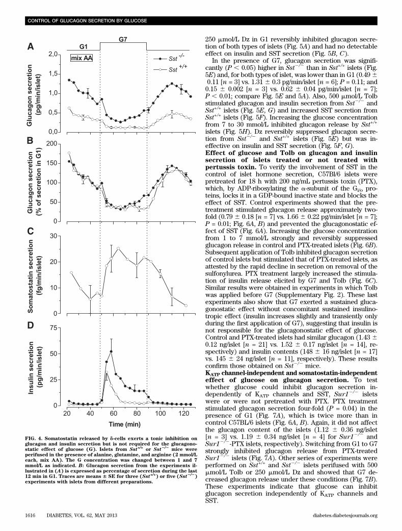
mice. Addi-tion of 500 mmol/L Tolb to G1 did not affect glucagon se-cretion from Sst+/+ islets, whereas it stimulated that ofSst2/2 islets (Fig. 5A), demonstrating that Tolb did notreproduce the glucagonostatic effect of glucose. BecauseTolb strongly stimulated SST release from Sst+/+ islets (Fig.
FIG. 2. Glucose (G) can inhibit glucagon secretion without functionalKATP channels. Islets from Sur1+/+ or Sur12/2
mice were perifused inthe presence of alanine, glutamine, and arginine (2 mmol/L each, mixAA). A–C: The G concentration was changed between 1 and 7 mmol/Lwhen indicated. B: Glucagon secretion from the experiments illustratedin (A) is expressed as percentage of secretion during the last 12 min inG1. C: The perifusion medium was supplemented with 500 mmol/L Tolb(○) or 250 mmol/L Dz (●) to maximally close or open KATP channels,respectively. Secretion in the presence of Dz is displayed with an ex-tended scale in the inset to better see the inhibitory effect of glucose.The dashed lines in the inset correspond to changes in glucose con-centrations. Traces are means 6 SE for seven (A and B: Sur1+/+), five(A and B: Sur12/2
), and three (C) experiments with islets from dif-ferent preparations.
CONTROL OF GLUCAGON SECRETION BY GLUCOSE
1614 DIABETES, VOL. 62, MAY 2013 diabetes.diabetesjournals.org
5B) and because SST-14 potently inhibited glucagon se-cretion from Sst2/2 islets (Fig. 5D), it is most likely that inSst+/+ islets, Tolb-induced SST secretion has counteractedthe direct stimulatory effect of Tolb on a-cells. By contrast,in Sst2/2 islets, glucagon secretion would be enhanced as
a result of the direct stimulatory effect of Tolb on a-cells. Itis worth noting that Tolb much more potently (nine-fold;P , 0.05) stimulated SST release by Sst+/+ islets than didG7 (compare Figs. 4C and 5B). The sulfonylurea equallyincreased insulin secretion of both types of islets (Fig. 5C).
FIG. 3. Effect of various concentrations of Tolb and Dz, and of glucose (G) on glucagon secretion. Islets from C57Bl/6 mice were perifused inthe presence of alanine, glutamine, and arginine (2 mmol/L each, mix AA). A–F: The G concentration was either 1 mmol/L (A, C, E) or 7 mmol/L(B, D, F) throughout. A and B: 500 mmol/L Tolb or 250 mmol/L Dz were applied when indicated. C–F: Various concentrations (2, 10, or 50 mmol/L) ofTolb or Dz were applied when indicated. Only the end of the perifusion experiment is shown, i.e., when secretion was more stable. G: The glucoseconcentration of the medium was changed between 7 and 30 mmol/L as indicated. Traces are means 6 SE for three to four experiments with isletsfrom different preparations.
R. CHENG-XUE AND ASSOCIATES
diabetes.diabetesjournals.org DIABETES, VOL. 62, MAY 2013 1615
250 mmol/L Dz in G1 reversibly inhibited glucagon secre-tion of both types of islets (Fig. 5A) and had no detectableeffect on insulin and SST secretion (Fig. 5B, C).
In the presence of G7, glucagon secretion was signifi-cantly (P , 0.05) higher in Sst2/2 than in Sst+/+ islets (Fig.5E) and, for both types of islet, was lower than in G1 (0.4960.11 [n = 3] vs. 1.31 6 0.3 pg/min/islet [n = 6]; P = 0.11; and0.15 6 0.002 [n = 3] vs. 0.62 6 0.04 pg/min/islet [n = 7];P , 0.01; compare Fig. 5E and 5A). Also, 500 mmol/L Tolbstimulated glucagon and insulin secretion from Sst2/2 andSst+/+ islets (Fig. 5E, G) and increased SST secretion fromSst+/+ islets (Fig. 5F). Increasing the glucose concentrationfrom 7 to 30 mmol/L inhibited glucagon release by Sst+/+
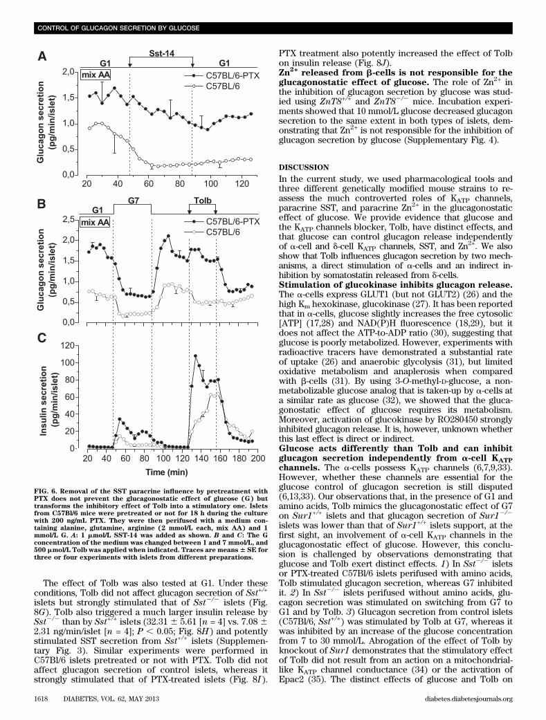
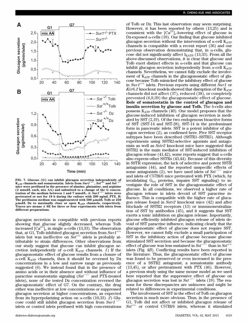
islets (Fig. 5H). Dz reversibly suppressed glucagon secre-tion from Sst2/2 and Sst+/+ islets (Fig. 5E) but was in-effective on insulin and SST secretion (Fig. 5F, G).Effect of glucose and Tolb on glucagon and insulinsecretion of islets treated or not treated withpertussis toxin. To verify the involvement of SST in thecontrol of islet hormone secretion, C57Bl/6 islets werepretreated for 18 h with 200 ng/mL pertussis toxin (PTX),which, by ADP-ribosylating the a-subunit of the Gi/o pro-teins, locks it in a GDP-bound inactive state and blocks theeffect of SST. Control experiments showed that the pre-treatment stimulated glucagon release approximately two-fold (0.79 6 0.18 [n = 7] vs. 1.66 6 0.22 pg/min/islet [n = 7];P = 0.01; Fig. 6A, B) and prevented the glucagonostatic ef-fect of SST (Fig. 6A). Increasing the glucose concentrationfrom 1 to 7 mmol/L strongly and reversibly suppressedglucagon release in control and PTX-treated islets (Fig. 6B).Subsequent application of Tolb inhibited glucagon secretionof control islets but stimulated that of PTX-treated islets, asattested by the rapid decline in secretion on removal of thesulfonylurea. PTX treatment largely increased the stimula-tion of insulin release elicited by G7 and Tolb (Fig. 6C).Similar results were obtained in experiments in which Tolbwas applied before G7 (Supplementary Fig. 2). These lastexperiments also show that G7 exerted a sustained gluca-gonostatic effect without concomitant sustained insulino-tropic effect (insulin increases slightly and transiently onlyduring the first application of G7), suggesting that insulin isnot responsible for the glucagonostatic effect of glucose.Control and PTX-treated islets had similar glucagon (1.43 60.12 ng/islet [n = 21] vs. 1.52 6 0.17 ng/islet [n = 14], re-spectively) and insulin contents (148 6 16 ng/islet [n = 17]vs. 145 6 24 ng/islet [n = 11], respectively). These resultsconfirm those obtained on Sst2/2 mice.KATP channel-independent and somatostatin-independenteffect of glucose on glucagon secretion. To testwhether glucose could inhibit glucagon secretion in-dependently of KATP channels and SST, Sur12/2 isletswere or were not pretreated with PTX. PTX treatmentstimulated glucagon secretion four-fold (P = 0.04) in thepresence of G1 (Fig. 7A), which is twice more than incontrol C57BL/6 islets (Fig. 6A, B). Again, it did not affectthe glucagon content of the islets (1.12 6 0.36 ng/islet[n = 3] vs. 1.19 6 0.34 ng/islet [n = 4] for Sur12/2 andSur12/2-PTX islets, respectively). Switching from G1 to G7strongly inhibited glucagon release from PTX-treatedSur12/2 islets (Fig. 7A). Other series of experiments wereperformed on Sst+/+ and Sst2/2 islets perifused with 500mmol/L Tolb or 250 mmol/L Dz and showed that G7 de-creased glucagon release under these conditions (Fig. 7B).These experiments indicate that glucose can inhibitglucagon secretion independently of KATP channels andSST.
FIG. 4. Somatostatin released by d-cells exerts a tonic inhibition onglucagon and insulin secretion but is not required for the glucagono-static effect of glucose (G). Islets from Sst+/+ or Sst2/2
mice wereperifused in the presence of alanine, glutamine, and arginine (2 mmol/Leach, mix AA). The G concentration was changed between 1 and 7mmol/L as indicated. B: Glucagon secretion from the experiments il-lustrated in (A) is expressed as percentage of secretion during the last12 min in G1. Traces are means 6 SE for three (Sst+/+) or five (Sst2/2
)experiments with islets from different preparations.
CONTROL OF GLUCAGON SECRETION BY GLUCOSE
1616 DIABETES, VOL. 62, MAY 2013 diabetes.diabetesjournals.org
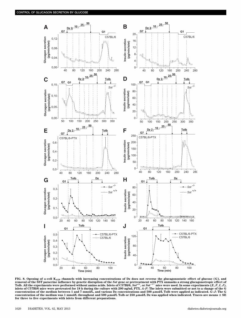
Effect of glucose and KATP channel modulators onislet hormone secretion in the absence of amino acids.Additional experiments were performed in amino acid–free media to verify key observations that were made inthe presence of amino acids. The absence of amino acidsdramatically reduced glucagon release. Dz (from 2 to 50mmol/L) did not increase glucagon secretion of C57Bl/6islets at G7, whereas a decrease in the glucose concen-tration to 1 mmol/L strongly stimulated glucagon release(Fig. 8A). Dz dose-dependently inhibited insulin release atG7 (Fig. 8B). Switching from G7 to G1 stimulated glucagonrelease of Sst2/2 islets, whereas the subsequent addition of
Dz tended to decrease glucagon secretion, as attested toby the reacceleration of secretion on Dz removal (Fig. 8C).Again, Dz dose-dependently inhibited insulin secretion(Fig. 8D). Tolb stimulated both glucagon and insulin re-lease (Fig. 8C, D). At G7, Dz did not affect glucagon se-cretion of PTX-pretreated C57Bl/6 islets, whereas Tolbpotently stimulated their glucagon and insulin release (Fig.8E, F). The observations that in the complete absence ofinfluence of SST (Fig. 8C, E), Dz did not reverse the glu-cagonostatic effect of G7 whereas Tolb strongly stimulatedglucagon release suggest that glucose inhibits glucagonrelease independently from a-cell KATP channels.
FIG. 5. Effects of KATP channel modulators, SST, and glucose (G) on islet hormone secretion. Islets from Sst+/+ or Sst2/2mice were perifused in the
presence of alanine, glutamine, and arginine (2 mmol/L each, mix AA). The G concentration of the medium was either 1 (A–D) or 7 mmol/L (E–G)throughout. A–C and E–G: 500 mmol/L Tolb or 250 mmol/L Dz was applied when indicated. D: 1 mmol/L SST-14 was added as shown. H: The Gconcentration was changed between 7 and 30 mmol/L as indicated. Traces are means 6 SE for seven (A–C: Sst+/+), six (A and C: Sst2/2
), and three(D–H) experiments with islets from different preparations.
R. CHENG-XUE AND ASSOCIATES
diabetes.diabetesjournals.org DIABETES, VOL. 62, MAY 2013 1617
The effect of Tolb was also tested at G1. Under theseconditions, Tolb did not affect glucagon secretion of Sst+/+
islets but strongly stimulated that of Sst2/2 islets (Fig.8G). Tolb also triggered a much larger insulin release bySst2/2 than by Sst+/+ islets (32.31 6 5.61 [n = 4] vs. 7.08 62.31 ng/min/islet [n = 4]; P , 0.05; Fig. 8H) and potentlystimulated SST secretion from Sst+/+ islets (Supplemen-tary Fig. 3). Similar experiments were performed inC57Bl/6 islets pretreated or not with PTX. Tolb did notaffect glucagon secretion of control islets, whereas itstrongly stimulated that of PTX-treated islets (Fig. 8I ).
PTX treatment also potently increased the effect of Tolbon insulin release (Fig. 8J).Zn
2+released from b-cells is not responsible for the
glucagonostatic effect of glucose. The role of Zn2+ inthe inhibition of glucagon secretion by glucose was stud-ied using ZnT8+/+ and ZnT82/2 mice. Incubation experi-ments showed that 10 mmol/L glucose decreased glucagonsecretion to the same extent in both types of islets, dem-onstrating that Zn2+ is not responsible for the inhibition ofglucagon secretion by glucose (Supplementary Fig. 4).
DISCUSSION
In the current study, we used pharmacological tools andthree different genetically modified mouse strains to re-assess the much controverted roles of KATP channels,paracrine SST, and paracrine Zn2+ in the glucagonostaticeffect of glucose. We provide evidence that glucose andthe KATP channels blocker, Tolb, have distinct effects, andthat glucose can control glucagon release independentlyof a-cell and d-cell KATP channels, SST, and Zn2+. We alsoshow that Tolb influences glucagon secretion by two mech-anisms, a direct stimulation of a-cells and an indirect in-hibition by somatostatin released from d-cells.Stimulation of glucokinase inhibits glucagon release.The a-cells express GLUT1 (but not GLUT2) (26) and thehigh Km hexokinase, glucokinase (27). It has been reportedthat in a-cells, glucose slightly increases the free cytosolic[ATP] (17,28) and NAD(P)H fluorescence (18,29), but itdoes not affect the ATP-to-ADP ratio (30), suggesting thatglucose is poorly metabolized. However, experiments withradioactive tracers have demonstrated a substantial rateof uptake (26) and anaerobic glycolysis (31), but limitedoxidative metabolism and anaplerosis when comparedwith b-cells (31). By using 3-O-methyl-D-glucose, a non-metabolizable glucose analog that is taken-up by a-cells ata similar rate as glucose (32), we showed that the gluca-gonostatic effect of glucose requires its metabolism.Moreover, activation of glucokinase by RO280450 stronglyinhibited glucagon release. It is, however, unknown whetherthis last effect is direct or indirect.Glucose acts differently than Tolb and can inhibitglucagon secretion independently from a-cell KATP
channels. The a-cells possess KATP channels (6,7,9,33).However, whether these channels are essential for theglucose control of glucagon secretion is still disputed(6,13,33). Our observations that, in the presence of G1 andamino acids, Tolb mimics the glucagonostatic effect of G7on Sur1+/+ islets and that glucagon secretion of Sur12/2
islets was lower than that of Sur1+/+ islets support, at thefirst sight, an involvement of a-cell KATP channels in theglucagonostatic effect of glucose. However, this conclu-sion is challenged by observations demonstrating thatglucose and Tolb exert distinct effects. 1) In Sst2/2 isletsor PTX-treated C57Bl/6 islets perifused with amino acids,Tolb stimulated glucagon secretion, whereas G7 inhibitedit. 2) In Sst2/2 islets perifused without amino acids, glu-cagon secretion was stimulated on switching from G7 toG1 and by Tolb. 3) Glucagon secretion from control islets(C57Bl/6, Sst+/+) was stimulated by Tolb at G7, whereas itwas inhibited by an increase of the glucose concentrationfrom 7 to 30 mmol/L. Abrogation of the effect of Tolb byknockout of Sur1 demonstrates that the stimulatory effectof Tolb did not result from an action on a mitochondrial-like KATP channel conductance (34) or the activation ofEpac2 (35). The distinct effects of glucose and Tolb on
FIG. 6. Removal of the SST paracrine influence by pretreatment withPTX does not prevent the glucagonostatic effect of glucose (G) buttransforms the inhibitory effect of Tolb into a stimulatory one. Isletsfrom C57Bl/6 mice were pretreated or not for 18 h during the culturewith 200 ng/mL PTX. They were then perifused with a medium con-taining alanine, glutamine, arginine (2 mmol/L each, mix AA) and 1mmol/L G. A: 1 mmol/L SST-14 was added as shown. B and C: The Gconcentration of the medium was changed between 1 and 7 mmol/L, and500 mmol/L Tolb was applied when indicated. Traces are means6 SE forthree or four experiments with islets from different preparations.
CONTROL OF GLUCAGON SECRETION BY GLUCOSE
1618 DIABETES, VOL. 62, MAY 2013 diabetes.diabetesjournals.org
glucagon secretion is compatible with previous reportsshowing that glucose slightly decreased, whereas Tolbincreased [Ca2+]c in single a-cells (13,33). The observationthat, at G1, Tolb inhibited glucagon secretion from Sur1+/+
islets but was ineffective on Sst+/+ islets is probably at-tributable to strain differences. Other observations fromour study suggest that glucose can inhibit glucagon se-cretion independently of a-cell KATP channels. 1) If theglucagonostatic effect of glucose results from a closure ofa-cell KATP channels, then it should be reversed by Dzconcentrations in a low and narrow range as previouslysuggested (6). We instead found that in the presence ofamino acids or in their absence and without influence ofparacrine somatostatin signaling (Sst2/2 and PTX-treatedislets), none of the tested Dz concentration reversed theglucagonostatic effect of G7. On the contrary, the drugeither was ineffective at low concentrations or suppressedglucagon secretion at higher concentrations, as expectedfrom its hyperpolarizing action on a-cells (10,33). 2) Glu-cose could still inhibit glucagon secretion from Sur12/2
islets or control islets perifused with high concentrations
of Tolb or Dz. This last observation may seem surprising.However, it has been reported by others (13,25) and isconsistent with the [Ca2+]c-lowering effect of glucose inDz-exposed a-cells (10). Our finding that glucose inhibitedglucagon secretion without the intervention of a-cell KATPchannels is compatible with a recent report (36) and ourprevious observation demonstrating that, in a-cells, glu-cose did not significantly affect IKATP (13,33). From all theabove-discussed observations, it is clear that glucose andTolb exert distinct effects in a-cells and that glucose caninhibit glucagon secretion independently from a-cell KATPchannels. Nevertheless, we cannot fully exclude the involve-ment of KATP channels in the glucagonostatic effect of glu-cose because Tolb mimicked the inhibitory effect of glucosein Sur1+/+ islets. Previous reports using different Sur1 orKir6.2 knockout models showed that disruption of the KATPchannels did not affect (37), reduced (38), or completelyprevented (6,9,39) the glucagonostatic effect of glucose.Role of somatostatin in the control of glucagon andinsulin secretion by glucose and Tolb. The d-cells alsopossess KATP channels (40). One model proposes that theglucose-induced inhibition of glucagon secretion is medi-ated by SST (2,19). Of the two endogenous bioactive formsof SST (SST-14 and SST-28), SST-14 is the predominantform in pancreatic islets. SST is a potent inhibitor of glu-cagon secretion (2), as confirmed here. Five SST receptorsubtypes have been described (SSTR1–SSTR5). Althoughexperiments using SSTR2-selective agonists and antago-nists as well as Sstr2 knockout mice have suggested thatSSTR2 is the main mediator of SST-induced inhibition ofglucagon release (41,42), some reports suggest that a-cellsalso express other SSTRs (43,44). Because of this diversityin SSTR expression, the lack of selective and potent SSTRantagonists (44), and the reported unspecific effects ofsome antagonists (2), we have used islets of Sst2/2 miceand islets of C57Bl/6 mice pretreated with PTX (which, byinvalidating Gi/o proteins, impairs SST signaling) to in-vestigate the role of SST in the glucagonostatic effect ofglucose. In all conditions, we observed a higher rate ofglucagon release in islets devoid of SST paracrine in-fluence. This is compatible with the higher rate of gluca-gon release found in Sstr2 knockout mice (42) and afterblockade of SSTR2 receptors (13) or immunoneutraliza-tion of SST by antibodies (45), and it suggests that SSTexerts a tonic inhibition on glucagon release. Importantly,glucose efficiently inhibited glucagon release of islets de-void of SST paracrine influence, indicating clearly that theglucagonostatic effect of glucose does not require SST.However, we cannot fully exclude a small participation ofSST in the inhibitory action of glucose because glucosestimulated SST secretion and because the glucagonostaticeffect of glucose was less sustained in Sst2/2 than in Sst+/+
islets (Fig. 4B). Conflicting results have been reported inthe literature. Thus, the glucagonostatic effect of glucosewas found to be preserved or even increased in the pres-ence of a SSTR2 antagonist, a somatostatin antibody(13,45), or after pretreatment with PTX (46). However,a previous study using the same mouse model as we usedhere reported that the suppressive effect of glucose onglucagon secretion was lost in Sst2/2 islets (19). The rea-sons for these discrepancies are unknown and might berelated to differences in experimental conditions.
The involvement of SST in the effect of Tolb on glucagonsecretion is much more obvious. Thus, in the presence ofG1, Tolb did not affect or inhibited glucagon release ofSst+/+ or control C57Bl/6 islets, whereas it stimulated
FIG. 7. Glucose (G) can inhibit glucagon secretion independently ofKATP channels and somatostatin. Islets from Sur12/2
, Sst+/+ and Sst2/2
mice were perifused in the presence of alanine, glutamine, and arginine(2 mmol/L each, mix AA) and submitted to a change of the G concen-tration of the medium between 1 and 7 mmol/L. A: Sur12/2
islets werepretreated or not for 18 h during the culture with 200 ng/mL PTX. B:The perifusion medium was supplemented with 500 mmol/L Tolb or 250mmol/L Dz to maximally close or open KATP channels, respectively.Traces are means 6 SE for three or four experiments with islets fromdifferent preparations.
R. CHENG-XUE AND ASSOCIATES
diabetes.diabetesjournals.org DIABETES, VOL. 62, MAY 2013 1619
FIG. 8. Opening of a-cell KATP channels with increasing concentrations of Dz does not reverse the glucagonostatic effect of glucose (G), andremoval of the SST paracrine influence by genetic disruption of the Sst gene or pretreatment with PTX unmasks a strong glucagonotropic effect ofTolb. All the experiments were performed without amino acids. Islets of C57Bl/6, Sst+/+, or Sst2/2
mice were used. In some experiments (E, F, I, J),islets of C57Bl/6 mice were pretreated for 18 h during the culture with 200 ng/mL PTX. A–F: The islets were submitted or not to a change of the Gconcentration of the medium between 1 and 7 mmol/L, and various Dz concentrations and 500 mmol/L Tolb were applied as indicated. G–J: The Gconcentration of the medium was 1 mmol/L throughout and 500 mmol/L Tolb or 250 mmol/L Dz was applied when indicated. Traces are means 6 SEfor three to five experiments with islets from different preparations.
CONTROL OF GLUCAGON SECRETION BY GLUCOSE
1620 DIABETES, VOL. 62, MAY 2013 diabetes.diabetesjournals.org
glucagon secretion of Sst2/2 or PTX-treated C57Bl/6 islets(even very potently in the absence of amino acids). Thisclearly indicates that SST is involved in the control ofglucagon release by Tolb. The stronger involvement of SSTin the glucagonostatic effect of Tolb than that of glucose iscompatible with our observation that the sulfonylureastimulated SST secretion much more potently than glu-cose. The difference in the effect of Tolb between isletswith or without paracrine SST signaling suggests that Tolbmodulates glucagon secretion by two distinct mechanisms,a direct stimulatory effect on a-cells that is detected in theabsence of SST (i.e., in Sst2/2 or PTX-treated islets) and anindirect inhibitory effect that is caused by the stimulationof SST release. The net effect of Tolb on glucagon se-cretion would thus result from a balance between thestimulatory and the inhibitory effects. That Tolb directlystimulates a-cells is supported by previous reports show-ing that the sulfonylurea increases [Ca2+]c (13,33) andglucagon secretion from isolated a-cells (8,16). Althoughthe a-cell KATP current is already small even at low glu-cose, a tiny additional reduction of the current elicited byTolb would strongly affect a-cell electrical activity becauseof the high resistance of its plasma membrane.
The opposite effects of a-cell and d-cell KATP channelmodulation for the control of glucagon secretion would alsoexplain why both Tolb and Dz, which have opposite effectson channel activity, inhibit glucagon secretion at G1. On theone hand, the glucagonostatic effect of Tolb would mainlybe mediated by the dominating effect of SST as explainedabove. On the other hand, the glucagonostatic effect of Dzwould essentially result from its dominating direct hyper-polarizing effect on a-cells.
The strong paracrine influence of SST released fromKATP channel–deficient d-cells might explain the reducedglucagon secretion of Sur12/2 islets. It is compatible withthe observation that pretreatment of these islets with PTXpotently stimulated glucagon secretion because of therelief of the inhibitory effect of SST.
At G7, Tolb stimulated glucagon secretion from isletswith or without paracrine SST signaling. It is possible that inconditions in which glucagon secretion is already inhibited,the direct stimulatory effect of Tolb on a-cells overwhelmsthe indirect inhibitory effect caused by the stimulation ofSST release. The glucose dependency of the effects of Tolband its two mechanisms of action, directly on a-cells andindirectly through d-cells, might explain why sulfonylureashave been reported to exert variable effects on glucagonrelease. Thus, glucagon secretion was stimulated (8,16),unaffected (47), or inhibited (38,39,48) by sulfonylureas.
Paracrine SST also influences insulin secretion. Thus, inmost tested conditions, glucose and Tolb induced a largerinsulin secretion in islets without paracrine SST signalingthan in control islets, confirming previous reports (19).KATP channel–independent and SST-independent effectof glucose. Experiments on islets with genetic or pharma-cological disruption of both the KATP channels and SST sig-naling (Fig. 7) revealed that glucose can inhibit glucagonsecretion independently from KATP channels and SST. This iscompatible with our previous observation demonstratingthat in isolated a-cells devoid of paracrine influence, glucosedecreased [Ca2+]c in the presence of a high concentration ofTolb (13,33). The nature of the underlying mechanism is,however, unknown.Glucose inhibits glucagon secretion independentlyfrom Zn
2+. It has been hypothesized that Zn2+ released
from b-cells could be responsible for the glucagonostatic
effect of glucose. By monitoring Zn2+ exocytosis fromZnT8+/+ and ZnT82/2 mice, we previously showed thatZnT8 is the main transporter responsible for Zn2+ accumu-lation in insulin granules because its ablation reduced thezinc exocytotic events by 99% (22). Here, we showed thatglucose similarly inhibited glucagon secretion of ZnT8+/+
and ZnT82/2 islets. This confirms previous reports (49,50)and excludes Zn2+ as an inhibitory paracrine signal medi-ating the glucagonostatic effect of glucose.Conclusion. SST exerts a tonic inhibition on insulin andglucagon secretion. Glucose can inhibit glucagon releaseindependently of Zn2+ released from b-cells, KATP channels,and SST. Participation of these last two factors in the glu-cagonostatic effect of glucose, however, cannot be ex-cluded. Closure of KATP channels controls glucagonsecretion by two mechanisms, a direct stimulation of a-cellsand an indirect inhibition via SST released from d-cells. Thenet effect on glucagon release results from a balance be-tween both effects. This might explain why Tolb reproducesthe glucagonostatic effect of glucose in some conditions,whereas it stimulates glucagon release in others. This lattersituation should be considered during treatment of type 2diabetic patients by sulfonylureas because stimulation ofglucagon secretion by the drugs could contribute to theunwanted hyperglucagonemia found in diabetes. Our studyalso calls for a careful examination of d-cell function in di-abetes.
ACKNOWLEDGMENTS
This work was supported by Grant 3.4554.10 from theFonds de la Recherche Scientifique Médicale (Brussels,Belgium), by Grant ARC (05/10-328) from the General Di-rection of Scientific Research of the French Community ofBelgium, by the Interuniversity Poles of Attraction Pro-gramme (PAI 6/40) from the Belgian Science Policy, byJuvenile Diabetes Research Foundation Project Grant2007-685, and by a European Foundation for the Study ofDiabetes/Boehringer Ingelheim grant.
P.G. is Research Director of the Fonds National de la Re-cherche Scientifique, Brussels. M.A.R. is Chargé deRecherches at INSERM, Paris, France. F.C. is employedby Mellitech SAS, Grenoble, France. No other potentialconflicts of interest relevant to this article were reported.
R.C.-X. and P.G. wrote the manuscript. R.C.-X. A.G.-R.,N.A., L.A.N., H.-Y.C., M.A.R., and P.G. researched data.M.A.R. contributed to discussion. M.A.R. and F.C.S. reviewedthe manuscript. M.A.R., F.C., and F.C.S. edited the manu-script. P.G. is the guarantor of this work, had full access toall the data, and takes full responsibility for the integrity ofdata and the accuracy of data analysis.
The authors thank F. Knockaert, V. Massé, andS. Godecharles (all three from Université Catholique de Lou-vain, Brussels, Belgium) for technical assistance, J. Bryan(Pacific Northwest Diabetes Research Institute, Seattle,Washington) for the gift of Sur12/2 mice, J.C. Henquin(Université Catholique de Louvain, Brussels, Belgium) forproviding access to his homemade insulin assay, and I. Rob-inson (National Institute for Medical Research, London,U.K.) and M. Low (Oregon Health and Science University,Portland, Oregon) for the gift of Sst2/2 mice.
REFERENCES
1. Unger RH, Cherrington AD. Glucagonocentric restructuring of diabetes:a pathophysiologic and therapeutic makeover. J Clin Invest 2012;122:4–12
R. CHENG-XUE AND ASSOCIATES
diabetes.diabetesjournals.org DIABETES, VOL. 62, MAY 2013 1621
2. Gromada J, Franklin I, Wollheim CB. a-cells of the endocrine pancreas: 35years of research but the enigma remains. Endocr Rev 2007;28:84–116
3. Pipeleers DG, Schuit FC, Van Schravendijk CF, Van de Winkel M. Interplayof nutrients and hormones in the regulation of glucagon release. Endo-crinology 1985;117:817–823
4. Rorsman P, Braun M, Zhang Q. Regulation of calcium in pancreatic a- andb-cells in health and disease. Cell Calcium 2012;51:300–308
5. Walker JN, Ramracheya R, Zhang Q, Johnson PR, Braun M, Rorsman P.Regulation of glucagon secretion by glucose: paracrine, intrinsic or both?Diabetes Obes Metab 2011;13(Suppl 1):95–105
6. MacDonald PE, De Marinis YZ, Ramracheya R, et al. A KATP channel-dependentpathway within a-cells regulates glucagon release from both rodent andhuman islets of Langerhans. PLoS Biol 2007;5:e143
7. Bokvist K, Olsen HL, Høy M, et al. Characterisation of sulphonylurea andATP-regulated K+ channels in rat pancreatic A-cells. Pflugers Arch 1999;438:428–436
8. Franklin I, Gromada J, Gjinovci A, Theander S, Wollheim CB. b-cell se-cretory products activate a-cell ATP-dependent potassium channels toinhibit glucagon release. Diabetes 2005;54:1808–1815
9. Shiota C, Rocheleau JV, Shiota M, Piston DW, Magnuson MA. Impairedglucagon secretory responses in mice lacking the type 1 sulfonylurea re-ceptor. Am J Physiol Endocrinol Metab 2005;289:E570–E577
10. Liu YJ, Vieira E, Gylfe E. A store-operated mechanism determines theactivity of the electrically excitable glucagon-secreting pancreatic a-cell.Cell Calcium 2004;35:357–365
11. Hjortoe GM, Hagel GM, Terry BR, Thastrup O, Arkhammar POG. Func-tional identification and monitoring of individual a- and b-cells in culturedmouse islets of Langerhans. Acta Diabetol 2004;41:185–193
12. Manning Fox JE, Gyulkhandanyan AV, Satin LS, Wheeler MB. Oscillatorymembrane potential response to glucose in islet b-cells: a comparison ofislet-cell electrical activity in mouse and rat. Endocrinology 2006;147:4655–4663
13. Vieira E, Salehi A, Gylfe E. Glucose inhibits glucagon secretion by a directeffect on mouse pancreatic a-cells. Diabetologia 2007;50:370–379
14. Bode HP, Weber S, Fehmann HC, Göke B. A nutrient-regulated cytosoliccalcium oscillator in endocrine pancreatic glucagon-secreting cells.Pflugers Arch 1999;437:324–334
15. Leclerc I, Sun G, Morris C, Fernandez-Millan E, Nyirenda M, Rutter GA.AMP-activated protein kinase regulates glucagon secretion from mousepancreatic a-cells. Diabetologia 2011;54:125–134
16. Olsen HL, Theander S, Bokvist K, Buschard K, Wollheim CB, Gromada J.Glucose stimulates glucagon release in single rat a-cells by mechanismsthat mirror the stimulus-secretion coupling in b-cells. Endocrinology 2005;146:4861–4870
18. Le Marchand SJ, Piston DW. Glucose suppression of glucagon secretion:metabolic and calcium responses from a-cells in intact mouse pancreaticislets. J Biol Chem 2010;285:14389–14398
19. Hauge-Evans AC, King AJ, Carmignac D, et al. Somatostatin secreted byislet d-cells fulfills multiple roles as a paracrine regulator of islet function.Diabetes 2009;58:403–411
20. Seghers V, Nakazaki M, DeMayo F, Aguilar-Bryan L, Bryan J. Sur1knockout mice. A model for KATP channel-independent regulation of in-sulin secretion. J Biol Chem 2000;275:9270–9277
21. Low MJ, Otero-Corchon V, Parlow AF, et al. Somatostatin is required formasculinization of growth hormone-regulated hepatic gene expression butnot of somatic growth. J Clin Invest 2001;107:1571–1580
22. Lemaire K, Ravier MA, Schraenen A, et al. Insulin crystallization dependson zinc transporter ZnT8 expression, but is not required for normalglucose homeostasis in mice. Proc Natl Acad Sci USA 2009;106:14872–14877
23. Quoix N, Cheng-Xue R, Guiot Y, Herrera PL, Henquin JC, Gilon P. TheGluCre-ROSA26EYFP mouse: a new model for easy identification of livingpancreatic a-cells. FEBS Lett 2007;581:4235–4240
24. Grimsby J, Sarabu R, Corbett WL, et al. Allosteric activators of glucoki-nase: potential role in diabetes therapy. Science 2003;301:370–373
25. Salehi A, Vieira E, Gylfe E. Paradoxical stimulation of glucagon secretionby high glucose concentrations. Diabetes 2006;55:2318–2323
26. Heimberg H, De Vos A, Pipeleers DG, Thorens B, Schuit F. Differences inglucose transporter gene expression between rat pancreatic a- and b-cellsare correlated to differences in glucose transport but not in glucose utili-zation. J Biol Chem 1995;270:8971–8975
27. Heimberg H, De Vos A, Moens K, et al. The glucose sensor protein glu-cokinase is expressed in glucagon-producing a-cells. Proc Natl Acad SciUSA 1996;93:7036–7041
28. Ravier MA, Rutter GA. Glucose or insulin, but not zinc ions, inhibit glu-cagon secretion from mouse pancreatic a-cells. Diabetes 2005;54:1789–1797
29. Quesada I, Todorova MG, Soria B. Different metabolic responses in a-, b-,and d-cells of the islet of Langerhans monitored by redox confocal mi-croscopy. Biophys J 2006;90:2641–2650
30. Detimary P, Dejonghe S, Ling ZD, Pipeleers D, Schuit F, Henquin JC. Thechanges in adenine nucleotides measured in glucose-stimulated rodentislets occur in b-cells but not in a-cells and are also observed in humanislets. J Biol Chem 1998;273:33905–33908
31. Schuit F, De Vos A, Farfari S, et al. Metabolic fate of glucose in purifiedislet cells. Glucose-regulated anaplerosis in b-cells. J Biol Chem 1997;272:18572–18579
32. Gorus FK, Malaisse WJ, Pipeleers DG. Differences in glucose handling bypancreatic A- and B-cells. J Biol Chem 1984;259:1196–1200
33. Quoix N, Cheng-Xue R, Mattart L, et al. Glucose and pharmacologicalmodulators of ATP-sensitive K+ channels control [Ca2+]c by differentmechanisms in isolated mouse a-cells. Diabetes 2009;58:412–421
34. Høy M, Olsen HL, Bokvist K, et al. Tolbutamide stimulates exocytosis ofglucagon by inhibition of a mitochondrial-like ATP-sensitive K+ (KATP)conductance in rat pancreatic A-cells. J Physiol 2000;527:109–120
35. Zhang CL, Katoh M, Shibasaki T, et al. The cAMP sensor Epac2 is a directtarget of antidiabetic sulfonylurea drugs. Science 2009;325:607–610
36. Le Marchand SJ, Piston DW. Glucose decouples intracellular Ca2+ activityfrom glucagon secretion in mouse pancreatic islet a-cells. PLoS ONE 2012;7:e47084
37. Miki T, Liss B, Minami K, et al. ATP-sensitive K+ channels in the hypo-thalamus are essential for the maintenance of glucose homeostasis. NatNeurosci 2001;4:507–512
38. Muñoz A, Hu M, Hussain K, Bryan J, Aguilar-Bryan L, Rajan AS. Regulationof glucagon secretion at low glucose concentrations: evidence for aden-osine triphosphate-sensitive potassium channel involvement. Endocrinol-ogy 2005;146:5514–5521
39. Gromada J, Ma XHM, Høy M, et al. ATP-sensitive K+ channel-dependentregulation of glucagon release and electrical activity by glucose in wild-type and SUR12/2 mouse a-cells. Diabetes 2004;53(Suppl 3):S181–S189
40. Göpel SO, Kanno T, Barg S, Rorsman P. Patch-clamp characterisation ofsomatostatin-secreting d-cells in intact mouse pancreatic islets. J Physiol2000;528:497–507
41. Cejvan K, Coy DH, Efendic S. Intra-islet somatostatin regulates glucagonrelease via type 2 somatostatin receptors in rats. Diabetes 2003;52:1176–1181
42. Strowski MZ, Parmar RM, Blake AD, Schaeffer JM. Somatostatin inhibitsinsulin and glucagon secretion via two receptors subtypes: an in vitrostudy of pancreatic islets from somatostatin receptor 2 knockout mice.Endocrinology 2000;141:111–117
43. Ludvigsen E, Olsson R, Stridsberg M, Janson ET, Sandler S. Expressionand distribution of somatostatin receptor subtypes in the pancreatic isletsof mice and rats. J Histochem Cytochem 2004;52:391–400
44. Strowski MZ, Blake AD. Function and expression of somatostatin re-ceptors of the endocrine pancreas. Mol Cell Endocrinol 2008;286:169–179
45. de Heer J, Rasmussen C, Coy DH, Holst JJ. Glucagon-like peptide-1, butnot glucose-dependent insulinotropic peptide, inhibits glucagon secretionvia somatostatin (receptor subtype 2) in the perfused rat pancreas. Dia-betologia 2008;51:2263–2270
46. Göpel S, Zhang Q, Eliasson L, et al. Capacitance measurements of exo-cytosis in mouse pancreatic a-, b- and d-cells within intact islets of Lang-erhans. J Physiol 2004;556:711–726
47. Gregorio F, Ambrosi F, Cristallini S, Pedetti M, Filipponi P, Santeusanio F.Therapeutical concentrations of tolbutamide, glibenclamide, gliclazide andgliquidone at different glucose levels: in vitro effects on pancreatic A- andB-cell function. Diabetes Res Clin Pract 1992;18:197–206
48. Göpel SO, Kanno T, Barg S, Weng XG, Gromada J, Rorsman P. Regulationof glucagon release in mouse a-cells by KATP channels and inactivation ofTTX-sensitive Na+ channels. J Physiol 2000;528:509–520
49. Hardy AB, Serino AS, Wijesekara N, Chimienti F, Wheeler MB. Regulationof glucagon secretion by zinc: lessons from the b-cell-specific Znt8knockout mouse model. Diabetes Obes Metab 2011;13(Suppl 1):112–117
50. Nicolson TJ, Bellomo EA, Wijesekara N, et al. Insulin storage and glucosehomeostasis in mice null for the granule zinc transporter ZnT8 and studiesof the type 2 diabetes-associated variants. Diabetes 2009;58:2070–2083
CONTROL OF GLUCAGON SECRETION BY GLUCOSE
1622 DIABETES, VOL. 62, MAY 2013 diabetes.diabetesjournals.org