Original Contribution ULTRASONOGRAPHIC QUALITATIVE CHARACTERS OF
RABBIT SPLEEN (ORYCTOLAGUS CUNICULUS)
R. Dimitrov1*, K. Stamatova1, A. Russenov2, D. Kostov1, D. Vladova3, M. Stefanov3
1Department of Veterinary Anatomy, Histology and Embryology, Faculty of Veterinary Medicine, Trakia University, Stara Zagora, Bulgaria
2Department of Internal Non-Infectious Diseases, Faculty of Veterinary Medicine, Trakia University, Stara Zagora, Bulgaria
3Department of Morphology, Physiology and Animal nutrition, Faculty of Agriculture, Trakia University, Stara Zagora, Bulgaria
ABSTRACT AIM: Finding some qualitative ultrasonographic features of normal rabbit spleen, in order to use the obtained results for imaging anatomical and diagnostic study of human and animal spleen lesions. MATERIALS: We investigated 9 sexually mature, healthy white New Zealand rabbit, aged 8 months, weighed from 2.8 kg to 3.2 kg. The animals were anesthetized. METHODS: The study was performed with Diagnostic Ultrasound System: model DC-6V Shenzhen Mindray Bio-Medical, Electrnics CO. Ltd (CHINA). We used 6.5 MHz microconvex and 5 MHz linear probes. The animals were positioned in supine recumbency. The approach was left percutaneous transabdominal hypochondrial left. The spleen of four animals was extirpated, following euthanasia. The obtained preparations were investigated in liquid isotonic medium. RESULTS: In the sagittal ultrasonographic study, the spleen was with elongated oval shape. In the transversal one the organ’s shape was triangular. The capsule was visualized as hyperechoic and heterogeneous striped finding. Parenchyma was hypoechoic, compared to the capsular structure. The blood vessels were visualized as oval anechoic findings. The investigation of extirpated spleen post mortem showed, that in sagittal aspect parenchyma is with homogeneous echogenicity. The capsule was relatively more hyperechoic and comaparatively more homogeneous structure than parenchyma. CONCLUSIONS: The qualitative ultrasonographic data for the rabbit spleen could be used as biological model in the imaging anatomical and diagnostic studies of some human and animal spleen lesions. Key words: spleen, anatomy, ultrasonography, rabbit
INTRODUCTION The spleen is a lymphoid organ, localized in the left cranial quarter of the abdomen. It is connected by gastrolienal ligament to the greater curvature of stomach and eneters in omental bursa. The spleen is with elongated shape, which varies in triangular in transversal sections. The organ’s shape, size and weight are variables, because of individual, age, and specimen, topographic and physiological features. In puberty the human, rat, mouse and canine spleen reaches its maximal _______________________________ *Correspondence to: Rosen Dimitrov , Department of Veterinary Anatomy, Histology and Embryology, Faculty of Veterinary Medicine, Trakia University, 6000 Stara Zagora, Bulgaria, Tel: + 359 42 699 647; E-mail: [email protected]
development and size and then it regresses with age. It is covered by elastic capsule, which comes from the organ stroma. The red and white pulps are situated in the spleen stroma. The rat and mouse spleen is used as biological model in the human immunodeficiency conditions’ investigations (1). The rabbit spleen is with elongated to oval shape, whose borders are parallel each other. Its dorsal end is situated at the last ribs’ level. Small part of it is located in front of the left kidney and the remainder is caudally to the rib arch. The spleen is localized under the dorsal abdominal wall, between the stomach, jejunum and caecum. Its hilus is elongated and
DIMITROV R., et al.
10 years - ANNIVERSARY EDITION TRAKIA JOURNAL OF SCIENCES, Vol. 10, No 1, 2012
65
separates gastric surface from intestinal one. The authors assume that its metric data are independent from age and body weight. The spleen is close to the caudomedial surface of the stomach (2, 3). The rabbit spleen is investigated anatomically by (4) in connection with macro- and microanatomical alterations in the organs’ structure in a diet. Many authors (5) investigate the normal human spleen structure via ultrasonography and computed tomography to apply the obtained results in the assessment of additional spleen tissues (accessory spleens, spleniculi). The ultrasonographic study of sheep, goat and buffalo spleen showed that the ultrasonographic approach is dorsal intercostals from 10th to 12th intercostals space. The visualization is achieved using 3.5 MHz convex transducer. The spleen capsule is hyperechoic, compared to the hypoechoic parenchyma, except the anechoic findings of the organs’ vessels (6). Ultrasonographic study of rabbit spleen is made by (7). The authors find the organ morphometric characters, investigating transversal ultrasonographic images during endotoxic shock. The transversal ultrasonographic image is triangular. The indirect literary data about rabbit spleen’s ultrasonographic anatomy and the direct ones about the human spleen motivated us to perform this imaging anatomical investigation. The aim of our study is to find some qualitative normal rabbit spleen’s ultrasonographic features for the imaging anatomical and diagnostic investigation of the human and animal spleen lesions.
MATERIALS AND METHODS Object We investigated 9 sexually mature and healthy white New Zealand rabbits, aged 8 months, weighed from 2.8 kg to 3.2 kg. The animals were anesthtized with 15 mg/kg Zoletil® 50 (tiletamine hydrochloride 125 mg and zolazepam hydrochloride 125 mg in 5 ml of the solution) Virbac, France. Transabdominal ultrasonography The study made with Diagnostic Ultrasound System: model DC-6V Shenzhen Mindray Bio-Medical, Electrnics CO. Ltd (CHINA) and
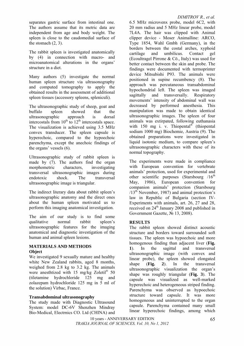
6.5 MHz microvenx probe, model 6C2, with 20 mm radius and 5 MHz linear probe, model 7L4A. The hair was clipped with Animal clipper device - Moser Animalline: ARCO, Type 1854, Wahl Gmbh (Germany), in the borders between the costal arches, xyphoid cartilage and umbilicus. Contact gel (Ecoultragel Pirrone & Cö., Italy) was used for better contact between the skin and probe. The findings were documented with termoprinter device Mitsubishi P93. The animals were positioned in supine recumbency (8). The approach was percutaneous transabdominal hypochondrial left. The spleen was imaged sagittally and transversally. Respiratory movements’ intensity of abdominal wall was decreased by performed anesthesia. This manipulation was made to obtain identical ultrasonographic images. The spleen of four animals was extirpated, following euthanasia with 150 mg i. v. Тhiopental® (thiopental sodium 1000 mg) Biochemie, Austria (9). The obtained preparations were investigated in liquid isotonic medium, to compare spleen’s ultrasonographic characters with these of its normal topography. The experiments were made in compliance with European convention for vertebrate animals’ protection, used for experimental and other scientific purposes (Starsbourg /16th May, 1986), European convention for companion animals’ protection (Starsbourg /13th November, 1987) and animal protection’s law in Republic of Bulgaria (section IV-Experiments with animals, art. 26, 27 and 28, received on 24th January 2008 and published in Government Gazette, № 13, 2008). RESULTS The rabbit spleen showed distinct acoustic structure and borders toward surrounded soft tissues. The spleen was hypoechoic and more homogenous finding than adjacent liver (Fig. 1). In the sagittal and transversal ultrasonographic image (with convex and linear probe), the spleen showed elongated shape (Fig. 2). In the transversal ultrasonographic visualization the organ’s shape was roughly triangular (Fig. 3). The capsule was visualized as well-marked hyperechoic and heterogeneous striped finding. Parenchyma was observed as hypoechoic structure toward capsule. It was more homogeneous and uninterrupted to the organ capsule. Parenchyma contained many small linear hyperechoic findings, among which
DIMITROV R., et al.
10 years - ANNIVERSARY EDITION TRAKIA JOURNAL OF SCIENCES, Vol. 10, No 1, 2012
66
small hypoechoic spaces were localized. The blood vessels were visualized as oval anechoic findings, localized in the spleen parenchyma. The vessels’ ultrasonographic image showed lack of visible wall. In the study with microconvex probe parenchyma showed more homogeneous character than this, investigated with linear probe, where the linear findings were more echoic and gave partly heterogeneous character of the parenchyma (Fig. 1, Fig. 2, Fig .3). The ultrasonographic study of extirpated post mortem spleen in isotonic liquid medium showed that in sagittal aspect the organ was visualized as echoic finding with distinct borders. Parenchyma showed homogeneous
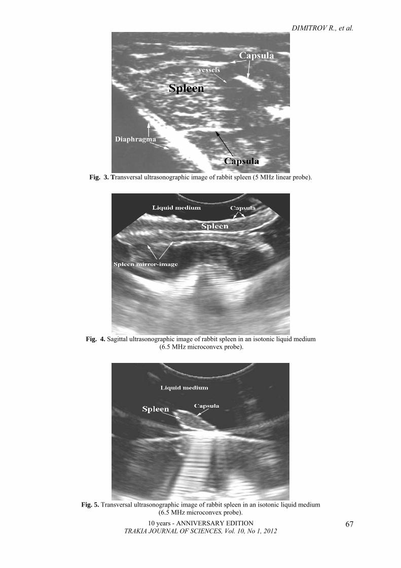
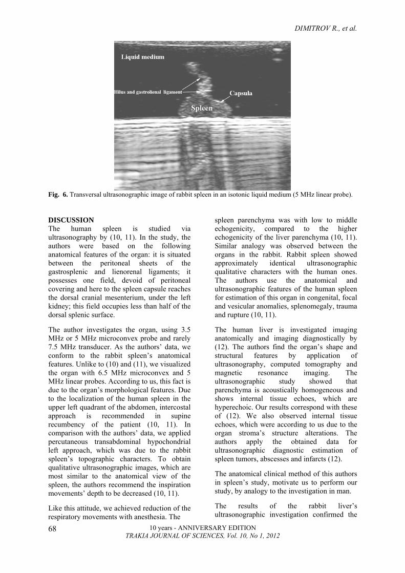
echogenicity, containing multiple hyperechoic stripes. The capsule was relatively hyperechoic and more homogeneous than parenchyma (Fig. 4). The observation in transversal aspect (with convex and linear probe), spleen showed approximately triangular transversal image, and hyperechoic capsule was well differentiated from the hypoechoic homogeneous parenchyma (Fig. 5 and Fig. 6). Furthermore, spleen hilus and gastrolienal ligament were well expressed on the visceral surface of the spleen, as they were hyperechogenic toward the organ capsule (Fig.6). In the spleen ultrasonographic study in isotonic medium, the mirror image of the organ was observed (Fig. 4, Fig. 5, Fig. 6).
Fig. 1. Sagital ultrasonographic image of rabbit spleen, liver and diaphragma (6.5 MHz microconvex probe).
Fig. 2. Sagittal ultrasonographic image of rabbit spleen (5 MHz linear probe).
DIMITROV R., et al.
10 years - ANNIVERSARY EDITION TRAKIA JOURNAL OF SCIENCES, Vol. 10, No 1, 2012
67
Fig. 3. Transversal ultrasonographic image of rabbit spleen (5 MHz linear probe).
Fig. 4. Sagittal ultrasonographic image of rabbit spleen in an isotonic liquid medium
(6.5 MHz microconvex probe).
Fig. 5. Transversal ultrasonographic image of rabbit spleen in an isotonic liquid medium
(6.5 MHz microconvex probe).
DIMITROV R., et al.
10 years - ANNIVERSARY EDITION TRAKIA JOURNAL OF SCIENCES, Vol. 10, No 1, 2012
68
Fig. 6. Transversal ultrasonographic image of rabbit spleen in an isotonic liquid medium (5 MHz linear probe). DISCUSSION The human spleen is studied via ultrasonography by (10, 11). In the study, the authors were based on the following anatomical features of the organ: it is situated between the peritoneal sheets of the gastrosplenic and lienorenal ligaments; it possesses one field, devoid of peritoneal covering and here to the spleen capsule reaches the dorsal cranial mesenterium, under the left kidney; this field occupies less than half of the dorsal splenic surface. The author investigates the organ, using 3.5 MHz or 5 MHz microconvex probe and rarely 7.5 MHz transducer. As the authors’ data, we conform to the rabbit spleen’s anatomical features. Unlike to (10) and (11), we visualized the organ with 6.5 MHz microconvex and 5 MHz linear probes. According to us, this fact is due to the organ’s morphological features. Due to the localization of the human spleen in the upper left quadrant of the abdomen, intercostal approach is recommended in supine recumbency of the patient (10, 11). In comparison with the authors’ data, we applied percutaneous transabdominal hypochondrial left approach, which was due to the rabbit spleen’s topographic characters. To obtain qualitative ultrasonographic images, which are most similar to the anatomical view of the spleen, the authors recommend the inspiration movements’ depth to be decreased (10, 11). Like this attitude, we achieved reduction of the respiratory movements with anesthesia. The
spleen parenchyma was with low to middle echogenicity, compared to the higher echogenicity of the liver parenchyma (10, 11). Similar analogy was observed between the organs in the rabbit. Rabbit spleen showed approximately identical ultrasonographic qualitative characters with the human ones. The authors use the anatomical and ultrasonographic features of the human spleen for estimation of this organ in congenital, focal and vesicular anomalies, splenomegaly, trauma and rupture (10, 11). The human liver is investigated imaging anatomically and imaging diagnostically by (12). The authors find the organ’s shape and structural features by application of ultrasonography, computed tomography and magnetic resonance imaging. The ultrasonographic study showed that parenchyma is acoustically homogeneous and shows internal tissue echoes, which are hyperechoic. Our results correspond with these of (12). We also observed internal tissue echoes, which were according to us due to the organ stroma’s structure alterations. The authors apply the obtained data for ultrasonographic diagnostic estimation of spleen tumors, abscesses and infarcts (12). The anatomical clinical method of this authors in spleen’s study, motivate us to perform our study, by analogy to the investigation in man. The results of the rabbit liver’s ultrasonographic investigation confirmed the
DIMITROV R., et al.
10 years - ANNIVERSARY EDITION TRAKIA JOURNAL OF SCIENCES, Vol. 10, No 1, 2012
69
anatomical data for this organ, generalized by (1, 2, 3). Our data for rabbit spleen ultrasonographic visualization and the used approach correspond with the data of Acorda et al. (6) for sheep, goat and buffalo spleen. Contrary to the frequency of the used microconvex probe, we used such with frequency 6.5 MHz, which according to us is due to the anatomical features of the rabbit spleen. Unlike to (7), who perform ultrasonographic study in rabbit spleen during endothoxemic shock, we investigated healthy rabbits. In comparison with the attitude of (13), that the observation of mirror images of investigated organs in living organism is artifact, and the images are pseudo, we confirm the following: The spleen’s mirror image in isotonic liquid medium is due to the fact that the vessel that has been looked at was glass-like and its bottom had a reflective surface. That was one of the peculiarities of this method. According to the opinion of (14) regarding the rabbit’s role as widely used experimental model in the human and veterinary practice, we propose the following: the qualitative ultarsonographic data for rabbit spleen to be used as a biological model in the imaging anatomical and diagnostic investigations of some human and animal spleen’s lesions. REFERENCES 1. Cesta, M., Normal structure, function, and
histology of the spleen. Toxicologic Рathology, 34: 455-465, 2006.
2. Barone, R., Chapitre VIII - Rate. In: Anatomie comparée des mammifères domestiques. Splanchnologie I. Tome troasième, Troisième edition, Editions Vigot, Paris, pp. 577-591, 1997.
3. Hristov, H., Kostov, D., Vladova, D., Topographical anatomy of some abdominal organs in rabbits. Trakia Journal of Sciences, 4: 7-10, 2006.
4. Al-Dahmesh, B., Dkhil, M., Al-Quraishy S., Chili pepper-induced injury to splenic tissue of rabbit. Journal of Medicinal Plants Research, 5: 2015-2020, 2011.
5. Peddu, P., Shah, M., Sidhu, P., Splenic abnormalities: a comparative review of ultrasound and computed tomography. Clinical radiology, 59: 777-792, 2004.
6. Acorda, J., Ancheta, M., Detera, M., Cabera, L., Maligaya, Rh., Comparative ultrasound features and echo histograms of the spleen in female goats (Capra hircus), sheep (Ovis aries) and buffaloes (Bubalus Bubalus). Philippine Journal of Veterinary and Animal Sciences, 35: 135-146, 2009.
7. Takeda, Y., Asaoka, H., Ueno, M., Jimma, F., Hidaka, M., Shibusawa, H., Kaneda, K., Saniabadi, A., Hiraishi, K., Kashiwagi, N., Assessment of rabbit spleen size using ultrasonography. The Journal of Veterinary Medical Science, 69: 841-842, 2007.
8. Moarabi, A., Mossalanejad, B., Ghadiri, A., Borujeni, M., Ultrasonographic evdlution of the urinary system in New Zealand White Rabbit and Totai Hare. Veterinary Research Forum, 2: 113-120, 2011.
10. Andrews, M., Ultrasound of the spleen. Word Journal of Surgery, 24: 183-187, 2000.
11. Benter, Th., Klühs, L., Teichgräber, U., Sonography of Spleen. Journal of Ultrasound Medicine, 30: 1281-1293, 2011.
12. Robertson, F., Leander, P., Ekberg, O., Radiology of the spleen. European Radiology, 11: 80-95, 2001.
13. Chakarski, V., Mincheva, E., Lazarova, I., Bueva, A., Popov, D., Oien, R., Katen, J., Atlas of abdominal echography. First edition, Medicine and Physical Education, Sofia, 1996.
14. Eken, E., Ģorumluoğlu, Ö., Paksoy, Y., Beşoluk, K., Kalayci, İ., A study of evaluation of 3D virtual rabbit kidney models by multidetector computed tomography images. Journal of Experimental and Clinical Anatomy, 3: 40-44, 2009.