215 ORIGINAL ARTICLE STATIC LOAD TEST ON DYNAMIC HIP SYSTEM (DHS) PLATES ANDERSON FREITAS, ALESSANDRO QUEIROZ DE MESQUITA, WALTER RODRIGO DAHER, DIOGO RANIER DE MACÊDO SOUTO, CARLOS HENRIQUE DA COSTA RANGEL Citation: Freitas A, Mesquita AQ, Daher WR, Souto DRM, Rangel CHC. Static load test on dynamic hip system (dhs) plates. Acta Ortop Bras. [online]. 2009; 17(4):215-8. Available from URL: http://www.scielo.br/aob. All the authors state no potential conflict of interest concerning this article. Study conducted at the Orthopaedics and Traumatology Service, Gama Regional Hospital - Gama (DF), Brazil Orthopaedics and Traumatology Service, Gama Regional Hospital - Gama (DF), Brazil. Correspondences to: Rua Fortaleza, 355, Setor Alto da Glória, Goiânia, Goiás. Brasil. CEP 74 815- 710. E-mail. [email protected]Received in: 06/03/08; approved in: 10/04/08 Acta Ortop Bras. 2009; 17(4):215-8 ABSTRACT Objective: To evaluate, both individually and comparatively, dynam- ic hip system-type plates marketed by two local manufacturers, to statistically analyze its results and show the lack of parameters for its manufacturing. Methods. Static tests of flexion were car- ried out in five DHS plates of the manufacturer I (I group I) and in equal quantity of the same model of the manufacturer II (I group II), being all made in stainless austenitic ASTM F 138 steel, with four holes and a 135° angle. A servo-hydraulic MTS machine, Test Star II model, was used with a load capacity of 10 tons and dislo- cation control. The data were obtained from the applied load (P) as a function of the vertical dislocation of the piston (L), whose speed was 5mm/min. The tests were shutdown after reaching the maximum vertical flexion specified by the tests’ standards. Results. GroupI: flexion resistance, 161,4 ± 17,2 kgf rigidity, 64,5 ± 1,8 kgf/mm,flexibility, > 25,4 mm. Group II: flexion resistance, 124,7 ± 4,4,rigidity 59,6 ± 2,3, flexibility > 25,4 mm. For statistical analy- sis, the Mann-Whitney test was adopted and the determination of significance was 5 % (p <0,05). When the results obtained are compared, a significant difference can be noticed. Conclusion. The DHS plates of group I presented stronger flexion resistance (kgf) and stiffness (kgf/mm). Keywords: Hip fractures. Bone nails. Bone screws. INTRODUCTION Intertrochanteric fractures represent a challenge to orthopaedic doctors 1 and are commonly found in the elderly population. 2-5 However, the fixation of these fractures has been evolving in the last few decades. 2-5 By investigating the factors that could influence the failure of intertro- chanteric fractures’ fixation with sliding screws (dynamic hip system - DHS), Rha 6 listed patient’s age, kind of fracture, osteoporosis degree, positioning of the sliding nail and the degree of fracture reduction. Lee et al. 7 conducted a biomechanical study on the differences between stainless steel and titanium employed on the manufacturing of DHS in view of femoral intertrochanteric fractures treatment. The authors found no statistical difference between both groups. Lundy et al. 8 submitted subtrochanteric fractures to three fixation methods and investigated the flexion strength of each employed plate. The authors found that 135° DHS-type plates (586N/mm) showed to be stronger than the DCS 95° (404N/mm) which, in turn, were shown to be stronger than those with 95° fixed angle (260N/mm). According to Schwartsmann et al. 2 a successful treatment for in- tertrochanteric fractures depends on five factors: quality of bone, kind of fracture, reduction achievement, and implant positioning; of these, a surgeon can only influence the latter three. In Brazil, Lima et al. 5 made a clinical and X-ray analysis of surgi- cal treatment outcomes on intertrochanteric fractures with open reduction and osteosynthesis by using the dynamic hip system (DHS). Forty-seven patients were studied, 10 males and 37 fe- males. As a result of the good outcomes reported, the authors recommended the use of DHS as an alternative for treating femo- ral intertrochanteric fractures. Also in Brazil, Sawaia and Belangero 9 conducted a comparative study between micro incision techniques and the open approach when treating intertrochanteric fractures. In both, the authors used DHS with three or four holes and concluded that the micro incision technique adds benefits to the traditional technique. Currently, several studies have demonstrated that the hip sliding screw system constitutes the method of choice for treating those fractures. 10-15 A sliding screw positioned at femoral head is intended to provide stability and compression to the fracture by means of a controlled collapse of proximal over distal fragment. 3,4,16,17 Its use is particularly featured by its ease to handle, by its relatively easy inser- tion technique, by its low cost and low rate of complications reported in literature. 3,4,17 Characteristics of the implant design such as fixed angle and intrin- sic rotational stability of the nail may lead to an improper position- ing, thus leading to a higher risk of failure. In order to reduce the complication rate when using this implant, some novelties have been introduced such as the dynamic MARTIN system (DMS), a DHS with variable angle, the sliding plate system MEDOFF (PSM), a DHS with ability to slide the shaft plate and the nail, intended to solve the issue of treating unstable fractures.
Transcript
215214 215214
Original article
Static load teSt on dYnamic hiP SYStem (dhS) PlateS
anderson FreiTas, alessandro queiroz de MesquiTa, WalTer rodrigo daher, diogo ranier de Macêdo souTo, carlos henrique da cosTa rangel
citation: freitas a, mesquita aq, daher Wr, souto drm, rangel cHc. static load test on dynamic hip system (dhs) plates. acta ortop bras. [online]. 2009; 17(4):215-8. available from urL: http://www.scielo.br/aob.
All the authors state no potential conflict of interest concerning this article.
Study conducted at the Orthopaedics and Traumatology Service, Gama Regional Hospital - Gama (DF), BrazilOrthopaedics and Traumatology Service, Gama Regional Hospital - Gama (DF), Brazil.
Correspondences to: Rua Fortaleza, 355, Setor Alto da Glória, Goiânia, Goiás. Brasil. CEP 74 815- 710. E-mail. [email protected]
Objective: To evaluate, both individually and comparatively, dynam-ic hip system-type plates marketed by two local manufacturers, to statistically analyze its results and show the lack of parameters for its manufacturing. Methods. Static tests of flexion were car-ried out in five DHS plates of the manufacturer I (I group I) and in equal quantity of the same model of the manufacturer II (I group II), being all made in stainless austenitic ASTM F 138 steel, with four holes and a 135° angle. A servo-hydraulic MTS machine, Test Star II model, was used with a load capacity of 10 tons and dislo-cation control. The data were obtained from the applied load (P) as a function of the vertical dislocation of the piston (L), whose
speed was 5mm/min. The tests were shutdown after reaching the maximum vertical flexion specified by the tests’ standards. Results. GroupI: flexion resistance, 161,4 ± 17,2 kgf rigidity, 64,5 ± 1,8 kgf/mm,flexibility, > 25,4 mm. Group II: flexion resistance, 124,7 ± 4,4,rigidity 59,6 ± 2,3, flexibility > 25,4 mm. For statistical analy-sis, the Mann-Whitney test was adopted and the determination of significance was 5 % (p <0,05). When the results obtained are compared, a significant difference can be noticed. Conclusion. The DHS plates of group I presented stronger flexion resistance (kgf) and stiffness (kgf/mm).
Keywords: Hip fractures. Bone nails. Bone screws.
introduction
Intertrochanteric fractures represent a challenge to orthopaedic doctors1 and are commonly found in the elderly population.2-5 However, the fixation of these fractures has been evolving in the last few decades.2-5
By investigating the factors that could influence the failure of intertro-chanteric fractures’ fixation with sliding screws (dynamic hip system - DHS), Rha6 listed patient’s age, kind of fracture, osteoporosis degree, positioning of the sliding nail and the degree of fracture reduction.Lee et al.7 conducted a biomechanical study on the differences between stainless steel and titanium employed on the manufacturing of DHS in view of femoral intertrochanteric fractures treatment. The authors found no statistical difference between both groups.Lundy et al.8 submitted subtrochanteric fractures to three fixation methods and investigated the flexion strength of each employed plate. The authors found that 135° DHS-type plates (586N/mm) showed to be stronger than the DCS 95° (404N/mm) which, in turn, were shown to be stronger than those with 95° fixed angle (260N/mm).According to Schwartsmann et al.2 a successful treatment for in-tertrochanteric fractures depends on five factors: quality of bone, kind of fracture, reduction achievement, and implant positioning; of these, a surgeon can only influence the latter three.In Brazil, Lima et al.5 made a clinical and X-ray analysis of surgi-cal treatment outcomes on intertrochanteric fractures with open
reduction and osteosynthesis by using the dynamic hip system (DHS). Forty-seven patients were studied, 10 males and 37 fe-males. As a result of the good outcomes reported, the authors recommended the use of DHS as an alternative for treating femo-ral intertrochanteric fractures.Also in Brazil, Sawaia and Belangero9 conducted a comparative study between micro incision techniques and the open approach when treating intertrochanteric fractures. In both, the authors used DHS with three or four holes and concluded that the micro incision technique adds benefits to the traditional technique. Currently, several studies have demonstrated that the hip sliding screw system constitutes the method of choice for treating those fractures.10-15 A sliding screw positioned at femoral head is intended to provide stability and compression to the fracture by means of a controlled collapse of proximal over distal fragment.3,4,16,17 Its use is particularly featured by its ease to handle, by its relatively easy inser-tion technique, by its low cost and low rate of complications reported in literature.3,4,17
Characteristics of the implant design such as fixed angle and intrin-sic rotational stability of the nail may lead to an improper position-ing, thus leading to a higher risk of failure. In order to reduce the complication rate when using this implant, some novelties have been introduced such as the dynamic MARTIN system (DMS), a DHS with variable angle, the sliding plate system MEDOFF (PSM), a DHS with ability to slide the shaft plate and the nail, intended to solve the issue of treating unstable fractures.
217216 217216
Flexion strengthLR
figure 3 – schematic illustration of the device assembly and the method-ology employed for determining the parameters for static flexion assay on angled plate.
P
supporting basefor application
rollers
base for loadapplication
L
P
angled plate
extensor nail
fixation device
76mm
135
A solution to the problem has been the core-medullar implants, the Proximal Femoral Nail (PFN), and, more recently, the Dynamic He-licoidal Hip System (DHHS), a DHS with spiral-form nail designed to spare bone on femoral neck, keeping bone stock.Therefore, and considering the importance of the topic, the present study was aimed to assess, both separately and comparatively, DHS plates supplied by two local manufacturers focusing the de-terminant factors for implants manufacturing.
material and methodS
Five DHS-type plates supplied by a manufacturer from São Paulo and widely available in market composed group I, and five widely employed DHS plates supplied by another local manufacturer composed group II. All models were manufactured with austenitic stainless steel ASTM F 138. (Figures 1A and 1B)
displacement) of 0.13mm, stiffness - ratio between flexion strength, as defined above, and total deflexion produced by that load (LR), flexibility - maximum vertical deflexion at the moment of load ap-plication a DHS can stand immediately before a visible fracture is produced under a magnification of at least 8 times.
figure 1a – angled dHs plate model (135º) with four holes, manufactured of austenitic stainless steel asTm f 138 used for static flexion assay, group i.
figure 1B – angled dHs plate model (135º) with four holes, manufactured of austenitic stainless steel asTm f 138 used for static flexion assay, group ii.
figure 2 – full assembly of the device onto the assay machine for angled dHs plate, group ii.
Static flexion assays were carried out with all plates, which had the same structural characteristics, i.e., four holes with fixed angle of 135º. Tests were conducted according to ASTM F 384 e NBR 13762 guidelines.18,19 For carrying assays out, the plates were pre-viously fixated to a stiff body of evidence by means of 4.5-mm wide screws. The body of evidence was especially made for this kind of assay, strictly following the specifications of the assay guidelines mentioned above. Also for complying with the specifications of the assay guidelines, an extensor nail was manufactured, which enabled the application of load at vertical orientation at a point 76 mm away from the curvature for the studied plate model.Static flexion assays were carried out on a servo-hydraulic ma-chine MTS, Test Star II model, with a load capacity of 10 tons and displacement control. Data for applied load (P) were obtained as a function of the vertical displacement of the piston (L). The verti-cal displacement speed was 5 mm/min. The static flexion assays were shutdown when maximum vertical deflexion (maximum vertical displacement) was achieved, according to assay guidelines. The full assembly of the DHS model on the assay machine was provided immediately before its performance. (Figure 2)The full assembly of assay devices, as well as the method em-ployed for determining the parameters of interest, according to assay guidelines are the following (Figure 3): flexion strength - required load to promote permanent vertical deflexion (vertical
Stifness =
217216 217216
Tables representing static flexion assays for DHS implant on group II were built from the values for flexion and stiffness strength pa-rameters (Table 2). Flexion strength ranged from -131.0 to -88.0 kgf, with mean +/- SD of 117.4 +/- 4.4 kgf. Stiffness ranged from 49.2 to 62.1 kgf/mm, with mean +/- SD of 57.5 +/- 2.3 kgf/mm. All tested plates showed flexibility superior to 25.4mm, and no fail-ures (breakage) were noted before the maximum vertical deflexion established by the static flexion assay guidelines for this kind of implant was reached. This result reveals a high flexibility of the studied implant.
table 2 – Values of the parameters obtained from static flexion assays for angled plate implants on group ii
PLATEFLEXION
STRENGTH, kgfSTIFFNESS, kgf/mm FLEXIBILITY, mm
1 -119.0 56.7 > 25.4
2 -126.0 62.1 > 25.4
3 -131.0 61.8 > 25.4
4 -123.0 58.0 > 25.4
5 -88.0 49.2 > 25.4
Mean ± Standard Deviation 117.4 ± 4.4 57.5 ± 2.3 ----
NOTE.: (-) sign resulting from load in compression.
When the results shown on Table 3 are assessed, a significant difference is noted on flexion strength between DHS plates, i.e., group I shows a significantly higher flexion strength compared to group II (p = 0.009).
table 3 – flexion strength (kgf) between groups i and ii.
Manufacturer Mean SD Median Minimum Maximum p value
I -161.4 19.3 -156 -195 -1470.009
II -117.4 17.0 -123 -131 -88
SD: Standard Deviation
A significant difference (p > 0.05) is also noted for stiffness between DHS plates, because group I shows a superior mean value for this parameter when compared to group II (p = 0.011).(Table 4)
table 4 – statistical analysis of stiffness (kgf/mm) between groups i and ii.
Manufacturer Mean SD Median Minimum Maximum p value
I 64.5 2.0 64.5 62.1 66.40.011
II 57.6 5.2 58.0 49.2 62.1SD: Standard Deviation
diScuSSion
Intertrochanteric fractures are undoubtedly of great importance in terms of public health, because population’s life expectancy has increased and, as a result, the frequency of fractures has also increased.1,3,20,21 On the other hand, the use of DHS-type plates does not require experimentation to prove its value in treating this kind of fracture. However, this study raises a question concern-ing standardization by manufacturers of the material employed in
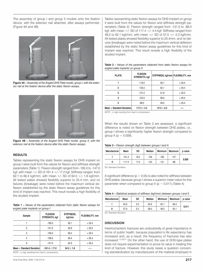
figure 4a – assembly of the angled dHs plate model, group i, with the exten-sor nail at the fixation device after the static flexion assays.
figure 4B – assembly of the angled dHs plate model, group ii, with the extensor nail at the fixation device after the static flexion assays.
The assembly of group I and group II models onto the fixation device, with the extensor nail attached, after assays performed. (Figure 4A and 4B)
reSultS
Tables representing the static flexion assays for DHS implant on group I were built from the values for flexion and stiffness strength parameters (Table 1). Flexion strength ranged from -195.0 to -147.0 kgf, with mean +/- SD of 161.4 +/- 17.2 kgf. Stiffness ranged from 62.1 to 66.4 kgf/mm, with mean +/- SD of 64.5 +/- 1.8 kgf/mm. All tested plates showed flexibility superior to 25.4 mm, and no failures (breakage) were noted before the maximum vertical de-flexion established by the static flexion assay guidelines for this kind of implant was reached. This result reveals a high flexibility of the studied implant.
table 1 – Values of the parameters obtained from static flexion assays for angled plate implants on group i
SampleFLEXION
STRENGTH, kgfSTIFFNESS,
kgf/mmFLEXIBILITY, mm
1 -195.0 62.1 > 25.4
2 -151.0 62.9 > 25.4
3 -156.0 66.4 > 25.4
4 -158.0 66.4 > 25.4
5 -147.0 64.5 > 25.4
Mean ± Standard Deviation -161.4 ± 17.2 64.5 ± 1.8 ----NOTE.: (-) sign resulting from load in compression.
219218 219218
the manufacturing of orthopaedic surgical implants. Furthermore, it provides inputs for professional societies, manufacturers, and surveillance and regulatory agencies to encourage new studies targeting the advancement of knowledge in such an important area of orthopaedics.Several variables can influence a successful treatment of intertro-chanteric fractures with sliding screws, namely: patient’s advanced age, kind of fracture (stable, unstable), osteoporosis severity, de-gree of fracture reduction, and the positioning of the sliding nail.6 Added to these, among others, are poor DHS insertion techniques and a failure when sliding the screw resulting in bending, and po-tentially leading to breakage for the plate being forcedly attached to the shaft with the use of clamps and the inappropriate use of a goniometer (1). According to Schwartsmann et al.2, intertrochanteric fractures have as interfering factors the quality of bone, the kind of fracture, the achievement of reduction, the selection and appropri-ate positioning of the implant, and, of these, a surgeon can only influence the latter three.In Brazil, study findings evidence DHS effectiveness on surgical treatment of intertrochanteric fractures.5,9 However, in the ortho-paedic routine treatment of these injuries, differences existent be-tween the implants employed in this surgical procedure are easily detected. Differences are found between plates’ holes (number and sizes), screws (thread steps), width, weight, length, flexion strength, stiffness. This strongly challenges a surgeon’s choice of implant and can interfere on treatment success.2
Lee et al.7, conducting a biomechanical study on a stainless steel DHS, found a mean maximum load of 92.59kgf for intertrochan-
teric fractures and 140.12kgf for subtrochanteric. The authors did not find breaks on metal implants even at maximum load (600kgf) conditions. Although experimental conditions are differ-ent, when these results are compared against the ones obtained here, we can see that the material employed by the authors can be regarded as similar to those submitted to test in this study. It is worthy to mention, however, that the determination of the maximum strength absorbed by implants does not constitute an object of this study.Considering the results achieved when DHS models supplied by two different manufacturers are compared, the need of establish-ing minimum quality parameters addressing material composi-tion, minimum values for biomechanical tests - stiffness and flex-ion strength - length, width, thickness, as well as holes’ diameter becomes evident, targeting the standardization of orthopaedic hip implants.
CONCLUSION
Within the conditions of this experiment and considering the results achieved here, we conclude that the DHS plates on group I, when compared to group II show stronger flexion (kgf) and stiffness (kgf/mm), evidencing the existence of differences between the implants employed in orthopaedic surgeries in Brazil.Therefore, we suggest that minimum limits for stiffness and flexion strength are established for orthopaedic implants, based on me-chanical and/ or biomechanical studies, targeting its standardiza-tion, as well as to provide a more confident selection of an implant and a successful surgical treatment.
1. Canto RST, Luciano RC, Souza MRP, Castro IJC, Martins APOB. Uso do DHS no tratamento das fraturas intertrocantéricas. Rev Bras Ortop. 1996;31:1007‑12.
2. Schwartsmann CR, Boschin LC. Quadril do adulto. In: Pardini Junior AG,.; Barros Filho T0E P,organizadores. Ortopedia e traumatologia ‑ princípios e prática. 3a. ed. Porto Alegre: Artes Médicas; 2003. p. 990.
4. Guyton JL. “Fractures of hip, acetabulum, and pelvis”. In: Canale ST. Campbell’s operative orthopaedics. St. Louis: Mosby; 1998. p. 2181‑279.
5. Lima ALP, Azevedo Filho AJ, Amaral NP, Franklin CE, Giordano V. Tratamento das fraturas intertrocanterianas com placa e parafuso deslizante. Rev Bras Ortop. 2003;38:271‑80.
6. Rha JD, Kim YH, Yoon SI, Park TS, Lee MH. Factors affecting sliding of the lag screw in intertrochanteric fractures. Int Orthop. 1993;17:320‑4.
7. Lee KS, Lee IH, Woo KJ, Park JH, Wie DG. Biomechanical study about difference between stainless steel and titanium dynamic hip screws in peritrochanteric fractures of the femur. J Korean Orthop Assoc. 1997;32:929‑36.
8. Lundy DW, Acevedo JI, Ganey TM, Ogden JA, Hutton WC. Mechanical comparison of plates used in the treatment of unstable subtrochanteric femur fractures. J Orthop Trauma. 1999;13:534‑8.
9. Sawaia R, Belangero WD. Estudo comparativo entre a técnica de miniincisão e a via de acesso a foco aberto para o tratamento das fraturas transtrocanterianas. Rev Bras Ortop. 2005;40: 106‑18.
10. Desjardins AL, Roy A, Paiement G, Newman N, Pedlow F, Desloges D et al. Unstable inter‑trochanteric fracture of the femur. A prospective randomized study comparing anatomical reduction and medial displacement osteotomy. J Bone Joint Surg Br. 1993;75:445‑7.
11. Kyle RF, Cabanela ME, Russell TA, Swiontkowski MF, Winquist RA, Zuckerman JD et al. Fractures of the proximal part of the femur. Instr Course Lect. 1995;44:227‑53.
12. Koval KJ, Zuckerman JD. Hip fractures. II. Evaluation and treatment of intertrochan‑teric fractures. J Am Acad Orthop Surg. 1994;2:150‑6.
13. O’Brien PJ, Meek RN, Blachut PA, Broekhuse HM, Sabharwal S. Fixation of inter‑trocantheric hip fractures: Gamma nail versus dynamic hip screw. A randomized prospective study. Can J Surg. 1995;38:516‑20.
14. Osnes EK, Lofthus CM, Falch JA, Meyer HE, Stensvold I, Kristiansen IS et al: More postoperative femoral fractures with the Gamma nail than the sliding screw plate in the treatment of trocantheric fractures. Acta Ortop Scand. 2001;72:252‑6.
15. Radford PJ, Needoff M, Webb JK. A prospective prolonged comparison of the dy‑namic hip screw and Gamma locking nail. J Bone Joint Surg Br. 1993;75:789‑93.
16. Melton LJ. Hip fractures: a worldwide problem today and tomorrow. Bone. 1993;14(Suppl 1):S1‑8.
17. DeLee JC. “Fraturas e luxações do quadril”. In: Rockwood CA, Green DP, Bucholz RW. Fraturas em adultos. São Paulo: Manole; 1993. p. 1453‑620.
18. Associação Brasileira de Normas Técnicas. Implantes Ortopédicos ‑ Ensaio Estático de Flexão de Placas Anguladas ‑ NBR 13762. Rio de Janeiro: Associação Brasileira de Normas Técnicas; 1996.
19. American Society for Testing and materials. standard Practice for Static Bend Testing of Nail Plates ‑ ASTM ‑ F 384‑73. Philadelphia: ASTMA; 1973.
20. Aharonoff GB, Dennis MG, Elshinawy A, Zuckerman JD, Koval KJ. Circumstances of falls causing hip fractures in the eldery. Clin Orthop Relat Res. 1998;(348):10–4.
21. Michelson JD, Myers A, Jinnah R, Cox Q, Van Natta M. Epidemiology of hip frac‑tures among the eldery. Risk factors for fracture type. Clin Orthop Relat Res. 1995;(311):129‑35.