42
Orthopedic Injuries- A Legal Perspective Mississippi – Alabama – Tennessee – North Carolina DIANE PRADAT PUMPHREY [email protected]
| Date post: | 17-Dec-2015 |
| Category: |
Documents |
| Upload: | adela-wiggins |
| View: | 219 times |
| Download: | 0 times |
Orthopedic Injuries-A Legal Perspective
Mississippi – Alabama – Tennessee – North Carolina
DIANE PRADAT PUMPHREY [email protected]
Common Orthopedic Complaints
The Spine
• The spine consists of 26 vertebrae or separate bones that are joined to permit the column to move forward, backward and side to side. The spine is typically S shaped to increase stability and strength.
• From the top, the spine consists of 7 cervical vertebrae, 12 thoracic vertebrae and 5 lumbar vertebrae.
• There are 5 smaller vertebrae that fuse to form the sacrum and 4 that fuse to form the coccyx.
• The vertebrae all have the same basic structure which includes an anterior body, central vertebral foramen and a posterior vertebral arch.
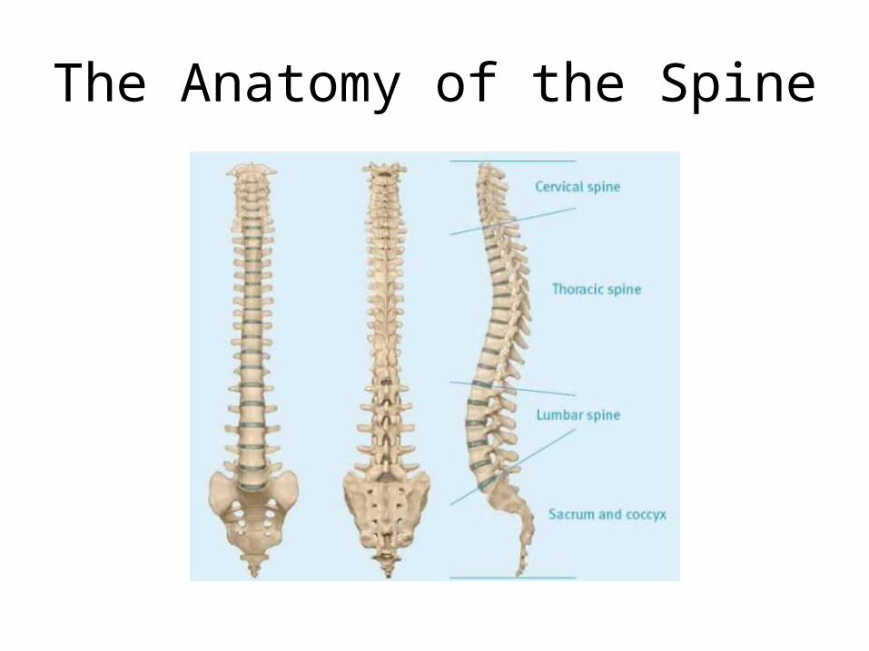
The Anatomy of the Spine
Common Spinal Problems
• Back pain – either acute or chronic• Acute pain due to injuries to muscles or soft
tissue.• Chronic pain generally dull, aching and deep that
lasts longer than three months. This can be due to specific anatomic abnormalities.
• Symptoms that require immediate attention are when there is bowel or bladder dysfunction, fever, weakness or numbness or pain that limits or prohibits everyday activities.
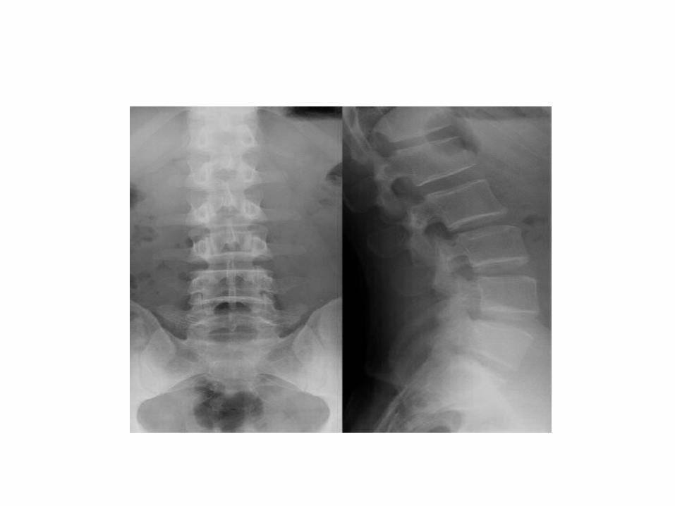
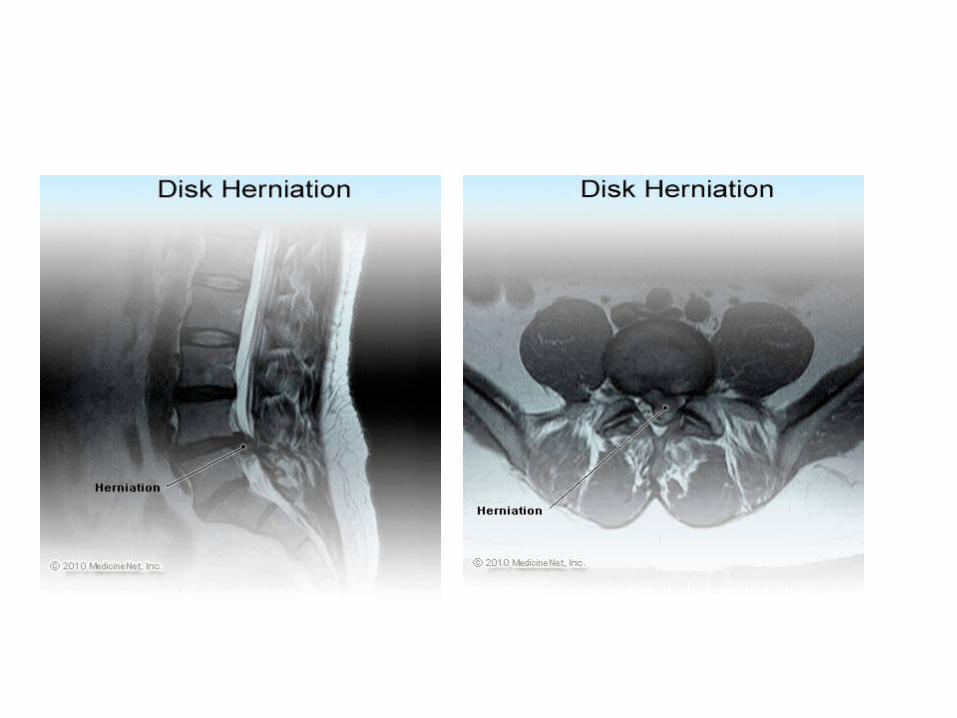
The Back Injury
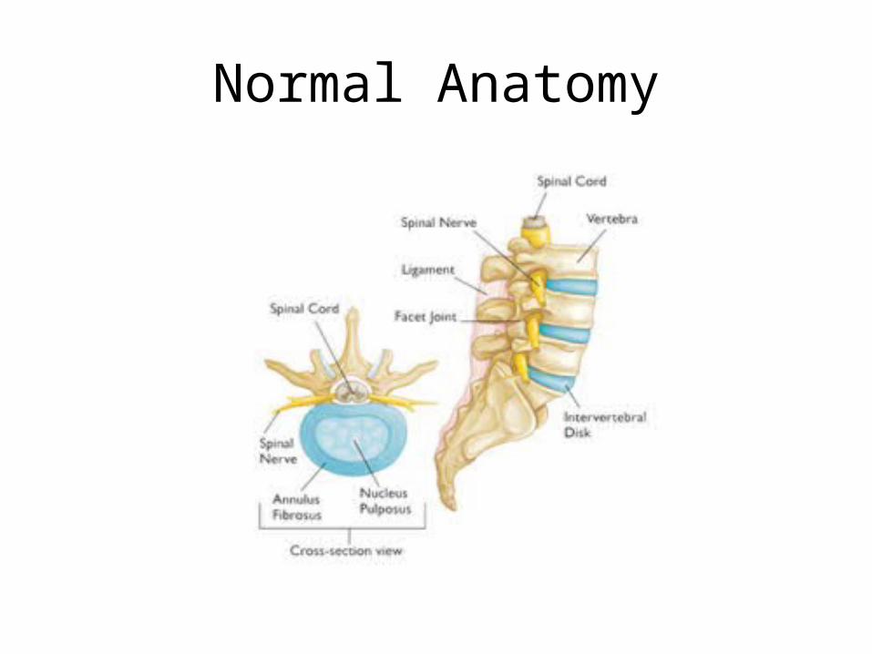
Normal Anatomy
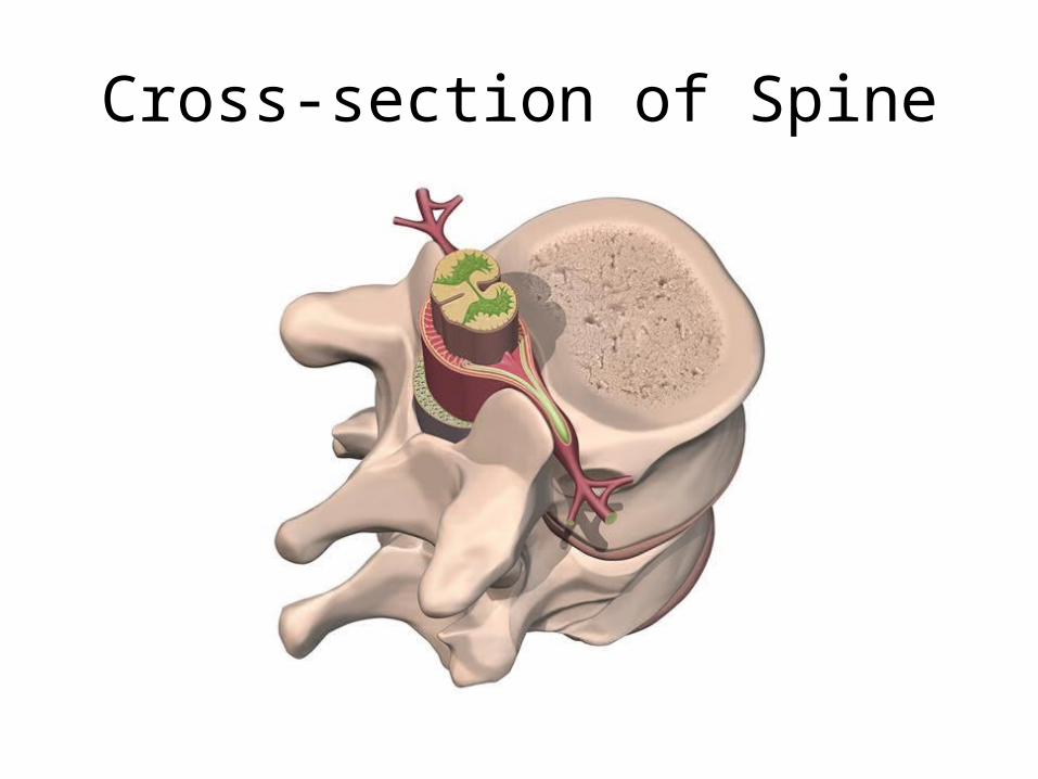
Cross-section of Spine
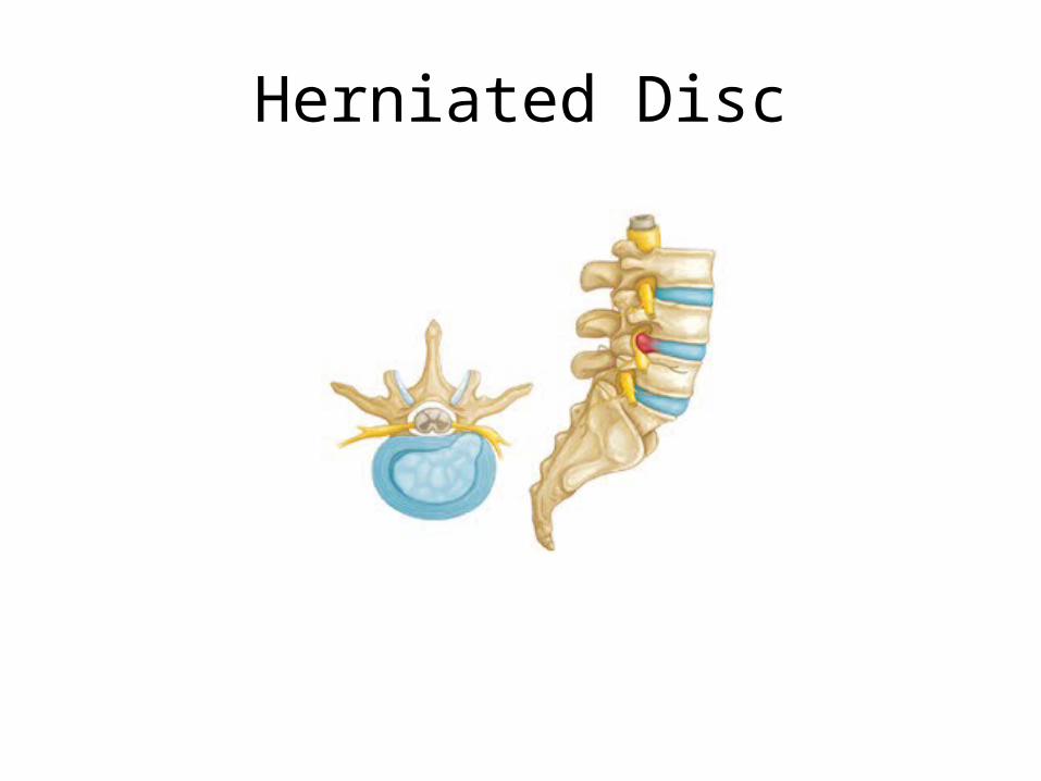
Herniated Disc

Treatment
• For strain – which is a stretch injury to the ligaments, tendons, and/or muscles of the low back – rest, medication, heat, massage and reconditioning exercises.
• For lumbar radiculopathy – nerve irritation that is caused by damage to the discs between the vertebrae – medical management to surgery.
• When there is unrelenting pain, severe impairment of function or incontinence, surgery may be necessary.
Surgical Procedures
• Laminotomy – removal of the herniated disc through a small hole in the bone of the lumbar spine surrounding the spinal cord.
• Laminectomy – removal of the herniated disc after removal of the bony wall.
• Percutaneous discectomy – removal of herniated disc with needle technique.
Neck Injury
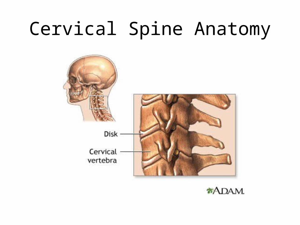
Cervical Spine Anatomy
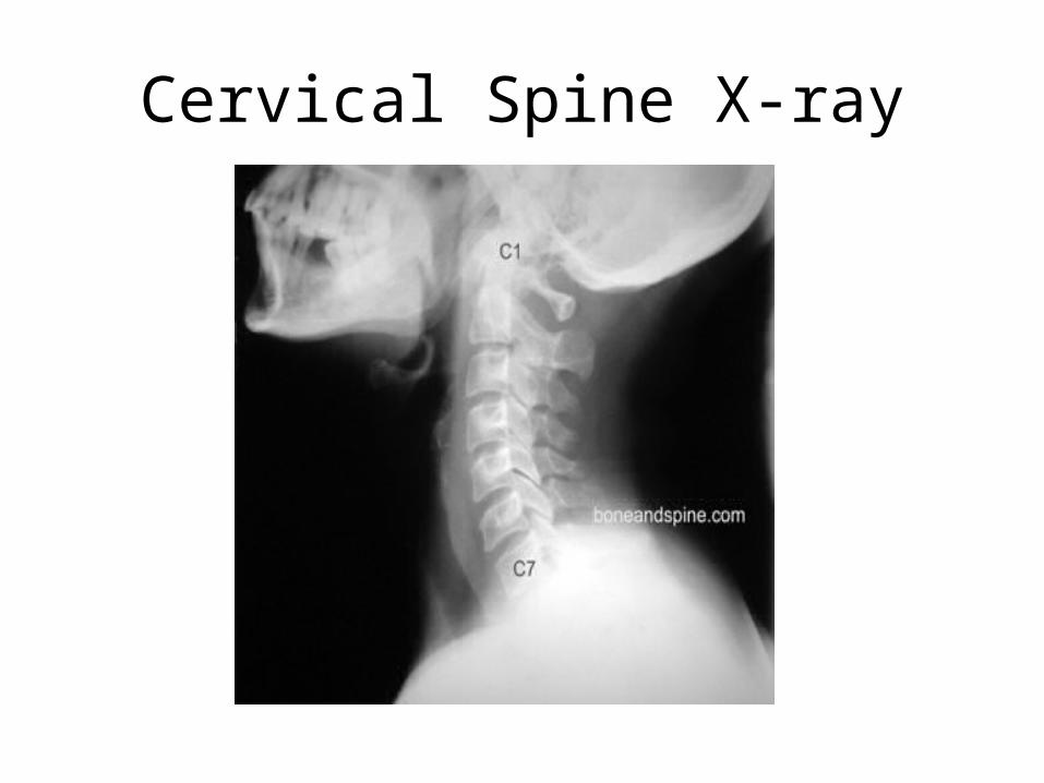
Cervical Spine X-ray

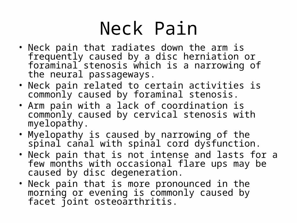
Neck Pain• Neck pain that radiates down the arm is frequently caused
by a disc herniation or foraminal stenosis which is a narrowing of the neural passageways.
• Neck pain related to certain activities is commonly caused by foraminal stenosis.
• Arm pain with a lack of coordination is commonly caused by cervical stenosis with myelopathy.
• Myelopathy is caused by narrowing of the spinal canal with spinal cord dysfunction.
• Neck pain that is not intense and lasts for a few months with occasional flare ups may be caused by disc degeneration.
• Neck pain that is more pronounced in the morning or evening is commonly caused by facet joint osteoarthritis.
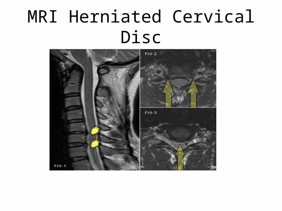
MRI Herniated Cervical Disc
Fall Injuries

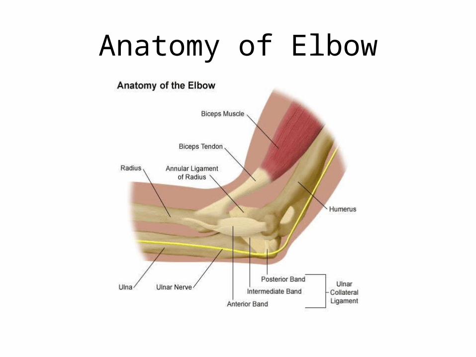
Anatomy of Elbow
Elbow Anatomy• The elbow is made up of three bones. They are the
humerus, radius and ulna. The elbow bends and straightens like a hinge. It allows the rotation of the forearm.
• The elbow is held together by ligaments, muscles and tendons and by the shape of the bones.
• The most common fracture that occurs is to the olecranon.
• The goal of rehabilitation in any elbow fracture is to regain full range of motion of the elbow. Most patients return to normal within about 4 months, but full healing can take more than a year.
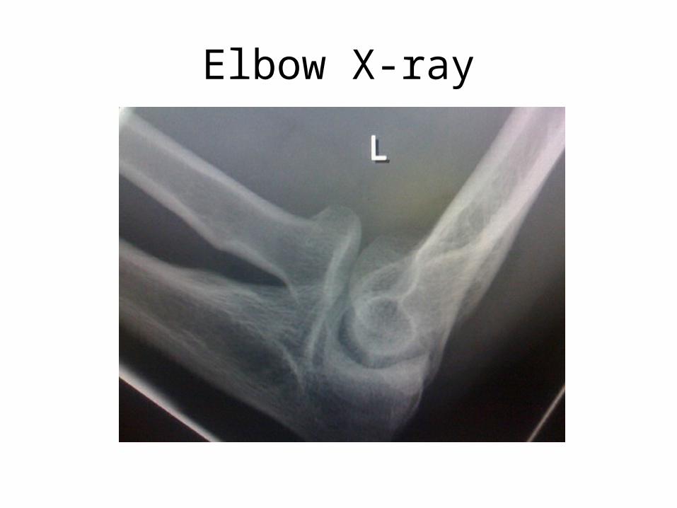
Elbow X-ray
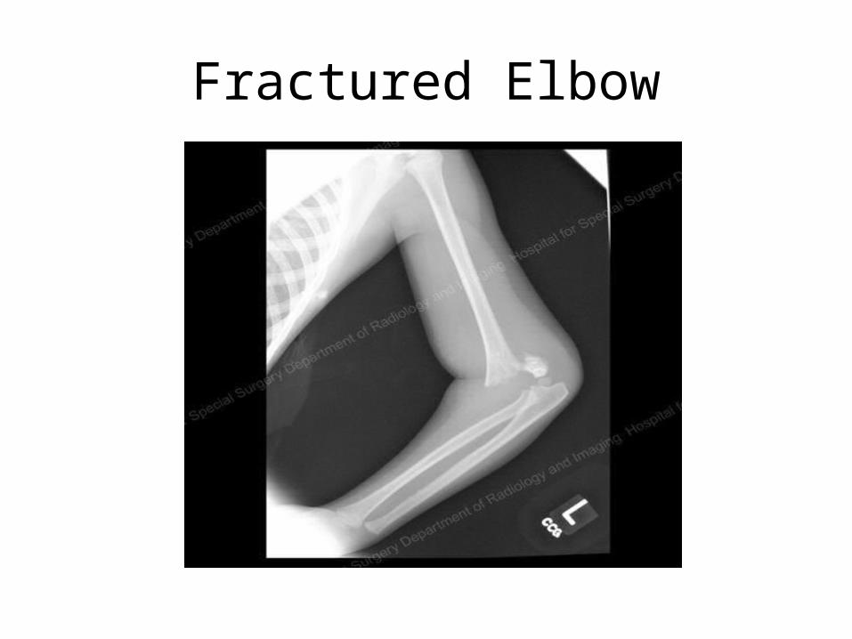
Fractured Elbow
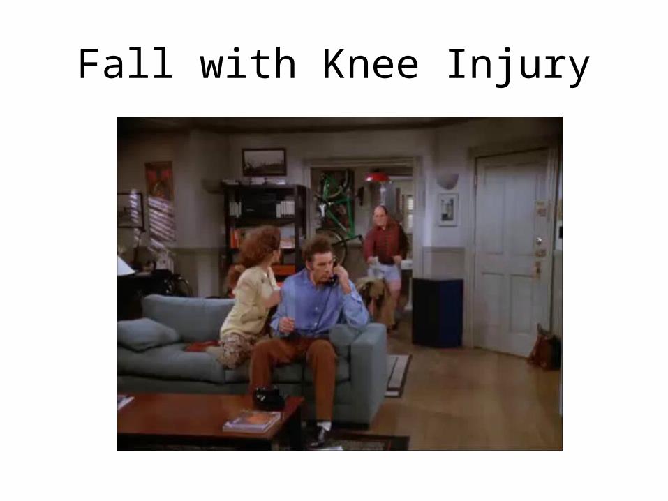
Fall with Knee Injury
Knee Anatomy and Function
• The knee is the largest joint in the body. It is made of the lower end of the thigh bone or the femur, which rotates on the upper end of the shinbone or tibia and the knee cap or patella. The knee cap slides in a groove on the end of the femur. The knee has three large ligaments that help control the motion of the connecting bones and help brace the joint.
• The other important part of the knee is the meniscus which is a wedge of cartilage between the femur and the tibia which cushions the knee and helps absorb shock during movement.
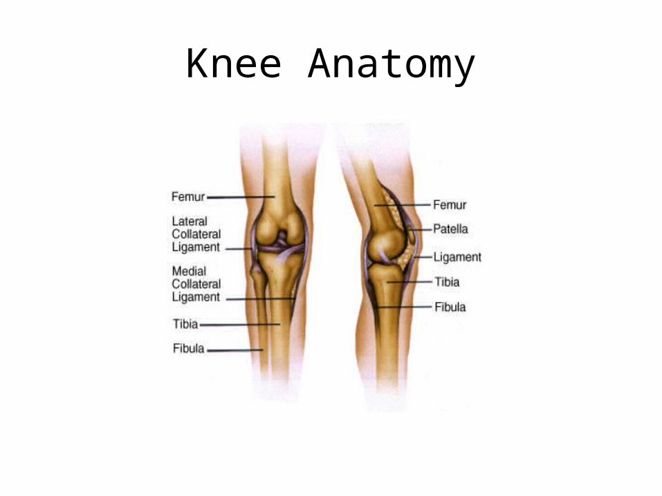
Knee Anatomy
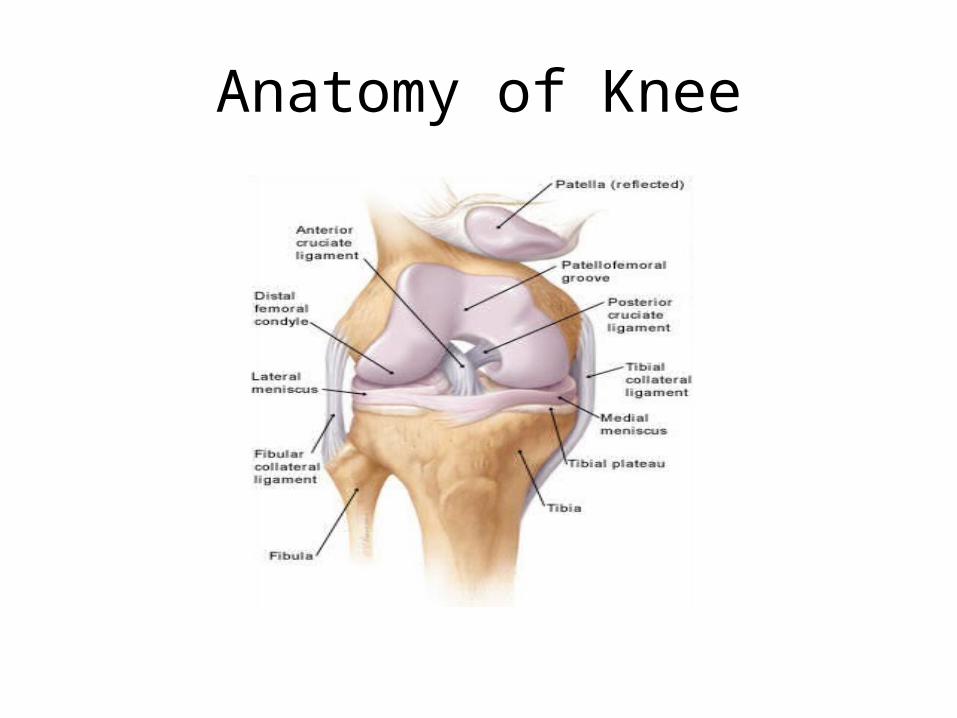
Anatomy of Knee
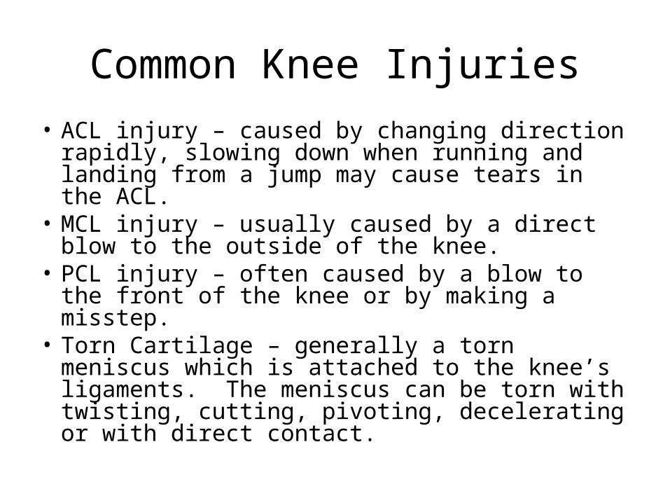
Common Knee Injuries• ACL injury – caused by changing direction rapidly,
slowing down when running and landing from a jump may cause tears in the ACL.
• MCL injury – usually caused by a direct blow to the outside of the knee.
• PCL injury – often caused by a blow to the front of the knee or by making a misstep.
• Torn Cartilage – generally a torn meniscus which is attached to the knee’s ligaments. The meniscus can be torn with twisting, cutting, pivoting, decelerating or with direct contact.
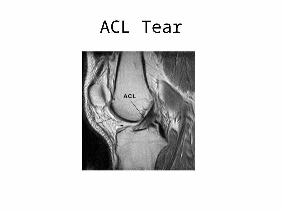
ACL Tear
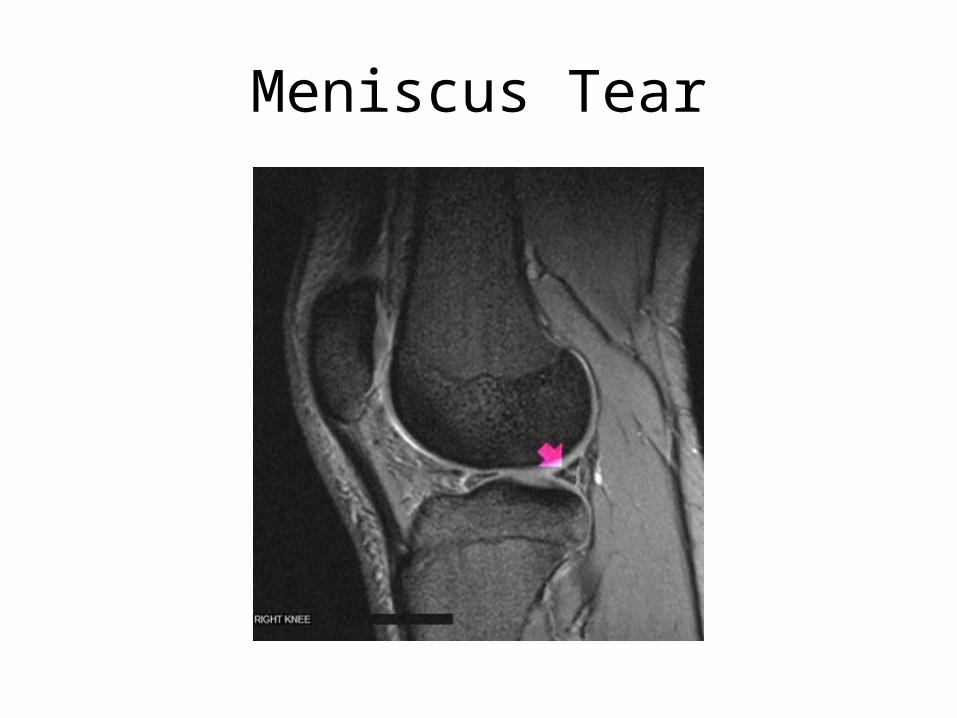
Meniscus Tear
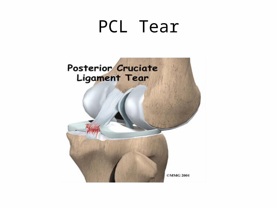
PCL Tear
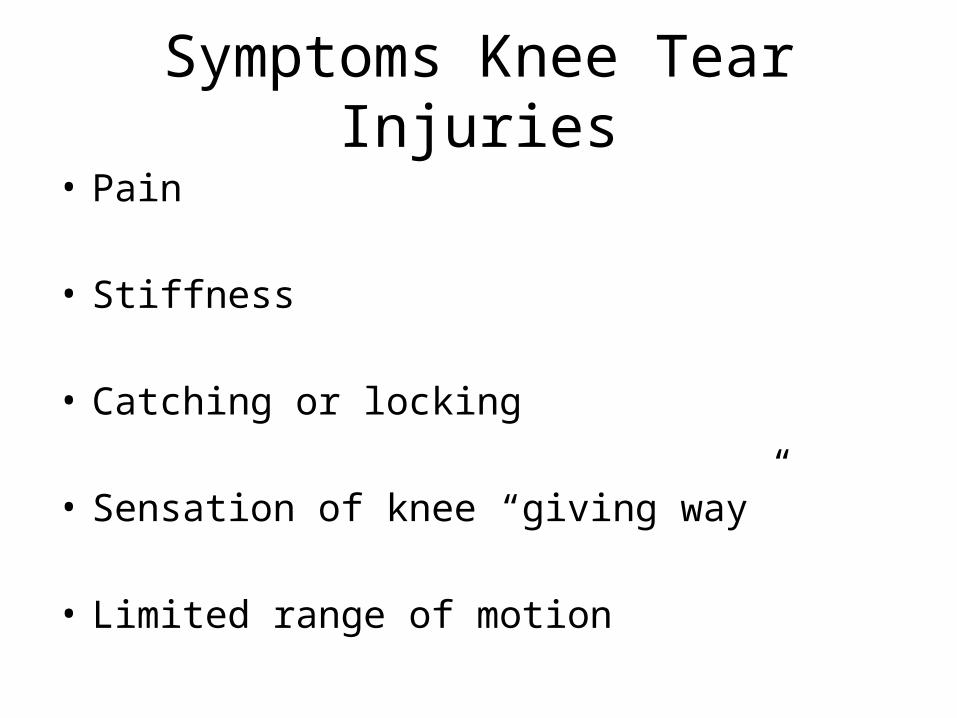
Symptoms Knee Tear Injuries• Pain
• Stiffness
• Catching or locking
• Sensation of knee “giving way”
• Limited range of motion
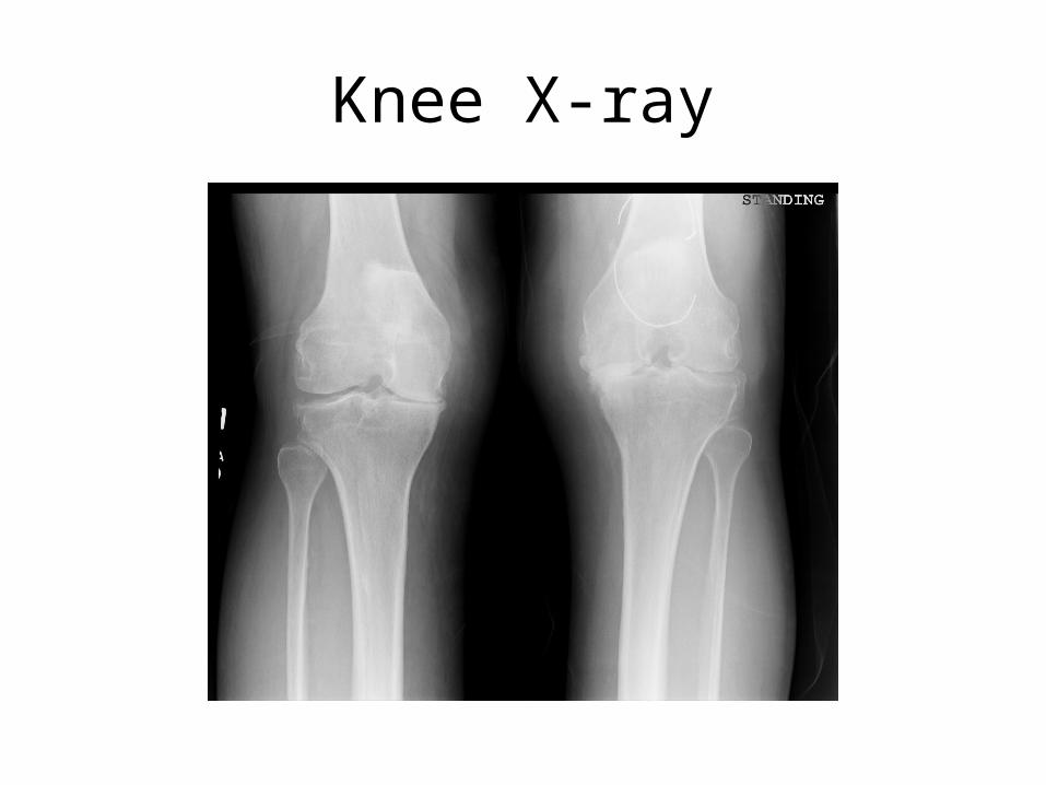
Knee X-ray
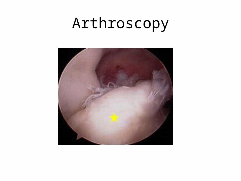
Arthroscopy
Questions


